Topic Menu
► Topic MenuTopic Editors


2. School of Human Sciences, Cell Communication in Disease Pathology, London Metropolitan University, 166-220 Holloway Road, London N7 8DB, UK
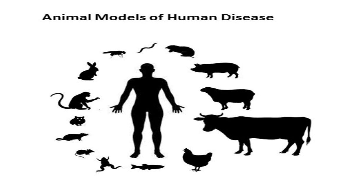
Animal Models of Human Disease
Topic Information
Dear Colleagues,
The use of animal models of human disease is critical for furthering our understanding of disease mechanisms, for the discovery of novel targets for treatment, and for translational research.
This Special Issue aims to collect state-of-the-art primary research studies and review articles from international experts and leading groups using animal models to study human diseases.
Submissions are welcome on a wide range of animal models and pathologies, including infectious disease, acute injury, regeneration, cancer, autoimmunity, and degenerative and chronic disease.
Prof. Dr. Sigrun Lange
Prof. Dr. Jameel M. Inal
Topic Editors
Keywords
- animal models
- human disease
- pathology
- pathobiology
- chronic disease
- acute injury
- regeneration
- infectious disease
- cancer
- autoimmunity
- neurodegenerative disease
- comparative animal models
- extracellular vesicles
- liquid biopsy
- biomarkers
Participating Journals
| Journal Name | Impact Factor | CiteScore | Launched Year | First Decision (median) | APC |
|---|---|---|---|---|---|
|
Biomedicines
|
3.9 | 6.8 | 2013 | 21 Days | CHF 2600 |
|
Cells
|
5.2 | 10.5 | 2012 | 15.5 Days | CHF 2700 |
|
Current Issues in Molecular Biology
|
3.0 | 3.7 | 1999 | 16.3 Days | CHF 2200 |
|
Diagnostics
|
3.3 | 5.9 | 2011 | 21.6 Days | CHF 2600 |
|
Genes
|
2.8 | 5.5 | 2010 | 14.6 Days | CHF 2600 |
|
International Journal of Molecular Sciences
|
4.9 | 9.0 | 2000 | 17.8 Days | CHF 2900 |
|
International Journal of Translational Medicine
|
- | 2.2 | 2021 | 28.2 Days | CHF 1200 |
![]()
Preprints.org is a multidisciplinary platform offering a preprint service designed to facilitate the early sharing of your research. It supports and empowers your research journey from the very beginning.
MDPI Topics is collaborating with Preprints.org and has established a direct connection between MDPI journals and the platform. Authors are encouraged to take advantage of this opportunity by posting their preprints at Preprints.org prior to publication:
- Share your research immediately: disseminate your ideas prior to publication and establish priority for your work.
- Safeguard your intellectual contribution: Protect your ideas with a time-stamped preprint that serves as proof of your research timeline.
- Boost visibility and impact: Increase the reach and influence of your research by making it accessible to a global audience.
- Gain early feedback: Receive valuable input and insights from peers before submitting to a journal.
- Ensure broad indexing: Web of Science (Preprint Citation Index), Google Scholar, Crossref, SHARE, PrePubMed, Scilit and Europe PMC.