


Tomography 2026, 12(6), 82; https://doi.org/10.3390/tomography12060082 (registering DOI) - 1 Jun 2026
Abstract
►
Show Figures
Purpose: To evaluate how often history taking and physical examination are omitted before MRI referral and whether their omission is associated with clinical reasoning quality and MRI diagnostic yield. Materials and Methods: In this prospective study, adults undergoing MRI at a tertiary academic
[...] Read more.
Purpose: To evaluate how often history taking and physical examination are omitted before MRI referral and whether their omission is associated with clinical reasoning quality and MRI diagnostic yield. Materials and Methods: In this prospective study, adults undergoing MRI at a tertiary academic hospital were surveyed before imaging to determine whether the referring clinician had taken their history and performed a physical examination. Multivariable regression was used to assess determinants of omission and associations with clinical reasoning quality (defined as agreement between the suspected diagnosis and MRI findings) and MRI positivity (defined as findings relevant to the indication). Results: Among 275 patients (median age 61 years; 50.0% male), history taking was omitted in 18.2% of cases and physical examination was omitted in 70.9%. History taking was less likely during surveillance than during new/first visits (odds ratio (OR) 0.140, p < 0.001) and more likely when MRI was requested by residents rather than medical specialists (OR 4.645, p = 0.018). Physical examination was more likely when MRI was requested by residents (OR 3.174, p = 0.007) or nurse specialists/physician assistants (OR 3.145, p = 0.033), and less likely during follow-up visits (OR 0.183, p < 0.001) and surveillance visits (OR 0.061, p < 0.001). Omission of physical examination was not associated with clinical reasoning quality (p = 0.370). Neither omission of history taking nor omission of physical examination was associated with MRI positivity (p = 0.430 and p = 0.286, respectively). Conclusions: History taking and physical examination were often omitted before MRI referral. Although no statistically significant association was observed between omission of bedside assessment and clinical reasoning quality or MRI positivity, reduced bedside assessment may limit the clinical context informing referral and interpretation.
Full article
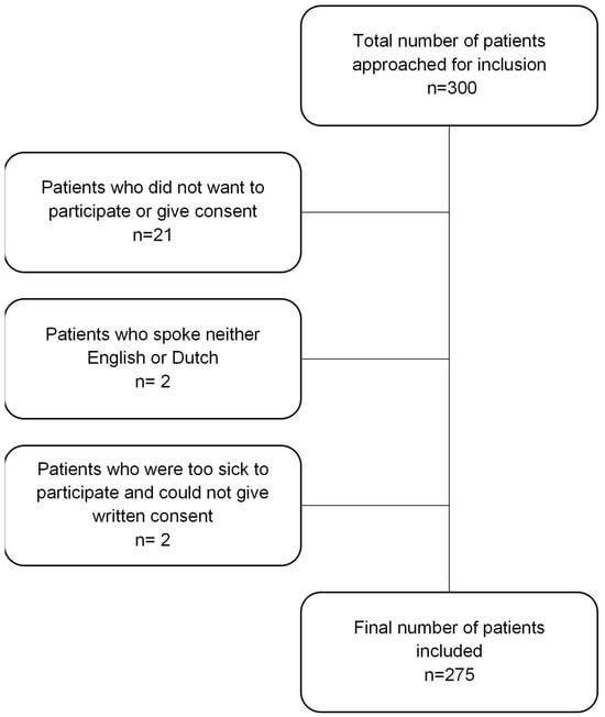
Figure 1


