New Insights into the Molecular Mechanisms of Neurodegeneration

A topical collection in Cells (ISSN 2073-4409). This collection belongs to the section "Cells of the Nervous System".

Viewed by 21861

Editors


E-Mail Website
Collection Editor
Department of Pharmacological and Biomolecular Sciences, Università degli Studi di Milano, Milan, Italy
Interests: adhesion molecules; cellular mechanobiology; mitochondrial dynamics; vesicular trafficking; islet of Langerhans; beta cells differentiation; diabetes mellitus; pancreatic tumors; glia–neuron interactions
Special Issues, Collections and Topics in MDPI journals

E-Mail Website
Collection Editor
Lab of Applied Biology, Dept. of Pharmacological and Biomolecular Sciences, Università degli Studi di Milano, Via Balzaretti, 9-20133 Milan, Italy
Interests: molecular mechanisms of neurodegeneration; motor neuron diseases; polyglutamine diseases; autophagy; lysosomal damage; protein quality control

E-Mail Website
Collection Editor
Lab of Applied Biology, Dept. of Pharmacological and Biomolecular Sciences, Università degli Studi di Milano, Via Balzaretti, 9-20133 Milan, Italy
Interests: molecular mechanisms of neurodegeneration; motor neuron diseases; amyotrophic lateral sclerosis; SBMA; neuromuscular diseases; myogenesis; neural differentiation

Topical Collection Information

Dear Colleagues,

Neurodegenerative disorders refer to a group of diseases characterized by the progressive dysfunction and death of selective neuronal populations of the central and peripheral nervous system.

Despite decades of research in this field, there is still no effective treatments to halt the neurodegenerative processes. The dissection of novel molecular mechanisms underlying the pathological processes is mandatory to identify promising therapeutic strategies that could prevent or modify the progression of these disorders.

While the neuronal populations affected and therefore the resulting clinical manifestations are different, neurodegenerative diseases often share common pathogenetic mechanisms. The abnormal aggregation of intracellular or extracellular proteins is the most common example. Protein aggregation triggers cellular homeostatic mechanisms aimed at coping with the specific defect and, if this approach fails, signalling pathways, ultimately leading to neuronal degeneration, are activated.

Therefore, the description of the early alterations that occur in neurons will be helpful in developing disease-specific therapies, while focusing on shared mechanisms, we will be able to identify targets that can be therapeutically exploited to deal with multiple pathologies.

This collection is interested in original works and reviews that provide new insights into the molecular mechanisms of neurodegeneration, revealing candidate biomarkers or therapeutic targets.

Dr. Carla Perego
Prof. Paola Rusmini
Prof. Dr. Mariarita Galbiati
Collection Editors

Manuscript Submission Information

Manuscripts should be submitted online at www.mdpi.com by registering and logging in to this website. Once you are registered, click here to go to the submission form. Manuscripts can be submitted until the deadline. All submissions that pass pre-check are peer-reviewed. Accepted papers will be published continuously in the journal (as soon as accepted) and will be listed together on the collection website. Research articles, review articles as well as short communications are invited. For planned papers, a title and short abstract (about 100 words) can be sent to the Editorial Office for announcement on this website.

Submitted manuscripts should not have been published previously, nor be under consideration for publication elsewhere (except conference proceedings papers). All manuscripts are thoroughly refereed through a single-blind peer-review process. A guide for authors and other relevant information for submission of manuscripts is available on the Instructions for Authors page. Cells is an international peer-reviewed open access semimonthly journal published by MDPI.

Please visit the Instructions for Authors page before submitting a manuscript. The Article Processing Charge (APC) for publication in this open access journal is 2700 CHF (Swiss Francs). Submitted papers should be well formatted and use good English. Authors may use MDPI's English editing service prior to publication or during author revisions.

Keywords

  • neurodegeneration
  • proteostasis
  • unfolded protein response
  • autophagy
  • lysosome homeostasis and dysfunction
  • mitochondria dynamics
  • mitochondrial quality control
  • mitochondria-associated membranes (MAMs)
  • neuromuscular junction
  • mRNA splicing
  • miRNA
  • Alzheimer’s disease
  • Parkinson’s disease
  • amyotrophic lateral sclerosis
  • polyglutamine diseases

Published Papers (6 papers)

2023

Jump to: 2022, 2021

13 pages, 6415 KiB  
Article
Genetic Deletion of Thorase Causes Purkinje Cell Loss and Impaired Motor Coordination Behavior
by Chao Li, Han Zhang, Kexin Tong, Menghua Cai, Fei Gao, Jia Yang, Yi Xu, Huaishan Wang, Hui Chen, Yu Hu, Wei He and Jianmin Zhang
Cells 2023, 12(16), 2032; https://doi.org/10.3390/cells12162032 - 10 Aug 2023
Viewed by 1252
Abstract
Thorase belongs to the AAA+ ATPase family, which plays a critical role in maintaining cellular homeostasis. Our previous work reported that Thorase was highly expressed in brain tissue, especially in the cerebellum. However, the roles of Thorase in the cerebellum have still not [...] Read more.
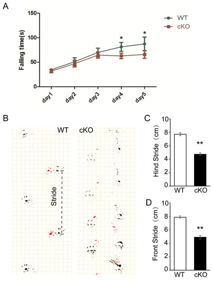
Thorase belongs to the AAA+ ATPase family, which plays a critical role in maintaining cellular homeostasis. Our previous work reported that Thorase was highly expressed in brain tissue, especially in the cerebellum. However, the roles of Thorase in the cerebellum have still not been characterized. In this study, we generated conditional knockout mice (cKO) with Thorase deletion in Purkinje cells. Thorase cKO mice exhibited cerebellar degenerative diseases-like behavior and significant impairment in motor coordination. Thorase deletion resulted in more Purkinje neuron apoptosis, leading to Purkinje cell loss in the cerebellum of Thorase cKO mice. We also found enhanced expression of the inflammatory protein ASC, IL-1β, IL-6 and TNF-α in the Thorase cKO cerebellum, which contributed to the pathogenesis of cerebellar degenerative disease. Our findings provide a better understanding of the role of Thorase in the cerebellum, which is a theoretical basis for Thorase as a therapeutic drug target for neurodegenerative diseases. Full article
Show Figures

Figure 1

2022

Jump to: 2023, 2021

16 pages, 2688 KiB  
Article
Paclitaxel Inhibits KCNQ Channels in Primary Sensory Neurons to Initiate the Development of Painful Peripheral Neuropathy
by Zizhen Wu, Gabor Toro, Guoying Xu, Danny Dang, Charmaine Prater and Qing Yang
Cells 2022, 11(24), 4067; https://doi.org/10.3390/cells11244067 - 15 Dec 2022
Cited by 5 | Viewed by 1857
Abstract
Cancer patients undergoing paclitaxel infusion usually experience peripheral nerve degeneration and serious neuropathic pain termed paclitaxel-induced peripheral neuropathy (PIPN). However, alterations in the dose or treatment schedule for paclitaxel do not eliminate PIPN, and no therapies are available for PIPN, despite numerous studies [...] Read more.
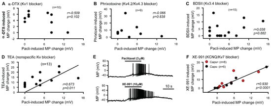
Cancer patients undergoing paclitaxel infusion usually experience peripheral nerve degeneration and serious neuropathic pain termed paclitaxel-induced peripheral neuropathy (PIPN). However, alterations in the dose or treatment schedule for paclitaxel do not eliminate PIPN, and no therapies are available for PIPN, despite numerous studies to uncover the mechanisms underlying the development/maintenance of this condition. Therefore, we aimed to uncover a novel mechanism underlying the pathogenesis of PIPN. Clinical studies suggest that acute over excitation of primary sensory neurons is linked to the pathogenesis of PIPN. We found that paclitaxel-induced acute hyperexcitability of primary sensory neurons results from the paclitaxel-induced inhibition of KCNQ potassium channels (mainly KCNQ2), found abundantly in sensory neurons and axons. We found that repeated application of XE-991, a specific KCNQ channel blocker, induced PIPN-like alterations in rats, including mechanical hypersensitivity and degeneration of peripheral nerves, as detected by both morphological and behavioral assays. In contrast, genetic deletion of KCNQ2 from peripheral sensory neurons in mice significantly attenuated the development of paclitaxel-induced peripheral sensory fiber degeneration and chronic pain. These findings may lead to a better understanding of the causes of PIPN and provide an impetus for developing new classes of KCNQ activators for its therapeutic treatment. Full article
Show Figures

Figure 1

29 pages, 4564 KiB  
Article
Aberrant DNA and RNA Methylation Occur in Spinal Cord and Skeletal Muscle of Human SOD1 Mouse Models of ALS and in Human ALS: Targeting DNA Methylation Is Therapeutic
by Lee J. Martin, Danya A. Adams, Mark V. Niedzwiecki and Margaret Wong
Cells 2022, 11(21), 3448; https://doi.org/10.3390/cells11213448 - 31 Oct 2022
Cited by 14 | Viewed by 3379
Abstract
Amyotrophic lateral sclerosis (ALS) is a fatal disease. Skeletal muscles and motor neurons (MNs) degenerate. ALS is a complex disease involving many genes in multiple tissues, the environment, cellular metabolism, and lifestyles. We hypothesized that epigenetic anomalies in DNA and RNA occur in [...] Read more.
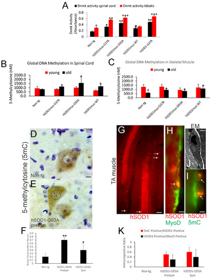
Amyotrophic lateral sclerosis (ALS) is a fatal disease. Skeletal muscles and motor neurons (MNs) degenerate. ALS is a complex disease involving many genes in multiple tissues, the environment, cellular metabolism, and lifestyles. We hypothesized that epigenetic anomalies in DNA and RNA occur in ALS and examined this idea in: (1) mouse models of ALS, (2) human ALS, and (3) mouse ALS with therapeutic targeting of DNA methylation. Human superoxide dismutase-1 (hSOD1) transgenic (tg) mice were used. They expressed nonconditionally wildtype (WT) and the G93A and G37R mutant variants or skeletal muscle-restricted WT and G93A and G37R mutated forms. Age-matched non-tg mice were controls. hSOD1 mutant mice had increased DNA methyltransferase enzyme activity in spinal cord and skeletal muscle and increased 5-methylcytosine (5mC) levels. Genome-wide promoter CpG DNA methylation profiling in skeletal muscle of ALS mice identified hypermethylation notably in cytoskeletal genes. 5mC accumulated in spinal cord MNs and skeletal muscle satellite cells in mice. Significant increases in DNA methyltransferase-1 (DNMT1) and DNA methyltransferase-3A (DNMT3A) levels occurred in spinal cord nuclear and chromatin bound extracts of the different hSOD1 mouse lines. Mutant hSOD1 interacted with DNMT3A in skeletal muscle. 6-methyladenosine (6mA) RNA methylation was markedly increased or decreased in mouse spinal cord depending on hSOD1-G93A model, while fat mass and obesity associated protein was depleted and methyltransferase-like protein 3 was increased in spinal cord and skeletal muscle. Human ALS spinal cord had increased numbers of MNs and interneurons with nuclear 5mC, motor cortex had increased 5mC-positive neurons, while 6mA was severely depleted. Treatment of hSOD1-G93A mice with DNMT inhibitor improved motor function and extended lifespan by 25%. We conclude that DNA and RNA epigenetic anomalies are prominent in mouse and human ALS and are potentially targetable for disease-modifying therapeutics. Full article
Show Figures

Figure 1

2021

Jump to: 2023, 2022

17 pages, 2616 KiB  
Article
Proteogenomics Reveals Orthologous Alternatively Spliced Proteoforms in the Same Human and Mouse Brain Regions with Differential Abundance in an Alzheimer’s Disease Mouse Model
by Esdras Matheus Gomes da Silva, Letícia Graziela Costa Santos, Flávia Santiago de Oliveira, Flávia Cristina de Paula Freitas, Vinícius da Silva Coutinho Parreira, Hellen Geremias dos Santos, Raphael Tavares, Paulo Costa Carvalho, Ana Gisele da Costa Neves-Ferreira, Andrea Siqueira Haibara, Patrícia Savio de Araujo-Souza, Adriana Abalen Martins Dias and Fabio Passetti
Cells 2021, 10(7), 1583; https://doi.org/10.3390/cells10071583 - 23 Jun 2021
Cited by 3 | Viewed by 3421
Abstract
Alternative splicing (AS) may increase the number of proteoforms produced by a gene. Alzheimer’s disease (AD) is a neurodegenerative disease with well-characterized AS proteoforms. In this study, we used a proteogenomics strategy to build a customized protein sequence database and identify orthologous AS [...] Read more.
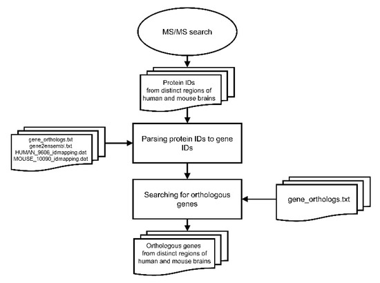
Alternative splicing (AS) may increase the number of proteoforms produced by a gene. Alzheimer’s disease (AD) is a neurodegenerative disease with well-characterized AS proteoforms. In this study, we used a proteogenomics strategy to build a customized protein sequence database and identify orthologous AS proteoforms between humans and mice on publicly available shotgun proteomics (MS/MS) data of the corpus callosum (CC) and olfactory bulb (OB). Identical proteotypic peptides of six orthologous AS proteoforms were found in both species: PKM1 (gene PKM/Pkm), STXBP1a (gene STXBP1/Stxbp1), Isoform 3 (gene HNRNPK/Hnrnpk), LCRMP-1 (gene CRMP1/Crmp1), SP3 (gene CADM1/Cadm1), and PKCβII (gene PRKCB/Prkcb). These AS variants were also detected at the transcript level by publicly available RNA-Seq data and experimentally validated by RT-qPCR. Additionally, PKM1 and STXBP1a were detected at higher abundances in a publicly available MS/MS dataset of the AD mouse model APP/PS1 than its wild type. These data corroborate other reports, which suggest that PKM1 and STXBP1a AS proteoforms might play a role in amyloid-like aggregate formation. To the best of our knowledge, this report is the first to describe PKM1 and STXBP1a overexpression in the OB of an AD mouse model. We hope that our strategy may be of use in future human neurodegenerative studies using mouse models. Full article
Show Figures

Figure 1

18 pages, 2156 KiB  
Review
“Let Food Be Thy Medicine”: Gluten and Potential Role in Neurodegeneration
by Aaron Lerner and Carina Benzvi
Cells 2021, 10(4), 756; https://doi.org/10.3390/cells10040756 - 30 Mar 2021
Cited by 19 | Viewed by 6501
Abstract
Wheat is a most favored staple food worldwide and its major protein is gluten. It is involved in several gluten dependent diseases and lately was suggested to play a role in non-celiac autoimmune diseases. Its involvement in neurodegenerative conditions was recently suggested but [...] Read more.
Wheat is a most favored staple food worldwide and its major protein is gluten. It is involved in several gluten dependent diseases and lately was suggested to play a role in non-celiac autoimmune diseases. Its involvement in neurodegenerative conditions was recently suggested but no cause-and-effect relationship were established. The present narrative review expands on various aspects of the gluten-gut-brain axes events, mechanisms and pathways that connect wheat and gluten consumption to neurodegenerative disease. Gluten induced dysbiosis, increased intestinal permeabillity, enteric and systemic side effects, cross-reactive antibodies, and the sequence of homologies between brain antigens and gluten are highlighted. This combination may suggest molecular mimicry, alluding to some autoimmune aspects between gluten and neurodegenerative disease. The proverb of Hippocrates coined in 400 BC, “let food be thy medicine,” is critically discussed in the frame of gluten and potential neurodegeneration evolvement. Full article
Show Figures

Figure 1

17 pages, 29587 KiB  
Article
Oxidative Stress Conditions Result in Trapping of PHF-Core Tau (297–391) Intermediates
by Mahmoud B. Maina, Youssra K. Al-Hilaly, Gunasekhar Burra, Janet E. Rickard, Charles R. Harrington, Claude M. Wischik and Louise C. Serpell
Cells 2021, 10(3), 703; https://doi.org/10.3390/cells10030703 - 22 Mar 2021
Cited by 8 | Viewed by 4257
Abstract
The self-assembly of tau into paired helical filaments (PHFs) in neurofibrillary tangles (NFTs) is a significant event in Alzheimer’s disease (AD) pathogenesis. Numerous post-translational modifications enhance or inhibit tau assembly into NFTs. Oxidative stress, which accompanies AD, induces multiple post-translational modifications in proteins, [...] Read more.
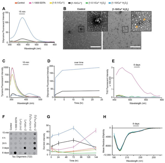
The self-assembly of tau into paired helical filaments (PHFs) in neurofibrillary tangles (NFTs) is a significant event in Alzheimer’s disease (AD) pathogenesis. Numerous post-translational modifications enhance or inhibit tau assembly into NFTs. Oxidative stress, which accompanies AD, induces multiple post-translational modifications in proteins, including the formation of dityrosine (DiY) cross-links. Previous studies have revealed that metal-catalysed oxidation (MCO) using Cu2+ and H2O2 leads to the formation of DiY cross-links in two misfolding proteins, Aβ and α-synuclein, associated with AD and Parkinson’s disease respectively. The effect of MCO on tau remains unknown. Here, we examined the effect of MCO and ultra-violet oxidation to study the influence of DiY cross-linking on the self-assembly of the PHF-core tau fragment. We report that DiY cross-linking facilitates tau assembly into tau oligomers that fail to bind thioflavin S, lack β-sheet structure and prevents their elongation into filaments. At a higher concentration, Cu2+ (without H2O2) also facilitates the formation of these tau oligomers. The DiY cross-linked tau oligomers do not cause cell death. Our findings suggest that DiY cross-linking of pre-assembled tau promotes the formation of soluble tau oligomers that show no acute impact on cell viability. Full article
Show Figures

Figure 1

Back to TopTop