
- 5.2Impact Factor
- 10.5CiteScore
- 16 daysTime to First Decision
Regulation of Cell Division
This special issue belongs to the section “Cell Proliferation and Division“.
Special Issue Information
Dear Colleagues,
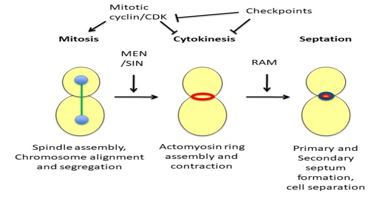
Cell division requires the coordination of chromosome segregation and cytokinesis. Since the discovery of cyclin-dependent kinase (CDK) in yeasts, research on many types of cells has led to insights on how mitosis, cytokinesis, and abscission are regulated. How cells trigger the assembly of the mitotic spindle, prevent anaphase and achieve the correct attachment of chromosomes to microtubules, prevent premature actomyosin ring contraction, and resolve intracellular bridges are essential questions in cell biology that have implications for human health.
This Special Issue will focus on the regulation of cell division. Topics will include regulatory pathways, mitotic checkpoints, and the regulation of actomyosin ring assembly, contraction, and abscission.
Dr. Katie Shannon
Guest Editor
Manuscript Submission Information
Manuscripts should be submitted online at www.mdpi.com by registering and logging in to this website. Once you are registered, click here to go to the submission form. Manuscripts can be submitted until the deadline. All submissions that pass pre-check are peer-reviewed. Accepted papers will be published continuously in the journal (as soon as accepted) and will be listed together on the special issue website. Research articles, review articles as well as short communications are invited. For planned papers, a title and short abstract (about 250 words) can be sent to the Editorial Office for assessment.
Submitted manuscripts should not have been published previously, nor be under consideration for publication elsewhere (except conference proceedings papers). All manuscripts are thoroughly refereed through a single-blind peer-review process. A guide for authors and other relevant information for submission of manuscripts is available on the Instructions for Authors page. Cells is an international peer-reviewed open access semimonthly journal published by MDPI.
Please visit the Instructions for Authors page before submitting a manuscript. The Article Processing Charge (APC) for publication in this open access journal is 2700 CHF (Swiss Francs). Submitted papers should be well formatted and use good English. Authors may use MDPI's English editing service prior to publication or during author revisions.
Keywords
- mitosis
- kinetochore
- chromosome segregation
- actomyosin ring
- cytokinesis
- septation
- mitotic exit network
- spindle assembly checkpoint
Benefits of Publishing in a Special Issue
- Ease of navigation: Grouping papers by topic helps scholars navigate broad scope journals more efficiently.
- Greater discoverability: Special Issues support the reach and impact of scientific research. Articles in Special Issues are more discoverable and cited more frequently.
- Expansion of research network: Special Issues facilitate connections among authors, fostering scientific collaborations.
- External promotion: Articles in Special Issues are often promoted through the journal's social media, increasing their visibility.
- e-Book format: Special Issues with more than 10 articles can be published as dedicated e-books, ensuring wide and rapid dissemination.
Published Papers
Get Alerted
Add your email address to receive forthcoming issues of this journal.