Article

Mycn Is Essential for Pubertal Mammary Gland Development and Promotes the Activation of Bcl11b-Maintained Quiescent Stem Cells

1
Westlake Laboratory of Life Sciences and Biomedicine, School of Life Sciences, Westlake University, Hangzhou 310024, China
2
Westlake Institute for Advanced Study, Fudan University, Hangzhou 310024, China
3
Westlake Disease Modeling Lab, Westlake Laboratory of Life Sciences and Biomedicine, Hangzhou 310024, China
4
Key Laboratory of Growth Regulation and Translational Research of Zhejiang Province, School of Life Sciences, Westlake University, Hangzhou 310024, China
*
Author to whom correspondence should be addressed.
Cells 2025, 14(16), 1239; https://doi.org/10.3390/cells14161239
Submission received: 6 July 2025 / Revised: 4 August 2025 / Accepted: 7 August 2025 / Published: 12 August 2025
(This article belongs to the Special Issue Regulation of Cell Division)

Abstract

This investigation examines the function of the mouse Mycn gene in regulating and activating quiescent mammary stem cells, which are vital for mammary gland development. The mammary gland, consisting of luminal and basal cells, progresses through complex developmental stages from embryonic development through puberty, adulthood, pregnancy, lactation, and involution. Quiescent stem cells, existing in a reversible non-proliferative state, are essential for gland maintenance, yet their activation mechanisms remain poorly understood. Mycn, a member of the Myc/MYC oncogene family, is recognized for its roles in embryonic development and cancer, notably aggressive neuroblastoma and triple-negative breast cancer. Through single-cell RNA sequencing (scRNA-seq), CRISPR knockout, and overexpression experiments, this study demonstrates that Mycn is highly enriched in the terminal end buds (TEBs) of the pubertal mammary gland, particularly in basal cells, and is critical for ductal development. Both deletion and overexpression of Mycn diminish the stemness and regenerative capacity of mammary stem cells. Mycn enhances cell proliferation while downregulating quiescent stem cell markers and regulators, including Bcl11b and Tspan8, affecting stem cell maintenance and differentiation. This research clarifies the regulatory role of Bcl11b in controlling Tspan8 expression and demonstrates that Mycn indirectly targets both under normal conditions. Maintaining appropriate levels of Mycn expression is essential for normal development and cancer prevention. These insights contribute to understanding diseases and aggressive cancers, including triple-negative breast cancer (TNBC), and suggest potential therapeutic approaches.

1. Introduction

Cancer treatment has evolved significantly through three main periods [1]: The first period is pre-1970, when traditional modalities established the foundation of cancer therapy. This included surgery, radiation therapy, chemotherapy, and bone marrow transplantation, which formed the cornerstone of oncological interventions for decades. The second period spans from 1970 to 2023, during which molecular understanding revolutionized treatment approach development. This era saw the clinical use of Tamoxifen, monoclonal antibodies, immune checkpoint inhibitors, and CAR-T therapy, all representing significant advances based on deeper comprehension of cancer biology. Future directions include new approaches, such as advanced immunotherapies and personalized cancer vaccines. CRISPR-mediated gene therapy, as well as AI-assisted diagnosis and treatment planning, also show great promise for improving cancer management in coming years [1].
Despite these significant advances in cancer treatment, certain types, including triple-negative breast cancer, remain difficult to cure, primarily due to the lack of targetable receptors and high molecular heterogeneity, which limit the effectiveness of current precision medicine approaches [2,3,4,5].
Despite significant advances in triple-negative breast cancer research in recent years, we have yet to fully overcome this challenging disease. At its core, this limitation reflects our insufficient understanding of how the regulatory networks that govern normal breast tissue developmental become dysregulated, as well as the molecular mechanisms underlying tumor initiation and progression. Elucidating the regulatory functions of key transcription factors such as Mycn in these processes may provide a breakthrough for developing novel therapeutic strategies [6,7,8,9,10].
In this study, we employ the mouse as an experimental model. The mammary gland is a specialized organ in mammals for milk production and neonatal nourishment [11]. The development of the mammary gland in mice encompasses a complex process that begins during the embryonic stages and continues through puberty, adulthood, pregnancy, lactation, and involution, cycling repeatedly [11].
The mammary gland primarily comprises two main cell types: an inner layer of luminal cells and an outer layer of elongated, contractile basal cells [11,12,13]. Basal cells are myoepithelial cells characterized by the expression of basal keratin 5 (K5) and keratin 14 (K14), possessing contractile properties and functioning to define the basal membrane and encapsulate the ductal-facing luminal cells. Luminal cells, on the other hand, express keratin 8 (K8) and keratin 18 (K18) [11,14]. These cells exhibit polarity and can be further clarified into estrogen receptor (ESR1 or ER)-positive and -negative ductal cells and alveolar cells, which facilitate milk production [11,15,16].
During embryonic development, mammary epithelial cells consist of a distinct population of undifferentiated and highly plastic progenitor cells that ultimately generate all postnatal mammary epithelial cells [17,18,19]. The postnatal mammary gland maintains the ability for rapid ductal expansion during puberty. At puberty onset, bulb-shaped structures called the terminal end buds (TEBs) form at the tips of growing epithelial ducts and drive ductal elongation, invasion, and branching of the mammary trees [11,20,21,22]. TEBs comprise two cell types: cap cells, which serve as progenitors of myoepithelial cells, and body cells, which function as progenitors of luminal cells in the subtending ducts [11,23,24,25,26,27,28]. When TEBs reach the fat pad edges, they regress. Upon completion of puberty, TEBs dissolve, establishing the mature adult mammary gland. This gland comprises an epithelial framework, which provides the foundation for the development of alveoli-like bud development during the estrus cycle and alveoli formation during pregnancy [11,28,29].
The dynamic changes in mammary epithelial cell proliferation, differentiation, and turnover during the process of mammary gland development and multiple pregnancies suggest the existence of mammary stem cells [11]. The mammary fat pad transplantation assay represents a gold-standard technique for studying mammary epithelial organogenesis and is visualized through carmine-alum whole-mount staining or bioluminescence imaging [30,31]. The implementation of classical mammary stem cell transplantation assays and lineage tracing analysis in model animals has demonstrated the presence of undifferentiated stem cells in the postnatal mammary organ [32,33].
In recent years, researchers have identified several markers to characterize mammary stem cells, including s-SHIP+ [28], Axin2+ [34], Cd1d+ [35], Procr+ [36], Bcl11bhigh [12], Lgr5+Tspan8high [13], and Dll1high [37]. These markers identify distinct types of stem cells that serve crucial roles in mammary gland development, homeostasis, and regeneration. The presence of these diverse stem cells demonstrates the heterogeneity within stem cell populations in the mammary gland system. This research primarily focuses on quiescent mammary stem cells and their biological functions.
Quiescence refers to a reversible state of the cell cycle, typically occurring in the G0 phase, though it occasionally occurs in the G2 phase [38,39]. While non-proliferation characterizes both terminally differentiated and senescent cells, only quiescent cells retain the capacity to rejoin the cell cycle under appropriate physiological conditions [40]. This quiescent state proves crucial for the longevity of proliferative cells, as it helps prevent the depletion of their proliferative capacity and reduces the risk of accumulating damage to DNA, proteins, and mitochondria, which could lead to malignant transformation or senescence [40].
Dysregulated exit from quiescence has been linked to hyperplasia and tumorigenesis, highlighting the importance of proper quiescence regulation [41,42,43]. The transcription factor Bcl11b has emerged as a critical regulator of this process in mammary tissue, functioning as a molecular switch between normal cellular aging and tumorigenesis [41]. Studies have demonstrated that Bcl11b deficiency accelerates the aging phenotype, characterized by upregulation of age-related signaling pathways (including MAPK, PI3K-Akt, NF-κB, and p53), enhanced cellular senescence, and activation of the senescence-associated secretory phenotype (SASP). Notably, Bcl11b loss leads to distinct early and late senescence states (e-Sen and l-Sen), with l-Sen cells exhibiting heightened NF-κB activity and increased susceptibility to malignant transformation. Mechanistically, Bcl11b directly suppresses NF-κB signaling and other aging-associated pathways, serving as a protective factor against premature senescence. The therapeutic relevance of this regulatory axis is underscored by experiments showing that inhibition of NF-κB/Jak-Stat signaling with TPCA-1 can rescue the aging phenotype and significantly reduce tumor incidence in Bcl11b-deficient models. These findings establish Bcl11b as a crucial determinant of mammary cell fate, governing the balance between quiescence, senescence, and neoplastic transformation, and suggest that targeting senescence programs may offer preventive strategies for age-related breast cancer [41].
Recent research has identified Bcl11bhigh [12], Lgr5+Tspan8high [13], and Nfatc1+ [44] basal cells as quiescent stem cells. Studies indicate that the Bcl11bhigh quiescent mammary epithelial cell population expresses elevated levels of Bcl11b and resides at the interface between luminal and basal cells, while the Lgr5+Tspan8high mammary stem cells represents a deeply quiescent subset that resides within the proximal region [13]. The dormant Nfatc1+ basal stem cells exhibit distinct characteristics from other known mammary stem cells [44]. Nevertheless, they all serve essential roles in long-term maintenance of the mammary gland.
The molecular mechanisms governing mammary stem cell activation from quiescence remain incompletely understood. Previous research identified Foxp1 as a direct repressor of Tspan8 in basal cells, potentially regulating mammary stem cell dormancy exit during differentiation and development [45]. In contrast to Bcl11b [12] or Foxp1 [45], Zeb1 distinctively promotes self-renewal in a specific subpopulation of basal stem cells while inhibiting their proliferation, as documented by the author [46]. Given the heterogeneous nature of mammary stem cells, the precise mechanism activating Bcl11bhigh quiescent stem cells remains unclear. Our recent research has identified Mycn, a cell proliferation regulator, as a crucial factor controlling the exit from mammary stem cell quiescence.
The discovery and understanding of the mouse Mycn gene in mice originated from the study of the human MYCN gene. MYCN belongs to the MYC oncogene family, which consists of three members (MYC, MYCN, and MYCL) and was first identified in neuroblastoma [47,48,49].
The MYC family serves a fundamental role in cellular processes and frequently contributes to cancer development. MYC proteins function as primary drivers of tumorigenesis, with dysregulated levels observed in numerous human tumors [50,51,52,53,54,55,56,57,58,59,60,61,62,63]. MYCN, encoded by the MYCN gene, plays an essential role in embryonic development, particularly in regulating growth, proliferation, and metabolism of progenitor cells across various developing organs and tissues [48,49,64]. Research demonstrates that the protein encoded by MYCN binds to specific promoters, influencing the expression of crucial target genes involved in multiple cellular functions, consistent with studies in mammalian cells [65]. Its dysregulation, particularly overexpression, correlates with various cancers, notably neuroblastoma, a pediatric cancer originating from immature nerve cells [66]. These findings underscore the importance of MYCN in regulating cellular processes that can become dysregulated in cancer.
The MYC family genes demonstrate crucial functions in stem cell biology. N-MYC (MYCN) and C-MYC have been identified as essential regulators of both neural and hematopoietic stem cell quiescence and activation, including proliferation, differentiation, and long-term self-renewal activity [67,68,69,70,71,72,73,74,75,76]. Despite the complex nature of MYC family regulation and function, their significance in cancer and cell biology has established them as central subjects in biological and oncological research. Ongoing efforts to understand and target the Myc family, particularly N-myc (Mycn in mouse), continue to enhance our understanding of cancer biology and facilitate novel therapeutic approaches.
Research indicates that the MYC family, including MYCN, correlates with breast cancer tumorigenesis [55,77,78,79,80,81], particularly in triple-negative breast cancer (TNBC) [82,83,84,85,86]. While the mechanisms of MYC in promoting breast cancer, including TNBC, have been extensively investigated, its role in normal mammary gland development requires further elucidation. Based on our research interests and preliminary findings, this study focuses primarily on examining mouse Mycn’s role in maintaining mammary gland homeostasis through the regulation of quiescent mammary stem cell activation.

2. Materials and Methods

2.1. Mice

The Bcl11bflox/flox mice (C57BL/6 background) were generously provided by Mark Leid’s lab and were described previously [87]. The Krt14-Cre (stock number 018964), mTmG (stock number 007676) mice were purchased from Jackson Laboratory. For analysis of pubertal and adult mammary glands, 5~6-week-old and 2~6-month-old female mice were used, respectively. For each experiment, the ages of the female mice used were indicated in the text. In addition, for the Bcl11b KO assay, the mice had a genotype of Krt14-Cre (+) mTmG (+, +) with or without a homogeneous Bcl11b (loxp, loxp) background for Bcl11b deletion. All mice were housed in specific pathogen-free conditions and bred at the Westlake University Laboratory Animal Resources Center. All animal experiments were approved by the local institutional animal ethics board (permission number: 24-149-CS-3).

2.2. Tissue Processing and Flow Cytometry

Mammary glands from pubertal (5–6 weeks) and adult (>2 months) virgin or pregnant C57BL/6 mice were dissected and processed according to the published protocol [12].

2.3. Colony Formation Assay

To perform the colony formation assay, 30 μL of growth factor reduced Matrigel (BD Bioscience, San Jose, CA, USA) was added into the 96-well round-bottom plate and then solidified at 37 °C for 10 min. A total of 4K/well cells were resuspended in 200 μL culture media (DMEM/F12 + 2% FBS+ 1%P/S + B27 + 10 mM HEPES) supplemented with EGF (10 ng/mL, BD Bioscience, San Jose, CA, USA), Rspo1 (250 ng/mL, R&D, Hong Kong SAR, China), and ROCK inhibitor Y27632 (10 μm, Sigma, Hong Kong SAR, China) and were overlaid on top of the matrigel. The plate was maintained in a 37 °C incubator with 5% CO2 for 1–2 weeks.

2.4. Transplantation Assay

Transplantation assays were performed as previously described [12]. The cells were sorted and then resuspended in injection media (DMEM/F12 + 50% Matrigel + 1% P/S) at a concentration of 50,000 cells/5 μL, followed by serial dilutions to 10,000 cells/5 μL, 1000 cells/5 μL, 200 cells/5 μL, and 50 cells/5 μL. The cells were then injected into the cleared fat pads of 3-week-old recipient mice (C57BL/6) at the specified dilutions. After six to eight weeks post-transplantation, the recipient mice were analyzed through whole-mount staining of the mammary glands with carmine. The frequency and confidence interval of MRUs (mammary repopulating units) were calculated using ELDA (http://bioinf.wehi.edu.au/software/elda/ (accessed on 15 October 2024)).

2.5. Real-Time PCR

Between 50 and 500 primary mammary cells or cultured epithelial cells were directly sorted into 400 μL Trizol (Life Technologies, Van Allen Way, Carlsbad, CA, USA). The detailed method was conducted as previously described [12]. Data analysis was performed using Graphad 8 and Excel with normalization to the expression of β-actin. The value of one reference sample was set to 1 to enable inter-sample comparisons. Statistical analysis employed two-tailed unpaired t-test. Data are presented as mean ± SEM. The SYBR Green primers are listed below:
  • β-Actin: Forward ACCTTCTACAATGAGCTGCG, Reverse CTGGATGGCTACGTACATGG;
  • Bcl11b: Forward AGGAGAGTATCTGAGCCAGTG, Reverse GTTGTGCAAATGTAGCTGGAAG;
  • Tspan8: Forward AGTTCCGTTTACCCAAAGACC, Reverse GCACCATAGAAAACACCAAACC;
  • Mycn: Forward GTCTGTTCCAGCTACTGCC, Reverse TCCTCTTCATCTTCCTCCTCG.

2.6. Paraffin Sections and Immunofluorescence

The method has been previously described [12].

2.7. Carmine Stain (Whole Mount)

The method has been previously described [12].

2.8. Mycn-Overexpressing Cell Line

Primary mammary basal cells from 5–6-week-old C57BL/6 mice were collected in Trizol (Life Technologies, Van Allen Way, Carlsbad, CA, USA), and RNA was extracted following the standard protocol for reverse transcription to obtain cDNA using Superscript III First Strand Synthesis kit (Life Technologies, Van Allen Way, Carlsbad, CA, USA) according to the manufacturer’s instructions. The Mycn (gene ID:18109) coding sequence was amplified from cDNA and subsequently cloned into pCDH plasmid (72484) as an overexpression construct. Lentivirus production involved co-transfecting 293T cells with the overexpression construct, pCMV-dR8.2 plasmid, and pCMV-VSVG plasmid at a ratio of 10:9:1. Viral supernatants were collected at 48 h and 72 h, then concentrated through ultracentrifugation at 20,000 rpm for 2 h at 4 °C. Viral titration was performed by infecting 293T cells with gradient virus for titer calculation. Successfully infected cells were isolated by FACS based on GFP expression.

2.9. CRISPR-Mediated Mycn Knockout Primary Basal Cells

To verify the function of the candidate gene, knockout cell lines were generated using the CRISPR-Cas9 system. gRNAs for Mycn knockout were designed using an online CRISPR Design Tool http://chopchop.cbu.uib.no/ (accessed on 10 October 2023). gRNA sequences were cloned into lentiCRISPRV2 plasmid (Addgene#52961) by BsmBI. The lentiCRISPRV2 plasmid containing Cas9 sequence and gRNA scaffold was packaged into lentivirus for primary mammary basal cells infection as described previously. The cells were selected using Puromycin (2 μg/mL) following lentivirus infection. Selected mammary basal cells were cultured on the cell culture plates coated with growth factor-reduced Matrigel Matrix (Corning) for colony formation. Individual colonies were selected under microscope and cultured for expansion. Indels were analyzed using the online tool TIDE (https://tide.nki.nl/, accessed on 10 October 2023, using R version 3.3.2.). After individual colonies were picked, they were expanded through further culture and subjected to sequencing, as well as Western blotting, to confirm successful Mycn knockout.

2.10. Single-Cell RNA Sequencing (scRNA-seq) and Data Analysis

Single cells dissociated from the nipples, ducts, and TEBs of mouse mammary glands were isolated through FACS (MoFlo Astrios, Beckman Coulter, Hong Kong SAR, China) into individual wells of 96-well PCR plates containing preloaded lysis buffer, an ERCC spike-in, and a barcode. Libraries were constructed according to previously established protocols [88]. The single-cell data were deposited in GEO: GSE251933.
An analysis of reported scRNA-seq data from CD49fhigh EpCAMlow Lin cells in WT-Basal and Bcl11b-KO C57BL/6 mice [41] revealed Bcl11b positively regulated genes (genes with p value < 0.05 & KO < WT intersecting with Bcl11b targets) and negatively regulated genes (genes with p value < 0.05 & KO > WT intersecting with Bcl11b targets), including Tspan8.

2.11. D-CUT&RUN

The ChIP-seq assay was performed to identify Mycn target genes, as previously described [89].

2.12. Data Analysis and Related Software

Stochastic analysis was conducted using Graphad Prism 9. Comparisons between two groups utilized were used for statistical analysis. Data were compared between two groups of samples using the unpaired, two-tailed Student’s t test. Sample sizes for each experiment were determined based on a minimum of n = 3 independent devices per experimental group. All data are presented as mean ± SEM. * p < 0.05, ** p < 0.01 and *** p < 0.001. Primer design was accomplished using Snapgene 7, and FACS data analysis was performed with Flowjo 10. Picture analysis utilized Image J 1.55 and Photoshop 2021. Adobe Illustrator CS6 was employed for image layout and editing.

3. Results

3.1. Mycn Was Spatially Enriched in TEB

Single-cell RNA sequencing (scRNA-seq) analysis was performed to examine the expression of Mycn in the TEB, duct, and nipple of the pubertal mammary gland (Figure 1A). The scRNA-seq data demonstrated significant Mycn enrichment in TEB basal cells, with lower levels in duct basal cells (Figure 1B), while Mycn expression in nipple basal cells was virtually undetectable (Figure 1B). Additionally, the expression of Ki67(Mki67) in scRNA-seq data revealed that the basal cells in the nipple exhibited relative quiescence, contrasting with the basal cells in the duct and TEB (Figure 1C). Furthermore, TEB basal cells displayed the highest proliferation characteristics (Figure 1C). These findings align with previous research, indicating that quiescent mammary stem cells primarily reside in the nipple region [13], and that TEB represents a highly proliferative region of the pubertal mammary gland [11,21]. High expression of Mycn in TEB basal cells was subsequently validated at both transcriptional and protein levels (Figure 1D–F). ScRNA-Seq data from Tabula muris indicated that Mycn-positive cells were present in the basal cells but absent in the luminal cells of the adult mouse mammary gland (Supplementary Figure S1A).
Subsequently, we verified the expression of Mycn in adult mammary ducts (Supplementary Figure S1B, the upper panel) and early pregnant alveolar structures (Supplementary Figure S1B, the lower panel). These observations suggest the persistent presence of Mycn-positive cells and their potential role in maintaining adult mammary homeostasis and reproductive cycles, warranting further investigation. A key question emerges regarding whether Mycn-positive cells in TEBs during puberty are associated with basal cells in alveolar structures during pregnancy. Notably, Lixia Bai and Larry R. Rohrschneider studied s-SHIPhigh cap cells enriched in TEB and discovered that these cells exhibited characteristics of activated stem cells with self-renewal and regenerative capabilities, proliferating rapidly in pubertal TEBs and pregnant alveolar structures [18]. This finding suggests a potential role for Mycn-positive basal cells in mouse reproductive cycles.
Unlike Mycn, which is expressed exclusively in basal cells, Foxp1 shows high expression in most basal cells and many luminal cells (Supplementary Figure S2). This broad expression complicates isolation of the specific effects of Foxp1 loss in mammary stem cells on gland expansion and luminal lineage differentiation.

3.2. Mycn Was Indispensable for Mammary Ductal Development

The pioneering studies by DeOme and his students, Les Faulkin and Charles Daniel, utilized serial transplantation of normal mammary glands into the cleared mammary fat pads of syngeneic mice (also called limiting dilution transplantation assay) to assess the ratio of mammary stem cells [90,91]. This technique allowed normal mammary cells to grow in their natural anatomical location and under a relatively normal physiological environment. Their experiments demonstrated that the mammary gland contained stem cells capable of developing into a fully functional ductal mammary tree and responding to hormonal signals through normal differentiation.
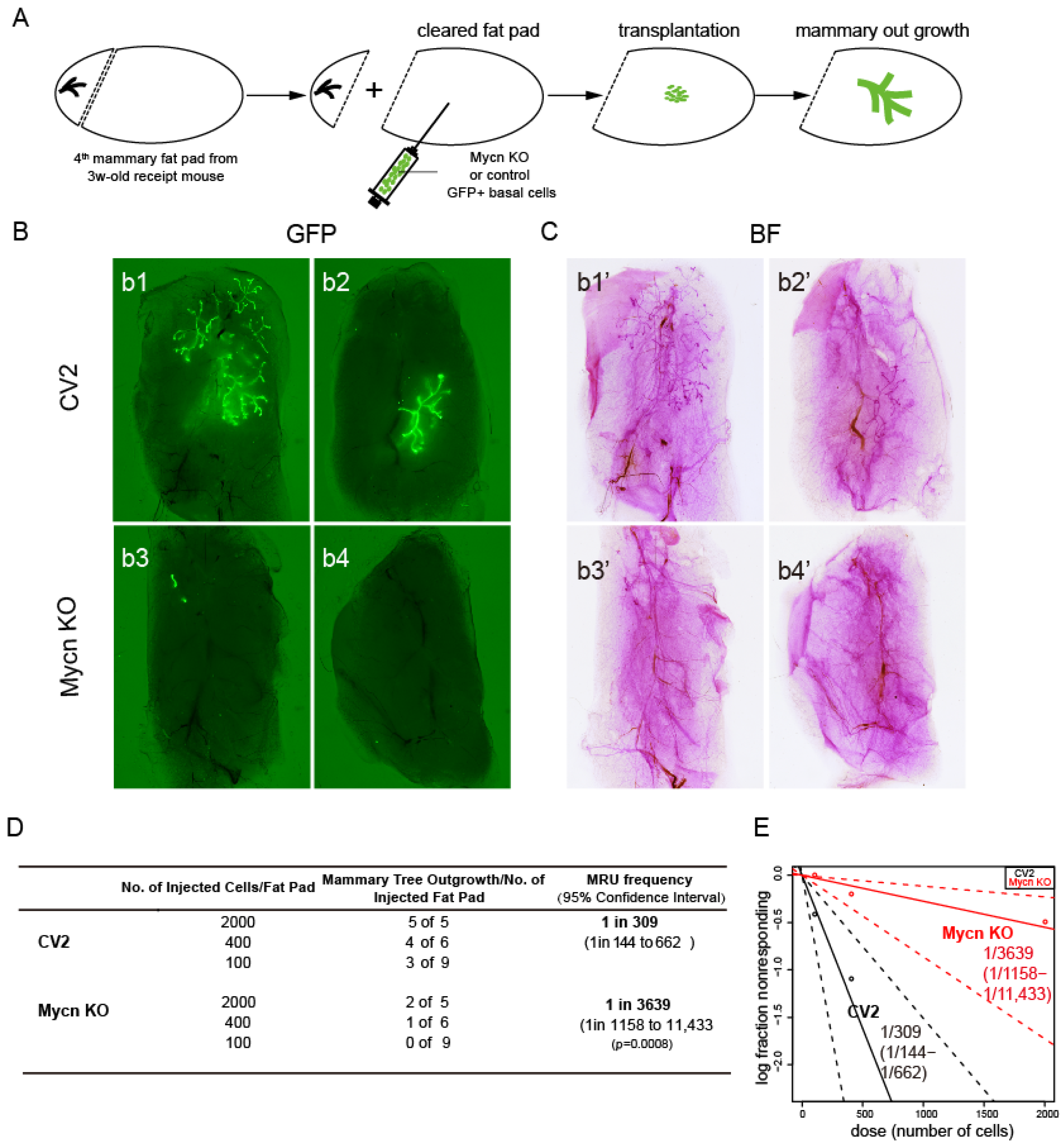
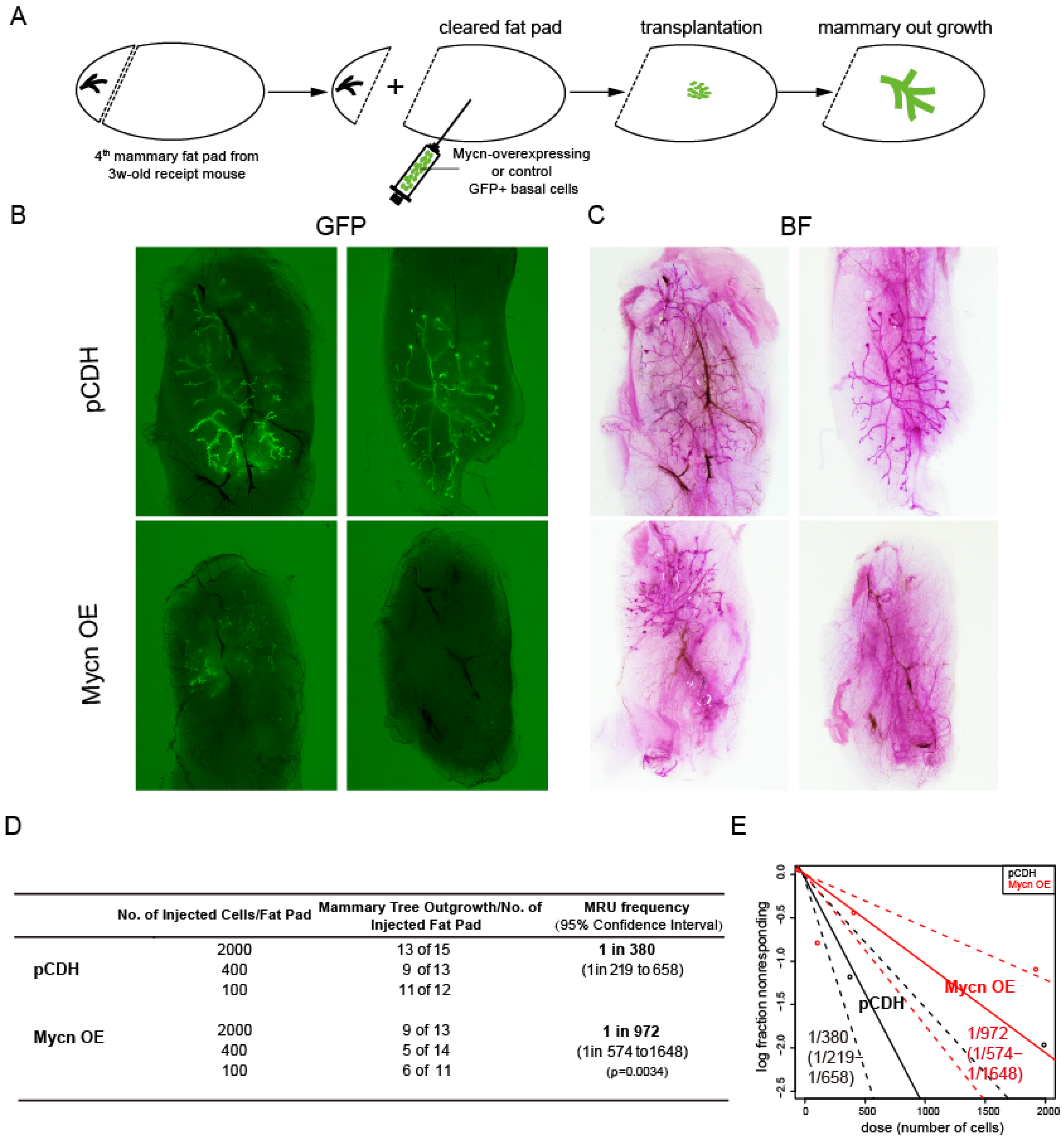
Given that MYCN plays significant roles in various cancer types [47,48,49,50], including triple-negative breast cancer [86], we sought to determine its importance in normal mammary gland development. To investigate the function of Mycn in murine mammary gland development, we employed CRISPR knockout (Figure 2A) and overexpression (Figure 3A) techniques in combination with a mammary cell transplantation assay.
To evaluate our hypothesis regarding the impact of Mycn knockout (KO) on mammary gland function, we examined the mammary tree reconstitution capacity of Mycn-mutant basal cells through a limiting dilution transplantation assay. The results demonstrated that Mycn deletion significantly reduced the frequency of mammary repopulating units (MRUs) in the mutant group (1/3639, ranging from 1/1158 to 1/11,433) compared to the control group (1/309, ranging from 1/144 to 1/662) (Figure 2B–D), with both groups labeled by GFP. Moreover, mammary trees reconstituted by Mycn KO basal cells exhibited significantly reduced size and fewer branches compared to the control group (Figure 2B,C). These observations suggest that Mycn plays a vital role in mammary gland development and mammary stem cell maintenance.
Given that Mycn is known to be overexpressed in various types of breast cancers, including certain triple-negative breast cancers [86], we sought to examine whether Mycn overexpression induces abnormal mammary development. The transplantation assay of Mycn-overexpressing basal cells produced results similar to those of Mycn KO cells, demonstrating reduced mammary regeneration capacity (Figure 3B–D). Specifically, Mycn overexpression decreased the MRU frequency in the control group from 1/380 (ranging from 1/219 to 1/658) to 1/972 (ranging from 1/574 to 1/1648). Whole-mount images further confirmed these findings (Figure 3B,C).
These findings indicate that both Mycn knockout and overexpression adversely affected the stemness of mammary stem cells, resulting in diminished mammary regeneration capacity. This effect may be attributed to the dose-dependent nature of Mycn activity. Therefore, we proceeded to investigate the molecular mechanisms through which Mycn-regulated stem cell function.

3.3. Mycn Promoted Cell Proliferation

Previous research has demonstrated that the human gene MYCN facilitates S-phase progression and cell proliferation, particularly in tumor cells. Specifically, MYCN functions as an RNA-binding accessory factor of the nuclear exosome-targeting complex, serving a critical role in these processes [92,93]. In neuroblastoma, MYCN-induced E2F5 enhances cell proliferation by regulating cell cycle progression [94]. Moreover, MYCN and MYC directly influence tumor proliferation and tumorigenesis through BMI1 in human neuroblastomas [93,94,95,96].
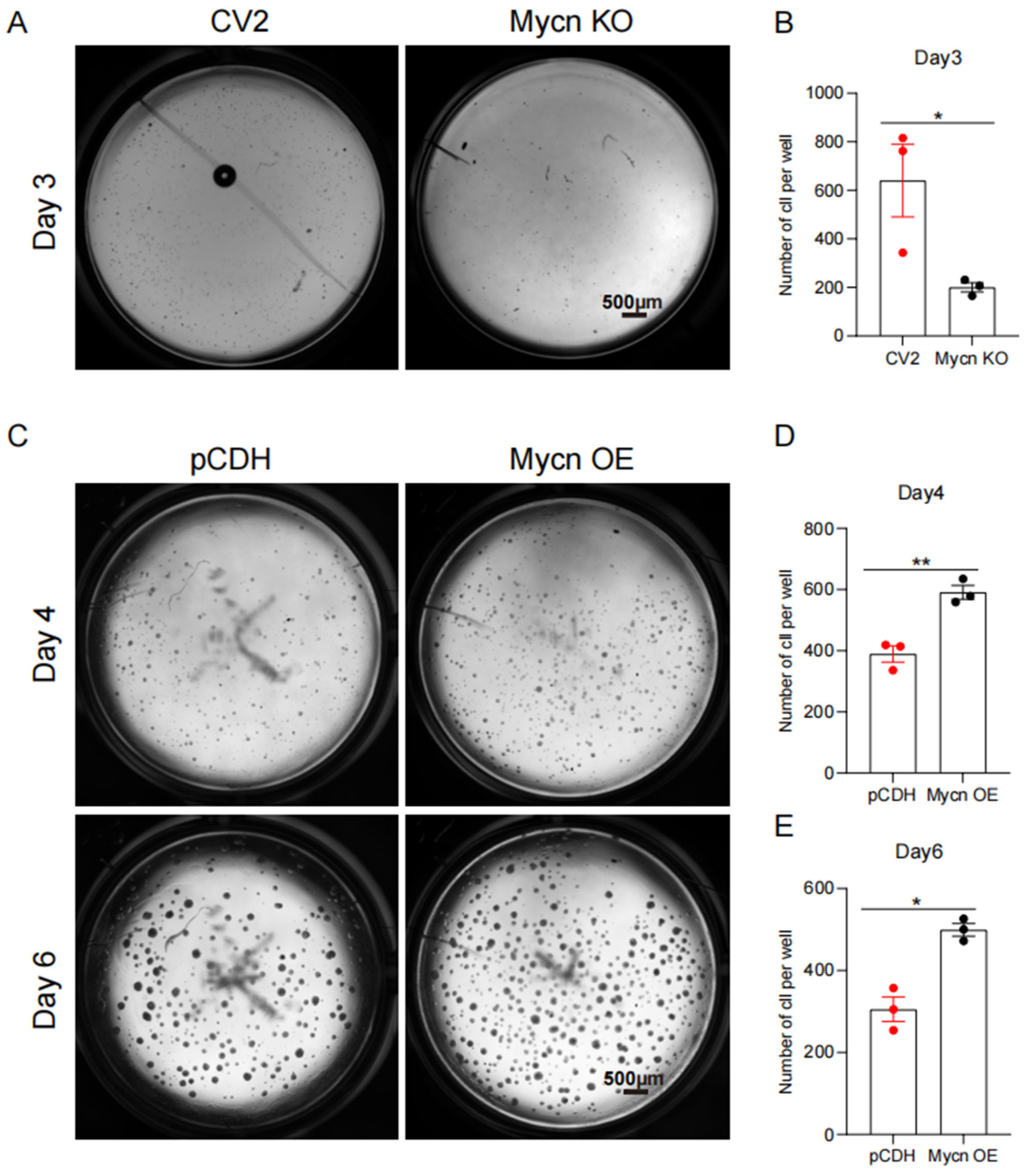
To examine the role of Mycn in regulating mammary basal cell proliferation, we conducted colony-formation assays using Mycn KO and Mycn-overexpressing basal cells. The results demonstrated that the Mycn knockout resulted in reduced proliferation of basal cells in vitro compared to the control group, as indicated by the decreased colony numbers (Figure 4A,B). This difference emerged within three days, supporting the hypothesis that Mycn plays a vital role in mammary basal cell proliferation. Additional validation through colony formation assays with Mycn-overexpressing basal cells revealed increased colony formation compared to control cells, at both Day 4 and Day 6 (Figure 4C–E). The concordance between these assays emphasizes the significance of Mycn in mammary basal cell proliferation.
Given Mycn’s essential role in cell proliferation, the impairment of mammary reconstitution capacity in Mycn KO basal cells during transplantation was anticipated. However, the observation that Mycn overexpression also negatively affects mammary regeneration presents an intriguing paradox.

3.4. Mycn Downregulated the Quiescent Stem Cell Regulators Bcl11b and Tspan8

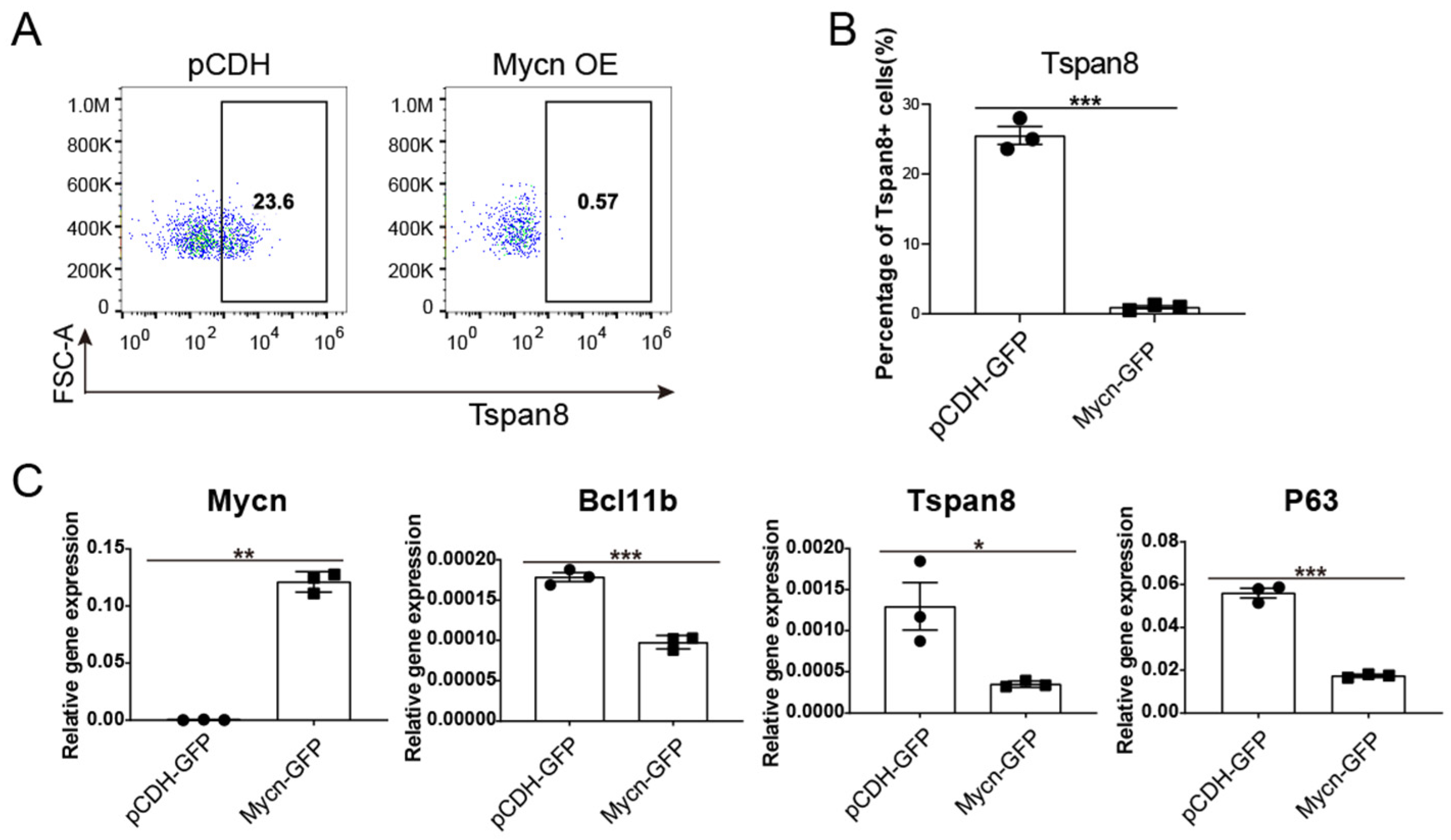
To elucidate the mechanism behind the decreased mammary regeneration capacity following Mycn overexpression, despite its proliferation-promoting role, quantitative PCR analysis was conducted to examine alterations in specific characteristic mammary stem/progenitor cell markers and regulators, including Bcl11b [12,97], Tspan8 [13,45], and Trp63 [18,98].
Bcl11b, as a transcription factor [99,100], has been identified as a crucial regulator of quiescent mammary stem cells, essential for maintaining long-term stemness, function, and differentiation [12]. Therefore, we investigated whether Mycn overexpression affected its expression. Additionally, we evaluated the expression level of Tspan8, another marker of quiescent mammary stem cells [13]. Previous research indicates that Tspan8 functions in maintaining mammary stem cell quiescence, and its deletion of Tspan8 rescued the defects in ductal morphogenesis caused by Foxp1 loss [45]. Furthermore, Trp63 promotes unipotent basal cell fate in the embryonic mammary gland [18], while its isoform, DeltaNp63, enhances stem cell activity in mammary gland development through decreased Fzd7 expression and Wnt signalling [98].
Upon examining these three markers under conditions of Mycn overexpression using FACS and qRT-PCR, we observed significant downregulation (Figure 5A–C). Given that Bcl11b KO activates mammary stem cells [12], and at the same time, deletion of Tspan8 also facilitated the activation of quiescent mammary stem cells [45], these findings suggest that Mycn overexpression promotes the exit from quiescent state in mammary stem cells. Moreover, the downregulation of these key markers and regulators due to Mycn overexpression could potentially impair both the maintenance of stem cell properties and differentiation processes. This observation may explain the reduced outgrowth of transplanted Mycn-overexpressing pubertal mammary basal cells, despite Mycn’s role in promoting cell proliferation in vitro.

3.5. Bcl11b KO Decreased the Expression Level of Tspan8 and Activated Cell Proliferation

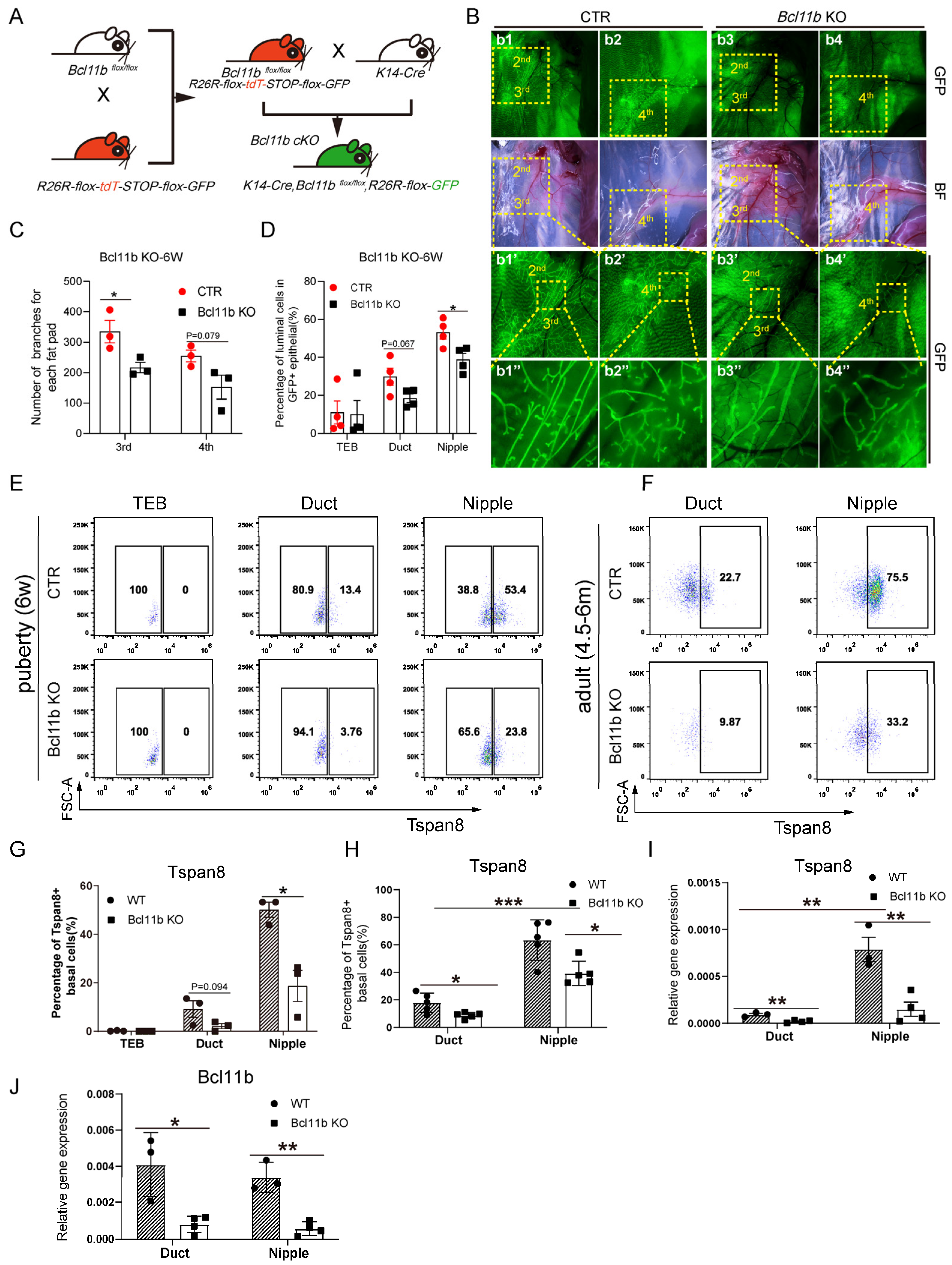
Following the observation that Mycn overexpression significantly reduced the expression of the quiescent stem cell regulators Bcl11b and Tspan8, we investigated whether the Bcl11b knockout (KO) also affected Tspan8 expression level. We utilized the K14-Cre/Bcl11bflox/flox/mTmG mouse model for our research. Through crossing Bcl11bflox/flox/mTmG mice with K14-Cre mice, we generated K14-Cre/Bcl11bflox/+/mTmG mice (Figure 6A), which were subsequently backcrossed to Bcl11bflox/flox/mTmG mice. After deleting Bcl11b in basal cells in K14-Cre/Bcl11bflox/flox/mTmG mice, we examined the developmental effects on mammary gland development. The mammary glands of the 6-week-old Bcl11b KO mice exhibited reduced branching (Figure 6b3–b3″,b4–b4″) compared to the controls (Figure 6b1–b1″,b2–b2″). The branching data are quantified in Figure 6C. Bcl11b KO also resulted in reduced luminal lineage within both duct and nipple areas (Figure 6D), consistent with the observation that “The Krt14-positive cells had impaired ability to give rise to luminal cells after Bcl11b deletion, reminiscent of a defect in lineage commitment and multipotency at some time during the mammary gland development” [12].
To evaluate the impact of Bcl11b KO on Tspan8 expression, we conducted FACS analysis to quantify the percentage of Tspan8-positive basal cells among GFP-positive basal cells, revealing a significant reduction in the nipple region and a moderate decrease in the ductal region following Bcl11b KO (Figure 6E,G). This pattern continued into adulthood (Figure 6F,H). Further analysis of Tspan8 transcriptional levels using qRT-PCR in adult mammary tissues confirmed a persistent change in expression (Figure 6I). Additionally, Bcl11b mRNA levels were analyzed to verify that the phenotypic changes resulted from Bcl11b KO, demonstrating a similar expression pattern to Tspan8 in both ductal and nipple basal cells of adult mammary glands (Figure 6J). An analysis of scRNA-seq data from adult (4-month-old) mice mammary glands indicated that Bcl11b KO in basal cells decreased Tspan8 expression (Supplementary Figure S3A), a trend consistent with findings in pubertal mammary tissues. Moreover, we also detected that Bcl11b KO prompted an increase in basal cell proliferation (Supplementary Figure S3B), which can be attributed to the disruption of stem cell quiescence and the activation of basal cell proliferation.
Collectively, these findings suggested that the transcription factor Bcl11b played a crucial role in maintaining the balance between differentiation and stem cell quiescence during mammary gland development, with its deletion disrupting this balance, leading to impaired branching and reduced Tspan8 level and activation of cell proliferation.

3.6. Mycn Coordinated Cell Proliferation and Differentiation

Given that Bcl11b regulated the balance between cell proliferation and differentiation and considering that Mycn has been demonstrated to suppress Bcl11b expression, we hypothesized that Mycn might regulate cell proliferation and differentiation processes during mammary gland development.
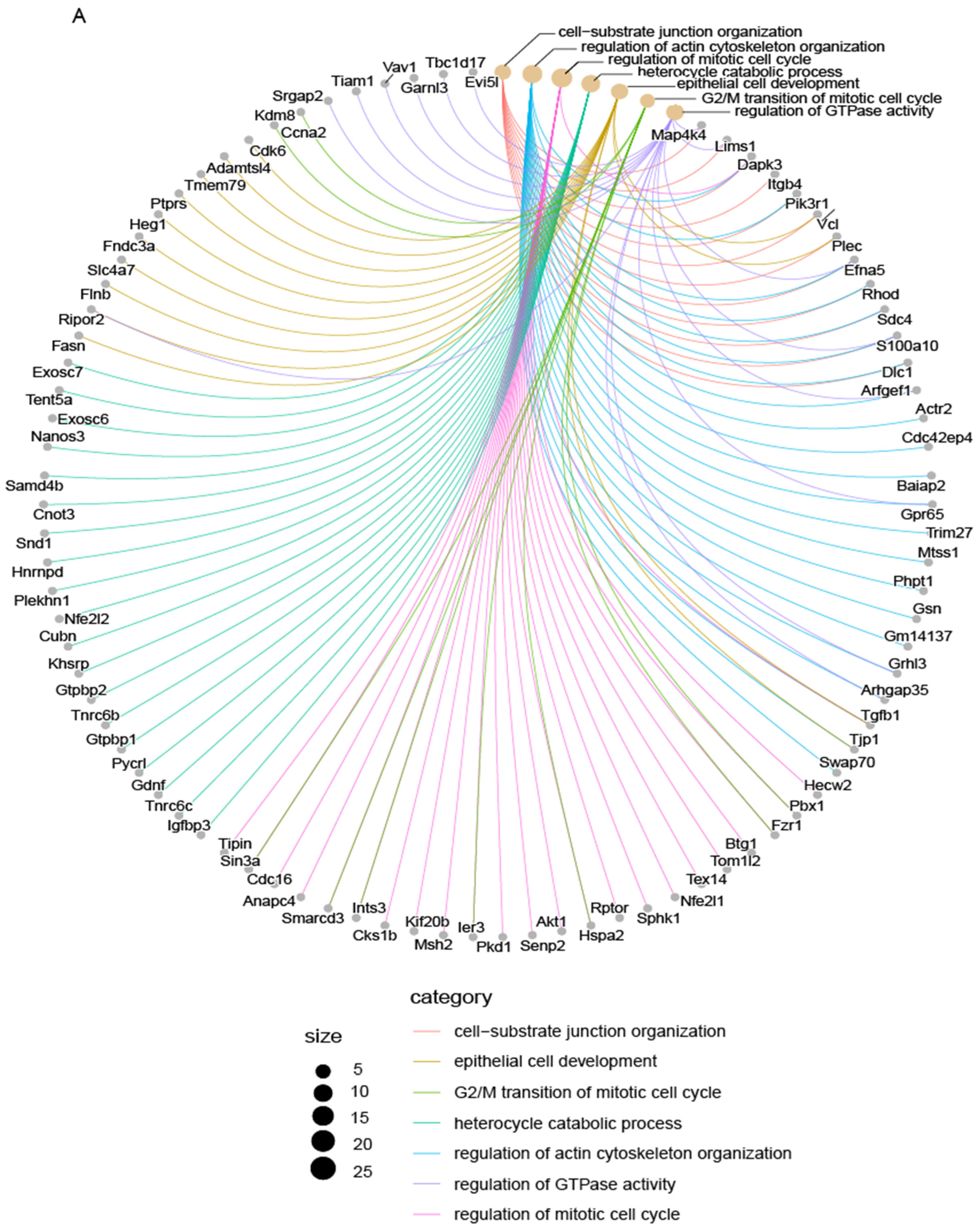
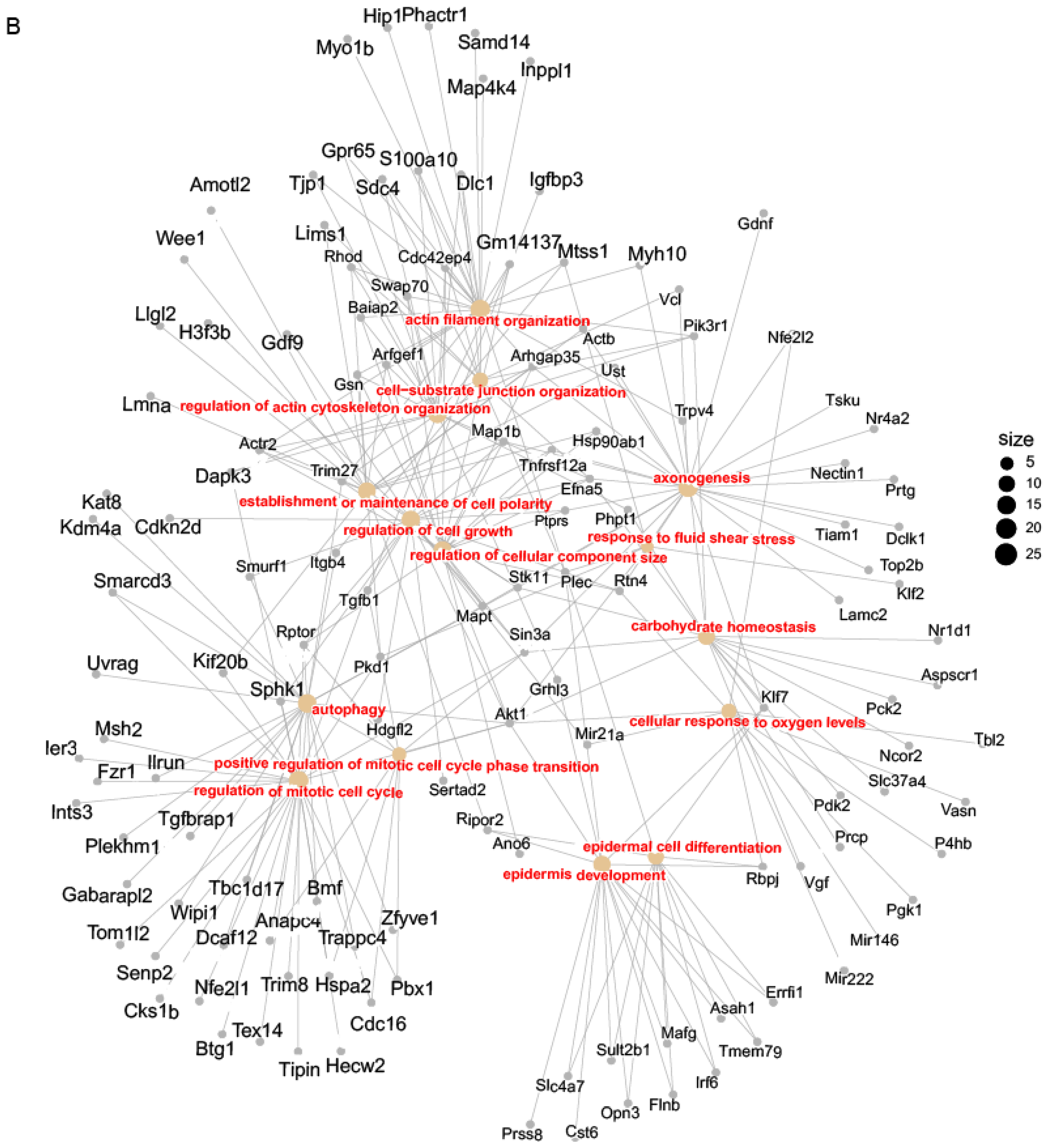
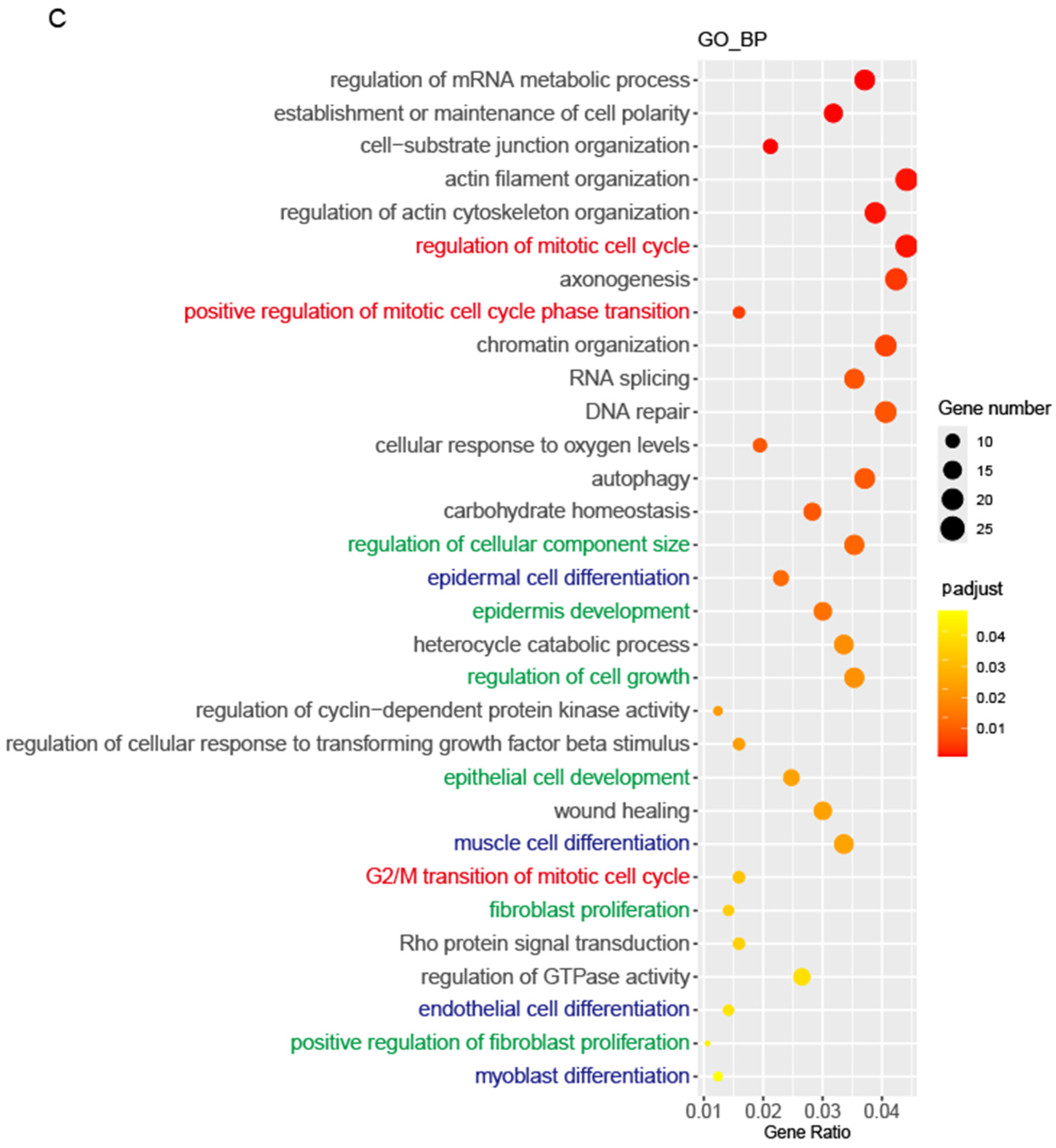
To test this hypothesis, we overexpressed Mycn gene in the mammary epithelial progenitor cell line Comma Dβ and performed a ChIP-Seq assay [89]. The analysis revealed Mycn binding sites across the genome, with 63.97% located in promoter regions in Comma Dβ cells (with 105 cells) [89]. A GO analysis determined that Mycn participated in the regulation of cell cycle, growth, proliferation, and differentiation (Figure 7A–C).
These findings align with both our previous data (Figure 1C and Figure 4A–E) and established research [89,94,95,96,101,102,103,104,105,106]. Notably, we observed that Mycn directly participates in the metaphase and anaphase stages of mitosis during primary mammary basal cell division when cultured in vitro (Supplementary Figure S4B). These observations demonstrate that Mycn coordinates cell proliferation and differentiation.

3.7. Mycn Indirectly Targeted Bcl11b and Tspan8

Previous research has established Tspan8’s critical role in cancer stem cell characteristic maintenance and the maintenance of stem cell characteristics through Sonic Hedgehog signaling pathway activation [107,108]. Research has demonstrated that the human TSPAN8 protein regulates MYC expression by binding to its promoter, enhancing STAT3’s chromatin occupancy, and influencing cancer-related gene transcription, including MYC [109]. These findings indicate a potential Mycn–Tspan8 interaction. Given Tspan8’s direct interaction with Myc, it appears plausible that Mycn might similarly bind and interact with Tspan8. Additionally, as Mycn functions as a transcription factor, it may directly interact with Bcl11b to regulate cell cycle, cell proliferation and differentiation.
Research has shown that both Bcl11b and Tspan8 regulate mammary stem cell quiescence maintenance, with their inhibition or deletion promoting mammary stem/progenitor cell activation or proliferation. Having observed Mycn’s downregulation of functional stem cell markers Bcl11b and Tspan8, we investigated whether this regulation occurred directly or indirectly. We employed the improved D-CUT&RUN technology to detect Mycn binding sites in mammary epithelial progenitor cells [89]. While direct interactions between Mycn and Bcl11b or Tspan8 were not detected, the analysis of existing research revealed Mycn’s interaction with both Bcl11b and Tspan8 in mouse and human neuroblastoma tumors through promoter binding (Supplementary Figure S5A,B). This may indicate the existence of different mechanisms of interaction between Mycn and Bcl11b and Tspan8 across different organs or between normal and pathological states.

4. Discussion

Mammary gland development is a highly regulated, dynamic process that occurs in distinct stages, including embryonic formation, postnatal growth, puberty, pregnancy, lactation, and involution. Puberty triggers the formation of terminal end buds (TEBs) at the tips of growing ducts, driving ductal elongation and branching. These TEBs eventually regress, forming a mature mammary gland. The mature gland’s epithelial framework supports alveolar development during pregnancy and lactation. Mammary stem cells, characterized by markers such as Bcl11bhigh, Lgr5+Tspan8high, and Nfatc1+, play key roles in gland maintenance and regeneration, though the mechanisms for activating quiescent stem cells remain unclear.
We employ scRNA-Seq technology to help discover the heterogeneity of different mammary subpopulations, including stem and progenitor cells. We acknowledge that there are both advantages and disadvantages for scRNA-Seq and bulk sequencing. Given that bulk sequencing provides cost-effectiveness and robustness for detecting population-level expression changes but masks cellular heterogeneity, while scRNA-Seq reveals cellular diversity and rare populations but faces challenges, including increased technical noise, higher costs, and analytical biases including dropout events [110,111], a combinatory strategy would be more effective in leveraging the strengths of both approaches: using bulk sequencing for initial subtype discovery across larger cohorts, followed by strategic single-cell validation on representative samples. This approach may ensure our genomic correlations authentically reflect underlying cellular architecture while maintaining resource efficiency [112].
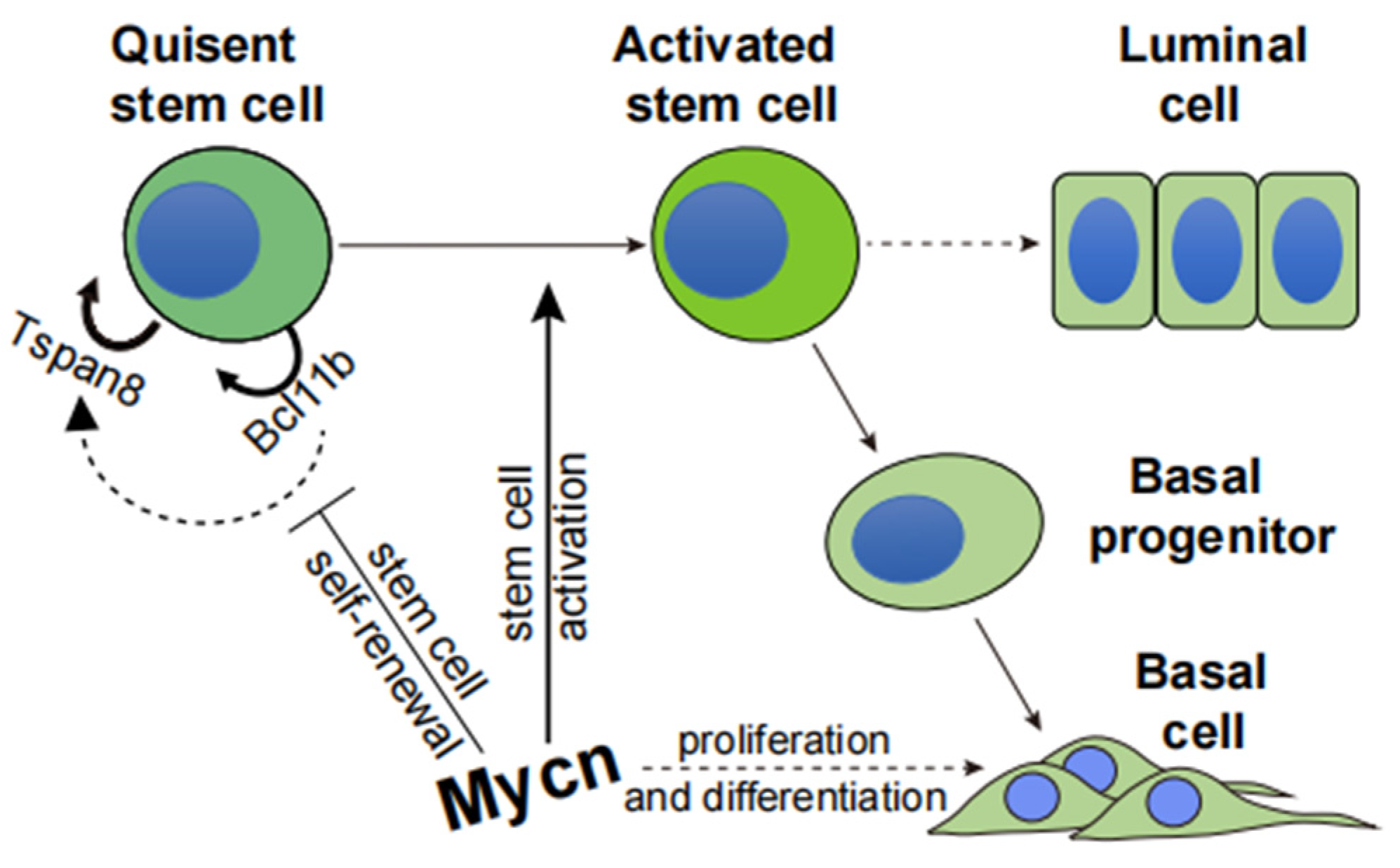
In our study, the transcription factor Mycn played a critical role in the exit from quiescence in mammary stem cells (Figure 8). Single-cell RNA sequencing (scRNA-seq) identified Mycn expression in TEB basal cells, validated through qPCR and immunofluorescence staining. This indicates Mycn’s significance in cell proliferation and differentiation within the TEB region. Colony formation and transplantation assays utilizing Mycn knockout (KO) and overexpressing basal epithelial cells established that Mycn promoted proliferation and differentiation by maintaining optimal expression levels, essential for mammary gland regeneration (Figure 8).
The data further indicate that Mycn’s interactions with Bcl11b and Tspan8 may differ between cancer and normal tissues, exhibiting a more direct interaction, observed in cancer tissues. This underscores the significance of Bcl11b and Tspan8 as downstream targets of Mycn. Notably, Bcl11b has been documented to function as a tumor suppressor in various cancers [41,113,114], aligning with the observed decrease in Bcl11b levels following Mycn overexpression in epithelial cells. However, TSPAN8 (human) has been shown to enhance breast cancer cell stemness through sonic hedgehog signaling activation, introducing additional complexity to understanding Mycn’s contribution to tumor progression. The potential roles and interactions of Bcl11b and Tspan8 with Mycn in cancers characterized by Mycn overexpression warrant further investigation.
Although this study primarily examined Mycn’s role in normal mammary gland development, additional research is necessary to investigate its role in differentiation using genetic mutation mouse models, as well as in vivo lineage tracing, given potential off-target effects of CRISPR. Future studies should also examine Mycn’s roles in cancer progression in both murine and human contexts.
In summary, Mycn serves essential functions in mammary gland development and regeneration by activating quiescent stem cells and promoting both proliferation and differentiation. These findings provide significant insights into cancers characterized by Mycn overexpression, such as triple-negative breast cancer (TNBC), and may guide potential therapeutic strategies.

5. Conclusions

Our research reveals the critical role of Mycn in pubertal mammary gland development, particularly in promoting the activation of quiescent mammary stem cells maintained by Bcl11b. Through single-cell RNA sequencing and functional experiments, we found that Mycn is spatially enriched in terminal end buds (TEBs), especially in basal cells, where it promotes cell proliferation and regulates ductal morphogenesis. At the mechanistic level, Mycn influences stem cell maintenance and differentiation by regulating the expression of key downstream target genes, including Bcl11b and Tspan8. The deletion of Bcl11b leads to significant downregulation of Tspan8 expression, further illustrating the functional interaction between these two regulatory factors in stem cell activation and lineage commitment.
More importantly, our research emphasizes that Mycn expression levels must be precisely regulated to maintain homeostasis during mammary gland development. We found that Mycn deficiency or low expression impairs the regenerative capacity of mammary stem cells, leading to underdeveloped mammary trees with reduced branching, affecting normal mammary structure and function. Conversely, Mycn overexpression, while promoting cell proliferation, simultaneously suppresses the expression of quiescent stem cell markers and regulators (such as Bcl11b and Tspan8), thus disrupting stem cell homeostasis, resulting in decreased regenerative capacity, and potentially inducing abnormal proliferation and tumorigenesis.
Given Mycn’s oncogenic potential in various cancers, including triple-negative breast cancer, our research further suggests that Mycn not only plays a core regulatory role in mammary development, but that its expression imbalance may also be an important driving factor in cancer occurrence. This provides new insights into understanding the molecular mechanisms of mammary development and the initiation pathways of breast cancer.
However, our research has certain limitations. The complex interaction network among Mycn, Bcl11b, and Tspan8 requires further investigation, especially regarding the regulatory mechanisms of direct binding sites or mediating factors. Additionally, although we observed Mycn-regulated phenotypes in mouse models, its translational relevance in human mammary development and disease needs further verification.
Future research should focus on analyzing the regulatory network among Mycn, Bcl11b, and Tspan8 at the transcriptional and epigenetic levels. Exploring whether pathways regulated by Mycn in mammary development are abnormally activated in breast cancer, as well as the expression correlation of Mycn, Bcl11b, and Tspan8 in human breast cancer samples, will be of significant value. Furthermore, using lineage tracing models can determine whether changes in Mycn expression levels affect the long-term fate of stem cells, potentially leading to stem cell depletion or malignant transformation. Finally, whether regulating Tspan8 or Bcl11b could serve as potential strategies for intervening in Mycn-driven abnormal proliferation or tumorigenesis is also worth further validation in animal models.
Ultimately, our research establishes the key regulatory role of Mycn in the delicate balance between quiescence, activation, and differentiation of pubertal mammary stem cells. Precise regulation of Mycn is crucial for normal mammary development, and its dysregulation may promote the occurrence of breast cancer. These findings open new directions for understanding mammary development mechanisms and developing targeted therapeutic strategies for breast cancer.

Supplementary Materials

The following supporting information can be downloaded at https://www.mdpi.com/article/10.3390/cells14161239/s1, Figure S1: The expression of Mycn in adult (duct) and pregnant (alveolar) stage in mouse mammary gland. (A) t-SNE plot showing the expression of Mycn in adult mouse mammary gland (data from Tabula Muris database, https://tabulamuris.sf.czbiohub.org/visualizations (accessed on 9 August 2024). (B) Representative immunofluorescence imaging of the expression of Mycn in TEB, adult duct, and alveolar basal cells. Magenta/GFP: Mycn; Red: αSMA; blue: DAPI. Figure S2: The expression of Foxp1 in pubertal mouse mammary. Violin plots show the single-cell expression levels of Foxp1 in pubertal mouse mammary tissue. Foxp1 is highly expressed in most basal cells and in a significant number of luminal cells. Figure S3: Bcl11b KO decreased the expression level of Tspan8 and activated cell proliferation. (A,B) Violin plots showing the single-cell expression level of Tspan8 (A) and Mki67 (B) in 4-month-old adult control and Bcl11b KO mice under C57BL/6 background. Figure S4: Detection of Mycn expression in cultured mammary basal cells. (A) Representative immunofluorescence images showing the expression of Mycn in Mycn-overexpressed primary basal cells from pubertal mouse mammary gland. GFP: Mycn; blue: DAPI. (B) Representative immunofluorescence images showing Mycn’s potential involvement in spindle behavior during primary basal cell division. Magenta: Mycn; purple: αSMA; blue: DAPI. Figure S5: ChIP-Seq data showing Mycn’s (MYCN in human) direct interaction with Bcl11b and Tspan8 (BCL11B and TSPAN8 in human) in both mouse and human neuroblastoma. (A) ChIP-Seq data for Mycn’s direct interaction with Bcl11b and Tspan8 in mouse neuroblastoma. The data originates from the published GEO database under the accession number GSE151426. (B) ChIP-Seq data for MYCN’s direct interaction with BCL11B and TSPAN8 in human neuroblastoma. The data originates from the published GEO database under the accession number GSE94782.

Author Contributions

Z.L. conceived this study and experimental design; Z.L. performed and analyzed the experiments. Z.L. wrote the manuscript. C.W., H.B., X.L. and M.L. contributed to single-cell RNA-Seq assay performance and data analysis. Y.Z. participated in plasmid construction. T.H. performed mouse genotyping. Y.M. helped with the procurement of experimental reagents and consumables. All authors have read and agreed to the published version of the manuscript.

Funding

This work was supported by the National Natural Science Foundation of China NSFC grant 32170803, 81872405 and by Westlake Education Foundation.

Institutional Review Board Statement

The animal study protocol was approved by the Institutional Animal Care and Use Committee (IACUC) of Westlake University (protocol code 24-149-CS-3, date of approval: 20 December 2024).

Informed Consent Statement

Not applicable.

Data Availability Statement

The data that support the findings in this study are openly available in GEO: The original scRNA-seq data generated in this study can be accessed via accession number GSE251933, while published datasets used for analysis are available under accession numbers GSE151426 and GSE94782.

Acknowledgments

We express special thanks to Jingjing Bao and Daijin Wu at the Westlake Animal Facility for mouse husbandry. We are grateful to Xilai Ding, Peng Guo, Jinhui Li, and Yanan Tang at the Flow Cytometry Facility of Westlake University for assistance in FACS sorting. We thank Yalin Wang, Yinping Gao, and Fang Xiao at the Microscopy Core Facility of Westlake University for tissue immunofluorescence imaging. We thank Zhenzhen Yu at the General Equipment & Autoclave Service Core Facility of Westlake University for experimental equipment management.

Conflicts of Interest

The authors declare no conflicts of interest.

References

  1. Sonkin, D.; Thomas, A.; Teicher, B.A. Cancer treatments: Past, present, and future. Cancer Genet. 2024, 286–287, 18–24. [Google Scholar] [CrossRef]
  2. Zagami, P.; Carey, L.A. Triple negative breast cancer: Pitfalls and progress. NPJ Breast Cancer 2022, 8, 95. [Google Scholar] [CrossRef]
  3. Wang, X.; Venet, D.; Lifrange, F.; Larsimont, D.; Rediti, M.; Stenbeck, L.; Dupont, F.; Rouas, G.; Garcia, A.J.; Craciun, L.; et al. Spatial transcriptomics reveals substantial heterogeneity in triple-negative breast cancer with potential clinical implications. Nat. Commun. 2024, 15, 10232. [Google Scholar] [CrossRef]
  4. Chen, Q.S.; Cai, R.Z.; Wang, Y.; Liang, G.H.; Zhang, K.M.; Yang, X.Y.; Yang, D.; Zhao, D.C.; Zhu, X.F.; Deng, R.; et al. Profiling triple-negative breast cancer-specific super-enhancers identifies high-risk mesenchymal development subtype and BETi-Targetable vulnerabilities. Mol. Cancer 2025, 24, 141. [Google Scholar] [CrossRef]
  5. Asleh, K.; Riaz, N.; Nielsen, T.O. Heterogeneity of triple negative breast cancer: Current advances in subtyping and treatment implications. J. Exp. Clin. Cancer Res. 2022, 41, 265. [Google Scholar] [CrossRef] [PubMed]
  6. Groelly, F.J.; Fawkes, M.; Dagg, R.A.; Blackford, A.N.; Tarsounas, M. Targeting DNA damage response pathways in cancer. Nat. Rev. Cancer 2023, 23, 78–94. [Google Scholar] [CrossRef] [PubMed]
  7. Wheeler, D.A.; Takebe, N.; Hinoue, T.; Hoadley, K.A.; Cardenas, M.F.; Hamilton, A.M.; Laird, P.W.; Wang, L.; Johnson, A.; Dewal, N.; et al. Molecular Features of Cancers Exhibiting Exceptional Responses to Treatment. Cancer Cell 2021, 39, 38–53.e7. [Google Scholar] [CrossRef]
  8. Bowling, E.A.; Wang, J.H.; Gong, F.; Wu, W.; Neill, N.J.; Kim, I.S.; Tyagi, S.; Orellana, M.; Kurley, S.J.; Dominguez-Vidaña, R.; et al. Spliceosome-targeted therapies trigger an antiviral immune response in triple-negative breast cancer. Cell 2021, 184, 384–403.e21. [Google Scholar] [CrossRef]
  9. Corces, M.R.; Granja, J.M.; Shams, S.; Louie, B.H.; Seoane, J.A.; Zhou, W.; Silva, T.C.; Groeneveld, C.; Wong, C.K.; Cho, S.W.; et al. The chromatin accessibility landscape of primary human cancers. Science 2018, 362, eaav1898. [Google Scholar] [CrossRef]
  10. Cossa, G.; Roeschert, I.; Prinz, F.; Baluapuri, A.; Vidal, R.S.; Schülein-Völk, C.; Chang, Y.C.; Ade, C.P.; Mastrobuoni, G.; Girard, C.; et al. Localized inhibition of protein phosphatase 1 by NUAK1 promotes spliceosome activity and reveals a MYC-sensitive feedback control of transcription. Mol. Cell 2021, 81, 2495. [Google Scholar] [CrossRef] [PubMed]
  11. Fu, N.Y.; Nolan, E.; Lindeman, G.J.; Visvader, J.E. Stem Cells and the Differentiation Hierarchy in Mammary Gland Development. Physiol. Rev. 2020, 100, 489–523. [Google Scholar] [CrossRef]
  12. Cai, S.; Kalisky, T.; Sahoo, D.; Dalerba, P.; Feng, W.; Lin, Y.; Qian, D.; Kong, A.; Yu, J.; Wang, F.; et al. A Quiescent Bcl11b High Stem Cell Population Is Required for Maintenance of the Mammary Gland. Cell Stem Cell 2017, 20, 247–260.e5. [Google Scholar] [CrossRef]
  13. Fu, N.Y.; Rios, A.C.; Pal, B.; Law, C.W.; Jamieson, P.; Liu, R.; Vaillant, F.; Jackling, F.; Liu, K.H.; Smyth, G.K.; et al. Identification of quiescent and spatially restricted mammary stem cells that are hormone responsive. Nat. Cell Biol. 2017, 19, 164–176. [Google Scholar] [CrossRef]
  14. Van Keymeulen, A.; Rocha, A.S.; Ousset, M.; Beck, B.; Bouvencourt, G.; Rock, J.; Sharma, N.; Dekoninck, S.; Blanpain, C. Distinct stem cells contribute to mammary gland development and maintenance. Nature 2011, 479, 189–193. [Google Scholar] [CrossRef]
  15. Wang, C.; Christin, J.R.; Oktay, M.H.; Guo, W. Lineage-Biased Stem Cells Maintain Estrogen-Receptor-Positive and -Negative Mouse Mammary Luminal Lineages. Cell Rep. 2017, 18, 2825–2835. [Google Scholar] [CrossRef]
  16. Van Keymeulen, A.; Fioramonti, M.; Centonze, A.; Bouvencourt, G.; Achouri, Y.; Blanpain, C. Lineage-Restricted Mammary Stem Cells Sustain the Development, Homeostasis, and Regeneration of the Estrogen Receptor Positive Lineage. Cell Rep. 2017, 20, 1525–1532. [Google Scholar] [CrossRef] [PubMed]
  17. Van Keymeulen, A.; Blanpain, C. Tracing epithelial stem cells during development, homeostasis, and repair. J. Cell Biol. 2012, 197, 575–584. [Google Scholar] [CrossRef]
  18. Wuidart, A.; Sifrim, A.; Fioramonti, M.; Matsumura, S.; Brisebarre, A.; Brown, D.; Centonze, A.; Dannau, A.; Dubois, C.; Van Keymeulen, A.; et al. Early lineage segregation of multipotent embryonic mammary gland progenitors. Nat. Cell Biol. 2018, 20, 666–676. [Google Scholar] [CrossRef] [PubMed]
  19. Lilja, A.M.; Rodilla, V.; Huyghe, M.; Hannezo, E.; Landragin, C.; Renaud, O.; Leroy, O.; Rulands, S.; Simons, B.D.; Fre, S. Clonal analysis of Notch1-expressing cells reveals the existence of unipotent stem cells that retain long-term plasticity in the embryonic mammary gland. Nat. Cell Biol. 2018, 20, 677–687. [Google Scholar] [CrossRef]
  20. McBryan, J.; Howlin, J. Pubertal Mammary Gland Development: Elucidation of In Vivo Morphogenesis Using Murine Models. Methods Mol. Biol. 2017, 1501, 77–114. [Google Scholar] [PubMed]
  21. Paine, I.S.; Lewis, M.T. The Terminal End Bud: The Little Engine that Could. J. Mammary Gland. Biol. Neoplasia 2017, 22, 93–108. [Google Scholar] [CrossRef]
  22. Slepicka, P.F.; Somasundara, A.V.H.; Dos Santos, C.O. The molecular basis of mammary gland development and epithelial differentiation. Semin. Cell Dev. Biol. 2021, 114, 93–112. [Google Scholar] [CrossRef]
  23. Williams, J.M.; Daniel, C.W. Mammary ductal elongation: Differentiation of myoepithelium and basal lamina during branching morphogenesis. Dev. Biol. 1983, 97, 274–290. [Google Scholar] [CrossRef]
  24. Smalley, M.; Ashworth, A. Stem cells and breast cancer: A field in transit. Nat. Rev. Cancer 2003, 3, 832–844. [Google Scholar] [CrossRef]
  25. Lewis, M.T.; Porter, W.W. Methods in mammary gland biology and breast cancer research: An update. J. Mammary Gland. Biol. Neoplasia 2009, 14, 365. [Google Scholar] [CrossRef] [PubMed]
  26. Kouros-Mehr, H.; Slorach, E.M.; Sternlicht, M.D.; Werb, Z. GATA-3 maintains the differentiation of the luminal cell fate in the mammary gland. Cell 2006, 127, 1041–1055. [Google Scholar] [CrossRef] [PubMed]
  27. Srinivasan, K.; Strickland, P.; Valdes, A.; Shin, G.C.; Hinck, L. Netrin-1/neogenin interaction stabilizes multipotent progenitor cap cells during mammary gland morphogenesis. Dev. Cell 2003, 4, 371–382. [Google Scholar] [CrossRef]
  28. Bai, L.; Rohrschneider, L.R. s-SHIP promoter expression marks activated stem cells in developing mouse mammary tissue. Genes Dev. 2010, 24, 1882–1892. [Google Scholar] [CrossRef] [PubMed]
  29. Sreekumar, A.; Roarty, K.; Rosen, J.M. The mammary stem cell hierarchy: A looking glass into heterogeneous breast cancer landscapes. Endocr. Relat. Cancer 2015, 22, T161–T176. [Google Scholar] [CrossRef]
  30. Sahu, S.; Albaugh, M.E.; Martin, B.K.; Patel, N.L.; Riffle, L.; Mackem, S.; Kalen, J.D.; Sharan, S.K. Growth factor dependency in mammary organoids regulates ductal morphogenesis during organ regeneration. Sci. Rep. 2022, 12, 7200. [Google Scholar] [CrossRef]
  31. Lawson, D.A.; Werb, Z.; Zong, Y.; Goldstein, A.S. The Cleared Mammary Fat Pad Transplantation Assay for Mammary Epithelial Organogenesis. Cold Spring Harb. Protoc. 2015, 2015, pdb.prot078071. [Google Scholar] [CrossRef]
  32. Shackleton, M.; Vaillant, F.; Simpson, K.J.; Stingl, J.; Smyth, G.K.; Asselin-Labat, M.L.; Wu, L.; Lindeman, G.J.; Visvader, J.E. Generation of a functional mammary gland from a single stem cell. Nature 2006, 439, 84–88. [Google Scholar] [CrossRef] [PubMed]
  33. Stingl, J.; Eirew, P.; Ricketson, I.; Shackleton, M.; Vaillant, F.; Choi, D.; Li, H.I.; Eaves, C.J. Purification and unique properties of mammary epithelial stem cells. Nature 2006, 439, 993–997. [Google Scholar] [CrossRef]
  34. van Amerongen, R.; Bowman, A.N.; Nusse, R. Developmental stage and time dictate the fate of Wnt/beta-catenin-responsive stem cells in the mammary gland. Cell Stem Cell 2012, 11, 387–400. [Google Scholar] [CrossRef]
  35. dos Santos, C.O.; Rebbeck, C.; Rozhkova, E.; Valentine, A.; Samuels, A.; Kadiri, L.R.; Osten, P.; Harris, E.Y.; Uren, P.J.; Smith, A.D.; et al. Molecular hierarchy of mammary differentiation yields refined markers of mammary stem cells. Proc. Natl. Acad. Sci. USA 2013, 110, 7123–7130. [Google Scholar] [CrossRef] [PubMed]
  36. Wang, D.; Cai, C.; Dong, X.; Yu, Q.C.; Zhang, X.O.; Yang, L.; Zeng, Y.A. Identification of multipotent mammary stem cells by protein C receptor expression. Nature 2015, 517, 81–84. [Google Scholar] [CrossRef]
  37. Chakrabarti, R.; Celia-Terrassa, T.; Kumar, S.; Hang, X.; Wei, Y.; Choudhury, A.; Hwang, J.; Peng, J.; Nixon, B.; Grady, J.J.; et al. Notch ligand Dll1 mediates cross-talk between mammary stem cells and the macrophageal niche. Science 2018, 360, eaan4153. [Google Scholar] [CrossRef] [PubMed]
  38. Otsuki, L.; Brand, A.H. Cell cycle heterogeneity directs the timing of neural stem cell activation from quiescence. Science 2018, 360, 99–102. [Google Scholar] [CrossRef]
  39. Urban, N.; Blomfield, I.M.; Guillemot, F. Quiescence of Adult Mammalian Neural Stem Cells: A Highly Regulated Rest. Neuron 2019, 104, 834–848. [Google Scholar] [CrossRef]
  40. Cheung, T.H.; Rando, T.A. Molecular regulation of stem cell quiescence. Nat. Rev. Mol. Cell Biol. 2013, 14, 329–340. [Google Scholar] [CrossRef]
  41. Bai, H.; Liu, X.; Lin, M.; Meng, Y.; Tang, R.; Guo, Y.; Li, N.; Clarke, M.F.; Cai, S. Progressive senescence programs induce intrinsic vulnerability to aging-related female breast cancer. Nat. Commun. 2024, 15, 5154. [Google Scholar] [CrossRef]
  42. Johnstone, M.E.; Leck, A.L.; Lange, T.E.; Wilcher, K.E.; Shephard, M.S.; Paranjpe, A.; Schutte, S.; Wells, S.I.; Kappes, F.; Salomonis, N.; et al. DEK promotes mammary hyperplasia and is associated with H3K27me3 epigenetic modifications. Life Sci. Alliance 2025, 8, e202503230. [Google Scholar] [CrossRef]
  43. Marescal, O.; Cheeseman, I.M. Cellular Mechanisms and Regulation of Quiescence. Dev. Cell 2020, 55, 259–271. [Google Scholar] [CrossRef] [PubMed]
  44. Liu, R.; Hu, H.; McNeil, M.; Xu, J.; Bi, X.; Lou, P.; Guerrero-Juarez, C.F.; Dai, X.; Plikus, M.V.; Shuai, J.; et al. Dormant Nfatc1 reporter-marked basal stem/progenitor cells contribute to mammary lobuloalveoli formation. iScience 2022, 25, 103982. [Google Scholar] [CrossRef] [PubMed]
  45. Fu, N.Y.; Pal, B.; Chen, Y.; Jackling, F.C.; Milevskiy, M.; Vaillant, F.; Capaldo, B.D.; Guo, F.; Liu, K.H.; Rios, A.C.; et al. Foxp1 Is Indispensable for Ductal Morphogenesis and Controls the Exit of Mammary Stem Cells from Quiescence. Dev. Cell 2018, 47, 629–644.e8. [Google Scholar] [CrossRef]
  46. Han, Y.; Villarreal-Ponce, A.; Gutierrez, G., Jr.; Nguyen, Q.; Sun, P.; Wu, T.; Sui, B.; Berx, G.; Brabletz, T.; Kessenbrock, K.; et al. Coordinate control of basal epithelial cell fate and stem cell maintenance by core EMT transcription factor Zeb1. Cell Rep. 2022, 38, 110240. [Google Scholar] [CrossRef]
  47. Kohl, N.E.; Kanda, N.; Schreck, R.R.; Bruns, G.; Latt, S.A.; Gilbert, F.; Alt, F.W. Transposition and amplification of oncogene-related sequences in human neuroblastomas. Cell 1983, 35 Pt 1, 359–367. [Google Scholar] [CrossRef] [PubMed]
  48. Kohl, N.E.; Gee, C.E.; Alt, F.W. Activated expression of the N-myc gene in human neuroblastomas and related tumors. Science 1984, 226, 1335–1337. [Google Scholar] [CrossRef]
  49. Brodeur, G.M.; Seeger, R.C.; Schwab, M.; Varmus, H.E.; Bishop, J.M. Amplification of N-myc in untreated human neuroblastomas correlates with advanced disease stage. Science 1984, 224, 1121–1124. [Google Scholar] [CrossRef]
  50. Nesbit, C.E.; Tersak, J.M.; Prochownik, E.V. MYC oncogenes and human neoplastic disease. Oncogene 1999, 18, 3004–3016. [Google Scholar] [CrossRef]
  51. Miranda Peralta, E.I.; Valles Ayoub, Y.; Hernandez Mendoza, L.; Rangel Ramirez, L.M.; Castrejon Rojas, A.; Collazo-Jaloma, J.; Gutierrez Romero, M.; Gonzalez Constance, R.; Gariglio Vidal, P. MYC protein and proteins antigenically related with MYC in acute lymphoblastic leukemia. Rev. Investig. Clin. 1991, 43, 139–145. [Google Scholar]
  52. Burmeister, T.; Schwartz, S.; Horst, H.A.; Rieder, H.; Gokbuget, N.; Hoelzer, D.; Thiel, E. Molecular heterogeneity of sporadic adult Burkitt-type leukemia/lymphoma as revealed by PCR and cytogenetics: Correlation with morphology, immunology and clinical features. Leukemia 2005, 19, 1391–1398. [Google Scholar] [CrossRef]
  53. Herms, J.W.; von Loewenich, F.D.; Behnke, J.; Markakis, E.; Kretzschmar, H.A. c-myc oncogene family expression in glioblastoma and survival. Surg. Neurol. 1999, 51, 536–542. [Google Scholar] [CrossRef]
  54. Sardi, I.; Dal Canto, M.; Bartoletti, R.; Guazzelli, R.; Travaglini, F.; Montali, E. Molecular genetic alterations of c-myc oncogene in superficial and locally advanced bladder cancer. Eur. Urol. 1998, 33, 424–430. [Google Scholar] [CrossRef]
  55. Mizukami, Y.; Nonomura, A.; Takizawa, T.; Noguchi, M.; Michigishi, T.; Nakamura, S.; Ishizaki, T. N-myc protein expression in human breast carcinoma: Prognostic implications. Anticancer Res. 1995, 15, 2899–2905. [Google Scholar]
  56. Ioannidis, P.; Mahaira, L.; Papadopoulou, A.; Teixeira, M.R.; Heim, S.; Andersen, J.A.; Evangelou, E.; Dafni, U.; Pandis, N.; Trangas, T. 8q24 Copy number gains and expression of the c-myc mRNA stabilizing protein CRD-BP in primary breast carcinomas. Int. J. Cancer 2003, 104, 54–59. [Google Scholar] [CrossRef] [PubMed]
  57. Naidu, R.; Wahab, N.A.; Yadav, M.; Kutty, M.K. Protein expression and molecular analysis of c-myc gene in primary breast carcinomas using immunohistochemistry and differential polymerase chain reaction. Int. J. Mol. Med. 2002, 9, 189–196. [Google Scholar] [CrossRef]
  58. Han, S.; Kim, H.Y.; Park, K.; Cho, H.J.; Lee, M.S.; Kim, H.J.; Kim, Y.D. c-Myc expression is related with cell proliferation and associated with poor clinical outcome in human gastric cancer. J. Korean Med. Sci. 1999, 14, 526–530. [Google Scholar] [CrossRef]
  59. Kawate, S.; Fukusato, T.; Ohwada, S.; Watanuki, A.; Morishita, Y. Amplification of c-myc in hepatocellular carcinoma: Correlation with clinicopathologic features, proliferative activity and p53 overexpression. Oncology 1999, 57, 157–163. [Google Scholar] [CrossRef]
  60. Treszl, A.; Adany, R.; Rakosy, Z.; Kardos, L.; Begany, A.; Gilde, K.; Balazs, M. Extra copies of c-myc are more pronounced in nodular melanomas than in superficial spreading melanomas as revealed by fluorescence in situ hybridisation. Cytom. B Clin. Cytom. 2004, 60, 37–46. [Google Scholar] [CrossRef] [PubMed]
  61. Baker, V.V.; Borst, M.P.; Dixon, D.; Hatch, K.D.; Shingleton, H.M.; Miller, D. c-myc amplification in ovarian cancer. Gynecol. Oncol. 1990, 38, 340–342. [Google Scholar] [CrossRef] [PubMed]
  62. Lui, W.O.; Tanenbaum, D.M.; Larsson, C. High level amplification of 1p32-33 and 2p22-24 in small cell lung carcinomas. Int. J. Oncol. 2001, 19, 451–457. [Google Scholar] [CrossRef]
  63. Gugger, M.; Burckhardt, E.; Kappeler, A.; Hirsiger, H.; Laissue, J.A.; Mazzucchelli, L. Quantitative expansion of structural genomic alterations in the spectrum of neuroendocrine lung carcinomas. J. Pathol. 2002, 196, 408–415. [Google Scholar] [CrossRef] [PubMed]
  64. Maris, J.M. Recent advances in neuroblastoma. N. Engl. J. Med. 2010, 362, 2202–2211. [Google Scholar] [CrossRef] [PubMed]
  65. Dang, C.V. MYC on the path to cancer. Cell 2012, 149, 22–35. [Google Scholar] [CrossRef]
  66. Malynn, B.A.; de Alboran, I.M.; O’Hagan, R.C.; Bronson, R.; Davidson, L.; DePinho, R.A.; Alt, F.W. N-myc can functionally replace c-myc in murine development, cellular growth, and differentiation. Genes Dev. 2000, 14, 1390–1399. [Google Scholar] [CrossRef] [PubMed]
  67. Laurenti, E.; Varnum-Finney, B.; Wilson, A.; Ferrero, I.; Blanco-Bose, W.E.; Ehninger, A.; Knoepfler, P.S.; Cheng, P.F.; MacDonald, H.R.; Eisenman, R.N.; et al. Hematopoietic stem cell function and survival depend on c-Myc and N-Myc activity. Cell Stem Cell 2008, 3, 611–624. [Google Scholar] [CrossRef]
  68. Peramangalam, P.S.; Surapally, S.; Veltri, A.J.; Zheng, S.; Burns, R.; Zhu, N.; Rao, S.; Muller-Tidow, C.; Bushweller, J.H.; Pulikkan, J.A. N-MYC regulates cell survival via eIF4G1 in inv(16) acute myeloid leukemia. Sci. Adv. 2024, 10, eadh8493. [Google Scholar] [CrossRef]
  69. King, B.; Boccalatte, F.; Moran-Crusio, K.; Wolf, E.; Wang, J.; Kayembe, C.; Lazaris, C.; Yu, X.; Aranda-Orgilles, B.; Lasorella, A.; et al. The ubiquitin ligase Huwe1 regulates the maintenance and lymphoid commitment of hematopoietic stem cells. Nat. Immunol. 2016, 17, 1312–1321. [Google Scholar] [CrossRef]
  70. Kerosuo, L.; Neppala, P.; Hsin, J.; Mohlin, S.; Vieceli, F.M.; Torok, Z.; Laine, A.; Westermarck, J.; Bronner, M.E. Enhanced expression of MycN/CIP2A drives neural crest toward a neural stem cell-like fate: Implications for priming of neuroblastoma. Proc. Natl. Acad. Sci. USA 2018, 115, E7351–E7360. [Google Scholar] [CrossRef]
  71. Zhang, J.T.; Weng, Z.H.; Tsang, K.S.; Tsang, L.L.; Chan, H.C.; Jiang, X.H. MycN Is Critical for the Maintenance of Human Embryonic Stem Cell-Derived Neural Crest Stem Cells. PLoS ONE 2016, 11, e0148062. [Google Scholar]
  72. Huang, M.; Weiss, W.A. Neuroblastoma and MYCN. Cold Spring Harb. Perspect. Med. 2013, 3, a014415. [Google Scholar] [CrossRef] [PubMed]
  73. Hatton, B.A.; Knoepfler, P.S.; Kenney, A.M.; Rowitch, D.H.; de Alboran, I.M.; Olson, J.M.; Eisenman, R.N. N-myc is an essential downstream effector of Shh signaling during both normal and neoplastic cerebellar growth. Cancer Res. 2006, 66, 8655–8661. [Google Scholar] [CrossRef]
  74. Wey, A.; Knoepfler, P.S. c-myc and N-myc promote active stem cell metabolism and cycling as architects of the developing brain. Oncotarget 2010, 1, 120–130. [Google Scholar] [CrossRef]
  75. Wilson, A.; Murphy, M.J.; Oskarsson, T.; Kaloulis, K.; Bettess, M.D.; Oser, G.M.; Pasche, A.C.; Knabenhans, C.; Macdonald, H.R.; Trumpp, A. c-Myc controls the balance between hematopoietic stem cell self-renewal and differentiation. Genes Dev. 2004, 18, 2747–2763. [Google Scholar] [CrossRef] [PubMed]
  76. Satoh, Y.; Matsumura, I.; Tanaka, H.; Ezoe, S.; Sugahara, H.; Mizuki, M.; Shibayama, H.; Ishiko, E.; Ishiko, J.; Nakajima, K.; et al. Roles for c-Myc in self-renewal of hematopoietic stem cells. J. Biol. Chem. 2004, 279, 24986–24993. [Google Scholar] [CrossRef] [PubMed]
  77. Deming, S.L.; Nass, S.J.; Dickson, R.B.; Trock, B.J. C-myc amplification in breast cancer: A meta-analysis of its occurrence and prognostic relevance. Br. J. Cancer 2000, 83, 1688–1695. [Google Scholar] [CrossRef]
  78. Shajahan-Haq, A.N.; Cook, K.L.; Schwartz-Roberts, J.L.; Eltayeb, A.E.; Demas, D.M.; Warri, A.M.; Facey, C.O.; Hilakivi-Clarke, L.A.; Clarke, R. MYC regulates the unfolded protein response and glucose and glutamine uptake in endocrine resistant breast cancer. Mol. Cancer 2014, 13, 239. [Google Scholar] [CrossRef]
  79. McNeil, C.M.; Sergio, C.M.; Anderson, L.R.; Inman, C.K.; Eggleton, S.A.; Murphy, N.C.; Millar, E.K.; Crea, P.; Kench, J.G.; Alles, M.C.; et al. c-Myc overexpression and endocrine resistance in breast cancer. J. Steroid Biochem. Mol. Biol. 2006, 102, 147–155. [Google Scholar] [CrossRef]
  80. Fallah, Y.; Brundage, J.; Allegakoen, P.; Shajahan-Haq, A.N. MYC-Driven Pathways in Breast Cancer Subtypes. Biomolecules 2017, 7, 53. [Google Scholar] [CrossRef]
  81. Dhanasekaran, R.; Deutzmann, A.; Mahauad-Fernandez, W.D.; Hansen, A.S.; Gouw, A.M.; Felsher, D.W. The MYC oncogene—The grand orchestrator of cancer growth and immune evasion. Nat. Rev. Clin. Oncol. 2022, 19, 23–36. [Google Scholar] [CrossRef] [PubMed]
  82. Horiuchi, D.; Kusdra, L.; Huskey, N.E.; Chandriani, S.; Lenburg, M.E.; Gonzalez-Angulo, A.M.; Creasman, K.J.; Bazarov, A.V.; Smyth, J.W.; Davis, S.E.; et al. MYC pathway activation in triple-negative breast cancer is synthetic lethal with CDK inhibition. J. Exp. Med. 2012, 209, 679–696. [Google Scholar] [CrossRef]
  83. Knudsen, E.S.; McClendon, A.K.; Franco, J.; Ertel, A.; Fortina, P.; Witkiewicz, A.K. RB loss contributes to aggressive tumor phenotypes in MYC-driven triple negative breast cancer. Cell Cycle 2015, 14, 109–122. [Google Scholar] [CrossRef]
  84. Zimmerli, D.; Brambillasca, C.S.; Talens, F.; Bhin, J.; Linstra, R.; Romanens, L.; Bhattacharya, A.; Joosten, S.E.P.; Da Silva, A.M.; Padrao, N.; et al. MYC promotes immune-suppression in triple-negative breast cancer via inhibition of interferon signaling. Nat. Commun. 2022, 13, 6579. [Google Scholar] [CrossRef]
  85. Tang, M.; O’Grady, S.; Crown, J.; Duffy, M.J. MYC as a therapeutic target for the treatment of triple-negative breast cancer: Preclinical investigations with the novel MYC inhibitor, MYCi975. Breast Cancer Res. Treat. 2022, 195, 105–115. [Google Scholar] [CrossRef]
  86. Schafer, J.M.; Lehmann, B.D.; Gonzalez-Ericsson, P.I.; Marshall, C.B.; Beeler, J.S.; Redman, L.N.; Jin, H.; Sanchez, V.; Stubbs, M.C.; Scherle, P.; et al. Targeting MYCN-expressing triple-negative breast cancer with BET and MEK inhibitors. Sci. Transl. Med. 2020, 12, eaaw8275. [Google Scholar] [CrossRef]
  87. Golonzhka, O.; Liang, X.; Messaddeq, N.; Bornert, J.M.; Campbell, A.L.; Metzger, D.; Chambon, P.; Ganguli-Indra, G.; Leid, M.; Indra, A.K. Dual role of COUP-TF-interacting protein 2 in epidermal homeostasis and permeability barrier formation. J. Investig. Dermatol. 2009, 129, 1459–1470. [Google Scholar] [CrossRef]
  88. Fu, A.; Yao, B.; Dong, T.; Chen, Y.; Yao, J.; Liu, Y.; Li, H.; Bai, H.; Liu, X.; Zhang, Y.; et al. Tumor-resident intracellular microbiota promotes metastatic colonization in breast cancer. Cell 2022, 185, 1356–1372.e26. [Google Scholar] [CrossRef] [PubMed]
  89. Bai, H.; Lin, M.; Meng, Y.; Bai, H.; Cai, S. An improved CUT&RUN method for regulation network reconstruction of low abundance transcription factor. Cell. Signal. 2022, 96, 110361. [Google Scholar] [CrossRef] [PubMed]
  90. Deome, K.B.; Faulkin, L.J., Jr.; Bern, H.A.; Blair, P.B. Development of mammary tumors from hyperplastic alveolar nodules transplanted into gland-free mammary fat pads of female C3H mice. Cancer Res. 1959, 19, 515–520. [Google Scholar]
  91. Faulkin, L.J., Jr.; Deome, K.B. Regulation of growth and spacing of gland elements in the mammary fat pad of the C3H mouse. J. Natl. Cancer Inst. 1960, 24, 953–969. [Google Scholar]
  92. Papadopoulos, D.; Solvie, D.; Baluapuri, A.; Endres, T.; Ha, S.A.; Herold, S.; Kalb, J.; Giansanti, C.; Schulein-Volk, C.; Ade, C.P.; et al. MYCN recruits the nuclear exosome complex to RNA polymerase II to prevent transcription-replication conflicts. Mol. Cell 2022, 82, 159–176.e12. [Google Scholar] [CrossRef]
  93. Papadopoulos, D.; Ha, S.A.; Fleischhauer, D.; Uhl, L.; Russell, T.J.; Mikicic, I.; Schneider, K.; Brem, A.; Valanju, O.R.; Cossa, G.; et al. The MYCN oncoprotein is an RNA-binding accessory factor of the nuclear exosome targeting complex. Mol. Cell 2024, 84, 2070–2086.e20. [Google Scholar] [CrossRef] [PubMed]
  94. Liu, K.; Wang, S.; Liu, Y.; Gu, J.; Gu, S.; Xu, Z.; Zhang, R.; Wang, Z.; Ma, H.; Chen, Y.; et al. Overexpression of MYCN promotes proliferation of non-small cell lung cancer. Tumour Biol. 2016, 37, 12855–12866. [Google Scholar] [CrossRef] [PubMed]
  95. Liu, Y.; Liu, D.; Wan, W. MYCN-induced E2F5 promotes neuroblastoma cell proliferation through regulating cell cycle progression. Biochem. Biophys. Res. Commun. 2019, 511, 35–40. [Google Scholar] [CrossRef]
  96. Huang, R.; Cheung, N.K.; Vider, J.; Cheung, I.Y.; Gerald, W.L.; Tickoo, S.K.; Holland, E.C.; Blasberg, R.G. MYCN and MYC regulate tumor proliferation and tumorigenesis directly through BMI1 in human neuroblastomas. FASEB J. 2011, 25, 4138–4149. [Google Scholar] [CrossRef] [PubMed]
  97. Miller, D.H.; Jin, D.X.; Sokol, E.S.; Cabrera, J.R.; Superville, D.A.; Gorelov, R.A.; Kuperwasser, C.; Gupta, P.B. BCL11B Drives Human Mammary Stem Cell Self-Renewal In Vitro by Inhibiting Basal Differentiation. Stem Cell Rep. 2018, 10, 1131–1145. [Google Scholar] [CrossRef]
  98. Chakrabarti, R.; Wei, Y.; Hwang, J.; Hang, X.; Andres Blanco, M.; Choudhury, A.; Tiede, B.; Romano, R.A.; DeCoste, C.; Mercatali, L.; et al. ΔNp63 promotes stem cell activity in mammary gland development and basal-like breast cancer by enhancing Fzd7 expression and Wnt signalling. Nat. Cell Biol. 2014, 16, 1004–1015. [Google Scholar] [CrossRef]
  99. Cismasiu, V.B.; Adamo, K.; Gecewicz, J.; Duque, J.; Lin, Q.; Avram, D. BCL11B functionally associates with the NuRD complex in T lymphocytes to repress targeted promoter. Oncogene 2005, 24, 6753–6764. [Google Scholar] [CrossRef]
  100. Du, H.; Wang, Z.; Guo, R.; Yang, L.; Liu, G.; Zhang, Z.; Xu, Z.; Tian, Y.; Yang, Z.; Li, X.; et al. Transcription factors Bcl11a and Bcl11b are required for the production and differentiation of cortical projection neurons. Cereb. Cortex 2022, 32, 3611–3632. [Google Scholar] [CrossRef]
  101. Li, H.L.; Dong, L.L.; Jin, M.J.; Li, Q.Y.; Wang, X.; Jia, M.Q.; Song, J.; Zhang, S.Y.; Yuan, S. A Review of the Regulatory Mechanisms of N-Myc on Cell Cycle. Molecules 2023, 28, 1141. [Google Scholar] [CrossRef]
  102. Ma, M.; Wu, W.; Li, Q.; Li, J.; Sheng, Z.; Shi, J.; Zhang, M.; Yang, H.; Wang, Z.; Sun, R.; et al. N-myc is a key switch regulating the proliferation cycle of postnatal cerebellar granule cell progenitors. Sci. Rep. 2015, 5, 12740. [Google Scholar] [CrossRef]
  103. Cowling, V.H.; Cole, M.D. An N-Myc truncation analogous to c-Myc-S induces cell proliferation independently of transactivation but dependent on Myc homology box II. Oncogene 2008, 27, 1327–1332. [Google Scholar] [CrossRef]
  104. Dominguez-Frutos, E.; Lopez-Hernandez, I.; Vendrell, V.; Neves, J.; Gallozzi, M.; Gutsche, K.; Quintana, L.; Sharpe, J.; Knoepfler, P.S.; Eisenman, R.N.; et al. N-myc controls proliferation, morphogenesis, and patterning of the inner ear. J. Neurosci. 2011, 31, 7178–7189. [Google Scholar] [CrossRef] [PubMed]
  105. Cavalheiro, G.R.; Matos-Rodrigues, G.E.; Zhao, Y.; Gomes, A.L.; Anand, D.; Predes, D.; de Lima, S.; Abreu, J.G.; Zheng, D.; Lachke, S.A.; et al. N-myc regulates growth and fiber cell differentiation in lens development. Dev. Biol. 2017, 429, 105–117. [Google Scholar] [CrossRef]
  106. Streeter, S.A.; Williams, A.G.; Evans, J.R.; Wang, J.; Guarnaccia, A.D.; Florian, A.C.; Al-Tobasei, R.; Liu, Q.; Tansey, W.P.; Weissmiller, A.M. Mitotic gene regulation by the N-MYC-WDR5-PDPK1 nexus. BMC Genom. 2024, 25, 360. [Google Scholar] [CrossRef] [PubMed]
  107. Zhu, R.; Gires, O.; Zhu, L.; Liu, J.; Li, J.; Yang, H.; Ju, G.; Huang, J.; Ge, W.; Chen, Y.; et al. TSPAN8 promotes cancer cell stemness via activation of sonic Hedgehog signaling. Nat. Commun. 2019, 10, 2863. [Google Scholar] [CrossRef] [PubMed]
  108. Yang, J.; Zhang, Z.; Lam, J.S.W.; Fan, H.; Fu, N.Y. Molecular Regulation and Oncogenic Functions of TSPAN8. Cells 2024, 13, 193. [Google Scholar] [CrossRef]
  109. Lu, X.; An, L.; Fan, G.; Zang, L.; Huang, W.; Li, J.; Liu, J.; Ge, W.; Huang, Y.; Xu, J.; et al. EGFR signaling promotes nuclear translocation of plasma membrane protein TSPAN8 to enhance tumor progression via STAT3-mediated transcription. Cell Res. 2022, 32, 359–374. [Google Scholar] [CrossRef]
  110. Liu, H.; Guo, Z.; Wang, P. Genetic expression in cancer research: Challenges and complexity. Gene Rep. 2024, 37, 102042. [Google Scholar] [CrossRef]
  111. Liu, H.; Li, Y.; Karsidag, M.; Tu, T.; Wang, P. Technical and Biological Biases in Bulk Transcriptomic Data Mining for Cancer Research. J. Cancer 2025, 16, 34–43. [Google Scholar] [CrossRef] [PubMed]
  112. Liu, H.; Dong, A.; Rasteh, A.M.; Wang, P.; Weng, J. Identification of the novel exhausted T cell CD8+ markers in breast cancer. Sci. Rep. 2024, 14, 19142. [Google Scholar] [CrossRef]
  113. Yang, W.J.; Sun, Y.F.; Jin, A.L.; Lv, L.H.; Zhu, J.; Wang, B.L.; Zhou, Y.; Zhang, C.Y.; Wang, H.; Hu, B.; et al. BCL11B suppresses tumor progression and stem cell traits in hepatocellular carcinoma by restoring p53 signaling activity. Cell Death Dis. 2020, 11, 895. [Google Scholar] [CrossRef] [PubMed]
  114. Kominami, R. Role of the transcription factor Bcl11b in development and lymphomagenesis. Proc. Jpn. Acad. Ser. B Phys. Biol. Sci. 2012, 88, 72–87. [Google Scholar] [CrossRef] [PubMed]
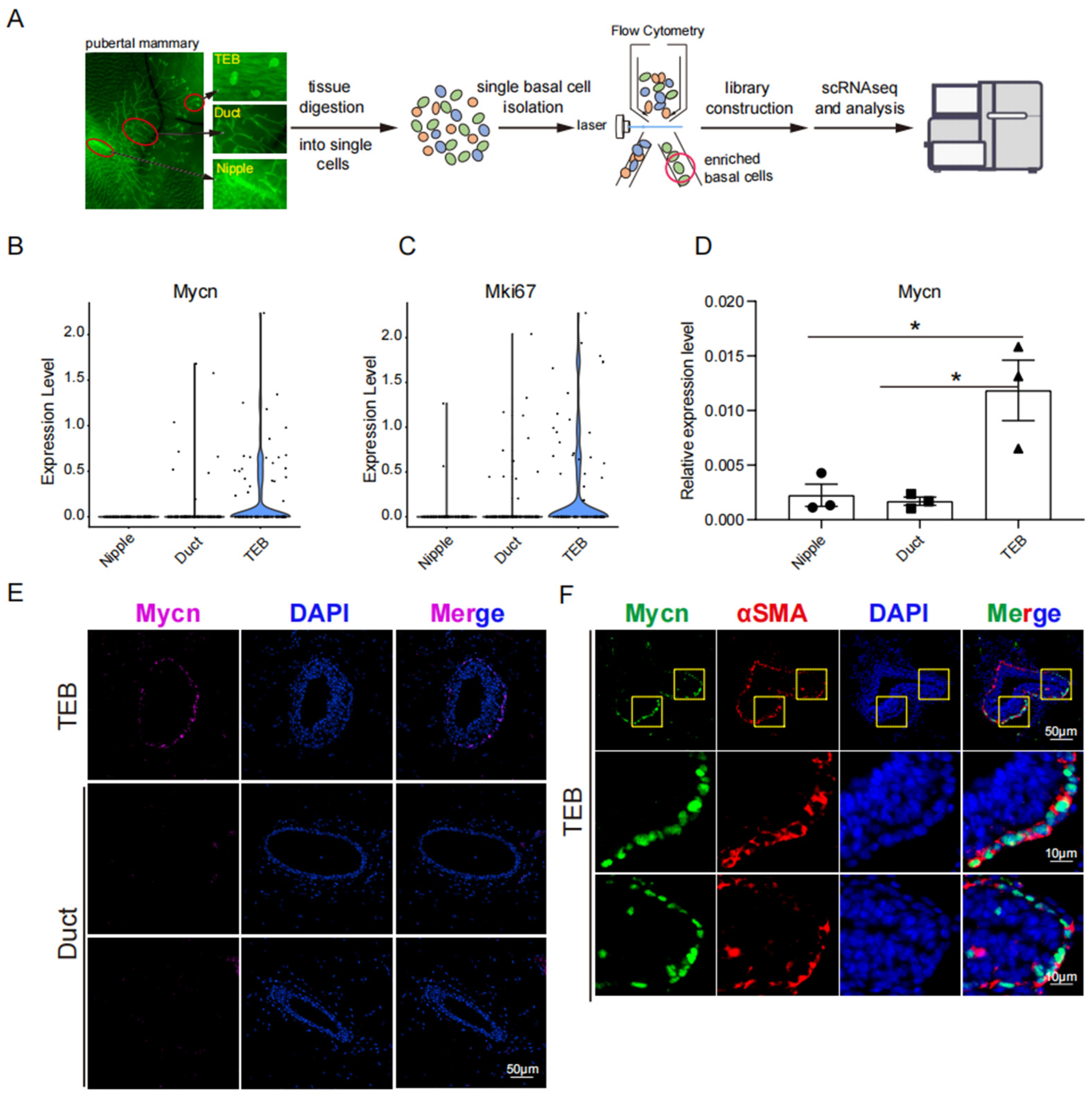
Figure 1. Expression of Mycn in the developing mouse mammary gland. (A) Schematic workflow for single-cell RNA-Seq in (B,C). Distinct mammary tissues, including nipples, ducts and TEBs, from 5-week-old K14-Cre/Rosa26-mTmG mice were micro-dissected and processed into single-cell suspensions for single-cell SMART-Seq. The area circled by the red circle indicates the isolated pure basal population. (B,C) Violin plots showing the expression of transcription factor Mycn (B) and the proliferation marker Mki67 (C) in basal cells in TEB, duct, and nipple. (D) Real-time PCR analysis showing the relative mRNA level of Mycn in TEB, duct, and nipple basal cells normalized to the expression of β-actin. Statistical analysis was performed using a two-tailed unpaired t-test. Data is presented as mean ± SEM, n = 3. * p < 0.05. Each circle, square, or triangle in this bar chart represents a sample from the corresponding group. (E,F) Representative immunofluorescence imaging of the expression of Mycn in duct and TEB. Magenta/GFP: Mycn; Red: αSMA; blue: DAPI. And the portion highlighted by the yellow box in the upper channel of the subfigure (F) is magnified in the lower channel. Scale bar, 10 μm or 50 μm.
Cells 14 01239 g001
Figure 2. Mycn-deficient basal cells were defective in mammary gland reconstitution activity. (A) Schematic diagram illustrating the process of mammary cell transplantation. The Mycn gene was knocked out (KO) using CRISPR in basal cells isolated from the mammary glands of 6-week-old C57BL/6 wild-type (WT) female mice. Subsequently, the Mycn KO and control (CV2) GFP-positive basal cells were injected into the cleared 4th mammary fat pads of 3-week-old recipient C57BL/6 WT female mice and collected 6–8 weeks after transplantation. (B) Representative immunofluorescence images under a stereomicroscope showing the mammary outgrowths of both the Mycn KO and control groups after transplantation. (C) Wholemount carmine stain images showing the mammary outgrowths in (B). (D) Data table showing the numbers of total injections, outgrowths, and repopulating frequency of Mycn KO and control basal populations in the cleared fat pad transplantation assay. (E) ELDA plot of limiting dilution transplant assay showing the frequency of the repopulating unit in the two different subpopulations of Mycn KO and control basal cells.
Cells 14 01239 g002
Figure 3. Mycn-overexpressing basal cells also displayed diminished mammary gland regeneration capacity. (A) Schematic diagram illustrating the transplantation process of of Mycn-overexpressing and control (pCDH) basal cells. These two groups of basal cells were injected into the cleared 4th mammary fat pads of 3-week-old recipient C57BL/6 WT female mice and analyzed 6–8 weeks after transplantation. (B) Representative immunofluorescence images viewed under a stereomicroscope showing the mammary outgrowths in both the Mycn overexpressing and control groups following transplantation. (C) Whole-mount carmine stain images visualizing the mammary outgrowths shown in (B). (D) Data table presenting injection numbers, outgrowths counts, and repopulating frequency of Mycn-overexpressing and control basal populations in the cleared fat pad transplantation assay. (E) ELDA plot of the limiting dilution transplant assay showing the frequency of the repopulating units within the distinct subpopulations of Mycn-overexpressing and control basal cells.
Cells 14 01239 g003
Figure 4. Mycn deficiency inhibited, while overexpression promoted, basal cell proliferation. (A) Colony formation assay for Mycn KO and CV2 basal cells. Five thousand cells per well were seeded for each group and cultured for 3 days. Scale bar, 500 μm. (B) Bar chart showing the average cell numbers for the cultured cells on Day3, as shown in (A). (C) Colony formation assay for Mycn-overexpressing and control (pCDH) basal cells. Five thousand cells per well were seeded for each group and cultured for 4 days and 6 days. Scale bar, 500 μm. (D) Bar chart showing the average cell numbers for the cultured cells on Day4, as shown in (C). (E) Bar chart showing the average cell numbers for the cultured cells on Day6, as shown in (C). Each red or black solid circle in these bar charts in subfigure (B,D,E) represents a sample from the corresponding group. All the above statistical analysis was performed using a two-tailed unpaired t-test. Data is presented as mean ± SEM, n = 3. * p < 0.05, ** p < 0.01.
Cells 14 01239 g004
Figure 5. Mycn overexpression downregulated the quiescent stem cell regulators Bcl11b and Tspan8. (A) Representative FACS plots showing the reduced expression level of Tspan8 in the Mycn-overexpressing group compared with the control group basal cells. (B) Statistic analysis of the percentage of Tspan8+ basal cells in both Mycn-overexpressing and control (pCDH) basal populations, as detected by FACS in (A). (C) Real-time PCR analysis showing the relative mRNA level of several key stem cell regulators in Mycn-overexpressing and control basal cells, normalized to the expression of β-actin. Each circular or square marker in these bar charts in subfigure (B,C) represents an individual sample from the corresponding group. Statistical analysis was performed using a two-tailed unpaired t test. Data is presented as mean ± SEM, n = 3. * p < 0.05, ** p < 0.01, *** p < 0.001.
Cells 14 01239 g005
Figure 6. Bcl11b loss downregulates Tspan8 expression. (A) Schematic diagram illustrating the breeding strategy to generate Krt14 promoter-specific Bcl11b conditional knockout (KO) mice. Through crossing Bcl11bflox/flox/mTmG mice with K14-Cre mice, offspring with the genotype K14-Cre/Bcl11bflox/+/mTmG were obtained. Subsequent backcrossing of this progeny with Bcl11bflox/flox/mTmG mice generated K14-Cre/Bcl11bflox/flox/mTmG mice, enabling specific ablation of Bcl11b in Krt14-positive basal epithelial cells of the murine mammary gland. (B) Representative immunofluorescence and bright field images of the 2nd, 3rd, and 4th mammary fat pads demonstrating reduced mammary branches after Bcl11b KO in 6-week-old female C57BL/6 mice compared with the control mice. (C) Bar chart depicting the effect of conditional Bcl11b KO on mammary branches of different mammary fat pads compared with the control group. Statistical analysis was performed using a two-tailed unpaired t-test. Data are presented as mean ± SEM, n = 3. * p < 0.05. (D) Bar chart illustrating the effect of conditional Bcl11b KO on mammary epithelial composition compared with the control group. Statistical analysis was performed using a two-tailed unpaired t-test. Data are presented as mean ± SEM, n = 3. * p < 0.05. (E,F) Representative FACS plots demonstrating the expression of Bcl11b KO on the expression of Tspan8 in the TEB, duct, and nipple basal cells of 6-week-old pubertal mice (E) or in the duct and nipple basal cells of 4.5–6-month-old adult mice (F) compared with the control mice. All the cells analyzed in FACS were GFP-positive basal epithelial cells. (G) Statistic analysis of the FACS of the percentage of GFP-labeled Tspan8+ basal cells in 6-week-old Bcl11b KO and control mice, as shown in (E). (H) Statistical analysis of the FACS of the percentage of GFP-labeled Tspan8+ basal cells in 4.5–6-month-old Bcl11b KO and control mice, as shown in (F). (I) Real-time PCR analysis showing the effect of Bcl11b KO on the relative mRNA level of Tspan8 in duct and nipple basal cells normalized to the expression of β-actin in adult 4.5–6-month-old mice compared with the control mice. Statistical analysis was performed using a two-tailed unpaired t-test. Data are presented as Mean ± SEM, n = 3–4. * p < 0.05, ** p < 0.01. (J) Real-time PCR analysis showing the relative mRNA level of Bcl11b in duct and nipple basal cells, normalized to the expression of β-actin in adult 4.5–6-month-old mice compared with the control mice. Statistical analysis was performed using a two-tailed unpaired t-test. Data is presented as mean ± SEM, n = 3–4. * p < 0.05, ** p < 0.01, *** p < 0.001.
Cells 14 01239 g006
Figure 7. Mycn regulated cell proliferation and differentiation simultaneously. Detection of Mycn binding sites using the D-CUT&RUN method in Comma Dβ cells. (A) Cnetplot showing KEGG pathway analysis of Mycn target genes in 2 × 10⁶ Comma Dβ cells. (B) Emapplot showing biological processes from GO pathway analysis of Mycn target genes in Comma Dβ cells. (C) Biological processes fromGO pathway analysis of Mycn target genes in Comma Dβ cells.
Cells 14 01239 g007aCells 14 01239 g007bCells 14 01239 g007c
Figure 8. The model for Mycn’s role in the activation of quiescent mammary stem cells. Mycn, a transcription factor and regulator of cell proliferation, serves a fundamental role in mammary gland development and regeneration. Mycn exhibits high expression in TEB basal cells, which are actively engaged in cell proliferation and differentiation. Mycn KO and Mycn-overexpressing basal epithelial cells demonstrated that Mycn enhances cell proliferation and differentiation, indicating that optimal Mycn expression levels are essential for mammary gland regeneration. Additionally, Mycn facilitates quiescent stem cell activation through downregulation of key quiescent stem cell markers and regulators, particularly Bcl11b and Tspan8. The ChIP seq data validated that Mycn coordinates cell proliferation and differentiation. In vivo, the specific knockout of Bcl11b in basal epithelial cells markedly reduced Tspan8 expression and enhanced cell proliferation. The research identified a significant signaling axis, Mycn/Bcl11b/Tspan8, which regulates the activation and differentiation of quiescent stem cells in the mammary gland. Thus, Mycn functions as a critical regulator in mammary gland development and regeneration, activating quiescent stem cells and promoting cell proliferation and differentiation through Bcl11b and Tspan8 downregulation. These findings present important implications for understanding and potentially treating diseases and cancers characterized by Mycn overexpression.
Cells 14 01239 g008
Disclaimer/Publisher’s Note: The statements, opinions and data contained in all publications are solely those of the individual author(s) and contributor(s) and not of MDPI and/or the editor(s). MDPI and/or the editor(s) disclaim responsibility for any injury to people or property resulting from any ideas, methods, instructions or products referred to in the content.