BioMed 2026, 6(3), 15; https://doi.org/10.3390/biomed6030015 - 29 Jun 2026
Abstract
►
Show Figures
Background: Parkinson’s Disease (PD) is a neurodegenerative disorder frequently accompanied by speech impairments, which could serve as non-invasive biomarkers for early detection. This study investigates the efficacy of machine learning models trained on voice and speech acoustic features for distinguishing PD patients from
[...] Read more.
Background: Parkinson’s Disease (PD) is a neurodegenerative disorder frequently accompanied by speech impairments, which could serve as non-invasive biomarkers for early detection. This study investigates the efficacy of machine learning models trained on voice and speech acoustic features for distinguishing PD patients from healthy controls (HC) using the publicly available Italian Parkinson’s Voice and Speech (IPVS) dataset. Methods: A comprehensive set of acoustic features was extracted, including perturbation, prosodic and temporal features, Mel-Frequency Cepstral Coefficients (MFCCs), and Gammatone Cepstral Coefficients (GTCCs). These features were evaluated individually and in combination using six supervised classifiers: Support Vector Machine (SVM), Decision Tree (DT), Random Forest (RF), K-Nearest Neighbors (KNN), XGBoost (XGB), and Multi-Layer Perceptron (MLP). Results: The best-performing configuration combination of GTCC and acoustic features with the SVM model achieved 94.68% accuracy, 94.37% sensitivity, 95.04% specificity, 95.71% precision, 96.04% F1-score, an MCC value of 0.89, and an ROC-AUC of 0.98. In the combination of all feature sets, the most impressive performance was observed with the MLP classifier. This achieved 93.08% test accuracy, 94.30% sensitivity, 91.18% specificity, 94.30% precision, 94.30 F1-score, an ROC-AUC of 0.97, and an MCC value of 0.85. Conclusions: The findings demonstrate that combining clinically relevant acoustic features with robust machine learning classifiers offers a reliable, interpretable, and computationally efficient solution for PD detection. The use of a publicly available dataset, open-source tools, and subject-wise validation contributes to the reproducibility and clinical relevance of the proposed approach. This study reinforces the potential of speech as a digital biomarker for early PD detection and supports the integration of voice-based assessments into diagnostic platforms.
Full article
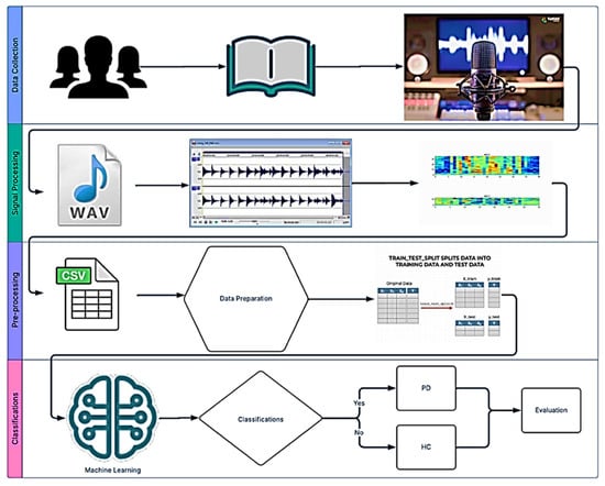
Figure 1


