


Abstract
Thoracic endovascular aortic repair (TEVAR) has transformed the management of type B aortic dissection (TBAD), yet its biological consequences remain substantially less understood than its mechanical effects. Stent-graft implantation occurs within an already injured and immunologically active aorta, creating a new interface between circulating blood, biomaterial, thrombus, endothelium, and the dissected aortic wall. This review examines the immune, thrombotic, cellular, and extracellular matrix responses associated with TEVAR and proposes a framework linking these processes to false-lumen healing and aortic remodeling. Evidence from clinical, translational, biomaterial, imaging, and experimental studies was critically integrated to examine pre-existing inflammatory activity in TBAD; blood–stent-graft interactions; complement, platelet, neutrophil, and monocyte/macrophage responses; post-implantation syndrome; false-lumen thrombosis and organization; endothelial and extracellular-matrix remodeling; and emerging approaches to longitudinal immune phenotyping. Available evidence supports a model in which post-TEVAR inflammation represents a composite response arising from pre-existing dissection biology, procedural injury, biomaterial exposure, altered mechanobiology, and therapeutically induced thrombosis rather than an isolated device reaction. Early immunothrombotic activation may contribute to false-lumen thrombosis and subsequent organization, whereas persistent immune activation, proteolysis, residual perfusion, and failure of inflammatory resolution may accompany adverse remodeling. Conventional markers such as fever, leukocytosis, C-reactive protein, and post-implantation syndrome provide limited mechanistic resolution. Integration of serial cellular and molecular phenotyping with quantitative imaging may better distinguish adaptive healing from persistent thrombo-inflammatory activity. TEVAR should be understood as both a mechanical intervention and a biological perturbation. The clinically relevant objective is not simply to quantify postoperative inflammation, but to define its origin, cellular composition, temporal evolution, and relationship to false-lumen organization and structural remodeling. Prospective longitudinal studies integrating immune phenotyping, device characteristics, quantitative imaging, and selected proteomic, single-cell, and spatial approaches are needed before immune signatures can guide prognosis or therapeutic intervention.
1. Type B Aortic Dissection as an Immunologically Active Vascular Disease
Type B aortic dissection (TBAD) is conventionally defined by its structural consequences: disruption of aortic wall integrity, separation of medial layers, formation of a false lumen, and the subsequent risk of malperfusion, progressive dilatation, rupture, or chronic aneurysmal degeneration. This mechanical description remains fundamental to clinical decision-making, but it incompletely captures the biology of the dissected aorta. Growing experimental and clinical evidence indicates that aortic dissection is accompanied by a coordinated inflammatory response involving the injured vascular wall, circulating innate immune cells, platelets, soluble inflammatory mediators, and extracellular matrix remodeling pathways [1,2,3]. Accordingly, the inflammatory state encountered after thoracic endovascular aortic repair (TEVAR) does not arise in an immunologically quiescent vascular system. Stent-graft implantation occurs within the aorta, in which tissue injury, thrombus formation, disturbed flow, and immune activation are already evolving [4,5]. This pre-existing biological context is essential when attempting to distinguish disease-driven inflammation from responses generated by the procedure or the implanted device.
The initiating structural event in dissection produces an unusually potent inflammatory environment. Intimal disruption and medial injury expose extracellular-matrix components and generate cellular stress, necrosis, and extracellular release of damage-associated molecular patterns (DAMPs) [6,7]. Molecules such as high-mobility group box 1, extracellular ATP, mitochondrial constituents, nucleic acids, and matrix-derived fragments can engage pattern-recognition pathways expressed by endothelial cells and resident or recruited leukocytes [8]. Toll-like receptor signaling and inflammasome-related pathways provide plausible molecular links between sterile tissue injury and the production of inflammatory cytokines, including interleukin (IL)-1β, IL-6, and tumor necrosis factor (TNF) [9,10]. These pathways should not be interpreted as a single linear cascade. Rather, they constitute an interconnected injury-sensing network in which mechanical disruption, cellular death, oxidative stress, coagulation, and inflammation amplify one another within the evolving dissection [11,12].
Neutrophils are among the earliest circulating cells to respond to this environment. Beyond conventional degranulation and reactive oxygen species production, activated neutrophils may contribute to vascular injury through proteolytic enzymes and the formation of neutrophil extracellular traps (NETs) [5,13,14,15]. NETs provide a particularly relevant conceptual link between inflammation and thrombosis because extracellular chromatin decorated with histones and granular proteins can serve as a scaffold for platelet adhesion, coagulation, and further leukocyte recruitment [16,17]. In aortic dissection, however, the biological meaning of neutrophil activation is unlikely to be uniformly detrimental. The same mechanisms that may aggravate endothelial and medial injury can also contribute to thrombus formation within the false lumen [18]. This duality becomes particularly important after TEVAR, when conversion of a persistently perfused false lumen into a thrombosed compartment is generally a prerequisite for favorable remodeling [19,20]. Thus, neutrophil activation and immunothrombosis should be considered not only as markers of injury but also as candidate components of the subsequent healing response.
Monocytes and macrophages represent a second major axis linking acute injury with longer-term remodeling. Recruitment of circulating monocytes into the injured aortic wall can amplify cytokine and chemokine signaling and contribute to extracellular matrix turnover via proteolytic pathways [21]. At the same time, macrophages are indispensable for the clearance of cellular debris, the processing of thrombotic material, the resolution of inflammation, matrix reorganization, and tissue repair [22]. The traditional binary distinction between pro-inflammatory and reparative macrophage phenotypes is unlikely to adequately represent this complexity. Contemporary vascular immunology instead supports a continuum of macrophage states determined by local cytokines, metabolic conditions, hypoxia, extracellular-matrix composition, cell–cell interactions, and the temporal phase of injury [23,24]. In the dissected aorta, macrophage biology may be an important determinant of whether acute inflammation resolves into organized healing or persists as chronic, maladaptive vascular wall remodeling [25,26].
Endothelial injury provides a further interface through which the structural, hemodynamic, and inflammatory consequences of aortic dissection converge. Disruption of intimal continuity exposes circulating blood to normally sequestered subendothelial and medial structures, while separation of the aortic wall into true and false lumina creates a markedly heterogeneous hemodynamic environment characterized by regional differences in flow velocity, wall shear stress, flow reversal, recirculation, and stasis [27,28]. These disturbances are not biologically neutral: endothelial cells exposed to mechanical and inflammatory stress can acquire an activated phenotype, with increased expression of adhesion molecules and chemokines that promote leukocyte adhesion and transmigration, thereby reinforcing local vascular inflammation [29,30]. At sites of endothelial disruption, exposure of thrombogenic surfaces simultaneously initiates platelet recruitment and activation and engages the coagulation system, creating a direct interface between tissue injury and thrombus formation [31,32]. Platelets, however, should not be regarded simply as terminal effectors of hemostasis in this setting. Through surface molecules, including P-selectin and CD40 ligand, and the release of chemokines and other granule-derived mediators, activated platelets communicate bidirectionally with endothelial cells, neutrophils, and monocytes, promoting leukocyte recruitment, heterotypic cell aggregate formation, NET generation, and coagulation [33]. They occupy a central position within the thrombo-inflammatory response, coupling vascular disruption to innate immune activation and thrombosis [34]. Complement signaling may provide an additional amplification pathway: activation of the alternative complement pathway and increased circulating concentrations of C3a and C5a have been demonstrated in thoracic aortic dissection, while experimental evidence indicates that complement signaling can enhance inflammatory cell recruitment and vascular wall injury [35,36]. Nevertheless, the extent to which complement activation contributes specifically to human type B aortic dissection, false-lumen biology, or the subsequent response to TEVAR remains insufficiently defined. Rather than representing independent processes, endothelial activation, platelet–leukocyte interactions, coagulation, and complement signaling are therefore better considered as interconnected components of an evolving vascular injury response whose balance may influence both acute tissue damage and subsequent thrombotic remodeling.
These inflammatory and thrombotic processes are accompanied by continuous remodeling of the extracellular matrix (ECM). Dysregulated matrix metalloproteinase activity, an imbalance with endogenous tissue inhibitors, and leukocyte-derived proteases can alter elastin and collagen architecture, smooth muscle cell function, and, ultimately, medial stability. Importantly, the relationship is bidirectional: matrix degradation not only reflects tissue injury but may also generate bioactive fragments that sustain innate immune signaling, thereby coupling structural failure to persistent inflammation. TBAD should therefore be regarded not as a sequence of isolated cellular events, but as a spatially and temporally evolving tissue response in which vascular injury, inflammation, thrombosis, and matrix remodeling are closely interdependent. The principal cellular and molecular components of this response, their major effector pathways, and their potential relevance to aortic injury and subsequent remodeling are summarized in Figure 1, providing a mechanistic framework for the discussion that follows.
Figure 1.
Type B Aortic Dissection as a Spatially Heterogeneous Immune–Thrombotic–Vascular Ecosystem. Figure legend/footnote. Type B aortic dissection (TBAD) creates a spatially and temporally heterogeneous biological environment in which structural injury to the aortic wall, innate immune activation, thrombosis, altered hemodynamics, endothelial dysfunction, and extracellular matrix remodeling are closely interconnected. Intimal and medial disruption promotes the release of damage-associated molecular patterns (DAMPs), activation of pattern-recognition and inflammatory pathways, and recruitment of neutrophils, monocytes, and platelets. Neutrophil activation, including reactive oxygen species generation, protease release, and neutrophil extracellular trap (NET) formation, intersects with platelet activation and coagulation to promote immunothrombosis within the false lumen. The false lumen itself represents a dynamic biological compartment that varies from persistent perfusion through partial thrombosis to thrombus organization and stabilization. Monocyte/macrophage states may contribute both to inflammatory and proteolytic injury and to phagocytosis, efferocytosis, thrombus organization, matrix reconstruction, and tissue repair. Concurrently, altered true- and false-lumen hemodynamics influence endothelial mechanotransduction, platelet–leukocyte interactions, and vascular cell phenotype, while extracellular matrix turnover may either contribute to progressive wall instability or evolve toward reparative remodeling and fibrosis. Thus, TBAD should be considered an evolving immune–thrombotic–vascular ecosystem in which the balance between persistent injury and inflammatory resolution may influence subsequent aortic stabilization or adverse remodeling. The pathways shown are conceptual and integrate established mechanisms with biologically plausible interactions; they should not be interpreted as a fully demonstrated linear sequence in human TBAD. Solid connectors indicate relationships supported by direct clinical evidence, whereas dashed connectors indicate mechanistically plausible or indirectly supported interactions. Abbreviations: ATP, adenosine triphosphate; DAMP, damage-associated molecular pattern; ECM, extracellular matrix; FL, false lumen; HMGB1, high-mobility group box 1; IL, interleukin; MMP, matrix metalloproteinase; MPO, myeloperoxidase; mtDNA, mitochondrial DNA; NET, neutrophil extracellular trap; NO, nitric oxide; PRR, pattern-recognition receptor; ROS, reactive oxygen species; TBAD, type B aortic dissection; TL, true lumen; TLR, Toll-like receptor; TNF, tumor necrosis factor; VSMC, vascular smooth muscle cell.
The false lumen introduces an additional layer of biological complexity that is not fully captured by its conventional radiological classification as patent, partially thrombosed, or completely thrombosed. These states are associated with distinct patterns of aortic growth and clinical outcomes, suggesting they are unlikely to represent biologically equivalent environments [37]. Rather than constituting a static anatomical endpoint, false-lumen thrombosis should be regarded as an evolving process encompassing coagulation, inflammatory cell recruitment, fibrin and cellular organization, proteolytic activity, and, ultimately, structural remodeling of both the thrombus and the adjacent aortic wall. Persistent false-lumen perfusion maintains communication between circulating blood and the injured vascular surface and has been associated with progressive aortic enlargement, whereas exclusion of the proximal entry tear by thoracic endovascular aortic repair (TEVAR) promotes false-lumen thrombosis, true-lumen expansion, and regression of the false-lumen compartment [23]. Importantly, partial thrombosis should not necessarily be interpreted as merely an intermediate state between patency and complete thrombosis; clinical observations have linked this phenotype to adverse late outcomes, suggesting that residual flow within a partially thrombosed compartment may create a particularly unfavorable local environment [38]. As thrombosis progresses, profound changes are also likely to occur in the local tissue milieu, including alterations in oxygen availability, cellular accessibility, inflammatory signaling, proteolytic activity, and extracellular matrix turnover. These biological processes, however, remain substantially less well characterized than their morphological consequences. The transition from a persistently perfused false lumen to an organized thrombosed compartment should therefore be considered not merely a hemodynamic or radiological event, but a process of active biological remodeling. Although complete false-lumen thrombosis after TEVAR is associated with more favorable aortic remodeling [39], the immune-cell populations, molecular pathways, and temporal signals governing thrombus organization and its interaction with the adjacent dissected wall remain insufficiently defined in humans. This represents an important mechanistic gap between the well-characterized imaging phenotype of false-lumen remodeling and the biological processes that may ultimately determine whether thrombosis progresses toward stable aortic healing or persistent maladaptive remodeling.
The inflammatory biology of type B aortic dissection (TBAD) should be considered not only in spatial but also in temporal terms. Acute dissection represents a phase of abrupt medial injury, exposure of normally sequestered matrix components, thrombus formation, and rapid activation of innate and systemic inflammatory pathways. As the disease evolves into the subacute and chronic phases, this early injury response is progressively replaced—or superimposed upon—by processes related to persistent false-lumen perfusion, thrombus maturation and organization, extracellular-matrix degradation and repair, fibrosis, and structural adaptation of the dissected aortic wall. Importantly, this transition is unlikely to be uniform across the aorta. Patent, partially thrombosed, and completely thrombosed false-lumen segments may coexist within the same patient and provide markedly different local biological environments, while the capacity for favorable aortic remodeling itself appears to diminish as dissection becomes chronic [40,41,42].
A measurable systemic inflammatory phenotype accompanies this process. Elevated leukocyte and neutrophil counts, C-reactive protein (CRP), neutrophil-to-lymphocyte ratio (NLR), systemic immune-inflammation index (SII), systemic inflammatory response index (SIRI), and selected cytokine profiles have all been associated with disease severity, mortality, or subsequent aorta-related adverse events in observational studies of aortic dissection [43,44,45]. In TBAD specifically, higher peak CRP has been associated with adverse long-term outcomes, whereas preprocedural composite indices derived from circulating leukocyte populations have shown prognostic associations with aortic-related events after thoracic endovascular aortic repair (TEVAR) [46]. More recently, characterization of circulating neutrophil subsets has suggested that the inflammatory signal may extend beyond simple changes in absolute leukocyte counts, with the expansion of immature neutrophil populations associated with adverse outcomes after TEVAR [47]. These observations support the existence of a clinically detectable systemic inflammatory phenotype in TBAD, but they should not be interpreted as demonstrating that peripheral inflammation is itself a causal driver of adverse aortic remodeling. Circulating biomarkers represent an integrated systemic readout, incorporating signals derived from the dissected aorta together with those generated by tissue injury, pain and neurohumoral stress, organ ischemia, comorbid disease, pharmacological treatment, and other systemic responses. Moreover, the extent to which peripheral inflammatory signatures reproduce the spatially heterogeneous immune landscape of the dissected aortic wall remains largely unresolved.
This distinction becomes particularly important when interpreting inflammation after TEVAR. Patients do not enter the procedure from a biologically neutral baseline. Rather, the magnitude and composition of pre-existing immune activation are likely to vary with the temporal phase of dissection, the anatomical extent of disease, false-lumen patency and thrombus burden, malperfusion, ongoing tissue injury, and the underlying vascular substrate. Indeed, systemic inflammatory responses after thoracic stent-graft implantation appear to be more pronounced in patients treated for acute rather than chronic aortic pathology, indicating that the biological state of the aorta at the time of intervention materially influences the subsequent inflammatory trajectory [48].
Accordingly, post-TEVAR inflammation is best regarded as a composite biological signal rather than as a device-specific response. Conceptually, at least three partially overlapping components should be distinguished: disease-derived inflammation, originating from the dissection and its associated tissue injury, thrombosis, ischemia, and remodeling; procedure-derived inflammation, induced by endovascular manipulation, contrast exposure, vascular injury, abrupt alterations in flow and pressure, and peri-procedural tissue stress; and device-derived inflammation, related to the implantation of a large, permanent blood- and tissue-contacting biomaterial within an already injured vascular environment. Experimental separation of these components is inherently difficult because they converge within the same early post-interventional window. The well-recognized increases in leukocyte count, CRP, fibrinogen, and other inflammatory markers after thoracic stent-graft implantation cannot, by themselves, establish a biomaterial-specific immune effect [49]. Evidence from the broader endovascular literature that the magnitude of the post-implantation inflammatory response may vary with graft material provides biological plausibility for a device contribution but does not resolve the attribution problem in TBAD [50]. Thus, the central challenge in defining the immunobiology of TEVAR is not simply to demonstrate that inflammation occurs after stent-graft implantation, but to determine which component of the observed response reflects ongoing disease, which represents procedural injury, and which—if any—can be attributed specifically to the implanted endograft.
Recognizing type B aortic dissection (TBAD) as an immunologically active vascular disease fundamentally changes the biological question posed by endovascular treatment. TEVAR is not deployed within an inert conduit that subsequently becomes inflamed simply because a foreign material has been introduced. Rather, the endograft is implanted into an aorta in which medial injury, innate immune activation, coagulation, false-lumen thrombosis, proteolysis, and extracellular-matrix remodeling are already underway. The intervention represents a major perturbation of an evolving immune–thrombotic–vascular system rather than the beginning of that process.
This distinction is important because TEVAR modifies several determinants of local biology almost simultaneously. Exclusion of the proximal entry tear abruptly alters pressure and flow within the false lumen, redistributes wall stress, promotes stasis and thrombosis, and changes the interface between circulating blood, thrombus, and the injured aortic wall [51]. These effects are central to subsequent aortic remodeling: persistent false-lumen perfusion and distal re-entry flow are associated with incomplete thrombosis and less favorable remodeling, whereas effective false-lumen exclusion and thrombosis generally accompany true-lumen expansion and regression of the false lumen [52,53]. The biological consequences of TEVAR cannot be reduced to the presence or absence of a postoperative inflammatory response. A more informative framework is to determine which inflammatory and reparative programs are engaged, from which cellular and anatomical compartments they originate, how their activity evolves over time, and whether they ultimately sustain tissue injury or instead facilitate thrombus organization, inflammatory resolution, matrix repair, and favorable aortic remodeling.
The systemic inflammatory response documented after thoracic endograft implantation—including increases in leukocyte count, C-reactive protein, fibrinogen, and D-dimer—supports the presence of substantial biological perturbation after TEVAR [54,55,56,57]. The post-TEVAR state should therefore be viewed as the convergence of pre-existing disease biology, abrupt hemodynamic reprogramming, newly induced thrombosis, procedural injury, and biomaterial exposure, rather than as a conventional foreign-body response occurring in isolation.
The cellular and molecular pathways operating within the dissected aorta should likewise not be considered as independent mechanisms. Vascular injury and damage-associated signaling recruit and activate innate immune cells; inflammatory and proteolytic pathways influence extracellular-matrix integrity; coagulation and platelet activation generate a thrombotic compartment that is itself biologically active; and subsequent thrombus organization, matrix deposition, and cellular repair contribute to either stabilization or progressive structural failure. The relative importance of these processes is likely to change with disease stage, anatomical location, and false-lumen status. Table 1 summarizes these pathways as an interconnected biological network rather than a catalog of isolated inflammatory mediators, distinguishing, where possible, their proposed contributions to acute tissue injury, thrombosis and thrombus organization, inflammatory resolution, extracellular matrix turnover, and subsequent aortic remodeling.
Table 1.
Immune and Vascular Components of the Preexisting Inflammatory Landscape in Type B Aortic Dissection.
2. Literature Search and Review Strategy
This review was developed as a focused, mechanistically oriented synthesis of the biological response to thoracic endovascular aortic repair (TEVAR) in type B aortic dissection (TBAD). The literature search was conducted primarily through PubMed/MEDLINE, with the objective of integrating clinical observations after TEVAR with evidence from vascular immunology, thrombosis, biomaterial science, vascular biology, and aortic remodeling. The review was designed to address biological mechanisms and translational knowledge gaps rather than to provide a quantitative estimate of treatment effects; accordingly, a narrative critical-synthesis approach was adopted.
Searches combined terms related to the underlying disease and intervention, including “type B aortic dissection”, “aortic dissection”, “thoracic endovascular aortic repair”, “TEVAR”, “thoracic stent graft”, and “endograft”, with mechanistic terms encompassing “inflammation”, “immune response”, “innate immunity”, “post-implantation syndrome”, “neutrophils”, “neutrophil extracellular traps”, “NETs”, “monocytes”, “macrophages”, “platelets”, “complement”, “coagulation”, “immunothrombosis”, “endothelial dysfunction”, “extracellular matrix”, “matrix metalloproteinases”, “proteolysis”, “false lumen”, “false-lumen thrombosis”, “thrombus organization”, “aortic remodeling”, and “vascular remodeling”. Where findings were inconsistent across studies, discordance was interpreted in relation to differences in study population, aortic pathology, disease chronicity, procedural characteristics, endograft platform, biomarker definition, sampling interval, and outcome ascertainment rather than resolved by formal evidence pooling. Additional searches combined TEVAR- or endograft-related terms with biomaterial-specific terms, including vascular biomaterial, polyester, Dacron, expanded polytetrafluoroethylene, ePTFE, protein adsorption, complement activation, platelet activation, and foreign-body response. The search covered PubMed/MEDLINE from database inception through 9 October 2026. No formal language restriction was applied during the literature search.
Because a major objective of the review was to examine the stent graft as a biologically active blood-contacting interface, additional searches incorporated “vascular biomaterial”, “blood–biomaterial interaction”, “vascular graft”, “polyester”, “Dacron”, “expanded polytetrafluoroethylene”, “ePTFE”, “protein adsorption”, “protein corona”, “complement activation”, “platelet activation”, and “foreign-body response”. This broader strategy was necessary because several mechanisms potentially relevant to thoracic endografts have been characterized more extensively in vascular biomaterials and other blood-contacting devices than specifically in TEVAR.
Priority was given to human studies directly addressing TBAD or TEVAR, including prospective and retrospective clinical studies, translational investigations, imaging studies, and systematic reviews or meta-analyses. The search included publications available in PubMed/MEDLINE from database inception through 9 October 2026. Studies were considered for inclusion when they addressed at least one of the predefined biological domains relevant to the review: inflammatory or immune responses in aortic dissection; biological responses to TEVAR or vascular endograft implantation; blood–biomaterial interactions; thrombosis or thrombus organization; endothelial or extracellular-matrix remodeling; or the relationship between these processes and subsequent aortic remodeling. Publications were excluded from the mechanistic synthesis when they addressed TEVAR exclusively from a technical or procedural perspective without biological or remodeling relevance, provided insufficient information to determine the biological context of the reported findings, or duplicated evidence more comprehensively reported elsewhere.
Titles and abstracts were initially evaluated for relevance to these predefined domains, followed by assessment of the full text of potentially informative publications. Human TBAD and TEVAR studies were assigned the greatest interpretative weight. Experimental and mechanistic studies were included when they addressed pathways insufficiently characterized in human TEVAR, whereas evidence from abdominal endovascular aortic repair, other vascular grafts, blood-contacting devices, or experimental biomaterials was incorporated selectively to establish biological plausibility. Reference lists of key original studies and relevant reviews were additionally examined to identify publications not retrieved through the primary keyword combinations. For interpretative purposes, evidence was considered across four levels. The greatest weight was assigned to observations demonstrated directly in human TBAD patients undergoing TEVAR, particularly when biological measurements could be related longitudinally to imaging or clinical outcomes. A second level comprised human evidence from aortic dissection not restricted to TEVAR and from other endovascular aortic interventions, principally EVAR, which was used when the underlying vascular or device-related mechanism was considered relevant but was not assumed to be equivalent. A third level included in vivo experimental models, ex vivo tissue studies, and vascular biomaterial research that provided mechanistic support for specific cellular or molecular pathways. Finally, biological interactions for which direct supporting evidence remains insufficient were treated explicitly as mechanistic hypotheses. Throughout the review, these levels were not considered interchangeable, and mechanistic plausibility was not presented as evidence of causality in human post-TEVAR remodeling. Because the objective was mechanistic integration rather than estimation of a pooled treatment effect, formal risk-of-bias scoring and quantitative meta-analysis were not undertaken.
3. The Stent Graft as a New Blood–Biomaterial–Aortic Wall Interface
The therapeutic rationale for thoracic endovascular aortic repair (TEVAR) in type B aortic dissection (TBAD) has traditionally been framed in predominantly mechanical terms: exclusion of the primary entry tear, reduction of false-lumen pressurization, restoration of true-lumen expansion, promotion of false-lumen thrombosis, and, ultimately, induction of favorable aortic remodeling. This paradigm is well supported by serial imaging studies demonstrating progressive true-lumen expansion and reduction in false-lumen volume after successful endovascular exclusion, although the magnitude and completeness of remodeling vary substantially depending on disease chronicity, distal re-entry flow, and the persistence of false-lumen perfusion [23,71]. The mechanical interpretation of TEVAR is therefore correct, but biologically incomplete.
Deployment of a thoracic endograft simultaneously introduces a large, permanent artificial surface into direct contact with circulating blood and places this material within an aortic segment already undergoing tissue injury, thrombosis, inflammation, and matrix remodeling. TEVAR consequently does more than redirect flow: it creates a new biological compartment in which circulating blood, synthetic graft fabric, metallic scaffold, newly forming thrombus, endothelium, and dissected aortic tissue interact within the same spatial environment. Blood contact with artificial vascular surfaces can initiate protein adsorption and engage platelets, coagulation, leukocytes, and complement, providing a mechanistic basis for considering the endograft as a biologically active interface rather than an entirely inert mechanical prosthesis [72]. Consistent with this concept, thoracic stent-graft implantation has been associated with activation of neutrophils, platelets, endothelium, and complement, as well as sustained increases in systemic inflammatory and coagulation markers [56]. The local consequences of these interactions within the dissected thoracic aorta, however, remain considerably less well characterized than the mechanical performance of the device.
This distinction is particularly relevant in TBAD because the endograft is not introduced into an immunologically quiescent or structurally intact arterial segment. Dissection disrupts endothelial continuity and medial architecture, exposes normally sequestered vascular and extracellular-matrix components, initiates thrombus formation, and establishes an inflammatory milieu before any endovascular intervention is undertaken. At the same time, true- and false-lumen flow, pressure gradients, recirculation, stasis, and wall stress are profoundly altered. TEVAR is therefore superimposed upon an established injury–inflammation–thrombosis–remodeling continuum, while abruptly changing the hemodynamic conditions that regulate that continuum. Direct intra-false-lumen pressure measurements confirm that entry-tear exclusion can produce immediate changes in false-lumen pressure that are subsequently associated with regional thrombosis and remodeling [64]. Thus, mechanical correction and biological response should not be regarded as sequential and independent events; they are likely to be closely coupled components of the same post-interventional process.
The host response to stent-graft implantation must consequently be interpreted against this pre-existing biological background. Post-TEVAR fever, leukocytosis, and elevations in C-reactive protein (CRP), fibrinogen, and D-dimer are well documented, and the magnitude of this response appears greater in acute than in chronic aortic disease and may increase with greater endograft burden. Moreover, evidence from abdominal endovascular repair indicates that graft composition itself can influence the intensity of post-implantation inflammation, with polyester-based devices producing greater systemic inflammatory responses than expanded polytetrafluoroethylene-based grafts in several clinical series [57,65]. Although these observations support a contribution from the implanted biomaterial, they do not permit attributing the postoperative inflammatory response specifically to the device, particularly in TBAD, where disease-related inflammation, procedural injury, contrast exposure, abrupt hemodynamic change, false-lumen thrombosis, and biomaterial exposure occur concurrently.
Accordingly, conventional systemic markers should be regarded as indicators of the integrated host response rather than direct measures of local device biocompatibility. Fever, leukocyte count, and CRP can quantify the systemic expression of post-implantation inflammation, but they cannot identify its anatomical source, discriminate disease-derived from procedure- or device-derived signaling or resolve the cellular and molecular events occurring at the newly established interface. A more mechanistically informative approach would therefore interrogate the blood–biomaterial–thrombus–aortic wall interface itself, including platelet and coagulation activation, complement engagement, neutrophil and monocyte responses, endothelial perturbation, thrombus maturation, and their relationship to subsequent tissue repair and aortic remodeling. Such a framework moves the biological interpretation of TEVAR beyond the question of whether inflammation occurs after implantation toward the more relevant questions of where it originates, which pathways sustain it, how it evolves over time, and whether individual components ultimately promote injury, resolution, or favorable remodeling.
3.1. Stent-Graft Composition Is Biologically Relevant
Contemporary thoracic endografts should not be regarded as biologically passive barriers but rather as complex composite implants whose interactions with the host are determined by several interdependent material and structural properties. Most devices combine a polymeric graft fabric—principally woven polyester or expanded polytetrafluoroethylene (ePTFE)—with a metallic supporting framework, commonly incorporating nitinol, together with device-specific sutures, fixation elements, connecting components, coatings, and delivery-related structures. Beyond determining radial force, conformability, sealing behavior, porosity, fatigue resistance, and long-term mechanical integrity, these characteristics define the blood-contacting and tissue-facing interfaces of the implant and may therefore influence protein adsorption, platelet adhesion and activation, complement engagement, leukocyte recruitment, and subsequent thrombo-inflammatory signaling. Contemporary analyses of endograft design consequently support the concept that fabric chemistry cannot be considered in isolation from scaffold configuration, device stiffness, surface architecture, and the overall quantity of prosthetic material introduced into the circulation [73].
Among these variables, graft fabric has historically generated the most consistent clinical signal. Much of this evidence derives from endovascular abdominal aortic aneurysm repair (EVAR), in which polyester-based endografts have repeatedly been associated with a more pronounced early inflammatory phenotype than PTFE-based devices. In a contemporary series of 367 elective EVAR procedures, Oddi et al. reported post-implantation syndrome (PIS) in 19.4% of patients receiving polyester devices, compared with only 1.2% of those treated with PTFE grafts, with concomitant differences in inflammatory burden and length of hospitalization [66]. More recent systematic evidence has reinforced this association: a 2024 systematic review encompassing 31 studies and 2847 patients found polyester graft use to be strongly overrepresented among patients who developed PIS, whereas PTFE devices were substantially less frequently represented in this group [74]. Collectively, these observations support the proposition that the implanted biomaterial can modify the magnitude of the acute host response, although they do not establish fabric composition as its sole—or necessarily dominant—biological determinant.
This distinction becomes particularly important when these observations are extrapolated from infrarenal EVAR to thoracic EVAR. The inflammatory phenotype following TEVAR reflects a substantially more complex interaction among the underlying aortic disease, procedural tissue injury, extent of aortic coverage, foreign-surface exposure, thrombus formation, and patient-specific inflammatory susceptibility. Indeed, recent TEVAR-specific evidence challenges an overly deterministic interpretation of graft composition. In a cohort of 369 TEVAR procedures, Leone et al. reported PIS in 26.8% of patients but found no relevant association between stent-graft composition and its occurrence; instead, thoracic aortic coverage exceeding 150 mm emerged as a strong independent risk factor, and greater coverage was associated with higher postoperative leukocyte and CRP responses [75]. These findings suggest that the biological consequences of endografting may depend at least as strongly on the extent and anatomical context of implantation as on the nominal identity of the graft fabric itself.
Polyester should not be characterized simply as “pro-inflammatory,” nor should ePTFE be regarded as biologically inert. A more defensible interpretation is that material composition constitutes one component of a multidimensional device–host interaction in which fabric chemistry, surface architecture, metallic scaffold, implanted surface area, aortic coverage, thrombus formation, and the pre-existing vascular inflammatory state collectively determine the magnitude and phenotype of the postoperative response [76,77]. This qualification is particularly important in type B aortic dissection (TBAD), where TEVAR is performed within an already injured and immunologically active aortic compartment characterized by endothelial disruption, medial damage, disturbed flow, inflammatory-cell recruitment, and evolving false-lumen thrombosis. The inflammatory signal observed after implantation in this setting cannot be attributed solely to the prosthesis; rather, it should be interpreted as the composite output of disease biology, procedural injury, device–blood interactions, thrombosis, and subsequent aortic remodeling. Defining the relative contribution of each of these components remains an important unresolved question for translational TEVAR research [78].
3.2. The Biological Interface Begins with Protein Adsorption, Not Cellular Adhesion
The earliest biological interaction with an implanted endograft does not occur between circulating leukocytes and a pristine synthetic surface. Rather, within seconds of blood contact, the exposed biomaterial becomes conditioned by the rapid adsorption of plasma proteins, effectively replacing the original physicochemical interface with a biologically active, protein-rich layer. This adsorbed compartment may contain fibrinogen, albumin, immunoglobulins, complement and coagulation proteins, among numerous other circulating constituents, and it is this newly formed interface—rather than the underlying polymer in isolation—that is subsequently encountered by platelets and immune cells [79]. Accordingly, the biological identity of a blood-contacting endograft should be considered an acquired and evolving property of the material–protein interface rather than an intrinsic property of the polymer alone.
The consequences of this initial conditioning extend well beyond passive surface coverage. The abundance, orientation, conformation, and biological accessibility of adsorbed proteins can influence platelet adhesion and activation, coagulation, complement engagement, and subsequent inflammatory-cell recruitment [80,81]. These processes are themselves sensitive to material characteristics, including surface chemistry, wettability, hydrophilicity, roughness, porosity, topography, and mechanical properties. Experimental vascular-graft studies illustrate this principle particularly clearly: modification of surface hydrophilicity can substantially alter platelet adsorption and thrombotic behavior, while contemporary antithrombogenic graft strategies are increasingly directed toward limiting nonspecific protein deposition and the cellular responses that follow it [5]. Moreover, the adsorbed layer should not be regarded as static. Plasma proteins compete for available binding sites and may undergo sequential adsorption, displacement, and conformational rearrangement—the classical Vroman-type behavior of blood–biomaterial interfaces—thereby creating a temporally evolving biological surface whose composition may differ substantially from that present immediately after implantation [82]. Thus, endografts with comparable macroscopic mechanical performance need not necessarily present equivalent biological interfaces to circulating blood.
The extent to which these immediate blood–biomaterial interactions contribute to the inflammatory phenotype observed after thoracic endovascular aortic repair remains largely unresolved. Protein adsorption, platelet adhesion, complement activation, and leukocyte engagement are well-established features of blood-contacting biomaterials; however, their direct relevance to contemporary thoracic endografts cannot simply be assumed. In particular, the composition, temporal evolution, and biological activity of the protein layer acquired by a thoracic stent-graft after deployment have not been systematically characterized in patients. The concept of an endograft-associated “protein corona” therefore provides a biologically compelling framework, but at present it should be regarded as a mechanistic hypothesis rather than a demonstrated pathway of post-implantation inflammation after TEVAR.
This distinction is especially important in type B aortic dissection (TBAD), because the endograft is introduced into a vascular compartment that is already biologically perturbed before any foreign material is deployed. Endothelial disruption, medial injury, abnormal shear and flow patterns, activation of coagulation and inflammatory pathways, and the presence of a variably perfused and thrombogenic false lumen together create a complex pre-existing substrate upon which device-related responses are superimposed. Consistent with this interpretation, contemporary clinical studies indicate that the magnitude of inflammation after TEVAR is influenced not only by the implanted device but also by the underlying aortic pathology and the extent of treatment. Aortic dissection has been associated with a greater postoperative inflammatory response than other thoracic aortic pathologies, while fresh parietal thrombus and the extent of stent-graft coverage have emerged as important correlates or predictors of post-implantation syndrome [83]. Notably, recent TEVAR-specific data found no independent relationship between stent-graft composition and post-implantation syndrome, whereas thoracic aortic coverage exceeding 150 mm was strongly associated with its occurrence and with greater postoperative leukocyte and C-reactive protein responses [84]. These observations argue against a simple material-centric model and instead support a more integrated concept in which device exposure, thrombus formation, procedural injury, and disease-specific vascular biology interact to determine the systemic inflammatory phenotype.
An additional limitation of the current literature is that clinical investigation begins predominantly downstream of the initiating interface. Post-TEVAR inflammation is generally defined or quantified using fever, leukocytosis, C-reactive protein, and, less consistently, circulating cytokines [85]. These measurements establish the presence of a systemic inflammatory response but provide limited insight into the molecular events occurring during the first minutes and hours at the blood–endograft boundary. Consequently, a substantial mechanistic interval remains between prosthesis implantation and the clinical phenotype conventionally termed post-implantation syndrome. Resolving this interval will require prospective studies that interrogate the earliest phases of device–host interaction, ideally integrating temporal proteomic characterization of the endograft-associated protein layer with markers of complement and coagulation activation, platelet responses, leukocyte phenotypes, and subsequent false-lumen thrombosis and aortic remodeling. Such an approach could determine whether the blood–endograft interface is itself an initiating driver of inflammation, a modifier of an already activated host response, or predominantly an epiphenomenon of the broader biological consequences of TEVAR.
3.3. Complement, Platelets, and Leukocytes: An Interconnected Foreign-Surface Response
Complement provides a plausible first layer of biological amplification at the blood–stent-graft interface. Exposure of circulating blood to a non-endothelialized artificial surface can initiate complement activation, generating C3a, C5a, and surface-bound complement products, as well as downstream inflammatory signals that recruit and activate leukocytes and modify endothelial and platelet behavior [86,87]. Complement activation in this setting should not, however, be conceptualized as an autonomous inflammatory pathway. Rather, it forms part of an interconnected thromboinflammatory network in which complement, contact activation, coagulation, platelets, and innate immune cells reciprocally reinforce one another [88]. Thus, even spatially restricted interactions on the graft surface could, in principle, translate into a broader systemic inflammatory signal. This framework is well established for blood-contacting biomaterials, although its specific contribution to the inflammatory response following thoracic endograft implantation remains insufficiently characterized.
Platelets represent a second major interface between material recognition, thrombosis, and inflammation. Rapid adsorption of fibrinogen and other adhesive plasma proteins onto artificial surfaces can create a substrate permissive for platelet adhesion and activation, while graft deployment simultaneously alters local geometry, shear forces, blood–surface interactions, and thrombogenic conditions [89]. Once activated, platelets cease to function merely as cellular mediators of hemostasis. Through P-selectin-dependent adhesion, chemokine and granule release, and direct interactions with circulating leukocytes, they can influence neutrophil recruitment and activation, promote monocyte inflammatory and procoagulant phenotypes, and further amplify endothelial and coagulation responses [90,91]. Platelet–neutrophil and platelet–monocyte aggregates are therefore particularly attractive candidate intermediates through which an initially localized blood–material interaction might be propagated into systemic thromboinflammation. Their role after TEVAR, however, has not been adequately defined by prospective cellular phenotyping.
Neutrophils may occupy a particularly important position within this proposed cascade because several signals present during acute type B aortic dissection and its endovascular treatment converge on neutrophil activation. Complement-derived mediators, activated platelets, damaged endothelium, tissue-derived damage-associated molecular patterns, thrombin generation, and the pre-existing inflammatory milieu of the dissected aorta collectively provide biologically credible stimuli for neutrophil recruitment and activation. The resulting response may include degranulation, reactive oxygen species generation, protease release, and formation of neutrophil extracellular traps (NETs), thereby linking inflammation directly to thrombosis [92]. NETs are particularly relevant because their extracellular DNA–histone framework can support platelet adhesion, concentrate procoagulant molecules, facilitate thrombin generation, and contribute to the development and structural organization of fibrin-rich thrombi [5,93]. Notably, recent integrated proteomic and single-cell analyses have demonstrated enhanced NET formation in both the systemic circulation and aortic microenvironment of patients with aortic dissection, with macrophage–neutrophil signaling through the CXCL3/CXCR2 axis implicated in disease progression. These observations considerably strengthen the biological relevance of NETs to aortic-dissection pathology, but they do not establish TEVAR or the stent-graft surface itself as the initiating stimulus. Stent-graft-induced NETosis should therefore remain framed as a compelling mechanistic hypothesis rather than an established component of post-implantation syndrome.
Monocytes and macrophages may become increasingly important as the response evolves from acute thromboinflammation toward thrombus organization and tissue remodeling. Activated platelets can induce inflammatory and procoagulant transcriptional programs in monocytes, while complement products, cytokines, endothelial activation, and chemotactic signals arising from injured aortic tissue provide additional mechanisms for their recruitment and functional reprogramming [94,95]. Once recruited into thrombus and adjacent tissue, monocyte-derived macrophages can participate in cellular debris clearance, extracellular matrix turnover, fibrinolysis, angiogenesis, and ultimately thrombus resolution and organization [96]. Such functions are particularly relevant to TBAD, in which the false lumen undergoes a dynamic transition from circulating blood to fresh thrombus and subsequently to variably organized tissue. It is therefore plausible that the early neutrophil-dominant inflammatory phase after TEVAR is followed by a more heterogeneous monocyte/macrophage response governing thrombus maturation and aortic remodeling. At present, however, this temporal immune trajectory remains largely inferential: serial studies integrating leukocyte phenotyping, platelet–leukocyte interactions, NET biomarkers, complement activation, false-lumen thrombosis, and subsequent aortic remodeling after TEVAR are notably lacking.
3.4. The Endothelium Is Both Target and Mediator
The biological relationship between a thoracic stent graft and the aortic wall differs fundamentally from that of a conventional intravascular device deployed within an otherwise intact, endothelialized vessel. In TBAD, the device traverses a spatially heterogeneous vascular environment: the proximal and distal landing zones may retain relatively preserved endothelial coverage, whereas the intervening dissected segment is characterized by intimal disruption, exposure of medial and extracellular-matrix components, mural and false-lumen thrombus, and variable residual communication between the true and false lumina. TEVAR therefore creates not a single blood–material interface, but a longitudinal mosaic of mechanically and biologically distinct microenvironments. At the landing zones, stent-graft apposition, oversizing, radial force, and device conformability modify local wall mechanics and concentrate mechanical stress, particularly at the boundaries between stented and native aorta [97,98]. Within the treated segment, exclusion of the proximal entry tear and expansion of the true lumen fundamentally reorganize blood–wall interactions, while residual distal fenestrations continue to determine pressure transmission, flow exchange, and thrombosis within the false lumen [99,100]. These effects are biologically relevant because vascular endothelial cells continuously interpret hemodynamic forces through mechanotransduction pathways that couple shear stress and mechanical deformation to nitric oxide bioavailability, redox signaling, junctional integrity, permeability, and inflammatory gene expression [101]. TEVAR should therefore be viewed not only as an anatomical intervention that redirects blood flow but also as a perturbation of one of the principal environmental signals governing vascular cell phenotype.
This perspective broadens the interpretation of post-TEVAR inflammation beyond direct recognition of the implanted material. A proportion of the host response may instead—or additionally—arise indirectly through hemodynamic reprogramming and mechanotransduction. Entry-tear exclusion alters true- and false-lumen pressure gradients, flow velocity, recirculation, residence time, oscillatory shear, and regional wall shear stress, while progressive false-lumen thrombosis further modifies this environment over time [102]. These parameters are not biologically neutral: disturbed or low shear can favor endothelial activation and dysfunction, alter platelet–wall interactions and leukocyte adhesion, and create local conditions permissive for thrombus initiation and propagation. Computational studies of TBAD increasingly support a close relationship between post-TEVAR flow patterns, shear-related indices, residual re-entry anatomy, and the subsequent distribution of false-lumen thrombosis [103]. What remains largely unresolved is whether these macroscopic hemodynamic changes translate into measurable spatial or temporal differences in endothelial and immune-cell phenotype in patients. Nevertheless, the available evidence argues against treating mechanical exclusion and inflammatory remodeling as independent consequences of TEVAR. They are more plausibly viewed as interdependent components of the same biological process, in which device-induced changes in geometry and force reshape local flow, altered flow modifies endothelial and thrombotic signaling, and the resulting cellular response may, in turn, influence thrombus organization and subsequent aortic remodeling. This reciprocal interaction between biomaterial properties, mechanical forces, and host thrombo-inflammatory responses provides a more integrated framework for understanding the biological consequences of thoracic stent-graft implantation, as summarized in Figure 2.
Figure 2.
The Stent Graft as a Dynamic Blood–Biomaterial–Aortic Wall Interface: Coupling Surface Chemistry, Mechanics, and Thrombo-Inflammation. Figure legend/footnote. Thoracic endovascular aortic repair (TEVAR) creates a dynamic interface between the implanted stent graft, circulating blood, newly forming thrombus, endothelium, and the dissected aortic wall. Immediately after implantation, adsorption of plasma proteins onto the graft surface establishes a biologically active interface capable of engaging platelets, coagulation, complement, and leukocytes. These pathways form an interconnected thrombo-inflammatory network rather than independent responses. Concurrently, device apposition, radial force, stiffness, conformability, and compliance mismatch modify local vascular mechanics, while exclusion of the primary entry tear abruptly redistributes true- and false-lumen pressure and flow, altering wall shear stress and endothelial mechanotransduction. Reduced false-lumen inflow promotes stasis and thrombosis, whereas residual distal perfusion may sustain thrombus turnover and biological activity. The evolving false-lumen thrombus is itself an active compartment in which early platelet–neutrophil interactions and immunothrombosis may transition toward monocyte/macrophage-mediated clearance, thrombus organization, extracellular-matrix remodeling, and fibrosis. The magnitude and phenotype of the post-TEVAR host response are therefore determined by the interaction of pre-existing disease biology, procedural injury, biomaterial properties, device burden and mechanics, altered hemodynamics, and thrombosis. Favorable trajectories may progress toward inflammatory resolution, thrombus organization, false-lumen regression, and aortic stabilization, whereas persistent false-lumen perfusion and unresolved thrombo-inflammatory and proteolytic activity may contribute to adverse remodeling. The temporal sequence and mechanistic pathways shown are conceptual; their relative contributions and causal relationships remain incompletely established in human TEVAR. Solid connectors indicate relationships supported by direct clinical evidence, whereas dashed connectors indicate mechanistically plausible or indirectly supported interactions. Abbreviations: CRP, C-reactive protein; ECM, extracellular matrix; ePTFE, expanded polytetrafluoroethylene; FL, false lumen; MMP, matrix metalloproteinase; MPO, myeloperoxidase; NET, neutrophil extracellular trap; NO, nitric oxide; PIS, post-implantation syndrome; ROS, reactive oxygen species; TBAD, type B aortic dissection; TEVAR, thoracic endovascular aortic repair; TL, true lumen; WSS, wall shear stress.
3.5. Device Burden: Does More Implanted Material Mean More Inflammation?
An intuitively attractive hypothesis is that the magnitude of the inflammatory response after endovascular aortic repair should increase in proportion to the amount of prosthetic material implanted. Greater thoracic aortic coverage necessarily introduces a larger blood-contacting foreign surface and extends the length of the aorta exposed to stent-graft apposition, radial force, altered compliance, flow redistribution, and false-lumen exclusion. From this perspective, total graft length, estimated fabric surface area, the number of implanted components, and the extent of device overlap represent plausible determinants of post-procedural inflammation. Indeed, early clinical observations after TEVAR suggested that inflammatory biomarker responses may be more pronounced in patients receiving multiple stent-graft components, providing some support for a relationship between procedural device burden and systemic inflammation [56]. However, subsequent evidence has not established a consistent linear dose–response relationship between the quantity of implanted material and post-implantation syndrome (PIS). In a frequently cited analysis in which both graft dimensions and newly formed thrombus were quantified, the amount of implanted graft material was not independently associated with PIS, whereas woven polyester grafts were associated with a substantially greater inflammatory response than expanded polytetrafluoroethylene (ePTFE) devices [65]. Subsequent observational studies and systematic analyses have broadly reinforced the importance of graft composition, repeatedly identifying polyester-based devices as associated with a higher incidence or intensity of post-implantation inflammation than ePTFE-based grafts [66]. Collectively, these findings suggest that surface quality may be at least as biologically important as surface quantity and challenge a purely volumetric interpretation of device-induced inflammation.
Longer treated segments generally reflect more extensive aortic disease and may require multiple components, longer instrumentation, more extensive manipulation of the diseased aortic wall, greater contrast exposure, and a larger volume of newly excluded aorta. In TBAD, extended coverage may additionally alter the proportion of false lumen exposed to residual flow and the volume and spatial distribution of newly forming thrombus—processes that themselves may contribute to inflammatory and coagulation responses. The difficulty of isolating these interdependent exposures may partly explain why individual studies have reported inconsistent associations among device dimensions, thrombus formation, and PIS. Contemporary systematic reviews similarly emphasize substantial heterogeneity in PIS definitions, devices, treated aortic pathologies, and measured inflammatory endpoints, limiting firm conclusions regarding individual procedural determinants [93].
A more informative framework would conceptualize the implanted endograft as a multidimensional exposure incorporating fabric chemistry and surface characteristics, total blood-contacting area, metallic scaffold architecture, component number and overlap, radial and longitudinal force distribution, conformability and apposition to the aortic wall, and the evolving proportion of the prosthetic surface that remains directly exposed to circulating blood versus becoming incorporated into thrombus or apposed to vascular tissue. Materials science further supports this distinction, as polyester and PTFE differ not only in chemical composition but also in structural, mechanical, and surface properties relevant to blood–material and vessel–device interactions [104]. Future mechanistic studies should therefore move beyond using graft length as a single surrogate variable and instead characterize the effective biological exposure generated by a particular device within a particular aortic anatomy. Such an approach may help reconcile the apparently discordant literature and determine whether post-implantation inflammation is governed primarily by the quantity of implanted material, its physicochemical properties, the host tissue environment, or—most plausibly—the interaction among all three.
3.6. Mechanical Forces Are Part of the Biological Exposure
The metallic scaffold warrants consideration not merely as a constituent material of the endograft, but as a mechanically active component that maintains a persistent interaction with the aortic wall. Thoracic endografts generate sustained radial and longitudinal forces to achieve apposition, preserve luminal patency, and resist migration; however, in TBAD these forces are transmitted to an aortic wall whose structural continuity and load-bearing capacity have already been disrupted by dissection. The resulting mechanical environment is determined not by radial force alone, but by the interaction among device oversizing, scaffold stiffness, spring-back force, conformability, native aortic curvature, landing-zone geometry, and the compliance mismatch between stented and non-stented segments. Contemporary biomechanical analyses support the clinical relevance of these interactions, demonstrating that local stress concentrations can develop at the margins of the endograft and that device-related mechanical forces contribute to distal stent-graft-induced new entry (dSINE) [105]. Importantly, the development of dissection-specific endografts designed specifically to reduce distal radial force further underscores that the scaffold should be regarded as a persistent mechanical intervention rather than as a passive carrier of graft fabric [64].
These mechanical effects have traditionally been considered primarily in relation to structural complications, particularly dSINE, endograft-induced aortic injury, and adverse remodeling. Their potential biological consequences deserve equal consideration. Vascular cells continuously sense tensile strain, compression, substrate stiffness, and shear through integrins, cytoskeletal structures, cell–cell junctions, mechanosensitive ion channels, and downstream transcriptional programs. Sustained or spatially concentrated mechanical stress may therefore influence endothelial integrity, vascular smooth muscle cell phenotype, extracellular matrix synthesis and degradation, and inflammatory signaling. In the dissected aorta, where extracellular-matrix architecture and normal force transmission are already profoundly altered, the biological consequences of chronic device–wall interaction may differ substantially from those occurring in a structurally intact vessel. Nevertheless, direct evidence demonstrating clinically meaningful immune activation specifically attributable to chronic endograft-associated mechanical stress remains lacking. The distinction between the chemistry of the implanted surface and the mechanics of the implanted device should therefore be regarded primarily as an analytical convenience: from the perspective of the host tissue, both exposures occur simultaneously and within the same spatially heterogeneous interface.
An analogous interdependence applies to the hemodynamic consequences of TEVAR. Successful proximal entry-tear exclusion produces an immediate redistribution of pressure and flow between the true and false lumina and promotes subsequent true-lumen expansion, false-lumen thrombosis, and reduction in false-lumen volume [64,106]. These changes constitute the anatomical basis of favorable aortic remodeling, but they simultaneously redefine the local biological environment. Direct intra-false-lumen pressure measurements demonstrate that entry closure can substantially modify pressure relationships throughout the dissected aorta, with the magnitude of these changes corresponding spatially to subsequent false-lumen thrombosis and remodeling [107]. Conversely, persistent distal re-entry, residual false-lumen perfusion, and partial thrombosis can maintain an adverse hemodynamic environment and compromise remodeling [84]. Thus, false-lumen thrombosis should not be regarded merely as the passive endpoint of successful flow exclusion. Thrombus formation itself changes the interface between circulating blood and the dissected wall, modifies transport and local flow conditions, and initiates cellular processes involved in thrombus organization and tissue repair; as remodeling progresses, the resulting changes in lumen geometry and wall configuration again alter local mechanical forces. TEVAR therefore initiates a dynamic feedback system in which device mechanics modify hemodynamics, hemodynamics influence thrombosis, thrombosis alters the local inflammatory and reparative milieu, and tissue remodeling subsequently reshapes the mechanical environment in which the device operates.
The biological exposure created by TEVAR cannot be reduced to the graft fabric or the metallic scaffold in isolation. It is more appropriately conceptualized as an evolving host–device ecosystem determined jointly by material composition and surface properties, scaffold architecture, implanted device burden, radial and longitudinal force transmission, device–wall apposition, aortic geometry, residual false-lumen communication, and the hemodynamic consequences of entry-tear exclusion. These determinants are likely to dominate different phases of the response: immediate blood–material interactions may be most relevant during the early post-implantation period, whereas altered force transmission, false-lumen thrombosis, and progressive remodeling may become increasingly important thereafter. Their individual contributions remain difficult to disentangle clinically because device selection, treatment length, anatomical complexity, procedural burden, and dissection severity are intrinsically interrelated. Table 2 integrates these determinants according to the principal biological interface involved, their proposed thrombo-inflammatory or remodeling consequences, and—critically—the strength and limitations of the evidence supporting their relevance to human TEVAR.
Table 2.
Stent-Graft-Related Determinants of the Host Immune Response: From Biomaterial Properties to Biological Consequences.
3.7. Device-Related or Procedure-Related Inflammation? A Persistent Attribution Problem
A major limitation of the current literature is the difficulty of attributing post-TEVAR inflammation specifically to the implanted device. TEVAR superimposes several biological perturbations on an aorta already characterized by dissection-associated inflammation: vascular access and instrumentation, mechanical interaction between the endograft and injured aortic wall, abrupt redistribution of true- and false-lumen hemodynamics, and newly induced false-lumen thrombosis [7]. Indeed, both the underlying aortic pathology and the amount of newly formed thrombus appear to influence the magnitude of the inflammatory response after TEVAR [104]. Consequently, postoperative increases in CRP, leukocyte count, or IL-6 cannot be interpreted as direct measures of a foreign-body response. Conversely, the inability of these nonspecific biomarkers to isolate a device-specific signal does not imply that the endograft is biologically neutral. Material-dependent differences in PIS provide indirect evidence for a device contribution, although they neither quantify its magnitude nor identify the responsible cellular pathways [93]. A more useful framework is therefore to consider post-TEVAR inflammation as a dynamic composite phenotype arising from four overlapping components: pre-existing dissection-associated inflammation, procedural tissue injury, biomaterial-driven immune activation, and thrombo-inflammation associated with false-lumen thrombosis (Figure 3). These signals are likely to differ in timing and magnitude and, importantly, to interact rather than simply coexist. Single-time-point measurements are therefore inherently inadequate to resolve their respective contributions, emphasizing the need for longitudinal immune profiling integrated with serial assessment of false-lumen thrombosis and subsequent aortic remodeling [115].
Figure 3.
Deconvolution of the Post-TEVAR Inflammatory Signal: Disease-, Procedure-, Device-, and Thrombosis-Derived Components. Figure legend/footnote. The systemic inflammatory response observed after thoracic endovascular aortic repair (TEVAR) should be interpreted as a composite biological signal rather than as a direct measure of device-induced inflammation. Four partially overlapping components contribute to this phenotype. Disease-derived inflammation is already present before intervention and reflects dissection-related intimal and medial injury, damage-associated molecular pattern signaling, endothelial activation, leukocyte recruitment, coagulation, extracellular-matrix injury, false-lumen thrombosis, and, when present, ischemia or malperfusion. Procedure-derived inflammation arises from vascular access, endovascular instrumentation, acute vascular-wall perturbation, contrast exposure, procedural stress, and abrupt redistribution of pressure and flow. Device-associated inflammation reflects the newly established blood–biomaterial and device–wall interfaces, including protein adsorption, platelet and coagulation activation, complement engagement, leukocyte interactions, and mechanical effects related to device apposition, radial force, stiffness, compliance mismatch, and extent of aortic coverage. Thrombosis-derived inflammation accompanies therapeutically induced false-lumen thrombosis and evolves from coagulation and early immunothrombotic activity toward thrombus organization or, in the setting of persistent perfusion and turnover, sustained inflammatory and proteolytic signaling. These components have distinct yet overlapping temporal trajectories; therefore, fever and conventional circulating markers such as CRP, leukocyte count, IL-6, fibrinogen, and D-dimer provide an integrated systemic readout and cannot identify the anatomical or mechanistic origin of the response. Longitudinal profiling beginning before TEVAR and integrating cellular immune phenotyping, endothelial, complement, platelet, and immunothrombotic markers, proteomics, and serial quantitative CTA may help deconvolve these signals and determine whether individual biological trajectories are associated with inflammatory resolution, false-lumen organization, and favorable remodeling or with persistent immune activation and adverse aortic remodeling. The temporal curves and relative contributions depicted are conceptual and are not intended to represent quantitatively established kinetics or causal relationships in humans. Abbreviations: CRP, C-reactive protein; CTA, computed tomography angiography; DAMP, damage-associated molecular pattern; ECM, extracellular matrix; FL, false lumen; IL, interleukin; NET, neutrophil extracellular trap; PIS, post-implantation syndrome; TBAD, type B aortic dissection; TEVAR, thoracic endovascular aortic repair; TL, true lumen.
This distinction defines an important unmet need in human TBAD research. Longitudinal profiling from the pre-implantation phase through early post-TEVAR inflammation and subsequent aortic remodeling, integrating cellular immune phenotyping with markers of endothelial injury, immunothrombosis, and proteomic signatures, may help distinguish disease-, procedure-, thrombus-, and device-related components of the host response [59,83]. Emerging studies already demonstrate that preoperative neutrophil phenotypes, postoperative systemic inflammatory indices, and circulating NET-associated markers carry prognostic information after TEVAR, while proteomic and single-cell approaches reveal substantial immune heterogeneity within aortic dissection [5,116]. The relevant question is therefore no longer simply whether the stent graft “induces inflammation,” but which biological responses are specifically modified by implantation, how they evolve over time, and whether they promote favorable false-lumen healing or maladaptive remodeling. Viewed in this context, the thoracic endograft is not merely a mechanical barrier across the entry tear, but the central component of an evolving blood–biomaterial–thrombus–aortic wall interface whose biology extends from immediate thrombo-inflammatory signaling to thrombus organization and chronic vascular remodeling. Defining this trajectory will be essential for distinguishing adaptive healing from persistent immune activation and, ultimately, for understanding TEVAR as both a mechanical and a biological intervention.
4. Acute Immune Activation After TEVAR: From Molecular Signaling to Post-Implantation Syndrome
The inflammatory response following thoracic endovascular aortic repair (TEVAR) is commonly referred to as post-implantation syndrome (PIS), typically characterized by fever, leukocytosis, and elevated C-reactive protein (CRP) levels in the absence of infection [117]. Although clinically useful, this construct may oversimplify the underlying biology. Inflammatory activation occurs across a broader spectrum after thoracic stent-graft implantation, whereas PIS may represent the clinically overt end of this continuum rather than a discrete biological event [104]. TEVAR simultaneously introduces several potentially interacting stimuli, including aortic-wall manipulation, endothelial injury, altered flow, foreign-surface exposure, and, in dissection, abrupt changes in false-lumen perfusion and thrombosis [111,118]. The resulting phenotype is therefore more appropriately viewed as an integrated thrombo-inflammatory response than as an isolated rise in conventional inflammatory markers.
This distinction is particularly relevant in type B aortic dissection (TBAD), where the preprocedural inflammatory state varies considerably with disease phase. Patients treated during acute dissection may already exhibit neutrophilia, elevated CRP, tissue injury, coagulation activation, and evolving false-lumen thrombosis, whereas subacute or chronic intervention occurs within a substantially different biological environment [59]. Accordingly, the postoperative signal represents a composite of disease-related inflammation and the additional perturbation imposed by TEVAR. Its magnitude and temporal trajectory may therefore be more biologically informative than a single postoperative CRP or leukocyte value—or the binary classification of PIS itself.
4.1. The Earliest Response: Innate Immune Activation Within Hours
Human data suggest that the inflammatory response to TEVAR begins within hours of stent-graft deployment, well before conventional acute-phase markers reach their postoperative peak. In patients undergoing elective TEVAR for TBAD, IL-1β and IL-6 exhibited a striking but transient monophasic increase, reaching concentrations approximately 4- to 27-fold above baseline before declining within 6 h and approaching preprocedural values by 24 h; notably, other measured cytokines did not show a comparable response [119]. This selective temporal pattern is biologically informative. IL-1β and IL-6 are central mediators of sterile inflammation, linking tissue injury and innate immune activation to the subsequent systemic acute-phase response, including hepatic CRP production. Accordingly, CRP measured one or two days after TEVAR should be interpreted primarily as a delayed, integrated consequence of earlier inflammatory signaling rather than as a direct marker of its initiation. Importantly, the rapid attenuation of IL-1β and IL-6 in uncomplicated elective cases also argues against equating early cytokine activation with pathological inflammation. The biologically relevant distinction may instead lie in whether this initial response resolves appropriately or becomes amplified and sustained.
4.2. Neutrophils as Early Effectors of the Peri-Implantation Response
Among circulating leukocytes, neutrophils are particularly well positioned to respond to the abrupt biological perturbation created by TEVAR. Periprocedural increases in neutrophil activation markers, including myeloperoxidase and lactoferrin, have been reported after thoracic stent-graft procedures, with activation detectable early enough to suggest contributions from procedural stimuli—including contrast exposure and vascular manipulation—as well as from the implanted device itself. Within the dissected aorta, neutrophils encounter a convergence of DAMPs, endothelial injury, platelet and complement activation, disturbed flow, and evolving false-lumen thrombosis, providing multiple and potentially synergistic triggers for activation. Once engaged, they may amplify thrombo-inflammation by releasing reactive oxygen species, proteases, granular enzymes, and neutrophil extracellular traps (NETs). Importantly, human studies now demonstrate increased NET formation in aortic dissection and associate circulating NET-related markers with adverse outcomes; however, these observations do not yet establish NETosis as a response specifically induced by TEVAR or as a defining mechanism of PIS [5,14]. Equally relevant, flow-cytometric profiling in TBAD has shown that increased preoperative CD10− immature neutrophils, rather than total neutrophil burden alone, independently predicted subsequent aorta-related adverse events after TEVAR [59]. Collectively, these findings suggest that neutrophil phenotype and functional state may be biologically more informative than conventional leukocyte counts or neutrophil-to-lymphocyte ratios.
4.3. Complement, Platelets, and Endothelium: A Coordinated Thrombo-Inflammatory Response
The acute biological response to TEVAR extends well beyond leukocyte and cytokine activation. Human studies of thoracic stent-graft implantation have demonstrated a coordinated response involving complement, platelets, and the vascular endothelium: terminal complement complex levels increase after intervention and may remain elevated for several days, while platelet counts fall in parallel with increased neutrophil-activating peptide-2 and soluble vascular cell adhesion molecule-1, consistent with concomitant platelet and endothelial activation [120]. These processes are unlikely to represent independent phenomena. Complement activation can promote leukocyte recruitment and endothelial responses, while activated platelets interact with neutrophils and monocytes, creating reciprocal links among innate immunity, coagulation, and vascular inflammation [73]. TEVAR should therefore be viewed as inducing a coordinated thrombo-inflammatory response, rather than an isolated postoperative cytokine reaction. This concept is particularly relevant in TBAD, where the intervention deliberately redirects flow and promotes false-lumen thrombosis; thus, thrombosis is not merely a consequence of treatment but an integral component of the biological environment in which post-TEVAR inflammation develops.
4.4. From Cytokine Signaling to the Acute-Phase Response
The inflammatory response after TEVAR unfolds as a temporally ordered, rather than synchronous, biological process. In a well-characterized cohort of 133 patients with type B acute aortic syndromes, IL-6 and D-dimer peaked approximately 24 h after intervention, followed by leukocyte count and CRP at 48 h and fibrinogen at 72 h, suggesting progression from early cytokine and coagulation activation toward a delayed systemic acute-phase response [121]. Longer serial observations similarly demonstrated a leukocyte peak around the second postoperative day, whereas CRP, fibrinogen, and D-dimer remained elevated for substantially longer periods [56]. These distinct trajectories are methodologically important: studies based on isolated sampling points may miss early cytokine peaks or underestimate later acute-phase responses. Future mechanistic studies should therefore align sampling with the expected kinetics of individual pathways rather than relying solely on routine postoperative blood-collection schedules.
The available human data support a temporally structured response to TEVAR in which molecular, cellular, thrombotic, and systemic inflammatory events evolve along partially overlapping trajectories rather than as a single synchronized reaction. To integrate these observations without implying a rigid sequence or a uniform response across patients, Figure 4 presents a conceptual framework that links the earliest peri-procedural events to subsequent systemic inflammation and the clinically recognizable PIS phenotype.
Figure 4.
Temporal Architecture of the Human Immune Response after TEVAR: From Early Molecular Activation to the Clinical PIS Phenotype. Figure legend/footnote. Conceptual temporal architecture of the inflammatory and thrombo-inflammatory response following thoracic endovascular aortic repair (TEVAR). The biological response begins from a disease-conditioned baseline rather than an immunologically quiescent state, particularly in acute type B aortic dissection (TBAD), where vascular-wall injury, damage-associated molecular pattern signaling, leukocyte activation, coagulation, endothelial dysfunction, and evolving false-lumen thrombosis may already be present. Stent-graft deployment superimposes procedural injury, blood–biomaterial interaction, abrupt hemodynamic redistribution, and therapeutically induced false-lumen thrombosis on this pre-existing milieu. Early post-implantation events include transient cytokine signaling, particularly involving IL-1β and IL-6, together with neutrophil activation and coordinated complement, platelet, endothelial, and coagulation responses. These molecular and cellular events precede the conventional systemic acute-phase response. Human TEVAR studies suggest that IL-6 and D-dimer may peak at approximately 24 h, followed by leukocyte and CRP responses at approximately 48 h and a later fibrinogen response, although the magnitude and timing vary among patients and studies. The clinically recognizable post-implantation syndrome (PIS), conventionally characterized by otherwise unexplained fever, leukocytosis, and elevated CRP, is therefore best understood as a downstream clinical phenotype arising from overlapping biological pathways rather than as a distinct mechanism. Beyond the acute phase, the response may evolve toward monocyte transition, endothelial recovery, thrombus organization, and vascular stabilization or toward persistent cellular, endothelial, thrombotic, and proteolytic activity potentially associated with incomplete healing and adverse remodeling. Importantly, normalization of systemic inflammatory markers does not necessarily indicate complete biological resolution within the aortic wall or false lumen. Accordingly, temporal characteristics—including response amplitude, time to peak, duration, area under the response curve, and resolution slope—may provide greater biological information than a single peak measurement or binary PIS classification. The curves depict conceptual, population-level temporal relationships derived from available human evidence and should not be interpreted as quantitatively established kinetics, fixed temporal boundaries, or proven causal sequences. Abbreviations: CRP, C-reactive protein; DAMP, damage-associated molecular pattern; ECM, extracellular matrix; FL, false lumen; IL, interleukin; MPO, myeloperoxidase; NET, neutrophil extracellular trap; PIS, post-implantation syndrome; ROS, reactive oxygen species; sVCAM-1, soluble vascular cell adhesion molecule-1; TBAD, type B aortic dissection; TEVAR, thoracic endovascular aortic repair.
4.5. Post-Implantation Syndrome: Phenotype Rather than Mechanism
Post-implantation syndrome (PIS) represents the clinically apparent end of the inflammatory spectrum following TEVAR, conventionally characterized by otherwise unexplained fever, leukocytosis, and elevated CRP in the absence of infection. Importantly, no universally accepted diagnostic definition exists, and differences in biomarker thresholds, observation windows, and inclusion of CRP contribute substantially to the variability in reported incidence [122]. Clinically, PIS is generally characterized by otherwise unexplained postoperative fever accompanied by leukocytosis and/or CRP elevation in the absence of an identifiable infectious source. However, no universally accepted diagnostic definition exists, and thresholds for temperature, leukocyte count, CRP concentration, and the required observation period vary across studies [123]. Accordingly, postoperative fever should not be attributed to PIS until clinically relevant alternative causes, particularly infection and procedure-related complications, have been reasonably excluded. This heterogeneity in diagnostic criteria substantially contributes to variability in reported PIS incidence and should be considered when comparing its clinical and prognostic significance across TEVAR studies [124]. In a well-characterized cohort of 133 patients undergoing TEVAR for type B acute aortic syndromes, PIS occurred in 15.8% and was associated with substantially higher peak leukocyte, CRP, fibrinogen, D-dimer, and IL-6 concentrations; notably, IL-6 increased significantly only in patients who subsequently developed PIS, suggesting that amplification of early cytokine signaling may precede the overt clinical phenotype [125]. Other TBAD series have reported incidences of approximately 29–32%, underscoring the influence of case mix, procedural characteristics, device platforms, and diagnostic criteria [126]. A recent systematic review encompassing 31 endovascular aortic studies similarly found PIS in approximately one-quarter of patients and confirmed substantial methodological heterogeneity [93].
PIS should therefore be regarded as a clinical inflammatory phenotype rather than a discrete biological entity. Dichotomization inevitably compresses a continuous and multidimensional host response into an arbitrary threshold-based diagnosis and may obscure substantial immune activation in patients who do not formally meet PIS criteria. The available human evidence instead supports an integrated response in which cytokine, leukocyte, complement, platelet, endothelial, coagulation, and fibrinolytic pathways evolve along distinct but overlapping trajectories. Table 3 synthesizes these observations by biological compartment examined, their temporal dynamics, and, importantly, the strength and limits of the mechanistic inference that can reasonably be drawn from current human evidence.
Table 3.
Human Evidence for the Acute Immune and Thrombo-Inflammatory Response to TEVAR: Biological Signals, Temporal Dynamics, and Interpretative Boundaries.
4.6. Why Do Some Patients Develop a More Pronounced Response?
The magnitude of the inflammatory response following TEVAR appears to be determined by the interplay among the patient’s biological state, the acuity and extent of aortic disease, and the cumulative burden of the intervention itself. This distinction is important because TEVAR is not performed against a uniform biological background. Acute aortic pathology is generally accompanied by greater tissue injury, thrombus turnover, and systemic inflammatory activation than chronic disease, potentially lowering the threshold for an amplified postoperative response. Consistent with this concept, preprocedural inflammatory status also appears relevant: contemporary TEVAR data identify higher baseline leukocyte counts as an independent predictor of PIS, suggesting that at least part of the postoperative phenotype may reflect amplification of an inflammatory state already present before device deployment [84,104].
Procedural burden provides an additional—and increasingly compelling—determinant. In a 547-patient TBAD cohort, PIS occurred in 28.9% of patients, and age, emergency intervention, operative duration, contrast-medium volume, and the number of main stent-graft prostheses were incorporated into a multivariable prediction model [126]. These observations are complemented by more recent TEVAR-specific data showing that thoracic aortic coverage exceeding 150 mm was independently associated with an approximately fivefold higher probability of PIS and with greater postoperative leukocyte and CRP responses [84]. Importantly, stent-graft composition itself was not independently associated with PIS in that study. Earlier human observations similarly demonstrated that the magnitude of the systemic inflammatory response increased with the number of implanted thoracic stent grafts, further linking procedural extent to postoperative inflammatory activation [56].
Greater aortic coverage simultaneously increases blood–device contact, modifies a larger vascular territory, and is likely to accompany more extensive disease and greater thrombus formation; multiple components increase foreign-surface exposure but also procedural manipulation; prolonged intervention increases cumulative vascular instrumentation; and contrast exposure introduces an additional biological perturbation. From this perspective, PIS is better understood as the clinically recognizable expression of an integrated host–disease–procedure interaction than as the consequence of any single inflammatory trigger.
4.7. Acute and Chronic TBAD Are Not Immunologically Equivalent
Disease timing should be regarded as a biological variable rather than a simple chronological descriptor. Human data demonstrate that acute aortic dissection is accompanied by substantially greater systemic inflammatory activity than chronic disease, including higher IL-6 and CRP concentrations, while longitudinal molecular imaging in acute TBAD shows that inflammatory activity within the dissected aorta evolves substantially during progression from the acute to chronic phase [14]. Importantly, this transition does not imply biological quiescence: chronic TBAD may retain metabolically active regions and ongoing platelet, coagulation, and fibrinolytic activity related to false-lumen morphology and subsequent aortic expansion [132]. The substrate encountered by TEVAR therefore changes qualitatively with time—from predominantly acute injury and innate inflammatory activation toward persistent thrombus turnover and structural remodeling. This temporal heterogeneity has direct methodological implications: pooling acute, subacute, and chronic TBAD may obscure biologically distinct responses and confound attribution of postoperative inflammation to the endograft itself. Accordingly, time from dissection onset to TEVAR should be incorporated into mechanistic studies as an integral component of the inflammatory phenotype.
4.8. Does a Stronger Inflammatory Response Predict a Worse Outcome?
Whether post-TEVAR inflammation is merely a marker of tissue injury or actively contributes to adverse aortic remodeling remains unresolved. Several observations nevertheless suggest biological relevance. In uncomplicated TBAD, a higher preoperative neutrophil-to-lymphocyte ratio independently predicted adverse events during follow-up, while systemic inflammatory response indices have similarly been associated with subsequent aortic-related events [54]. More refined cellular phenotyping further strengthens this signal: increased circulating CD10− immature neutrophils independently predicted aorta-related adverse events after TEVAR, suggesting that an inflammatory phenotype—not simply leukocyte burden—may identify a biologically vulnerable host [59].
Conversely, the prognostic significance of the overt PIS phenotype remains inconsistent. Contemporary thoracic data associate PIS primarily with prolonged hospitalization rather than with mortality, whereas the broader endovascular literature has failed to demonstrate a reproducible relationship with long-term cardiovascular events or survival [133]. This apparent discrepancy may reflect a fundamental limitation of conventional inflammatory endpoints: peak inflammatory magnitude is not necessarily equivalent to biological consequence. A brisk, self-limited response accompanying effective false-lumen thrombosis may represent adaptive healing, whereas persistent endothelial activation, thrombo-inflammation, or failure of inflammatory resolution could plausibly favor adverse remodeling. The clinically relevant phenotype may therefore reside less in the presence of PIS or peak CRP than in the trajectory, cellular composition, and resolution of the inflammatory response, as well as in the evolution of false-lumen thrombosis.
5. Immunothrombosis, False-Lumen Healing, and Long-Term Aortic Remodeling
Post-TEVAR inflammation should not necessarily be interpreted as an adverse biological response. The available human evidence does not establish that these immune responses independently drive false-lumen organization or favorable remodeling. Early immune and thrombotic activation may participate in thrombus formation and organization, but may also occur downstream of the profound changes in false-lumen flow, pressure, and stasis induced by TEVAR [134,135]. Conversely, persistent inflammatory activity may contribute to ongoing vascular injury, but may equally represent a consequence of residual false-lumen perfusion, continued tissue damage, incomplete thrombus organization, or greater underlying disease severity [5]. Current observations should therefore be interpreted primarily as associations embedded within a coupled mechanical–biological process rather than as evidence of a unidirectional causal pathway.
In TBAD, favorable remodeling requires false-lumen thrombosis and subsequent reorganization of the injured vascular compartment—processes inherently linked to innate immunity, cellular clearance, extracellular-matrix turnover, and tissue repair. Conversely, persistent inflammatory and proteolytic activity may sustain vascular injury and promote progressive remodeling. The biologically relevant distinction may therefore be less between inflammation and its absence than between a self-limited reparative response and inflammation that fails to resolve. This proposed divergence—from controlled thrombo-inflammatory healing toward persistent vascular injury and adverse remodeling—is summarized in Figure 5.
Figure 5.
Divergent Biological Trajectories after TEVAR: Resolution and Aortic Healing versus Persistent Thrombo-Inflammation and Adverse Remodeling. Figure legend/footnote. Thoracic endovascular aortic repair (TEVAR) excludes the proximal entry tear, reduces false-lumen (FL) inflow and pressure, and promotes stasis and thrombosis. The resulting FL thrombus should be regarded as a provisional biological scaffold rather than as the endpoint of healing. Early immunothrombosis involves reciprocal interactions among coagulation, platelets, neutrophils, monocytes/macrophages, endothelium, and inflammatory mediators and may subsequently diverge toward two conceptual biological trajectories. In a resolving trajectory, declining neutrophil and platelet–leukocyte activation is accompanied by macrophage-mediated clearance and efferocytosis, thrombus organization, fibrocellular infiltration, extracellular-matrix (ECM) reconstruction, and controlled fibrosis, ultimately favoring FL regression, true-lumen (TL) expansion, and aortic stabilization. Conversely, persistent FL perfusion through distal re-entry or residual channels may sustain blood–wall–thrombus interactions, ongoing thrombus turnover, endothelial and leukocyte activation, oxidative and proteolytic injury, and an imbalance between ECM degradation and repair, potentially contributing to incomplete thrombus organization and adverse aortic remodeling. Macrophages are depicted as context- and time-dependent regulators rather than as binary pro- versus anti-inflammatory phenotypes. Importantly, radiological evidence of FL thrombosis does not necessarily indicate biological maturity of the thrombus; anatomically similar thrombosis patterns may encompass different degrees of cellular clearance, organization, inflammatory resolution, and matrix reconstruction. Integration of longitudinal immune and thrombotic phenotyping with quantitative aortic imaging may therefore provide greater biological information than isolated biomarkers or single-time-point imaging. The pathways, temporal sequence, and molecular relationships depicted are conceptual and hypothesis-generating; several components have not been directly demonstrated longitudinally within the human post-TEVAR false lumen and should not be interpreted as established causal mechanisms. This framework emphasizes that successful post-TEVAR remodeling may depend not simply on the induction of FL thrombosis, but on its progression toward inflammatory resolution, biological organization, and structural stabilization. Abbreviations: CTA, computed tomography angiography; DAMP, damage-associated molecular pattern; ECM, extracellular matrix; FL, false lumen; MMP, matrix metalloproteinase; MPO, myeloperoxidase; NET, neutrophil extracellular trap; ROS, reactive oxygen species; TEVAR, thoracic endovascular aortic repair; TGF-β, transforming growth factor beta; TIMP, tissue inhibitor of metalloproteinases; TL, true lumen; VSMC, vascular smooth muscle cell.
This framework also challenges the interpretation of false-lumen thrombosis as a purely radiological endpoint. Thrombus status is strongly associated with subsequent aortic behavior, yet partial thrombosis may coexist with continued false-lumen expansion and late adverse events [136,137]. Moreover, metabolic imaging demonstrates persistent biological activity within chronically dissected aortas and links this activity with thrombotic and fibrinolytic biomarkers [132]. Thus, entry-tear exclusion creates the hemodynamic conditions for remodeling but does not itself complete the reparative process. The subsequent fate of the false lumen likely reflects the interplay among thrombosis, inflammation, matrix remodeling, and resolution, rather than thrombosis alone.
5.1. False-Lumen Thrombosis as a Form of Therapeutically Induced Immunothrombosis
The concept of immunothrombosis provides a useful framework for interpreting false-lumen thrombosis after TEVAR. Rather than viewing coagulation and inflammation as parallel processes, immunothrombosis recognizes their reciprocal coupling through platelets, neutrophils, monocytes, endothelium, complement, and tissue factor-dependent coagulation [126,138]. Platelets are central to this interface, providing a procoagulant surface while recruiting and activating innate immune cells; importantly, platelet–leukocyte interactions may contribute not only to thrombus propagation but also to inflammatory resolution and tissue repair [139]. Thrombin further extends this crosstalk beyond fibrin generation through protease-activated receptor signaling in platelets and vascular cells, linking coagulation directly to inflammatory and remodeling pathways [140].
TEVAR creates conditions in which such interactions may become particularly relevant. Exclusion of the proximal entry tear profoundly alters false-lumen hemodynamics and promotes thrombosis, whereas residual flow and partial thrombosis remain closely associated with subsequent remodeling [100]. The post-TEVAR false lumen may therefore be conceptualized as a newly established thrombo-inflammatory niche, in which altered flow initiates thrombosis and cellular–molecular interactions subsequently influence thrombus propagation, organization, and resolution. This interpretation remains a mechanistic framework rather than an established human pathway: direct demonstration of a coordinated immunothrombotic program within the post-TEVAR false lumen is still lacking.
5.2. Thrombus Formation Is Only the Beginning of False-Lumen Healing
Complete false-lumen thrombosis is commonly regarded as a surrogate of successful exclusion, yet thrombus formation should not be equated with biological healing. The newly formed thrombus represents a provisional matrix composed predominantly of fibrin, erythrocytes, platelets, and recruited inflammatory cells; its long-term significance depends on whether this structure undergoes effective organization and incorporation into a stable remodeled compartment. Importantly, false-lumen status itself carries prognostic information, but conventional imaging provides limited insight into the cellular maturity of the thrombus [59].
Thrombus organization requires transition from acute coagulation and neutrophil-rich inflammation toward monocyte/macrophage recruitment, cellular clearance, neovascularization, matrix deposition, and fibrosis [141]. The false-lumen thrombus may therefore be considered a temporary biological scaffold rather than a final therapeutic endpoint. As illustrated in Figure 6, therapeutically induced thrombosis potentially initiates a continuum extending from immunothrombosis through thrombus organization to structural stabilization.
Figure 6.
From Therapeutically Induced Immunothrombosis to False-Lumen Healing: Cellular and Molecular Maturation of the Post-TEVAR Thrombus. Figure legend/footnote. Conceptual model of the cellular and molecular maturation of false-lumen (FL) thrombus after thoracic endovascular aortic repair (TEVAR). Stage I, proximal entry-tear exclusion reduces FL inflow and velocity, alters local shear conditions, and promotes stasis, coagulation, and early fibrin deposition, thereby creating the hemodynamic substrate for thrombosis without itself constituting biological healing. Stage II, the newly formed thrombus represents an acute immunothrombotic compartment containing fibrin, erythrocytes, activated platelets, neutrophils, and recruited monocytes, with reciprocal interactions among coagulation, innate immunity, and platelet activation. Neutrophil-derived reactive oxygen species, myeloperoxidase, proteases, and potentially neutrophil extracellular traps may contribute to this early environment, although direct evidence for TEVAR-specific NET involvement remains limited. Stage III, successful progression toward resolution is proposed to involve declining neutrophil-dominant activity, monocyte/macrophage recruitment, phagocytosis and efferocytosis, controlled fibrinolysis, and clearance of cellular and fibrin-rich material, with macrophages acting as context- and time-dependent intermediaries between inflammation and tissue repair. Stage IV: stromal/fibroblast cell infiltration, reparative vascular smooth muscle cell activity, neovascularization, collagen deposition, and extracellular matrix (ECM) organization progressively convert the provisional thrombus into an organized fibrocellular tissue compartment. Stage V, a mature organization is characterized by collagen-rich ECM, reduced inflammatory-cell density, regulated matrix turnover, and controlled fibrotic consolidation, potentially contributing to structural stabilization of the excluded FL. Successful maturation requires a regulated balance between proteolysis and matrix reconstruction rather than complete suppression of ECM turnover. Residual FL perfusion or distal re-entry may interrupt this sequence by sustaining thrombus turnover, leukocyte recruitment, damage-associated molecular pattern signaling, and proteolysis, resulting in incomplete organization and a persistently active FL. Importantly, radiological evidence of thrombosis does not necessarily establish biological maturity: similarly, thrombosed FLs may differ substantially in cellular composition, inflammatory resolution, and matrix organization. Accordingly, mechanical entry-tear exclusion and FL thrombosis should be distinguished from the subsequent biological processes required for durable tissue organization and structural stabilization. The cellular sequence, molecular transitions, and temporal intervals depicted are conceptual and hypothesis-generating; direct longitudinal characterization of human post-TEVAR FL tissue remains limited, and the pathways should not be interpreted as an established linear or causal sequence. Abbreviations: CTA, computed tomography angiography; DAMP, damage-associated molecular pattern; ECM, extracellular matrix; FL, false lumen; MMP, matrix metalloproteinase; MPO, myeloperoxidase; NET, neutrophil extracellular trap; ROS, reactive oxygen species; TEVAR, thoracic endovascular aortic repair; TGF-β, transforming growth factor beta; TIMP, tissue inhibitor of metalloproteinase; TL, true lumen; VSMC, vascular smooth muscle cell.
This distinction may partly explain why apparently similar radiological patterns do not invariably translate into equivalent outcomes. Persistent communication through distal re-entry sites, residual channels, or incomplete thrombus organization may maintain blood–wall interaction and ongoing biological activity despite exclusion of the proximal entry site. Conversely, a progressive organization could increasingly isolate the injured false-lumen Surface, thereby favoring fibrosis and resolution. Thus, the prognostic significance of false-lumen thrombosis may depend not only on its extent, but also on its biological maturity—a property that current clinical imaging captures only indirectly.
5.3. Macrophages as Determinants of Resolution and Repair
Macrophages are plausible determinants of the transition from acute thrombosis to organization and repair. Their function is considerably more complex than a binary pro- versus anti-inflammatory classification. Contemporary human single-cell studies demonstrate marked macrophage heterogeneity in dissected aortic tissue, including monocyte-derived populations that express programs associated with both inflammation and tissue repair [9]. Macrophages may consequently participate sequentially in phagocytic clearance, proteolysis, angiogenesis, extracellular-matrix remodeling, and fibrosis.
A central component of this transition is the clearance of apoptotic and damaged cells. In thrombus resolution biology, monocyte/macrophage recruitment facilitates the removal of cellular and fibrin-rich material, neovascularization, and matrix remodeling [23]. Failure of effective clearance could, conversely, perpetuate DAMP release and inflammatory signaling. Within the dissected aortic wall, macrophages are also closely linked to MMP-mediated extracellular-matrix remodeling, illustrating their potential to contribute to both structural injury and subsequent repair [26,61].
Whether these processes occur in the same manner within the human post-TEVAR false lumen remains unknown. Peripheral monocyte phenotyping may capture systemic activation but cannot reliably reconstruct tissue-conditioned macrophage states. Resolving this question will therefore require integration of circulating immune phenotypes with appropriately selected human tissue, spatial or single-cell approaches, and translational models of post-TEVAR thrombus organization.
5.4. Extracellular-Matrix Turnover: The Structural Consequence of Immune Resolution—Or Its Failure
The long-term objective of TEVAR is not false-lumen thrombosis per se, but durable stabilization and favorable remodeling of the dissected aorta. This outcome ultimately depends on the restoration of extracellular matrix (ECM) integrity through coordinated regulation of vascular smooth muscle cells, fibroblasts, collagen and elastin turnover, transforming growth factor signaling, and proteolytic pathways, particularly matrix metalloproteinases (MMPs) [142].
Inflammation and matrix remodeling are closely interdependent. Neutrophil- and macrophage-derived proteases, MMPs, cathepsins, and oxidative stress can accelerate degradation of structural matrix components, whereas products of tissue injury may sustain innate immune signaling. A persistent imbalance between matrix degradation and repair may therefore establish a self-reinforcing inflammatory–proteolytic state that favors continued aortic-wall degeneration [143].
Successful healing requires interrupting this cycle and transitioning from acute thrombo-inflammation to cellular clearance, matrix organization, and reparative stromal responses. In this setting, fibrosis is not necessarily maladaptive: within an excluded false lumen, controlled fibrotic organization of thrombus may represent a desirable biological endpoint, replacing an unstable blood-filled channel with a mechanically consolidated compartment. Conversely, persistent false-lumen perfusion through distal re-entry tears may maintain hemodynamic and biological stimuli, limit thrombus organization, and permit continued aortic enlargement despite proximal entry exclusion [23,51,64].
Thus, false-lumen thrombosis should be interpreted as an intermediate stage rather than the endpoint of repair. Following TEVAR, the false lumen may progress along a continuum from adaptive immunothrombosis, cellular clearance, and ECM organization toward stable remodeling—or, alternatively, persistent thrombo-inflammatory activity, proteolysis, and adverse aortic expansion. Table 4 integrates these processes into a proposed biological continuum of post-TEVAR false-lumen healing.
Table 4.
From False-Lumen Immunothrombosis to Aortic Remodeling: Biological Determinants of Resolution and Failure after TEVAR.
5.5. Endothelial Healing and Restoration of Vascular Quiescence
Successful aortic remodeling requires not only stabilization of the false lumen but also adaptation of the true-lumen blood–vascular interface to the altered mechanical environment created by TEVAR. Functionally intact endothelium normally maintains an antithrombotic and anti-inflammatory surface through nitric oxide signaling, restriction of platelet adhesion, and regulation of leukocyte recruitment [146,147]. This equilibrium may be perturbed by the underlying dissection, procedural injury, and the abrupt changes in wall mechanics and flow imposed by the endograft [111]. Restoration of vascular quiescence should therefore be regarded as a plausible component of late healing; however, direct evidence defining the extent, spatial distribution, and functional significance of endothelial recovery over thoracic stent-grafts in humans remains limited. Accordingly, endothelialization of the endograft itself should not be assumed to represent an established endpoint of successful TEVAR remodeling.
6. From Post-Implantation Inflammation to Precision Immune Phenotyping: Unanswered Questions and Future Directions
Recognition that TEVAR elicits an inflammatory response is no longer sufficient. The more consequential question is what this response represents biologically: transient adaptation to vascular injury and thrombus formation, a productive component of false-lumen healing, or persistence of a maladaptive inflammatory state. Fever, leukocytosis, C-reactive protein (CRP), and post-implantation syndrome (PIS) established that endovascular aortic repair is not biologically inert [57,65], but they provide limited mechanistic resolution. They cannot identify the cellular source or anatomical compartment of inflammation, distinguish adaptive immunothrombosis from continuing tissue injury, or determine whether the response is resolving appropriately. The field should therefore move from documenting inflammation toward defining the longitudinal immune–vascular trajectory that accompanies successful or failed aortic remodeling.
6.1. Time, Rather than Peak Inflammation, May Define the Biological Phenotype
Post-TEVAR inflammation is intrinsically dynamic, with postoperative inflammatory markers evolving substantially over the first days after intervention [119,120]. A single measurement at 24 or 48 h cannot distinguish an intense but self-limited response from lower-amplitude inflammation that fails to resolve. Future prospective studies should therefore incorporate a true pre-TEVAR baseline followed by early post-implantation sampling, assessment during the first postoperative days, and measurements aligned with subsequent imaging follow-up. Such a design would capture the transition from baseline disease activity through procedural activation to resolution or persistence.
This temporal approach shifts emphasis from absolute biomarker concentrations toward response kinetics. Time-to-peak, response amplitude, area under the inflammatory curve, resolution slope, and return toward baseline may prove more informative than binary PIS definitions. Importantly, these trajectories should be interpreted against the inflammatory background of acute TBAD itself rather than attributed automatically to the implanted device.
6.2. From Inflammatory Markers to Coordinated Immune Programs
CRP and leukocyte counts should remain the clinical foundation of post-TEVAR inflammatory assessment because they are accessible, reproducible, and inexpensive [26]. Their limitation is biological resolution. Mechanistic studies should therefore interrogate coordinated pathways rather than simply expand the number of measured biomarkers.
Particular attention should be directed toward the interface between complement, platelets, neutrophils, monocytes, coagulation, endothelial activation, and extracellular-matrix turnover. Blood–biomaterial interactions can activate several of these systems simultaneously, with substantial reciprocal signaling among complement, coagulation, platelets, and leukocytes [148]. Cytokines such as IL-6 and IL-1β may characterize early inflammatory signaling, while complement is particularly relevant because it occupies a functional interface between foreign-surface recognition, coagulation, platelet activation, endothelial responses, and leukocyte recruitment [149]. The objective should not be to identify a single superior inflammatory marker, but to determine whether reproducible multicellular programs of activation and resolution distinguish favorable from adverse remodeling.
Neutrophils are particularly attractive in this context. Absolute counts conceal substantial heterogeneity in maturation, activation, degranulation, oxidative activity, platelet interaction, and propensity for NET formation. Combining conventional hematology with cellular phenotyping and carefully validated NET-associated measurements could therefore distinguish transient neutrophil mobilization from persistent thrombo-inflammatory activation. The critical question is not simply whether NET-associated signals increase after TEVAR, but whether their temporal evolution accompanies effective false-lumen thrombosis followed by resolution or persists in association with incomplete thrombosis and adverse remodeling. Imaging-derived remodeling endpoints should be complemented by prespecified clinical outcomes, including aortic reintervention, dissection-related hospitalization, rupture, and aorta-related mortality, with follow-up extending beyond the early remodeling period whenever feasible. Candidate immune signatures should ultimately be evaluated for incremental prognostic value beyond disease chronicity, baseline aortic dimensions, entry-tear and distal re-entry anatomy, false-lumen perfusion, malperfusion, treatment extent, device characteristics, blood-pressure control, and relevant pharmacological therapy.
Monocytes may provide a complementary window into the transition from inflammation toward repair. Longitudinal assessment of monocyte subsets, activation, chemokine-receptor expression, and phagocytic programs could identify cellular transitions associated with thrombus organization and inflammatory resolution. Circulating monocytes, however, should not be treated as surrogates for tissue macrophages, whose phenotype is shaped locally by hypoxia, fibrin, erythrocyte degradation, extracellular-matrix fragments, dying cells, and the mechanical environment of the dissected aorta.
6.3. A Tiered Strategy for Precision Phenotyping
High-parameter or spectral flow cytometry is particularly suited to longitudinal investigation because it can characterize neutrophil maturation and activation, monocyte subsets, platelet–leukocyte interactions, and selected lymphocyte or endothelial-associated phenotypes repeatedly within the same patient [150,151]. A focused, hypothesis-driven panel is likely to be more reproducible and interpretable than maximal phenotyping without a predefined biological model.
Proteomics can provide a complementary discovery layer spanning inflammation, complement, coagulation, endothelial activation, tissue injury, matrix turnover, angiogenesis, and fibrosis. Single-cell transcriptomics may subsequently resolve cellular states and transitions invisible to conventional cytometry. These technologies should, however, be deployed selectively. In relatively small TEVAR cohorts, high-dimensional datasets are particularly vulnerable to overfitting, making prespecified analysis, correction for multiple testing, validation, and independent replication essential.
6.4. Restoring the Missing Spatial Dimension
The principal limitation of peripheral blood is anatomical ambiguity. A circulating inflammatory signal cannot establish whether its source is the stent-graft interface, organizing false-lumen thrombus, residual dissected wall, distal re-entry zone, or a systemic postoperative response. Rare human tissue obtained during clinically indicated open conversion, secondary surgery, explantation, or autopsy may therefore have disproportionate mechanistic value.
Spatially resolved approaches are particularly compelling because the post-TEVAR aorta should not be considered a biologically homogeneous compartment. The proximal landing zone, graft-covered true lumen, thrombosed false lumen, residual distal dissection, and regions surrounding re-entry communications experience different combinations of flow, wall stress, thrombus, hypoxia, foreign material, and inflammatory signaling [51,103]. Histology, multiplex imaging, spatial transcriptomics, and complementary experimental models could determine whether systemic immune phenotypes correspond to discrete local vascular niches.
Validation of the proposed framework will require complementary experimental systems rather than reliance on any single model. In vitro studies could isolate specific components of the post-TEVAR interface by exposing endothelial cells, monocytes/macrophages, neutrophils, platelets, or vascular smooth muscle cells to clinically relevant graft materials under controlled static and flow conditions, with systematic variation in shear stress, hypoxia, thrombin generation, and thrombus-derived mediators. Such models would permit direct assessment of complement activation, platelet–leukocyte interactions, NET formation, inflammatory signaling, endothelial phenotype, protease activity, and matrix-regulatory pathways while separating biomaterial effects from those of altered flow or thrombus.
In vivo models of aortic dissection combined with endovascular exclusion could subsequently determine whether these responses occur within an integrated vascular system and whether their temporal evolution influences thrombus organization, inflammatory resolution, extracellular-matrix reconstruction, and aortic-wall stability. Finally, histopathological analysis of human tissue obtained during clinically indicated open conversion, reintervention, explantation, or autopsy could provide the essential translational bridge by defining the cellular composition and spatial organization of the graft–thrombus–wall interface. Integration of such tissue findings with pre-existing longitudinal imaging would be particularly informative, allowing local immune-cell states, thrombus maturity, proteolytic activity, fibrosis, and matrix architecture to be related retrospectively to false-lumen regression, persistent expansion, or aneurysmal degeneration. Concordance across reductionist in vitro systems, physiologically integrated in vivo models, and spatially resolved human histopathology would provide substantially stronger evidence for causality than associations derived from circulating biomarkers alone.
6.5. Coupling Immune Trajectories to Quantitative Aortic Remodeling
Immune measurements become clinically meaningful only when interpreted alongside the evolving anatomy. Binary classification of false-lumen patency is unlikely to capture the full remodeling phenotype. Serial studies have demonstrated substantial longitudinal changes in true- and false-lumen volumes after TEVAR, while volumetric approaches can provide information beyond conventional diameter measurements [114,152]. Prospective studies should therefore quantify true- and false-lumen volumes, extent of thrombosis, residual perfusion, aortic diameter, distal re-entry, and longitudinal rates of expansion or regression [153].
The most informative analytical strategy may be to relate biological change to anatomical change. Rather than asking whether a single postoperative cytokine concentration predicts late aortic enlargement, studies should determine whether defined immune trajectories parallel the rate of false-lumen thrombosis, thrombus organization, true-lumen expansion, false-lumen regression, or continued aortic growth. Persistent false-lumen communication and branch-vessel involvement are associated with reduced complete false-lumen thrombosis and less favorable remodeling after TEVAR [51]. Integration with flow imaging and computational hemodynamics could further connect residual flow and local mechanical forces with systemic and, where available, spatial immune states [103].
Such an approach reframes post-TEVAR inflammation from an isolated postoperative phenomenon into one component of a dynamic immune–thrombotic–mechanical system. The ultimate objective is not increasingly detailed immune profiling for its own sake, but identification of biological trajectories that distinguish adaptive healing from failure of resolution before adverse remodeling becomes anatomically irreversible.
6.6. Graft Material as a Prespecified Biological Exposure
Device composition should be treated as a prespecified biological exposure rather than a post hoc explanation for PIS. Differences in inflammatory responses between polyester- and ePTFE-based endografts have been reported, although much of this evidence derives from abdominal EVAR and should not be extrapolated uncritically to thoracic stent-grafts [66]. Meaningful TEVAR comparisons should therefore systematically characterize not only graft fabric and metallic scaffold, but also implanted surface burden, graft length, component overlap, oversizing, landing zones, and procedural complexity. These variables are closely interdependent: extensive disease requiring multiple overlapping components represents a fundamentally different biological and mechanical intervention from limited single-device treatment.
Accordingly, any material-specific effect should be sought beyond conventional PIS and examined through pathway-level responses, including complement and platelet activation, leukocyte phenotype, cytokine kinetics, and proteomic signatures [87,129,148,149]. Whether such device-associated signals independently influence false-lumen organization or subsequent aortic remodeling remains unresolved and warrants prospective investigation.
6.7. A Longitudinal Framework for Precision Immune–Aortic Phenotyping
The principal methodological challenge is therefore not to expand the number of biomarkers measured, but to reconstruct the temporal biological trajectory that follows TEVAR. This trajectory begins before intervention, within the pre-existing inflammatory and thrombo-inflammatory milieu of TBAD, is acutely reshaped by endograft implantation, vascular manipulation, and false-lumen thrombosis, and subsequently evolves toward either resolution with thrombus organization and favorable aortic remodeling or persistent biological activity accompanied by incomplete healing and adverse remodeling [5,63]. Importantly, inflammation in acute TBAD is itself dynamic rather than static; longitudinal molecular imaging has demonstrated marked early aortic inflammatory activity followed by a time-dependent decline, supporting the concept that biological measurements should be interpreted according to the phase of disease rather than as isolated observations [154].
Capturing this continuum requires biologically timed serial sampling, prospectively integrated with quantitative aortic imaging [155]. Table 5 proposes such a framework, assigning each sampling interval a defined biological question rather than treating postoperative inflammation as a single, homogeneous event. Within this model, temporal trajectories may prove more informative than isolated biomarker concentrations. The magnitude and cellular composition of the early response, its subsequent rate and completeness of resolution, and its temporal relationship to false-lumen thrombosis, true-lumen expansion, and changes in aortic and false-lumen dimensions could collectively identify biologically distinct post-TEVAR phenotypes [54]. Such longitudinal phenotyping may ultimately help distinguish a transient, potentially reparative inflammatory response from persistent immune–thrombotic activity associated with incomplete aortic healing and adverse remodeling.
The framework is intentionally hierarchical rather than prescriptive. Conventional biomarkers and standardized cellular phenotyping could provide the longitudinal backbone, while proteomics, single-cell approaches, and spatial tissue analysis should be deployed selectively for discovery and mechanistic validation. Sampling intervals will necessarily vary across studies; preserving the biological sequence is more important than adherence to fixed time points.
Ultimately, candidate immune signatures should be judged not by statistical association alone, but by whether they provide information beyond baseline disease severity, dissection anatomy, procedural burden, device characteristics, and established imaging predictors. The objective is therefore not a universal “post-TEVAR biomarker,” but a reproducible immune–aortic phenotype linking baseline state, acute perturbation, resolution kinetics, and structural outcome.
6.8. From Association Toward Mechanism—And Eventually Intervention
Longitudinal immune profiling can define biological trajectories after TEVAR but cannot, by itself, establish causality. An inflammatory phenotype associated with adverse remodeling may contribute directly to tissue injury, arise secondarily from persistent false-lumen perfusion, or simply reflect greater baseline disease burden. Establishing a mechanism will therefore require temporal precedence, reproducibility, anatomical localization, and, where feasible, experimental validation. This caution is particularly important because the post-TEVAR inflammatory response is influenced by both patient- and procedure-related factors, while its relationship with subsequent adverse outcomes remains incompletely established [156].
These uncertainties also argue against indiscriminate suppression of post-TEVAR inflammation. Early immunothrombotic responses may participate in false-lumen thrombosis, cellular clearance, thrombus organization, and subsequent tissue repair, whereas persistent immune activation and proteolytic matrix injury may characterize unsuccessful healing [128].
Table 5.
Proposed Longitudinal Framework for Precision Immune–Aortic Phenotyping after TEVAR: From Pre-Implantation State to Late Aortic Remodeling.
These observations also raise the possibility that selected biological pathways could eventually become therapeutic targets, although the available evidence is not sufficient to support post-TEVAR immunomodulatory treatment at present. Candidate strategies might include selective modulation of excessive IL-1/IL-6-related inflammatory signaling, complement activation, NET-associated immunothrombosis, oxidative stress, or persistent matrix-metalloproteinase and other proteolytic activity. An alternative and potentially more physiologically appropriate strategy may be to promote inflammatory resolution, effective efferocytosis, endothelial recovery, and regulated matrix reconstruction rather than simply suppressing inflammatory activity.
Any such approach would require particular caution. Inflammation and thrombosis are not uniformly pathological after TEVAR: platelet–leukocyte interactions, innate immune recruitment, macrophage-mediated clearance, and controlled extracellular-matrix turnover participate in false-lumen thrombosis, thrombus organization, removal of cellular debris, and subsequent tissue repair. Indiscriminate or prematurely administered anti-inflammatory therapy could therefore attenuate pathways required for successful biological maturation of the excluded false lumen and paradoxically impair durable aortic healing. The therapeutic objective, if pharmacological modulation ultimately becomes feasible, should consequently not be global suppression of the post-implantation response but selective correction of persistent or dysregulated pathways after the transition from adaptive immunothrombosis to failed inflammatory resolution has been demonstrated. Defining that transition—and identifying the patients in whom it occurs—must precede interventional trials.
6.9. Beyond Mechanical Exclusion
Post-TEVAR inflammation may therefore be better understood as part of a broader healing trajectory rather than as an isolated procedural complication.
An important unresolved question is why aortic degeneration may continue in some patients despite technically successful proximal entry-tear exclusion and appropriate control of conventional cardiovascular risk factors. Several mechanical determinants are well established. Persistent distal re-entry, branch-vessel communications, residual false-lumen perfusion or pressurization, incomplete thrombosis, unfavorable true- and false-lumen geometry, and heterogeneous wall stress can maintain mechanical loading of the residual dissected aorta and are consistently associated with incomplete remodeling or continued aortic enlargement. These mechanisms remain the primary and clinically established explanation for late disease progression after TEVAR.
Mechanical failure, however, may not fully account for the heterogeneity of long-term remodeling. Anatomically similar residual dissections exposed to apparently comparable hemodynamic conditions can follow different trajectories, raising the possibility that the biological state of the residual aortic wall and false-lumen compartment modifies the response to persistent mechanical stress. Candidate mechanisms include failure of inflammatory resolution, continued macrophage- and neutrophil-associated proteolytic activity, maladaptive extracellular-matrix turnover, incomplete maturation of false-lumen thrombus, endothelial dysfunction, oxidative stress, vascular smooth-muscle-cell dysfunction, and persistent damage-associated signaling. At present, these mechanisms remain incompletely demonstrated in human post-TEVAR TBAD and should not be regarded as established causes of late aortic enlargement. Their potential importance lies instead in providing a biologically plausible explanation for interindividual variation that is not fully captured by conventional anatomical or hemodynamic descriptors. Future studies should therefore test whether persistent biological activity independently predicts aortic growth after adjustment for residual false-lumen perfusion, re-entry anatomy, blood pressure, and other established mechanical determinants.
The central translational challenge is to distinguish adaptive immunothrombosis that progresses toward resolution, thrombus organization, and aortic stabilization from persistent thrombo-inflammatory activity associated with incomplete healing and adverse remodeling [26]. Integrating longitudinal immune phenotyping with quantitative aortic imaging may ultimately identify these divergent trajectories and move the field beyond mechanical exclusion toward a mechanistic understanding of why some treated aortas achieve durable stabilization whereas others continue to progress.
7. Limitations
Several limitations of the available evidence should frame interpretation of this review. First, direct mechanistic data obtained specifically in human TBAD after TEVAR remain limited, and much of the current understanding is derived from observational studies using peripheral inflammatory markers that cannot localize biological activity to the dissected aortic wall, false lumen, thrombus, or blood–device interface. Second, evidence from EVAR, other vascular implants, experimental models, and general biomaterial research was necessarily incorporated to establish biological plausibility where TEVAR-specific data were unavailable; although these sources provide important mechanistic context, their findings cannot be assumed to translate directly to the anatomically and biologically distinct setting of TBAD. Third, substantial heterogeneity in disease chronicity, dissection anatomy, false-lumen status, procedural extent, endograft design and material, sampling intervals, biomarker definitions, and imaging endpoints limits direct comparison across studies and precludes firm attribution of postoperative inflammatory signals to disease-, procedure-, device-, or thrombosis-related mechanisms. Finally, this review represents a mechanistically oriented narrative synthesis rather than a systematic review or meta-analysis, and the proposed immune–thrombotic framework should therefore be regarded as a hypothesis-generating model intended to structure future prospective investigation rather than as a validated prognostic or therapeutic construct.
8. Conclusions
Thoracic endovascular aortic repair should no longer be viewed exclusively as a mechanical intervention that excludes the primary entry tear and redirects aortic flow. In type B aortic dissection, stent-graft implantation is superimposed on an already active vascular environment characterized by tissue injury, disturbed hemodynamics, thrombosis, innate immune activation, and extracellular matrix remodeling, while simultaneously creating a new and persistent interface between blood, biomaterial, thrombus, and the injured aortic wall. The resulting inflammatory response is therefore unlikely to represent a single device-driven phenomenon. Rather, it reflects the convergence of disease-related inflammation, procedural injury, foreign-surface interactions, altered mechanobiology, and therapeutically induced false-lumen thrombosis. Importantly, inflammation in this setting should not be considered intrinsically detrimental: a transient and appropriately resolved thrombo-inflammatory response may contribute to thrombus organization, tissue repair, and favorable aortic remodeling, whereas persistent or dysregulated activation may accompany ongoing vascular injury and adverse structural evolution. This distinction also exposes the limitations of post-implantation syndrome, CRP, and leukocyte counts as stand-alone biological endpoints. The next step should therefore be to move from describing the magnitude of postoperative inflammation toward defining its origin, cellular composition, temporal trajectory, and relationship to anatomical remodeling. Prospective studies integrating pre-TEVAR baselines, serial immune and thrombotic phenotyping, device characteristics, quantitative imaging, and, where feasible, proteomic, single-cell, and spatial approaches will be required to determine whether reproducible immune–aortic trajectories can distinguish adaptive healing from failure of biological resolution. Such evidence is necessary before inflammatory pathways can reasonably be targeted therapeutically or immune signatures can inform clinical decision-making. Ultimately, understanding TEVAR as both a mechanical correction and a biological perturbation may provide a more complete framework for explaining why anatomically similar interventions lead to markedly different patterns of false-lumen healing and long-term aortic remodeling.
Author Contributions
Conceptualization, Z.K., M.K., T.U., C.P., V.N., P.S., B.K., A.T. and G.M.R.; methodology, Z.K., M.K., T.U., C.P., V.N., P.S., B.K., A.T. and G.M.R.; investigation, Z.K., M.K., T.U., C.P., V.N., P.S., B.K., A.T. and G.M.R.; writing—original draft preparation, Z.K., M.K., T.U., C.P., V.N., P.S., B.K., A.T. and G.M.R.; writing—review and editing, Z.K., M.K., T.U., C.P., V.N., P.S., B.K., A.T. and G.M.R.; visualization, T.U.; supervision, G.M.R.; project administration, T.U. All authors have read and agreed to the published version of the manuscript.
Funding
This research received no external funding.
Institutional Review Board Statement
Not applicable.
Informed Consent Statement
Not applicable.
Data Availability Statement
No new data were created or analyzed in this study. Data sharing is not applicable to this article.
Acknowledgments
During the preparation of this manuscript, the authors used Grammarly [version 1.179.1.0] for language corrections. The Figures were created by using www.figurelabs.ai, with ID certificates enclosed to each of them. Graphical abstract certificate ID: FL-PUB-20261009-J5BS93. The authors have reviewed and edited the output and take full responsibility for the content of this publication.
Conflicts of Interest
The authors declare no conflicts of interest.
Abbreviations
The following abbreviations are used in this manuscript:
| ATP | adenosine triphosphate |
| AUC | area under the curve |
| CBC | complete blood count |
| citH3 | citrullinated histone H3 |
| CRP | C-reactive protein |
| CTA | computed tomography angiography |
| DAMP | damage-associated molecular pattern |
| ECM | extracellular matrix |
| ePTFE | expanded polytetrafluoroethylene |
| EVAR | endovascular aneurysm repair |
| FL | false lumen |
| HMGB1 | high-mobility group box 1 |
| IL | interleukin |
| MMP | matrix metalloproteinase |
| MPO | myeloperoxidase |
| MR | magnetic resonance |
| mtDNA | mitochondrial DNA |
| NE | neutrophil elastase |
| NET | neutrophil extracellular trap |
| NLR | neutrophil-to-lymphocyte ratio |
| NO | nitric oxide |
| PIS | post-implantation syndrome |
| PRR | pattern-recognition receptor |
| PTFE | polytetrafluoroethylene |
| ROS | reactive oxygen species |
| scRNA-seq | single-cell RNA sequencing |
| SII | systemic immune-inflammation index |
| SINE | stent-graft-induced new entry |
| SIRI | systemic inflammatory response index |
| SMC | smooth muscle cell |
| TBAD | type B aortic dissection |
| TEVAR | thoracic endovascular aortic repair |
| TGF-β | transforming growth factor beta |
| TIMP | tissue inhibitor of metalloproteinase |
| TL | true lumen |
| TLR | Toll-like receptor |
| TNF | tumor necrosis factor |
| VSMC | vascular smooth muscle cell |
| WBC | white blood cell count |
References
- Tieu, B.C.; Lee, C.; Sun, H.; Lejeune, W.; Recinos, A., 3rd; Ju, X.; Spratt, H.; Guo, D.C.; Milewicz, D.; Tilton, R.G.; et al. An adventitial IL-6/MCP1 amplification loop accelerates macrophage-mediated vascular inflammation leading to aortic dissection in mice. J. Clin. Investig. 2009, 119, 3637–3651. [Google Scholar] [CrossRef] [Scilit] [PubMed]
- Luo, F.; Zhou, X.L.; Li, J.J.; Hui, R.T. Inflammatory response is associated with aortic dissection. Ageing Res. Rev. 2009, 8, 31–35. [Google Scholar] [CrossRef] [Scilit] [PubMed]
- Wu, D.; Choi, J.C.; Sameri, A.; Minard, C.G.; Coselli, J.S.; Shen, Y.H.; LeMaire, S.A. Inflammatory Cell Infiltrates in Acute and Chronic Thoracic Aortic Dissection. Aorta 2013, 1, 259–267. [Google Scholar] [CrossRef] [Scilit] [PubMed]
- Zhao, Y.F.; Zuo, Z.A.; Li, Z.Y.; Yuan, Y.; Hong, S.C.; Fu, W.G.; Zhou, B.; Wang, L.X. Integrated multi-omics profiling reveals neutrophil extracellular traps potentiate Aortic dissection progression. Nat. Commun. 2024, 15, 10736–10751. [Google Scholar] [CrossRef] [Scilit] [PubMed]
- Kimura, N.; Machii, Y.; Hori, D.; Mieno, M.; Eguchi, N.; Shiraishi, M.; Yamaguchi, A.; Matsumoto, K.; Tanaka, M. Influence of false lumen status on systemic inflammatory response triggered by acute aortic dissection. Sci. Rep. 2025, 15, 475–486. [Google Scholar] [CrossRef] [Scilit] [PubMed]
- Xu, C.; Chen, W.; Nie, X.; Xu, R.; Feng, X.; Chen, Z.; Wang, D. The innate immune axis drives aortic dissection pathogenesis through inflammation and presents novel therapeutic targets. Front. Immunol. 2025, 16, 1654622–1654630. [Google Scholar] [CrossRef] [Scilit] [PubMed]
- Antonello, J.; Roy, P. Damage-associated molecular patterns (DAMPs) in vascular diseases. J. Biol. Chem. 2025, 301, 110241–110253. [Google Scholar] [CrossRef] [Scilit] [PubMed]
- Liu, Y.; Zou, L.; Tang, H.; Li, J.; Liu, H.; Jiang, X.; Jiang, B.; Dong, Z.; Fu, W. Single-Cell Sequencing of Immune Cells in Human Aortic Dissection Tissue Provides Insights into Immune Cell Heterogeneity. Front. Cardiovasc. Med. 2022, 9, 791875–791891. [Google Scholar] [CrossRef] [Scilit] [PubMed]
- Xiang, J.; Yue, H.; Jiang, D.; Zheng, S.; Wu, Z. UCP1 prevents the formation of thoracic aortic aneurysms and dissection by inhibiting the TLR4/NLRP3/IL-1β signaling pathway and VSMC phenotype switching in mice. Biochem. Pharmacol. 2025, 237, 116929–116942. [Google Scholar] [CrossRef] [Scilit] [PubMed]
- Le, S.; Zhang, H.; Huang, X.; Chen, S.; Wu, J.; Chen, S.; Ding, X.; Chen, S.; Zhao, J.; Xu, H.; et al. PKM2 Activator TEPP-46 Attenuates Thoracic Aortic Aneurysm and Dissection by Inhibiting NLRP3 Inflammasome-Mediated IL-1β Secretion. J. Cardiovasc. Pharmacol. Ther. 2020, 25, 364–376. [Google Scholar] [CrossRef] [Scilit] [PubMed]
- Wu, D.; Ren, P.; Zheng, Y.; Zhang, L.; Xu, G.; Xie, W.; Lloyd, E.E.; Zhang, S.; Zhang, Q.; Curci, J.A.; et al. NLRP3 (Nucleotide Oligomerization Domain-Like Receptor Family, Pyrin Domain Containing 3)-Caspase-1 Inflammasome Degrades Contractile Proteins: Implications for Aortic Biomechanical Dysfunction and Aneurysm and Dissection Formation. Arterioscler. Thromb. Vasc. Biol. 2017, 37, 694–706. [Google Scholar] [PubMed]
- Wen, D.; Zhou, X.L.; Li, J.J.; Luo, F.; Zhang, L.; Gao, L.G.; Wang, L.P.; Song, L.; Sun, K.; Zou, Y.B.; et al. Plasma concentrations of interleukin-6, C-reactive protein, tumor necrosis factor-α and matrix metalloproteinase-9 in aortic dissection. Clin. Chim. Acta 2012, 413, 198–202. [Google Scholar] [CrossRef] [Scilit] [PubMed]
- Yang, M.; Zhou, X.; Pearce, S.W.A.; Yang, Z.; Chen, Q.; Niu, K.; Liu, C.; Luo, J.; Li, D.; Shao, Y.; et al. Causal Role for Neutrophil Elastase in Thoracic Aortic Dissection in Mice. Arterioscler. Thromb. Vasc. Biol. 2023, 43, 1900–1920. [Google Scholar] [CrossRef] [Scilit] [PubMed]
- Yang, S.; Xiao, Y.; Du, Y.; Chen, J.; Ni, Q.; Guo, X.; Xue, G.; Xie, X. Diagnostic and Prognostic Value of Neutrophil Extracellular Trap Levels in Patients with Acute Aortic Dissection. Front. Cardiovasc. Med. 2022, 8, 683445–683457. [Google Scholar] [CrossRef] [Scilit] [PubMed]
- Chen, H.; Yang, K.; Zhang, S.; Yesitayi, G.; Ling, Y.; Gao, R.; Lyu, Y.; Wei, W.; Shi, J.; Li, Y.; et al. Caspase-12 exhibits non-redundant functions in response to endoplasmic reticulum stress to promote GSDMD-mediated NETosis, leading to thoracic aortic dissection. Transl. Res. 2025, 278, 48–60. [Google Scholar] [CrossRef] [Scilit] [PubMed]
- Fuchs, T.A.; Brill, A.; Duerschmied, D.; Schatzberg, D.; Monestier, M.; Myers, D.D., Jr.; Wrobleski, S.K.; Wakefield, T.W.; Hartwig, J.H.; Wagner, D.D. Extracellular DNA traps promote thrombosis. Proc. Natl. Acad. Sci. USA 2010, 107, 15880–15885. [Google Scholar] [CrossRef] [Scilit] [PubMed]
- Brill, A.; Fuchs, T.A.; Savchenko, A.S.; Thomas, G.M.; Martinod, K.; De Meyer, S.F.; Bhandari, A.A.; Wagner, D.D. Neutrophil extracellular traps promote deep vein thrombosis in mice. J. Thromb. Haemost. 2012, 10, 136–144. [Google Scholar] [CrossRef] [Scilit] [PubMed]
- Gould, T.J.; Vu, T.T.; Swystun, L.L.; Dwivedi, D.J.; Mai, S.H.; Weitz, J.I.; Liaw, P.C. Neutrophil extracellular traps promote thrombin generation through platelet-dependent and platelet-independent mechanisms. Arterioscler. Thromb. Vasc. Biol. 2014, 34, 1977–1984. [Google Scholar] [CrossRef] [Scilit] [PubMed]
- Huptas, S.; Mehta, R.H.; Kühl, H.; Tsagakis, K.; Reinsch, N.; Kahlert, P.; Jakob, H.G.; Erbel, R.; Eggebrecht, H. Aortic remodeling in type B aortic dissection: Effects of endovascular stent-graft repair and medical treatment on true and false lumen volumes. J. Endovasc. Ther. 2009, 16, 28–38. [Google Scholar] [CrossRef] [Scilit] [PubMed]
- Ahmad, W.; Brunkwall, J.; Bunck, A.C.; Dorweiler, B.; Mylonas, S. Favorable Remodeling after TEVAR in Uncomplicated Acute and Subacute Type B Aortic Dissection in Comparison to Conservative Treatment: A Midterm Analysis. J. Endovasc. Ther. 2024, 31, 964–974. [Google Scholar] [CrossRef] [Scilit] [PubMed]
- Zhang, B.; Zeng, K.; Guan, R.C.; Jiang, H.Q.; Qiang, Y.J.; Zhang, Q.; Yang, M.; Deng, B.P.; Yang, Y.Q. Single-Cell RNA-Seq Analysis Reveals Macrophages Are Involved in the Pathogenesis of Human Sporadic Acute Type A Aortic Dissection. Biomolecules 2023, 13, 399–424. [Google Scholar] [CrossRef] [Scilit] [PubMed]
- Lu, M.J.; Zhang, J.Q.; Nie, Z.Y.; Yan, T.H.; Cao, Y.B.; Zhang, L.C.; Li, L. Monocyte/macrophage-mediated venous thrombus resolution. Front. Immunol. 2024, 15, 1429523–1429539. [Google Scholar] [CrossRef] [Scilit] [PubMed]
- Wang, Y.; Jia, X.; Zhang, Y.; Zhang, B.; Zhou, Y.; Li, X.; Zhu, X.; Xia, J.; Ren, J.; Zou, C.; et al. ACKR1hiECs Promote Aortic Dissection Through Adjusting Macrophage Behavior. Circ. Res. 2025, 136, 211–228. [Google Scholar] [CrossRef] [Scilit] [PubMed]
- Li, Z.; Chen, H.; Liu, X.; Shao, Y.; Peng, Y.; Wu, Q.; Shi, H.; Zhang, C. Integration of single-cell and bulk RNA sequencing data identified an aortic dissection-enriched ANGPTL4+ macrophage subpopulation. Front. Immunol. 2026, 17, 1874233–1874252. [Google Scholar] [CrossRef] [Scilit] [PubMed]
- Yuan, Z.; Chen, R.; Fan, R.; He, Y.; Sun, S.; Ji, L.; Qiu, C.; Wu, Z.; Li, D.; Zeng, Q.; et al. Multi-level validation of high expression of PLAUR, FOSL2, and SLC11A1 in aortic dissection and their clinical implications. Int. Immunopharmacol. 2025, 165, 115428–115443. [Google Scholar] [CrossRef] [Scilit] [PubMed]
- He, W.; Yu, S.; Li, J.; Li, S.; Chen, Z.; Zhang, J.; Liu, Y.; Zhou, M.; Yang, T.; Cheng, W.; et al. From inflammation to remodelling: A novel BASP1+ monocyte subset as a catalyst for acute aortic dissection. J. Adv. Res. 2025, 78, 647–666. [Google Scholar] [CrossRef] [Scilit] [PubMed]
- Chu, S.; Kilinc, O.; Pradella, M.; Weiss, E.; Baraboo, J.; Maroun, A.; Jarvis, K.; Mehta, C.K.; Malaisrie, S.C.; Hoel, A.W.; et al. Baseline 4D Flow-Derived in vivo Hemodynamic Parameters Stratify Descending Aortic Dissection Patients with Enlarging Aortas. Front. Cardiovasc. Med. 2022, 9, 905718–905729. [Google Scholar] [CrossRef] [Scilit] [PubMed]
- Veger, H.T.C.; Pasveer, E.H.; Westenberg, J.J.M.; Wever, J.J.; van Eps, R.G.S. Wall Shear Stress Assessment of the False Lumen in Acute Type B Aortic Dissection Visualized by 4-Dimensional Flow Magnetic Resonance Imaging: An Ex-Vivo Study. Vasc. Endovasc. Surg. 2021, 55, 696–701. [Google Scholar] [CrossRef] [Scilit] [PubMed]
- Han, L.; Dai, L.; Zhao, Y.F.; Li, H.Y.; Liu, O.; Lan, F.; Jiang, W.J.; Zhang, H.J. CD40L promotes development of acute aortic dissection via induction of inflammation and impairment of endothelial cell function. Aging 2018, 10, 371–385, Correction in Aging 2018, 10, 3627. [Google Scholar] [CrossRef] [Scilit] [PubMed]
- Jia, L.X.; Zhang, W.M.; Li, T.T.; Liu, Y.; Piao, C.M.; Ma, Y.C.; Lu, Y.; Wang, Y.; Liu, T.T.; Qi, Y.F.; et al. ER stress dependent microparticles derived from smooth muscle cells promote endothelial dysfunction during thoracic aortic aneurysm and dissection. Clin. Sci. 2017, 131, 1287–1299. [Google Scholar] [CrossRef] [Scilit] [PubMed]
- Zhang, S.; Qian, H.; Yang, Q.; Hu, J.; Gan, C.; Meng, W. Relationship between the extent of dissection and platelet activation in acute aortic dissection. J. Cardiothorac. Surg. 2015, 10, 162–168. [Google Scholar] [CrossRef] [Scilit] [PubMed]
- Paparella, D.; Rotunno, C.; Guida, P.; Malvindi, P.G.; Scrascia, G.; De Palo, M.; de Cillis, E.; Bortone, A.S.; de Luca Tupputi Schinosa, L. Hemostasis alterations in patients with acute aortic dissection. Ann. Thorac. Surg. 2011, 91, 1364–1369. [Google Scholar] [CrossRef] [Scilit] [PubMed]
- Koupenova, M.; Clancy, L.; Corkrey, H.A.; Freedman, J.E. Circulating Platelets as Mediators of Immunity, Inflammation, and Thrombosis. Circ. Res. 2018, 122, 337–351. [Google Scholar] [CrossRef] [Scilit] [PubMed]
- Zaid, Y.; Merhi, Y. Implication of Platelets in Immuno-Thrombosis and Thrombo-Inflammation. Front. Cardiovasc. Med. 2022, 9, 863846–863853. [Google Scholar] [CrossRef] [Scilit] [PubMed]
- Piao, C.; Zhang, W.M.; Deng, J.; Zhou, M.; Liu, T.T.; Zheng, S.; Jia, L.X.; Song, W.C.; Liu, Y.; Du, J. Activation of the alternative complement pathway modulates inflammation in thoracic aortic aneurysm/dissection. Am. J. Physiol. Cell Physiol. 2024, 326, C647–C658. [Google Scholar] [CrossRef] [Scilit] [PubMed]
- Zhong, X.; Li, Y.; Xie, Y.; Jiang, H.; Liu, Z.; Dong, W.; Wang, H.; Zeng, H. Complement C3/C3a-CCL9 feedback loop orchestrates inflammatory crosstalk to accelerate aortic dissection. Biochem. Pharmacol. 2026, 243, 117421–117435. [Google Scholar] [CrossRef] [Scilit] [PubMed]
- Sueyoshi, E.; Sakamoto, I.; Hayashi, K.; Yamaguchi, T.; Imada, T. Growth rate of aortic diameter in patients with type B aortic dissection during the chronic phase. Circulation 2004, 110, II256–II261. [Google Scholar] [CrossRef] [Scilit] [PubMed]
- Tsai, T.T.; Evangelista, A.; Nienaber, C.A.; Myrmel, T.; Meinhardt, G.; Cooper, J.V.; Smith, D.E.; Suzuki, T.; Fattori, R.; Llovet, A.; et al. International Registry of Acute Aortic Dissection. Partial thrombosis of the false lumen in patients with acute type B aortic dissection. N. Engl. J. Med. 2007, 357, 349–359. [Google Scholar] [CrossRef] [Scilit] [PubMed]
- Tolenaar, J.L.; Kern, J.A.; Jonker, F.H.; Cherry, K.J.; Tracci, M.C.; Angle, J.F.; Sabri, S.; Trimarchi, S.; Strider, D.; Alaiwaidi, G.; et al. Predictors of false lumen thrombosis in type B aortic dissection treated with TEVAR. Ann. Cardiothorac. Surg. 2014, 3, 255–263. [Google Scholar] [CrossRef] [Scilit] [PubMed]
- Evangelista, A.; Isselbacher, E.M.; Bossone, E.; Gleason, T.G.; Eusanio, M.D.; Sechtem, U.; Ehrlich, M.P.; Trimarchi, S.; Braverman, A.C.; Myrmel, T.; et al. IRAD Investigators. Insights From the International Registry of Acute Aortic Dissection: A 20-Year Experience of Collaborative Clinical Research. Circulation 2018, 137, 1846–1860. [Google Scholar] [PubMed]
- Fanelli, F.; Cannavale, A.; O’Sullivan, G.J.; Gazzetti, M.; Cirelli, C.; Lucatelli, P.; Santoni, M.; Catalano, C. Endovascular Repair of Acute and Chronic Aortic Type B Dissections: Main Factors Affecting Aortic Remodeling and Clinical Outcome. JACC Cardiovasc. Interv. 2016, 9, 183–191. [Google Scholar] [PubMed]
- Miletic, K.G.; Kindzelski, B.A.; Hodges, K.E.; Beach, J.; Tong, M.Z.; Bakaeen, F.; Johnston, D.R.; Desai, M.; Lyden, S.; Roselli, E.E. Impact of Endovascular False Lumen Embolization on Thoracic Aortic Remodeling in Chronic Dissection. Ann. Thorac. Surg. 2021, 111, 495–501. [Google Scholar] [CrossRef] [Scilit] [PubMed]
- Su, S.; Liu, J.; Chen, L.; Xie, E.; Geng, Q.; Zeng, H.; Yuan, Q.; Yang, F.; Luo, J. Systemic immune-inflammation index predicted the clinical outcome in patients with type-B aortic dissection undergoing thoracic endovascular repair. Eur. J. Clin. Investig. 2022, 52, e13692. [Google Scholar] [CrossRef] [Scilit] [PubMed]
- Zhao, Y.; Hong, X.; Xie, X.; Guo, D.; Chen, B.; Fu, W.; Wang, L. Preoperative systemic inflammatory response index predicts long-term outcomes in type B aortic dissection after endovascular repair. Front. Immunol. 2022, 13, 992463–992473. [Google Scholar] [CrossRef] [Scilit] [PubMed]
- del Porto, F.; Proietta, M.; Tritapepe, L.; Miraldi, F.; Koverech, A.; Cardelli, P.; Tabacco, F.; de Santis, V.; Vecchione, A.; Mitterhofer, A.P.; et al. Inflammation and immune response in acute aortic dissection. Ann. Med. 2010, 42, 622–629. [Google Scholar] [CrossRef] [Scilit] [PubMed]
- Sakakura, K.; Kubo, N.; Ako, J.; Wada, H.; Fujiwara, N.; Funayama, H.; Ikeda, N.; Nakamura, T.; Sugawara, Y.; Yasu, T.; et al. Peak C-reactive protein level predicts long-term outcomes in type B acute aortic dissection. Hypertension 2010, 55, 422–429. [Google Scholar] [CrossRef] [Scilit] [PubMed]
- Abu Bokha, A.; Li, C.H.; Song, M.Y.; Wei, X.; Li, R. Preoperative Immature Neutrophils Predict Clinical Outcomes in Patients with Uncomplicated Type-B Aortic Dissection after Thoracic Endovascular Aortic Repair. Int. J. Gen. Med. 2023, 16, 3637–3644. [Google Scholar] [CrossRef] [Scilit] [PubMed]
- Eggebrecht, H.; Mehta, R.H.; Metozounve, H.; Huptas, S.; Herold, U.; Jakob, H.G.; Erbel, R. Clinical implications of systemic inflammatory response syndrome following thoracic aortic stent-graft placement. J. Endovasc. Ther. 2008, 15, 135–143. [Google Scholar] [CrossRef] [Scilit] [PubMed]
- Zimmer, S.; Heiss, M.M.; Schardey, H.M.; Weilbach, C.; Faist, E.; Lauterjung, L. Inflammatorisches Syndrom nach endovaskulären Aortenstentimplantationen--Eine vergleichende Studie [Inflammatory syndrome after endovascular implantation of an aortic stent--a comparative study]. Langenbecks Arch. Chir. Suppl. Kongressbd 1998, 115, 13–17. [Google Scholar] [PubMed]
- Sartipy, F.; Lindström, D.; Gillgren, P.; Ternhag, A. The Impact of Stent Graft Material on the Inflammatory Response after EVAR. Vasc. Endovasc. Surg. 2015, 49, 79–83. [Google Scholar] [CrossRef] [Scilit] [PubMed]
- Conrad, M.F.; Crawford, R.S.; Kwolek, C.J.; Brewster, D.C.; Brady, T.J.; Cambria, R.P. Aortic remodeling after endovascular repair of acute complicated type B aortic dissection. J. Vasc. Surg. 2009, 50, 510–517. [Google Scholar] [CrossRef] [Scilit] [PubMed]
- Jubouri, M.; Patel, R.; Tan, S.Z.; Al-Tawil, M.; Bashir, M.; Bailey, D.M.; Williams, I.M. Fate and Consequences of the False Lumen after Thoracic Endovascular Aortic Repair in Type B Aortic Dissection. Ann. Vasc. Surg. 2023, 94, 32–37. [Google Scholar] [CrossRef] [Scilit] [PubMed]
- Miura, S.; Kurimoto, Y.; Maruyama, R.; Yamamoto, M.; Fusegawa, M.; Sasaki, K.; Masuda, T.; Nishioka, N.; Naraoka, S. Thoracic Endovascular Aortic Repair on Zone 2 Landing for Uncomplicated Type B Aortic Dissection with Measurement of Intra-False Lumen Pressure. Ann. Vasc. Surg. 2024, 98, 137–145. [Google Scholar] [CrossRef] [Scilit] [PubMed]
- Voûte, M.T.; Bastos Gonçalves, F.M.; van de Luijtgaarden, K.M.; Klein Nulent, C.G.; Hoeks, S.E.; Stolker, R.J.; Verhagen, H.J. Stent graft composition plays a material role in the postimplantation syndrome. J. Vasc. Surg. 2012, 56, 1503–1509. [Google Scholar] [CrossRef] [Scilit] [PubMed]
- Oddi, F.M.; Vacca, F.; Ciattaglia, R.; Fresilli, M.; Fazzini, S.; Ippoliti, A. Polyester Stent Graft Devices and Higher Risk of Post-Implantation Syndrome after EVAR: Single-Center Analysis of 367 Patients. Ann. Vasc. Surg. 2021, 75, 455–460. [Google Scholar] [CrossRef] [Scilit] [PubMed]
- Zilber, Z.A.; Boddu, A.; Malaisrie, S.C.; Hoel, A.W.; Mehta, C.K.; Vassallo, P.; Burris, N.S.; Roldán-Alzate, A.; Collins, J.D.; François, C.J.; et al. Noninvasive Morphologic and Hemodynamic Evaluation of Type B Aortic Dissection: State of the Art and Future Perspectives. Radiol. Cardiothorac. Imaging 2021, 3, e200456. [Google Scholar] [CrossRef] [Scilit] [PubMed]
- Zhang, J.; Wang, L.; Fu, W.; Wang, C.; Guo, D.; Jiang, J.; Wang, Y. Smooth muscle cell phenotypic diversity between dissected and unaffected thoracic aortic media. J. Cardiovasc. Surg. 2013, 54, 511–521. [Google Scholar]
- Yang, H.; Luo, C.; Yu, Z.; Lan, W.; Kong, X.; Liu, Z.; Gu, X. Indole-3-propionic acid attenuates aortic dissection by inhibiting neutrophil extracellular traps via the AHR/Nrf2/ROS axis. Int. Immunopharmacol. 2026, 188, 117320–117335. [Google Scholar] [CrossRef] [Scilit] [PubMed]
- Wu, J.; Song, J.; Li, X.; Yang, J.; Yu, C.; Zhou, C.; Sun, T.; Fan, R. Is Partially Thrombosed False Lumen Really a Predictor for Adverse Events in Uncomplicated Type B Aortic Dissection: A Systematic Review and Meta-Analysis? Front. Cardiovasc. Med. 2022, 8, 788541–788550. [Google Scholar] [CrossRef] [Scilit] [PubMed]
- He, R.; Zhang, F.; Chen, H.; Guo, J. Aortic dissection as a disease of vascular wall homeostasis: Integrating vasa vasorum-inflammation-metabolism axis for mechanistic insight and clinical translation. Front. Immunol. 2026, 17, 1878406–1878440. [Google Scholar] [CrossRef] [Scilit] [PubMed]
- Li, X.; Liu, D.; Zhao, L.; Wang, L.; Li, Y.; Cho, K.; Tao, C.; Jiang, B. Targeted depletion of monocyte/macrophage suppresses aortic dissection with the spatial regulation of MMP-9 in the aorta. Life Sci. 2020, 254, 116927–116936. [Google Scholar] [CrossRef] [Scilit] [PubMed]
- Tang, Q.H.; Chen, J.; Yang, H.; Qin, Z.; Lin, Q.N.; Qin, X. Factors Affecting False Lumen Thrombosis In Type B Aortic Dissection. Arq. Bras. Cardiol. 2023, 120, e20220939. [Google Scholar] [PubMed]
- Bondesson, J.; Suh, G.Y.; Dake, M.D.; Lee, J.T.; Cheng, C.P. Cardiac Pulsatile Helical Deformation of the Thoracic Aorta Before and after Thoracic Endovascular Aortic Repair of Type B Dissections. J. Endovasc. Ther. 2025, 32, 332–341. [Google Scholar] [CrossRef] [Scilit] [PubMed]
- Shao, L.; Hu, F.; Zhao, L.N.; Luo, J.P.; Zou, P.T.; Liu, X.; Zheng, S.Y.; Chen, C.; Ye, L.X.; Zhou, Y.X.; et al. Single-cell transcriptomics reveals cellular heterogeneity and phenotypic transitions of smooth muscle cells in aortic dissection. iMeta 2026, 5, e70124. [Google Scholar] [CrossRef] [Scilit] [PubMed]
- Manabe, T.; Imoto, K.; Uchida, K.; Doi, C.; Takanashi, Y. Decreased tissue inhibitor of metalloproteinase-2/matrix metalloproteinase ratio in the acute phase of aortic dissection. Surg. Today 2004, 34, 220–225. [Google Scholar] [CrossRef] [Scilit] [PubMed]
- Wang, L.; Zhang, J.; Fu, W.; Guo, D.; Jiang, J.; Wang, Y. Association of smooth muscle cell phenotypes with extracellular matrix disorders in thoracic aortic dissection. J. Vasc. Surg. 2012, 56, 1698–1709. [Google Scholar] [CrossRef] [Scilit] [PubMed]
- Patterson, B.O.; Cobb, R.J.; Karthikesalingam, A.; Holt, P.J.; Hinchliffe, R.J.; Loftus, I.M.; Thompson, M.M. A systematic review of aortic remodeling after endovascular repair of type B aortic dissection: Methods and outcomes. Ann. Thorac. Surg. 2014, 97, 588–595. [Google Scholar] [CrossRef] [Scilit] [PubMed]
- Xu, H.; Li, Z.; Dong, H.; Zhang, Y.; Wei, J.; Watton, P.N.; Guo, W.; Chen, D.; Xiong, J. Hemodynamic parameters that may predict false-lumen growth in type-B aortic dissection after endovascular repair: A preliminary study on long-term multiple follow-ups. Med. Eng. Phys. 2017, 50, 12–21. [Google Scholar] [CrossRef] [Scilit] [PubMed]
- Tang, Q.H.; Long, Z.; Dai, P.; Yang, H.; Qin, Z.; Su, X.A.; Wang, Y.L.; Mao, X.N.; Chen, J.; Zhang, H.Y.; et al. Hemodynamic factors of aortic dilatation after thoracic endovascular aortic repair for type-B aortic dissection. Front. Bioeng. Biotechnol. 2026, 14, 1780047–1780147. [Google Scholar] [CrossRef] [Scilit] [PubMed]
- Zhao, Y.; Jiang, J.; Yuan, Y.; Shu, X.; Wang, E.; Fu, W.; Wang, L. Prognostic Value of the Systemic Immune Inflammation Index after Thoracic Endovascular Aortic Repair in Patients with Type B Aortic Dissection. Dis. Markers 2023, 2023, 2126882–2126891. [Google Scholar] [CrossRef] [Scilit] [PubMed]
- Li, Z.; Wang, X.; He, Y.; Xiang, Y.; Wu, Z.; Zhang, H.; Li, D. Long-Term Aortic Remodeling after Thoracic Endovascular Aortic Repair of Acute, Subacute, and Chronic Type B Dissections. Front. Cardiovasc. Med. 2022, 9, 819501–819512. [Google Scholar] [CrossRef] [Scilit] [PubMed]
- Christensen, K.; Larsson, R.; Emanuelsson, H.; Elgue, G.; Larsson, A. Coagulation and complement activation. Biomaterials 2001, 22, 349–355. [Google Scholar] [CrossRef] [Scilit] [PubMed]
- Sultan, S.; Acharya, Y.; Soliman, O.; Parodi, J.C.; Hynes, N. TEVAR and EVAR, the unknown knowns of the cardiovascular hemodynamics; and the immediate and long-term consequences of fabric material on major adverse clinical outcome. Front. Surg. 2022, 9, 940304–940312. [Google Scholar] [CrossRef] [Scilit] [PubMed]
- D’Oria, M.; Manoranjithan, S.; Scoville, C.; Vogel, T.R.; Cheung, S.; Calvagna, C.; Lepidi, S.; Bath, J. Systematic review of risk factors and outcomes of post-implantation syndrome following endovascular aortic repair. J. Vasc. Surg. 2024, 79, 1240–1250. [Google Scholar] [CrossRef] [Scilit] [PubMed]
- Leone, N.; Bartolotti, L.A.M.; Migliari, M.; Ferri, A.; Baresi, G.F.; Andreoli, F.; Saitta, G.; Gennai, S. Risk Factors and Perioperative Outcomes of Postimplantation Syndrome after Thoracic Endovascular Aortic Repair. J. Endovasc. Ther. 2026, 33, 2421–2428. [Google Scholar] [CrossRef] [Scilit] [PubMed]
- Avci-Adali, M.; Grözinger, G.; Cabane, V.; Schreve, M.; Wendel, H.P. Improving Bioactive Characteristics of Small Diameter Polytetrafluoroethylene Stent Grafts by Electrospinning: A Comparative Hemocompatibility Study. Bioengineering 2023, 10, 411–427. [Google Scholar] [CrossRef] [Scilit] [PubMed]
- Chatzelas, D.A.; Pitoulias, A.G.; Telakis, Z.C.; Kalogirou, T.E.; Tachtsi, M.D.; Christopoulos, D.C.; Pitoulias, G.A. Incidence and risk factors of postimplantation syndrome after elective endovascular aortic aneurysm repair. Int. Angiol. 2022, 41, 196–204. [Google Scholar] [CrossRef] [Scilit] [PubMed]
- Yin, Z.Q.; Han, H.; Yan, X.; Zheng, Q.J. Research Progress on the Pathogenesis of Aortic Dissection. Curr. Probl. Cardiol. 2023, 48, 101249–101261. [Google Scholar] [CrossRef] [Scilit] [PubMed]
- Wakabayashi, N.; Yoshida, T.; Oyama, K.; Naruse, D.; Tsutsui, M.; Kikuchi, Y.; Koga, D.; Kamiya, H. Polyvinyl alcohol coating prevents platelet adsorption and improves mechanical property of polycaprolactone-based small-caliber vascular graft. Front. Cardiovasc. Med. 2022, 9, 946899–946909. [Google Scholar] [CrossRef] [Scilit] [PubMed]
- Rohringer, S.; Specht, S.J.; Schmitt, A.M.; Topcu, S.; Schneider, K.H.; Enayati, M.; Grasl, C.; Ehrmann, K.; Baudis, S.; Kiss, H.; et al. Autologous and synthetic vascular grafts trigger different host responses in the anastomotic regions and in the perivascular adipose tissue during the early healing phase. Mater. Today Bio 2025, 33, 102089–102102. [Google Scholar] [CrossRef] [Scilit] [PubMed]
- Ferreira, H.P.; Moroni, L.; Bergmeister, H.; Gonçalves, I.C. Engineering antithrombogenic surfaces in synthetic small-diameter vascular grafts: A review of passive strategies. Acta Biomater. 2026, 209, 64–88. [Google Scholar] [CrossRef] [Scilit] [PubMed]
- Brash, J.L.; Horbett, T.A.; Latour, R.A.; Tengvall, P. The blood compatibility challenge. Part 2: Protein adsorption phenomena governing blood reactivity. Acta Biomater. 2019, 94, 11–24. [Google Scholar] [CrossRef] [Scilit] [PubMed]
- Volevski, L.A.; Vasiloi, I.; Abudureheman, N.; Talipov, I.; König, A.; Dielmann, K.; Glück, A.C.; Andrási, T.B. Impact of the underlying aortic pathology on postimplantation syndrome after endovascular thoracic aortic repair. J. Cardiovasc. Surg. 2023, 64, 93–99. [Google Scholar] [CrossRef] [Scilit] [PubMed]
- Giuffrè, G.; Pennetta, F.F.; Palmier, M.; Houérou, T.L.; Gaudin, A.; Costanzo, A.; Fabre, D.; Haulon, S. Impact of false lumen occluders on aortic remodeling after endovascular repair of chronic type B dissection. J. Vasc. Surg. 2026, 83, 1298–1308. [Google Scholar] [CrossRef] [Scilit] [PubMed]
- Senkulak, T.; Oberhuber, A.; Yordanov, M.; Rukosujew, A.; Ibrahim, A. Fever Management after TEVAR in Patients with Aortic Dissection. Zentralbl. Chir. 2024, 149, 231–239. [Google Scholar] [CrossRef] [Scilit] [PubMed]
- Umnyakova, E.; Felsch, J.; Ricklin, D. Surface-targeted complement modulation at biomedical interfaces. Trends Pharmacol. Sci. 2026, 47, 890–905. [Google Scholar] [CrossRef] [Scilit] [PubMed]
- Kizhakkedathu, J.N.; Conway, E.M. Biomaterial and cellular implants: Foreign surfaces where immunity and coagulation meet. Blood 2022, 139, 1987–1998. [Google Scholar] [CrossRef] [Scilit] [PubMed]
- Mödinger, Y.; Teixeira, G.Q.; Neidlinger-Wilke, C.; Ignatius, A. Role of the Complement System in the Response to Orthopedic Biomaterials. Int. J. Mol. Sci. 2018, 19, 3367–3384. [Google Scholar] [CrossRef] [Scilit] [PubMed]
- Kuchinka, J.; Willems, C.; Telyshev, D.V.; Groth, T. Control of Blood Coagulation by Hemocompatible Material Surfaces-A Review. Bioengineering 2021, 8, 215–241. [Google Scholar] [CrossRef] [Scilit] [PubMed]
- Nording, H.; Langer, H.F. Complement links platelets to innate immunity. Semin. Immunol. 2018, 37, 43–52. [Google Scholar] [CrossRef] [Scilit] [PubMed]
- Mandel, J.; Casari, M.; Stepanyan, M.; Martyanov, A.; Deppermann, C. Beyond Hemostasis: Platelet Innate Immune Interactions and Thromboinflammation. Int. J. Mol. Sci. 2022, 23, 3868–3898. [Google Scholar] [CrossRef] [Scilit] [PubMed]
- Martinod, K.; Deppermann, C. Immunothrombosis and thromboinflammation in host defense and disease. Platelets 2021, 32, 314–324. [Google Scholar] [CrossRef] [Scilit] [PubMed]
- Thakur, M.; Junho, C.V.C.; Bernhard, S.M.; Schindewolf, M.; Noels, H.; Döring, Y. NETs-Induced Thrombosis Impacts on Cardiovascular and Chronic Kidney Disease. Circ. Res. 2023, 132, 933–949. [Google Scholar] [CrossRef] [Scilit] [PubMed]
- Rolling, C.C.; Barrett, T.J.; Berger, J.S. Platelet-monocyte aggregates: Molecular mediators of thromboinflammation. Front. Cardiovasc. Med. 2023, 10, 960398–960410. [Google Scholar] [CrossRef] [Scilit] [PubMed]
- Han, J.; Bloxham, C.J.; Kirmes, K.; Viggiani, G.; Unkelbach, L.P.; von Scheidt, M.; Kühne, S.G.; Bernlochner, I.; Condorelli, G.; Raake, P.W.J.; et al. Platelet-leukocyte aggregates in cardiovascular disease: Prognostic significance and therapeutic potential. Cardiovasc. Res. 2025, 121, 1679–1696. [Google Scholar] [CrossRef] [Scilit] [PubMed]
- Kimball, A.S.; Obi, A.T.; Luke, C.E.; Dowling, A.R.; Cai, Q.; Adili, R.; Jankowski, H.; Schaller, M.; Holinstadt, M.; Jaffer, F.A.; et al. Ly6CLo Monocyte/Macrophages are Essential for Thrombus Resolution in a Murine Model of Venous Thrombosis. Thromb. Haemost. 2020, 120, 289–299. [Google Scholar] [CrossRef] [Scilit] [PubMed]
- Bianchi, D.; Conti, M.; Bissacco, D.; Domanin, M.; Trimarchi, S.; Auricchio, F. Impact of thoracic endovascular aortic repair on aortic biomechanics: Integration of in silico and ex vivo analysis using porcine model. Int. J. Numer. Method. Biomed. Eng. 2023, 39, e3594. [Google Scholar] [CrossRef] [Scilit] [PubMed]
- Shehab, M.; Wanhainen, A.; Tegler, G.; Lindstrom, D.; Yoon, W.; Mani, K. Six Years’ Experience with a Novel Dissection-Specific Stent-Graft to Prevent Distal Stent-Graft-Induced New Entry. J. Endovasc. Ther. 2026, 33, 397–405. [Google Scholar] [CrossRef] [Scilit] [PubMed]
- Jafarinia, A.; Armour, C.H.; Gibbs, R.G.J.; Xu, X.Y.; Hochrainer, T. Shear-driven modelling of thrombus formation in type B aortic dissection. Front. Bioeng. Biotechnol. 2022, 10, 1033450–1033462. [Google Scholar] [CrossRef] [Scilit] [PubMed]
- Wang, J.; Chen, B.; Gao, F. Exploring hemodynamic mechanisms and re-intervention strategies for partial false lumen thrombosis in Stanford type B aortic dissection after thoracic aortic endovascular repair. Int. J. Cardiol. 2024, 417, 132494–132505. [Google Scholar] [CrossRef] [Scilit] [PubMed]
- Harrison, D.G.; Widder, J.; Grumbach, I.; Chen, W.; Weber, M.; Searles, C. Endothelial mechanotransduction, nitric oxide and vascular inflammation. J. Intern. Med. 2006, 259, 351–363. [Google Scholar] [CrossRef] [Scilit] [PubMed]
- Chong, M.Y.; Gu, B.; Armour, C.H.; Dokos, S.; Ong, Z.C.; Xu, X.Y.; Lim, E. An integrated fluid-structure interaction and thrombosis model for type B aortic dissection. Biomech. Model. Mechanobiol. 2022, 21, 261–275. [Google Scholar] [CrossRef] [Scilit] [PubMed]
- Li, J.; Wang, S.; Wang, S.; Sun, C.; Dai, X.; Liu, H. Biomechanical mechanisms behind the distal false lumen enlargement after TEVAR for type b aortic dissections: A computational one-way fluid-structure interaction study. J. Biomech. 2025, 188, 112756–112765. [Google Scholar] [CrossRef] [Scilit] [PubMed]
- Hynes, N.; Acharya, Y.; Sultan, S. The contemporary design of endovascular aneurysm stent-graft materials: PTFE versus polyester. Front. Surg. 2022, 9, 984727–984734. [Google Scholar] [CrossRef] [Scilit] [PubMed]
- Nakamura, S.; Kudo, T.; Yokota, J.; Hata, M.; Kuratani, T. Stent Graft-Induced New Entry Created at Locations Other Than Both Proximal and Distal Ends. JACC Case Rep. 2025, 30, 105162–105165. [Google Scholar] [CrossRef] [Scilit] [PubMed]
- Acuna Higaki, A.; Hameed, I.; Erez, E.; Hu, K.G.; Asmelash, S.; Chatterjee, D.; Shou, B.; Zafar, M.; Assi, R.; Vallabhajosyula, P. Endovascular Repair vs Medical Therapy for Uncomplicated Acute Type B Dissection: Aortic Remodeling. Ann. Thorac. Surg. Short. Rep. 2025, 3, 555–560. [Google Scholar] [CrossRef] [Scilit]
- Nienaber, C.A.; Yuan, X.; Aboukoura, M.; Blanke, P.; Jakob, R.; Janosi, R.A.; Lovato, L.; Riambau, V.; Trebacz, J.; Trimarchi, S.; et al. Improved Remodeling with TEVAR and Distal Bare-Metal Stent in Acute Complicated Type B Dissection. Ann. Thorac. Surg. 2020, 110, 1572–1579. [Google Scholar] [CrossRef] [Scilit] [PubMed]
- Latour, R.A. Fundamental Principles of the Thermodynamics and Kinetics of Protein Adsorption to Material Surfaces. Colloids Surf. B Biointerfaces 2020, 191, 110992–111022. [Google Scholar] [CrossRef] [Scilit] [PubMed]
- Horbett, T.A. Fibrinogen adsorption to biomaterials. J. Biomed. Mater. Res. A 2018, 106, 2777–2788. [Google Scholar] [CrossRef] [Scilit] [PubMed]
- Nagaraja, S.; Sullivan, S.J.L.; Stafford, P.R.; Lucas, A.D.; Malkin, E. Impact of nitinol stent surface processing on in-vivo nickel release and biological response. Acta Biomater. 2018, 72, 424–433. [Google Scholar] [CrossRef] [Scilit] [PubMed]
- Gorla, R.; Erbel, R.; Eagle, K.A.; Bossone, E. Systemic inflammatory response syndromes in the era of interventional cardiology. Vasc. Pharmacol. 2018, 107, 53–66. [Google Scholar] [CrossRef] [Scilit] [PubMed]
- Li, M.; Ma, T.; Cai, Y.; Li, J.; Meng, Z.; Dong, Z.; Wang, S. Numerical simulation of the distal stent graft-induced new entry after TEVAR. Int. J. Numer. Method. Biomed. Eng. 2024, 40, e3819. [Google Scholar] [CrossRef] [Scilit] [PubMed]
- Wu, Q.; He, J.; Li, H.; Xie, L.; Zeng, W.; Lin, X.; Qiu, Z.; Chen, L. Outcomes of post-implantation syndrome after endovascular repair for Stanford type B aortic dissection. J. Vasc. Surg. 2024, 79, 1326–1338. [Google Scholar] [CrossRef] [Scilit] [PubMed]
- Wan Ab Naim, W.N.; Sun, Z.; Liew, Y.M.; Chan, B.T.; Jansen, S.; Lei, J.; Ganesan, P.B.; Hashim, S.A.; Sridhar, G.S.; Lim, E. Comparison of diametric and volumetric changes in Stanford type B aortic dissection patients in assessing aortic remodeling post-stent graft treatment. Quant. Imaging Med. Surg. 2021, 11, 1723–1736. [Google Scholar] [CrossRef] [Scilit] [PubMed]
- Carbone, A.; Turchino, D.; Fanti, C.; Bottino, R.; Ferrara, F.; Mannina, C.; Lerakis, S.; Comentale, G.; Rega, S.; Cittadini, A.; et al. Post-implantation syndrome in patients undergoing thoracic and abdominal endovascular aortic repair: Comprehensive systematic review and meta-analysis. Curr. Probl. Cardiol. 2025, 50, 103055–103068. [Google Scholar] [CrossRef] [Scilit] [PubMed]
- Zhao, Z.; Chen, T.; Liu, Q.; Hu, J.; Ling, T.; Tong, Y.; Han, Y.; Zhu, Z.; Duan, J.; Jin, Y.; et al. Development and Validation of a Diagnostic Model for Stanford Type B Aortic Dissection Based on Proteomic Profiling. J. Inflamm. Res. 2025, 18, 533–547. [Google Scholar] [CrossRef] [Scilit] [PubMed]
- Arnaoutoglou, E.; Kouvelos, G.; Koutsoumpelis, A.; Patelis, N.; Lazaris, A.; Matsagkas, M. An Update on the Inflammatory Response after Endovascular Repair for Abdominal Aortic Aneurysm. Mediat. Inflamm. 2015, 2015, 945035–945041. [Google Scholar] [CrossRef] [Scilit] [PubMed]
- Mannina, C.; Kini, A.; Carbone, A.; Neibart, E.; Bossone, E.; Prandi, F.R.; Tadros, R.; Esposito, G.; Erbel, R.; Sharma, S.K.; et al. Management of Systemic Inflammatory Response Syndrome after Cardiovascular Interventions. Diagnostic, Prognostic, and Therapeutic Implications. Am. J. Cardiol. 2024, 221, 84–93. [Google Scholar] [CrossRef] [Scilit] [PubMed]
- Cheuk, B.L.; Chan, Y.C.; Cheng, S.W. Changes in inflammatory response after endovascular treatment for type B aortic dissection. PLoS ONE 2012, 7, e37389. [Google Scholar] [CrossRef] [Scilit] [PubMed]
- Ødegård, A.; Aasland, J.; Myhre, H.O.; Mollnes, T.E.; Videm, V. The inflammatory response to stent grafting of the thoracic aorta. Int. Angiol. 2007, 26, 219–227. [Google Scholar] [PubMed]
- Gorla, R.; Erbel, R.; Kahlert, P.; Tsagakis, K.; Jakob, H.; Mahabadi, A.A.; Schlosser, T.; Eagle, K.; Bossone, E.; Jánosi, R.A. Clinical features and prognostic value of stent-graft-induced post-implantation syndrome after thoracic endovascular aortic repair in patients with type B acute aortic syndromes. Eur. J. Cardiothorac. Surg. 2016, 49, 1239–1247. [Google Scholar] [CrossRef] [Scilit] [PubMed]
- Blann, A.D.; Seigneur, M.; Steiner, M.; Boisseau, M.R.; McCollum, C.N. Circulating endothelial cell markers in peripheral vascular disease: Relationship to the location and extent of atherosclerotic disease. Eur. J. Clin. Investig. 1997, 27, 916–921. [Google Scholar] [CrossRef] [Scilit] [PubMed]
- Xie, L.F.; Lin, X.F.; Wu, Q.S.; Xie, Y.L.; Zhang, Z.F.; Qiu, Z.H.; Chen, L.W. Risk prediction and prognostic analysis of post-implantation syndrome after thoracic endovascular aortic repair. Sci. Rep. 2024, 14, 17376–17390. [Google Scholar] [CrossRef] [Scilit] [PubMed]
- Bradley, N.A.; Roxburgh, C.; Khan, F.; Guthrie, G. Postimplantation syndrome in endovascular aortic aneurysm repair—A systematic review. Vasa 2021, 50, 174–185. [Google Scholar] [CrossRef] [Scilit] [PubMed]
- Li, D.; Ye, L.; He, Y.; Cao, X.; Liu, J.; Zhong, W.; Cao, L.; Zeng, R.; Zeng, Z.; Wan, Z.; et al. False Lumen Status in Patients with Acute Aortic Dissection: A Systematic Review and Meta-Analysis. J. Am. Heart Assoc. 2016, 5, e003172, Correction in J. Am. Heart Assoc. 2016, 12, e002068. [Google Scholar] [CrossRef] [Scilit] [PubMed]
- Engelmann, B.; Massberg, S. Thrombosis as an intravascular effector of innate immunity. Nat. Rev. Immunol. 2013, 13, 34–45. [Google Scholar] [CrossRef] [Scilit] [PubMed]
- Zhu, Y.; Luo, S.; Ding, H.; Liu, Y.; Huang, W.; Xie, N.; Li, J.; Xue, L.; Luo, J. Predictors associated with an increased prevalence of postimplantation syndrome after thoracic endovascular aortic repair for type B aortic dissection†. Eur. J. Cardiothorac. Surg. 2019, 55, 998–1005. [Google Scholar] [CrossRef] [Scilit] [PubMed]
- Lappegård, K.T.; Fung, M.; Bergseth, G.; Riesenfeld, J.; Lambris, J.D.; Videm, V.; Mollnes, T.E. Effect of complement inhibition and heparin coating on artificial surface-induced leukocyte and platelet activation. Ann. Thorac. Surg. 2004, 77, 932–941. [Google Scholar] [CrossRef] [Scilit] [PubMed]
- Gorbet, M.B.; Sefton, M.V. Biomaterial-associated thrombosis: Roles of coagulation factors, complement, platelets and leukocytes. Biomaterials 2004, 25, 5681–5703. [Google Scholar] [CrossRef] [Scilit]
- Zhu, H.; Zhang, L.; Liang, T.; Li, Y.; Zhou, J.; Jing, Z. Elevated preoperative neutrophil-to-lymphocyte ratio predicts early adverse outcomes in uncomplicated type B aortic dissection undergoing TEVAR. BMC Cardiovasc. Disord. 2021, 21, 95–104. [Google Scholar] [CrossRef] [Scilit] [PubMed]
- Blackman, J.; McGuinness, B.; Kotha, V.K. Postimplantation syndrome following stentless thoracic endovascular aortic repair of chronic aortic dissection. J. Vasc. Surg. Cases Innov. Tech. 2025, 11, 101933–101938. [Google Scholar] [CrossRef] [Scilit] [PubMed]
- Sakalihasan, N.; Nienaber, C.A.; Hustinx, R.; Lovinfosse, P.; El Hachemi, M.; Cheramy-Bien, J.P.; Seidel, L.; Lavigne, J.P.; Quaniers, J.; Kerstenne, M.A.; et al. (Tissue PET) Vascular metabolic imaging and peripheral plasma biomarkers in the evolution of chronic aortic dissections. Eur. Heart J. Cardiovasc. Imaging 2015, 16, 626–633. [Google Scholar] [CrossRef] [Scilit] [PubMed]
- Soares Ferreira, R.; Oliveira-Pinto, J.; Ultee, K.; Voûte, M.T.; Oliveira, N.F.G.; Hoeks, S.; Verhagen, H.J.M.; Bastos Gonçalves, F. Long Term Outcomes of Post-Implantation Syndrome after Endovascular Aneurysm Repair. Eur. J. Vasc. Endovasc. Surg. 2021, 62, 561–568. [Google Scholar] [CrossRef] [Scilit] [PubMed]
- Cosset, B.; Boussel, L.; Davila Serrano, E.; Millon, A.; Douek, P.; Farhat, F.; Sigovan, M. Hemodynamic Changes Before and after Endovascular Treatment of Type B Aortic Dissection by 4D Flow MRI. Front. Cardiovasc. Med. 2022, 9, 873144–873158. [Google Scholar] [CrossRef] [Scilit] [PubMed]
- Jiang, X.; Li, D.; Wu, P.; Li, X.; Zheng, T. A two-fluid blood stasis model for false lumen thrombosis after type B dissection repair. Comput. Methods Biomech. Biomed. Eng. 2022, 25, 1499–1508. [Google Scholar] [CrossRef] [Scilit] [PubMed]
- Higashigaito, K.; Sailer, A.M.; van Kuijk, S.M.J.; Willemink, M.J.; Hahn, L.D.; Hastie, T.J.; Miller, D.C.; Fischbein, M.P.; Fleischmann, D. Aortic growth and development of partial false lumen thrombosis are associated with late adverse events in type B aortic dissection. J. Thorac. Cardiovasc. Surg. 2021, 161, 1184–1190. [Google Scholar] [CrossRef] [Scilit] [PubMed]
- Trimarchi, S.; Tolenaar, J.L.; Jonker, F.H.; Murray, B.; Tsai, T.T.; Eagle, K.A.; Rampoldi, V.; Verhagen, H.J.; van Herwaarden, J.A.; Moll, F.L.; et al. Importance of false lumen thrombosis in type B aortic dissection prognosis. J. Thorac. Cardiovasc. Surg. 2013, 145, S208–S222. [Google Scholar] [CrossRef] [Scilit] [PubMed]
- Marcos-Jubilar, M.; Lecumberri, R.; Páramo, J.A. Immunothrombosis: Molecular Aspects and New Therapeutic Perspectives. J. Clin. Med. 2023, 12, 1399–1410. [Google Scholar] [CrossRef] [Scilit] [PubMed]
- Rayes, J.; Bourne, J.H.; Brill, A.; Watson, S.P. The dual role of platelet-innate immune cell interactions in thrombo-inflammation. Res. Pract. Thromb. Haemost. 2019, 4, 23–35. [Google Scholar] [CrossRef] [Scilit] [PubMed]
- Han, X.; Nieman, M.T.; Kerlin, B.A. Protease-activated receptors: An illustrated review. Res. Pract. Thromb. Haemost. 2020, 5, 17–26. [Google Scholar] [CrossRef] [Scilit] [PubMed]
- Nicklas, J.M.; Gordon, A.E.; Henke, P.K. Resolution of Deep Venous Thrombosis: Proposed Immune Paradigms. Int. J. Mol. Sci. 2020, 21, 20802104–20802128. [Google Scholar] [CrossRef] [Scilit] [PubMed]
- Zhou, S.; Ma, B.; Luo, M. Matrix metalloproteinases in aortic dissection. Vasc. Pharmacol. 2024, 156, 107420–107429. [Google Scholar] [CrossRef] [Scilit] [PubMed]
- Cifani, N.; Proietta, M.; Tritapepe, L.; Di Gioia, C.; Ferri, L.; Taurino, M.; Del Porto, F. Stanford-A acute aortic dissection, inflammation, and metalloproteinases: A review. Ann. Med. 2015, 47, 441–446. [Google Scholar] [CrossRef] [Scilit] [PubMed]
- Leu, H.J.; Feigl, W.; Susani, M.; Odermatt, B. Differentiation of mononuclear blood cells into macrophages, fibroblasts and endothelial cells in thrombus organization. Exp. Cell Biol. 1988, 56, 201–210. [Google Scholar] [CrossRef] [Scilit] [PubMed]
- Schilperoort, M.; Ngai, D.; Sukka, S.R.; Avrampou, K.; Shi, H.; Tabas, I. The role of efferocytosis-fueled macrophage metabolism in the resolution of inflammation. Immunol. Rev. 2023, 319, 65–80. [Google Scholar] [CrossRef] [Scilit] [PubMed]
- Radomski, M.W.; Vallance, P.; Whitley, G.; Foxwell, N.; Moncada, S. Platelet adhesion to human vascular endothelium is modulated by constitutive and cytokine induced nitric oxide. Cardiovasc. Res. 1993, 27, 1380–1382. [Google Scholar] [CrossRef] [Scilit] [PubMed]
- Van Belle, E.; Tio, F.O.; Couffinhal, T.; Maillard, L.; Passeri, J.; Isner, J.M. Stent endothelialization. Time course, impact of local catheter delivery, feasibility of recombinant protein administration, and response to cytokine expedition. Circulation 1997, 95, 438–448. [Google Scholar] [PubMed]
- Ekdahl, K.N.; Lambris, J.D.; Elwing, H.; Ricklin, D.; Nilsson, P.H.; Teramura, Y.; Nicholls, I.A.; Nilsson, B. Innate immunity activation on biomaterial surfaces: A mechanistic model and coping strategies. Adv. Drug Deliv. Rev. 2011, 63, 1042–1050. [Google Scholar] [CrossRef] [Scilit] [PubMed]
- Gorbet, M.; Sperling, C.; Maitz, M.F.; Siedlecki, C.A.; Werner, C.; Sefton, M.V. The blood compatibility challenge. Part 3: Material associated activation of blood cascades and cells. Acta Biomater. 2019, 94, 25–32. [Google Scholar] [CrossRef] [Scilit] [PubMed]
- Heubeck, A.; Savage, A.; Henderson, K.; Roll, C.; Hernandez, V.; Torgerson, T.; Bumol, T.; Reading, J. Cross-platform immunophenotyping of human peripheral blood mononuclear cells with four high-dimensional flow cytometry panels. Cytometry A 2023, 103, 500–517. [Google Scholar] [CrossRef] [Scilit] [PubMed]
- Spurgeon, B.E.J.; Frelinger, A.L., 3rd. OMIP-097: High-parameter phenotyping of human platelets by spectral flow cytometry. Cytometry A 2023, 103, 935–940. [Google Scholar] [CrossRef] [Scilit] [PubMed]
- Sobocinski, J.; Lombardi, J.V.; Dias, N.V.; Berger, L.; Zhou, Q.; Jia, F.; Resch, T.; Haulon, S. Volume analysis of true and false lumens in acute complicated type B aortic dissections after thoracic endovascular aortic repair with stent grafts alone or with a composite device design. J. Vasc. Surg. 2016, 63, 1216–1224. [Google Scholar] [CrossRef] [Scilit] [PubMed]
- Sigman, M.M.; Palmer, O.P.; Ham, S.W.; Cunningham, M.; Weaver, F.A. Aortic morphologic findings after thoracic endovascular aortic repair for type B aortic dissection. JAMA Surg. 2014, 149, 977–983. [Google Scholar] [CrossRef] [Scilit] [PubMed]
- Kuzniar, M.; Wanhainen, A.; Tegler, G.; Hansen, T.; Mani, K. Longitudinal Assessment of Inflammatory Activity in Acute Type B Aortic Dissection with Integrated Fluorodeoxyglucose Positron Emission Tomography/Magnetic Resonance Imaging. Eur. J. Vasc. Endovasc. Surg. 2023, 66, 323–331. [Google Scholar] [CrossRef] [Scilit] [PubMed]
- Lee, S.J.; Kang, W.C.; Ko, Y.G.; Woo, Y.; Ahn, C.M.; Won, J.Y.; Lee, D.Y.; Hong, S.J.; Kim, J.S.; Kim, B.K.; et al. Aortic Remodeling and Clinical Outcomes in Type B Aortic Dissection According to the Timing of Thoracic Endovascular Aortic Repair. Ann. Vasc. Surg. 2020, 67, 322–331. [Google Scholar] [CrossRef] [Scilit] [PubMed]
- Sönmez, E.; Jalalzai, İ.; Arslan, Ü. Inflammation-Based Cell Ratios Beyond White Blood Cell Count for Predicting Postimplantation Syndrome after EVAR and TEVAR. Int. J. Mol. Sci. 2025, 26, 9753–9769. [Google Scholar] [CrossRef] [Scilit] [PubMed]
- He, Y.B.; Jin, H.Z.; Zhao, J.L.; Wang, C.; Ma, W.R.; Xing, J.; Zhang, X.B.; Zhang, Y.Y.; Dai, H.D.; Zhao, N.S.; et al. Single-cell transcriptomic analysis reveals differential cell subpopulations and distinct phenotype transition in normal and dissected ascending aorta. Mol. Med. 2022, 28, 158–178. [Google Scholar] [CrossRef] [Scilit] [PubMed]
- Ryu, Y.; Han, G.H.; Jung, E.; Hwang, D. Integration of Single-Cell RNA-Seq Datasets: A Review of Computational Methods. Mol. Cells 2023, 46, 106–119. [Google Scholar] [CrossRef] [Scilit] [PubMed]
- Li, Y.H.; Cao, Y.; Liu, F.; Zhao, Q.; Adi, D.; Huo, Q.; Liu, Z.; Luo, J.Y.; Fang, B.B.; Tian, T.; et al. Visualization and Analysis of Gene Expression in Stanford Type A Aortic Dissection Tissue Section by Spatial Transcriptomics. Front. Genet. 2021, 12, 698124–698140. [Google Scholar] [CrossRef] [Scilit] [PubMed]
- Tao, J.; Yang, H.; Yong, J.; Chen, X.; Zhao, Q.; Wu, X.; Yan, L.; Pang, L.; Luo, F.; Yu, M.; et al. Integrated Single-Cell and Spatial Analysis Reveals a Metabolic-Immune Axis Driving Aortic Dissection. Adv. Sci. 2026, 13, e75509. [Google Scholar] [CrossRef] [Scilit] [PubMed]
- Xie, L.; Lin, X.; Xie, Y.; Luo, S.; Wu, Q.; Zhang, Z.; Qiu, Z.; Chen, L. The Value of Different Systemic Inflammatory Response Indicators in the Long-term Prognosis of Type B Aortic Dissection Patients Undergoing Thoracic Endovascular Aortic Repair. J. Endovasc. Ther. 2026, 33, 1493–1503. [Google Scholar] [CrossRef] [Scilit] [PubMed]
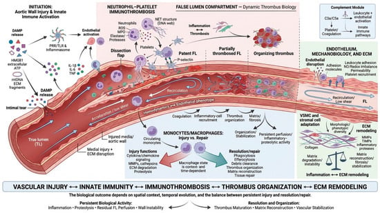
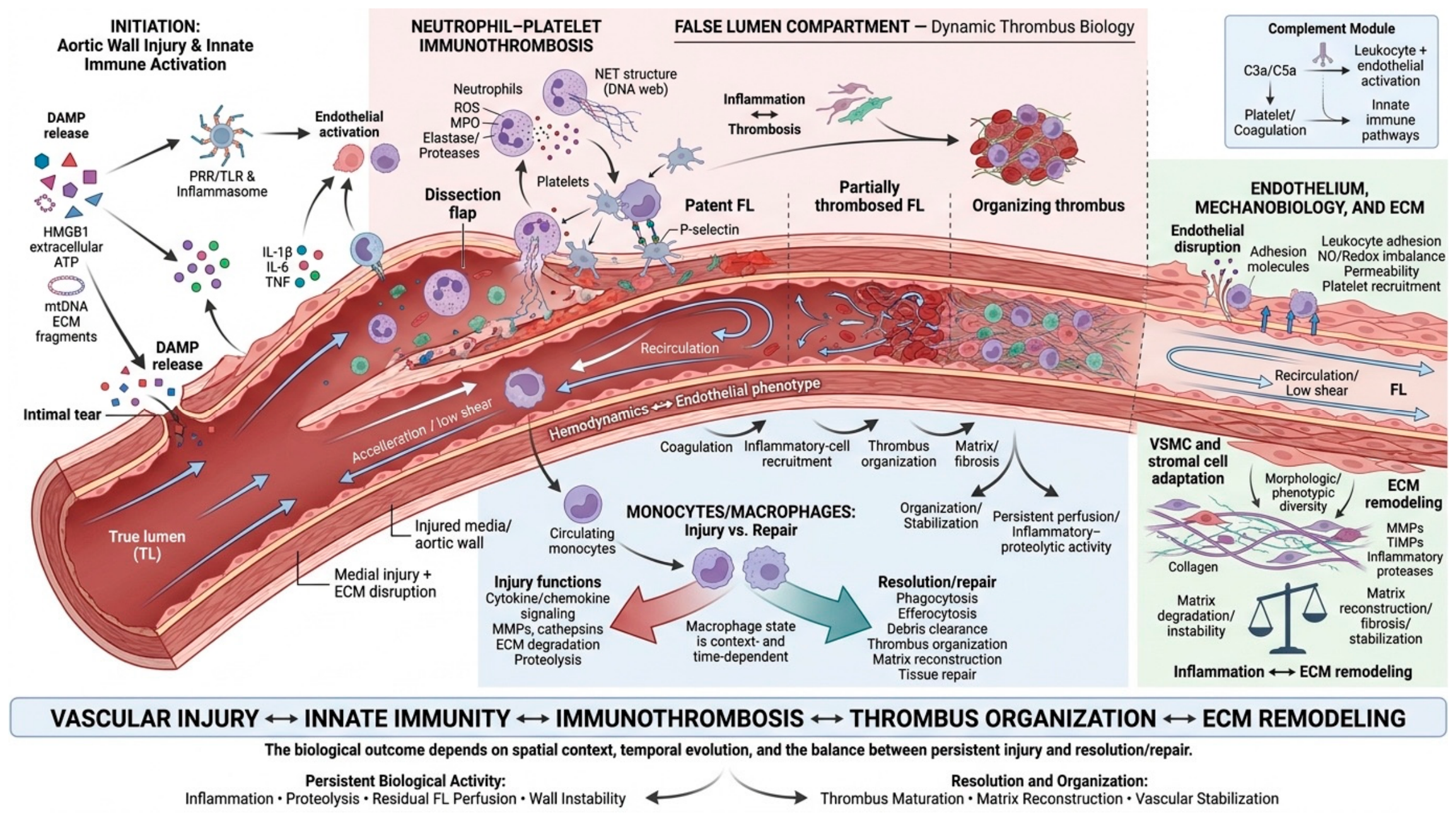
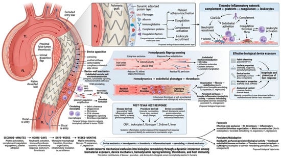
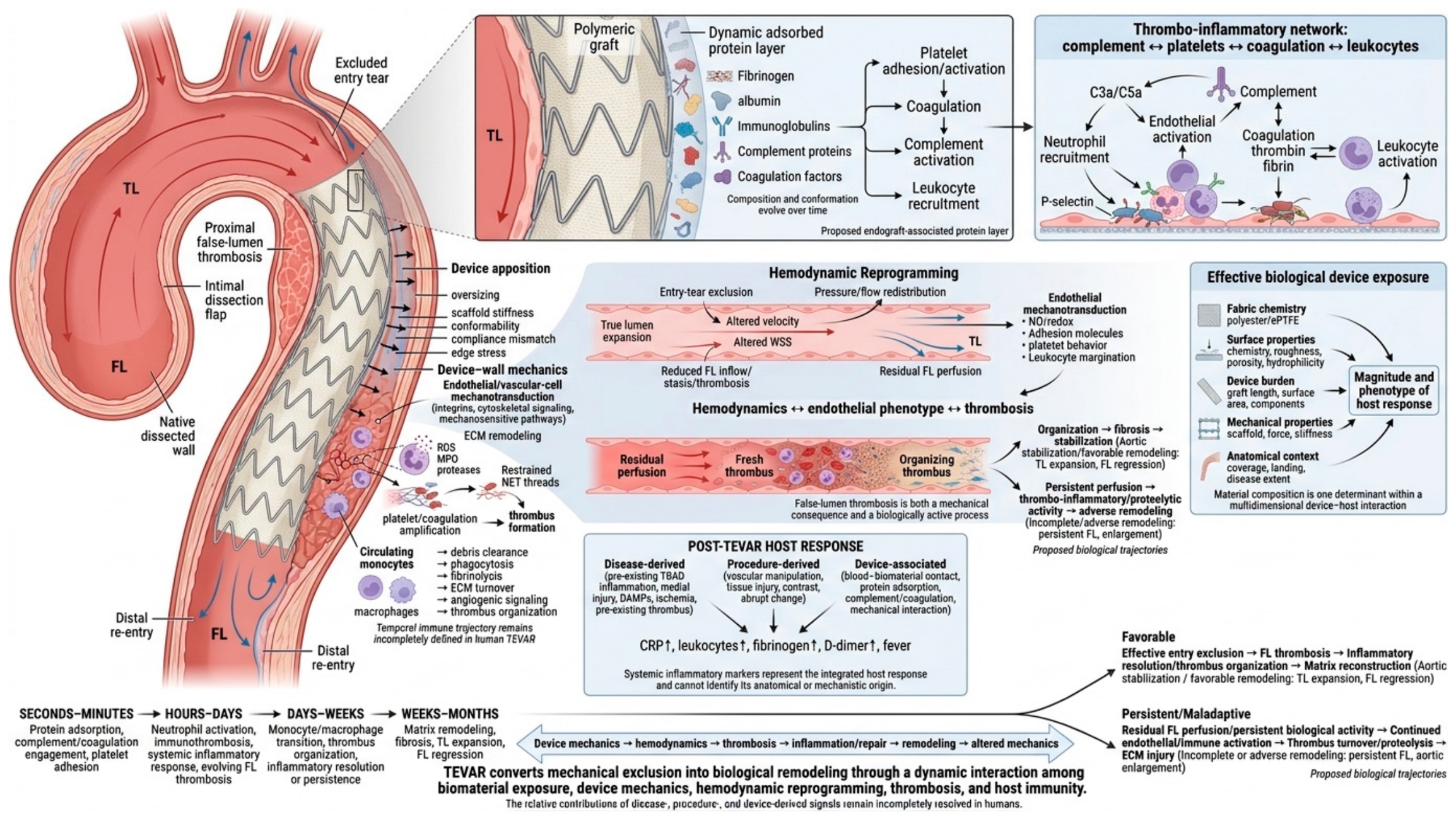
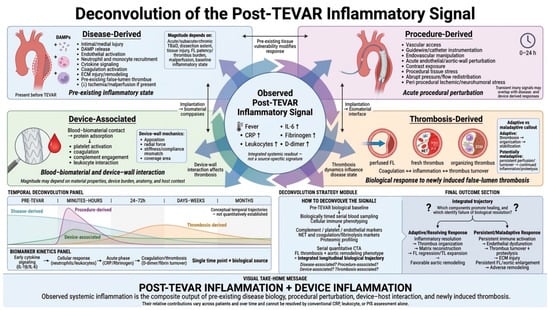
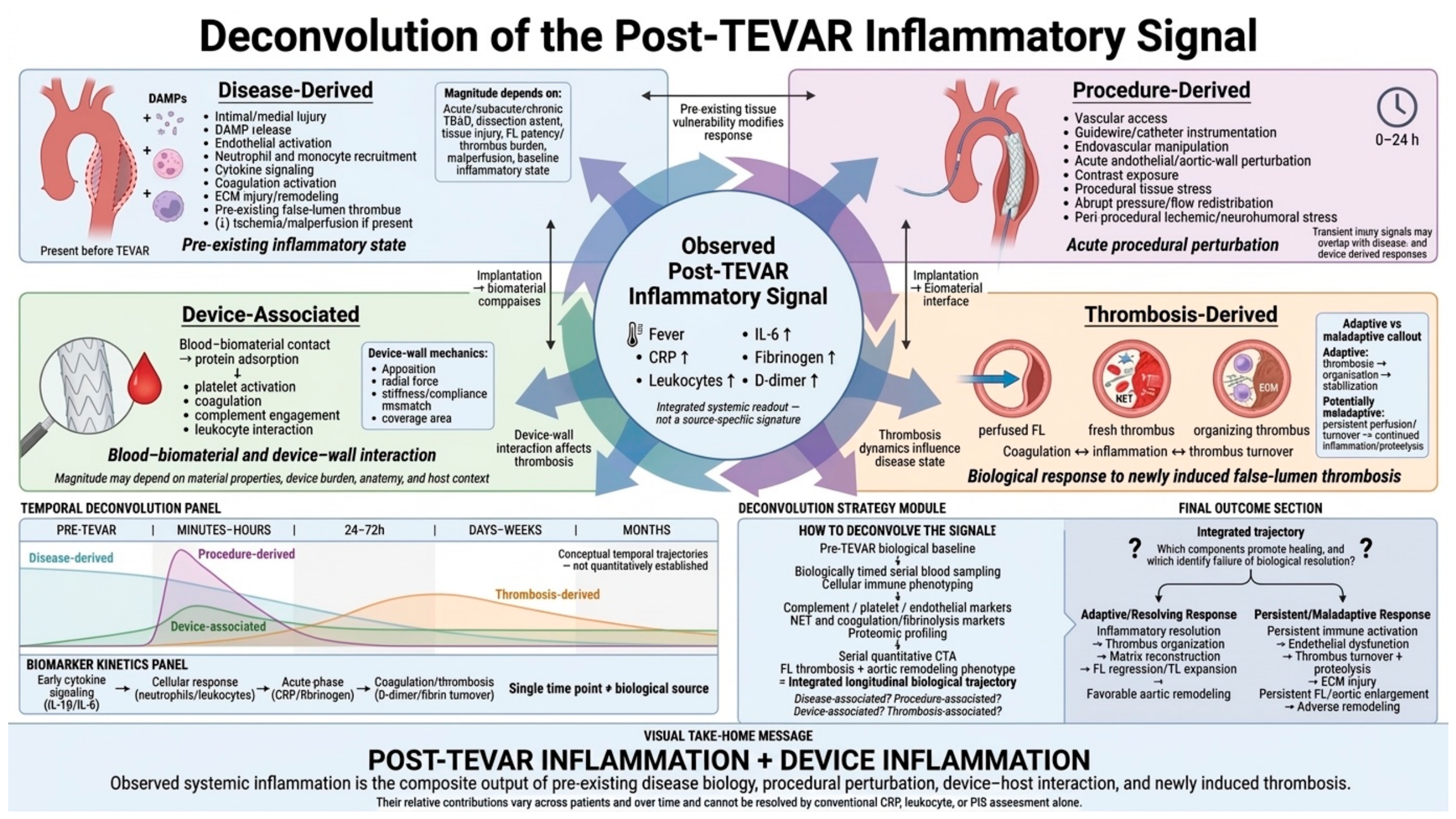
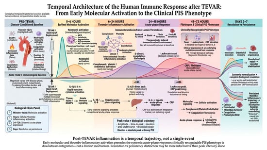
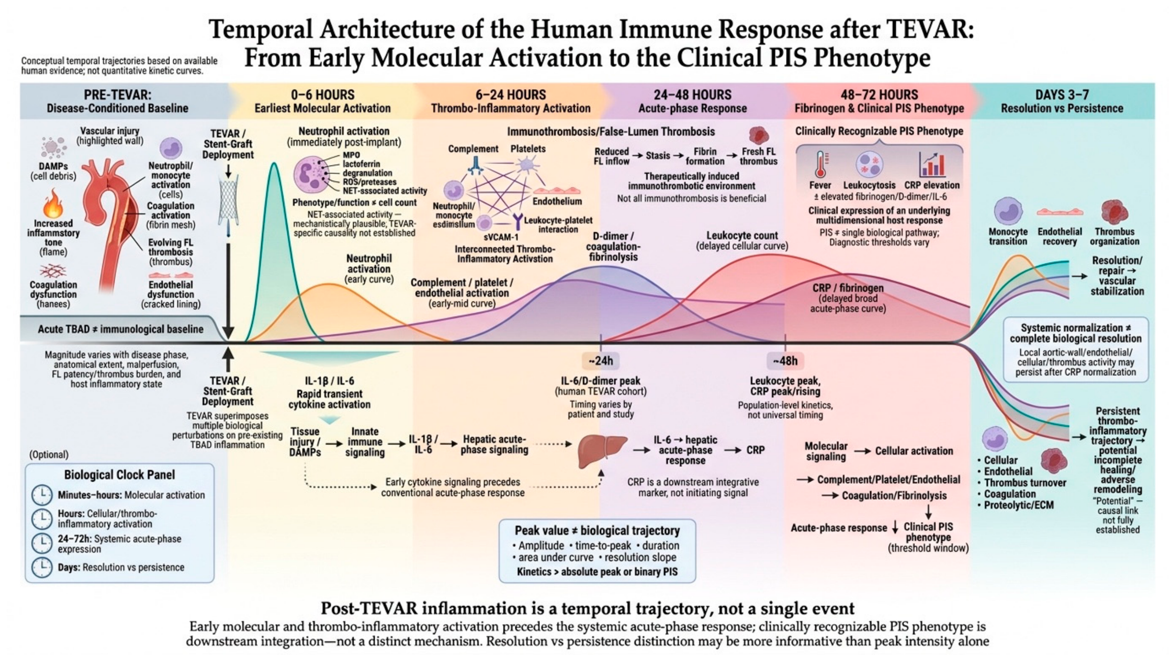
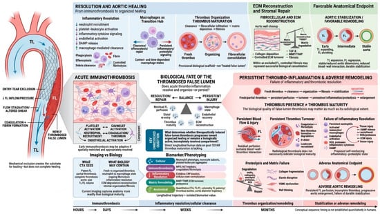
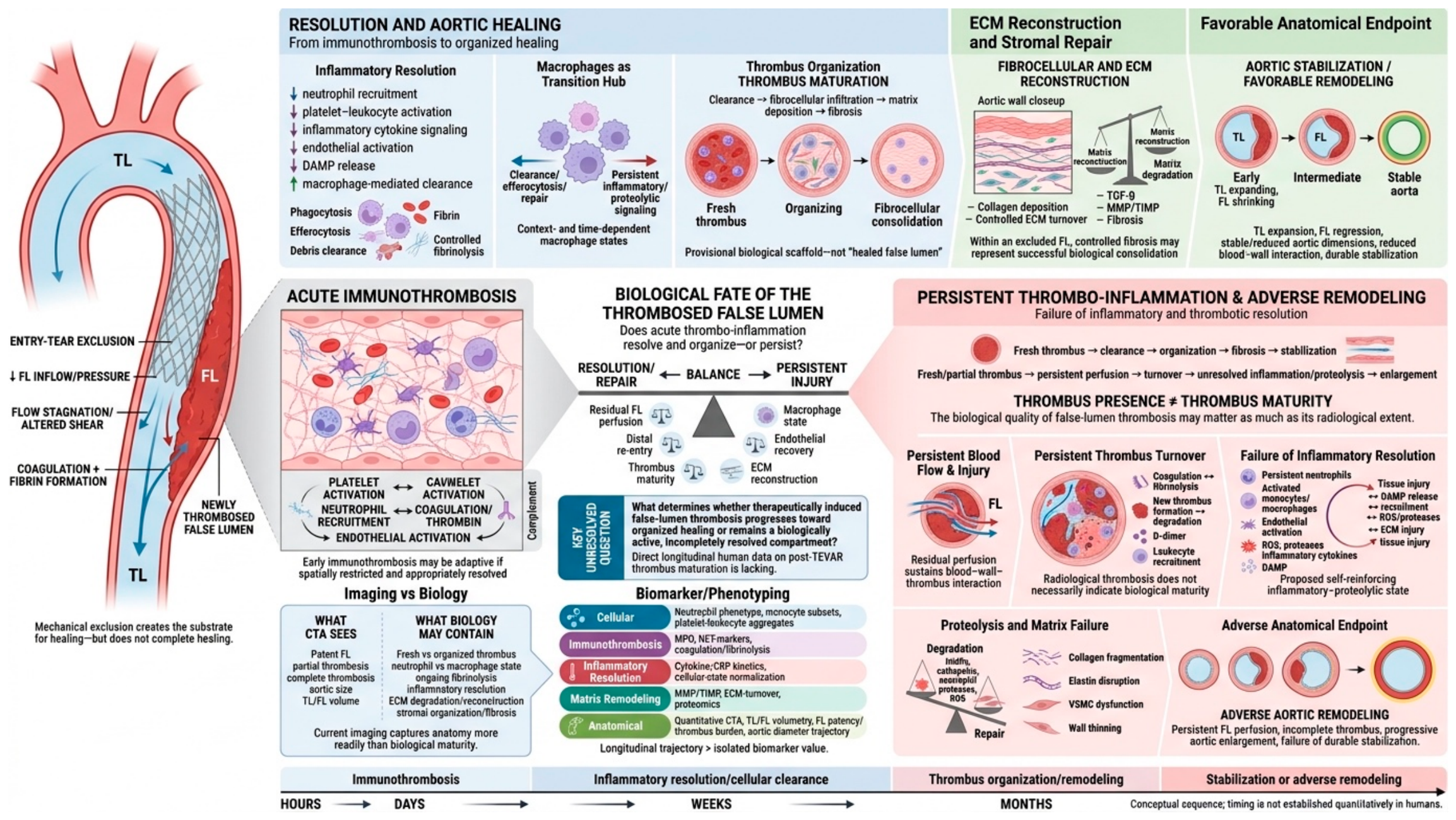
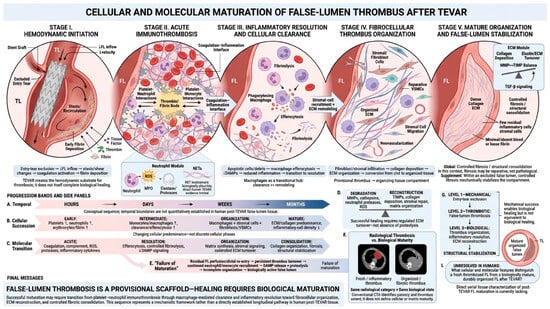
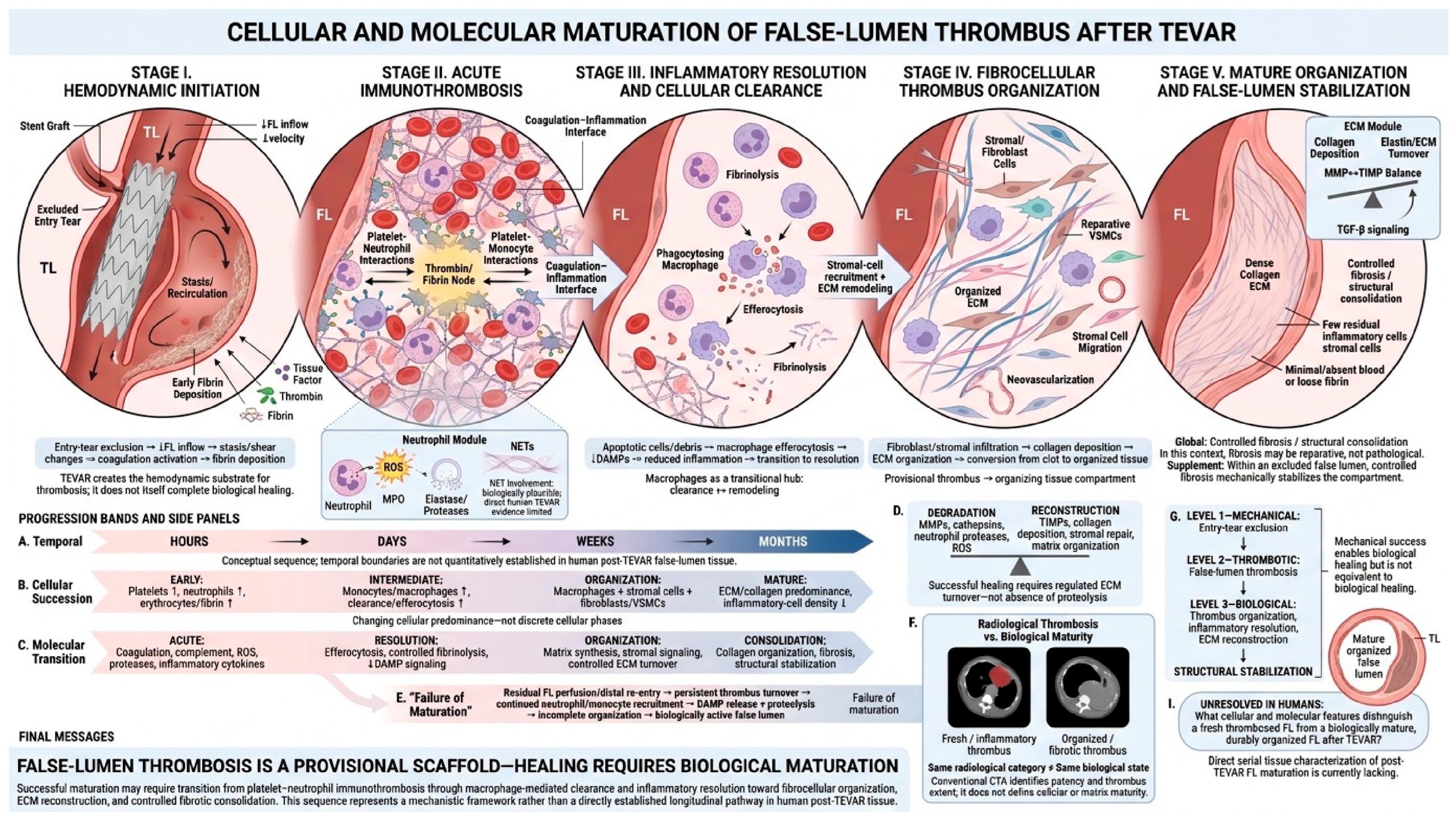

Disclaimer/Publisher’s Note: The statements, opinions and data contained in all publications are solely those of the individual author(s) and contributor(s) and not of MDPI and/or the editor(s). MDPI and/or the editor(s) disclaim responsibility for any injury to people or property resulting from any ideas, methods, instructions or products referred to in the content. |
© 2026 by the authors. Licensee MDPI, Basel, Switzerland. This article is an open access article distributed under the terms and conditions of the Creative Commons Attribution (CC BY) license.