


Abstract
Due to the limitations of conventional antibiotics, antimicrobial peptides (AMPs) have emerged as promising therapeutic alternatives for the prevention and treatment of oral infections. This study systematically evaluated in vitro evidence regarding the antimicrobial and anti-biofilm activity of natural AMPs against oral pathogens. A systematic search using the PICOT strategy was conducted in PubMed, EMBASE, and Scopus, retrieving 7711 articles. After title and abstract screening, 109 studies were selected for full-text analysis, resulting in 26 articles that met the eligibility criteria. Among the AMPs evaluated, nisin (n = 15) and LL-37 (n = 5) were the most frequently investigated, while other peptides included lactoferrin, lactoferricin, melittin, lysozyme, histatin-5, cystatin C, chromogranin A, parasin-1, protamine, AmyI-1-18, and DCD-1L. Natural AMPs of human and animal origin demonstrated antimicrobial activity against bacteria associated with oral infections, particularly Streptococcus mutans and Enterococcus faecalis. These peptides were tested in different formulations, including solutions, incorporation into dental materials and polymers, and application in sonodynamic antimicrobial therapy. Overall, the findings indicate that natural AMPs represent a promising class of biomolecules for controlling oral biofilms; however, further clinical studies are required to validate their long-term efficacy and safety.
1. Introduction
The oral cavity constitutes a complex environment that facilitates microbial colonization due to continuous nutrient availability, stable temperature, and high humidity [1]. Under physiological conditions, this environment supports a diverse microbiota of approximately 1000 species, which form cooperative, self-regulating biofilms that maintain symbiosis with the host. Disruption of local homeostasis leads to dysbiosis, characterized by uncontrolled proliferation of specific bacterial species. Dysbiosis is linked to the development of infectious and autoimmune diseases, as well as oral carcinomas. Major oral pathologies associated with microorganism-induced inflammation include dental caries, periodontal disease, pulpitis, apical periodontitis, and implant-related infections [1].
Systemic antibiotics for oral infections must be used judiciously, as overuse promotes bacterial resistance and disrupts both oral and intestinal microbiomes [2]. Most oral infections are localized and can typically be managed through mechanical or surgical interventions; systemic antimicrobial therapy is primarily indicated as an adjunct rather than a substitute. Furthermore, excessive or inappropriate antibiotic use in the oral cavity may lead to fungal overgrowth, disruption of the oral microbiota, and masking of clinical symptoms, thereby complicating disease diagnosis and management. Moreover, recent studies have identified antimicrobial resistance genes within the oral microbiome, conferring resistance to clinically relevant antibiotics such as beta-lactams, fluoroquinolones, lincosamides, and macrolides [3,4]. Due to the limitations of traditional antibiotics, antimicrobial peptides (AMPs) have emerged as promising therapeutic alternatives. AMPs are protein molecules consisting of short amino acid chains with amphipathic and cationic properties. They exhibit antibacterial, antibiofilm, antiviral, and antifungal activities [2,5]. AMPs are classified by origin as either natural or synthetic [6].
Natural AMPs function as components of the defense system in multicellular organisms, providing protection against infections and performing immunomodulatory roles. Their mechanisms of action include disrupting the plasma membrane, penetrating bacterial cells, and interfering with protein and cell wall synthesis [2,7]. The therapeutic and biomimetic potential of natural AMPs has attracted significant interest in recent decades. In dentistry, these biomolecules have been investigated primarily as adjunctive strategies for caries control and prevention, as well as supportive therapies in endodontic treatment, coronal restoration, tissue remineralization, and the development and improvement of dental materials [8]. Therefore, this study systematically evaluates in vitro evidence on the antimicrobial and antibiofilm activities of natural antimicrobial peptides for the prevention and treatment of oral infections.
2. Materials and Methods
Protocol and Registration
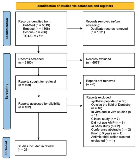
This systematic review was structured according to PRISMA—Preferred Reporting Items for Systematic Reviews and Meta-Analyses (Figure 1). This study was registered prospectively at Open Science Framework on 10 December 2025.
Figure 1.
PRISMA flow diagram of study selection.
Search Strategies
The systematic search was conducted using the PICOT strategy, where the guiding question was “In oral biofilms, can the use of natural AMPs promote antibacterial activity or reduce the growth/viability of biofilms in in vitro studies?”. Focus was given to studies of natural antimicrobial peptides, isolated or in combination with other antimicrobial agents. The bibliographic databases searched included PubMed, EMBASE, and Scopus. The database search was conducted on 13 October 2025. The review team collaboratively developed the search strategy, employing both controlled vocabulary (MeSH and DeCS terms) and free-text keywords. The final search strategies incorporated terms related to “natural antimicrobial peptides,” “AMPs,” “antibacterial activity,” “endodontic infection,” “dental caries,” “periodontal infection,” “catelhecidin,” and “dentistry” (Table S1).
Eligibility Criteria
Eligible studies for this review evaluated the antimicrobial and/or antibiofilm activity of natural antimicrobial peptides. Only in vitro studies were included. No language restrictions were imposed. The search was limited to the past five years to ensure the inclusion of recent scientific evidence representative of the current state of the art, particularly in a rapidly evolving field characterized by advances in peptide development, formulation strategies, delivery systems, and incorporation into dental materials, while avoiding the inclusion of potentially outdated data.
Studies were excluded if they did not assess the antimicrobial and/or antibiofilm activity of natural antimicrobial peptides or if they focused on synthetic antimicrobial peptides. Additionally, review articles, editorials, opinion pieces, conference abstracts, and case reports were excluded. Studies that did not investigate the antimicrobial and/or antibiofilm effects of natural antimicrobial peptides in oral infections were also excluded.
Study Selection and Data Collection
The research results were exported to Rayyan (Qatar Computing Research Institute, HBKU, Doha, Qatar), a free platform for reference management, duplicate detection, and study screening by title and abstract. All records from the databases were imported to facilitate an organized and blind selection process among reviewers. Study selection occurred in two phases: first, two independent reviewers screened titles and abstracts to identify potentially eligible studies, resolving disagreements through discussion or with a third reviewer. Next, the full texts of pre-selected articles were evaluated according to the established inclusion and exclusion criteria. Discrepancies during data extraction were resolved by consensus, and Gwet’s AC1 coefficient was used to measure inter-reviewer agreement [9]. Data extraction was performed independently by two reviewers using a standardized form developed by the research team that included information on publication details, the type of antimicrobial peptide investigated, the biological parameters evaluated, and the primary outcomes.
Methodological quality analysis
The methodological quality of the in vitro studies was assessed using the Science in Risk Assessment and Policy (SciRAP in vitro tool, v1.0) [10]. Two independent reviewers assessed the risk of bias in the included articles, resolving disagreements through discussion or with a third reviewer.
3. Results
The study selection process is detailed in the PRISMA flow diagram (Figure 1), which delineates the stages of identification, screening, eligibility assessment, and inclusion. The database search yielded 7711 articles. After duplicate removal, 6880 articles remained for initial evaluation. Title and abstract screening excluded 6772 articles. Full-text screening was performed on 109 articles; 81 were excluded for not meeting inclusion criteria, including narrative reviews (n = 3), in vivo studies (n = 11), synthetic peptide evaluations (n = 31), observational studies (n = 9), in silico studies (n = 2), absence of antimicrobial peptide (n = 2), lack of antimicrobial or antibiofilm evaluation (n = 1), and studies outside the last five years (n = 2). Two studies were excluded because only abstracts were available (Figure 1). Ultimately, 26 studies met the eligibility criteria and were included in this review. The level of agreement between reviewers was 0.82 using Gwet’s AC1 coefficient.
Among the AMPs evaluated, nisin (n = 15) and LL-37 (n = 5) were the most extensively studied. Table S2 summarizes the key mechanisms, primary oral indications, and strongest evidence supporting these peptides. Additional natural antimicrobial peptides identified included Lactoferrin (n = 2), Lactoferricin (n = 2), Melittin (n = 2), Lysozyme (n = 2), Histatin-5 (n = 1), Cystatin C (n = 1), Chromogranin A (n = 1), Parasin-1 (n = 1), Protamine (n = 1), AmyI-1-18 (n = 1), and DCD-1L (n = 1). Five studies compared the antimicrobial and/or antibiofilm activities of two or more peptides within the same investigation.
Minimum Inhibitory Concentration (MIC) and Minimum Bactericidal Concentration (MBC) were the primary methods used to assess the antimicrobial activity of the evaluated natural antimicrobial peptides. Most studies investigated the antibacterial effects of these peptides against Streptococcus mutans or Enterococcus faecalis, which are implicated in caries formation and secondary or persistent endodontic infections, respectively.
The principal methodological characteristics and findings of the 26 included studies are summarized in Table S3. Of the 26 studies included in this review, only five utilized multispecies biofilm models [11,12,13,14,15], highlighting the predominance of monospecies systems in the current literature. These multispecies studies assessed a limited range of antimicrobial peptides, primarily nisin, LL-37, and lactoferricin, and consistently reported reduced antibiofilm efficacy compared to monospecies models. When used alone, these peptides generally exhibited limited activity against complex biofilms, likely due to increased microbial diversity, interspecies interactions, and restricted penetration through the extracellular matrix. In contrast, studies combining antimicrobial peptides with conventional antibiotics or adjunctive agents reported enhanced or synergistic effects, such as improved short-term biofilm susceptibility and reduced antibiotic tolerance [12,13,14].
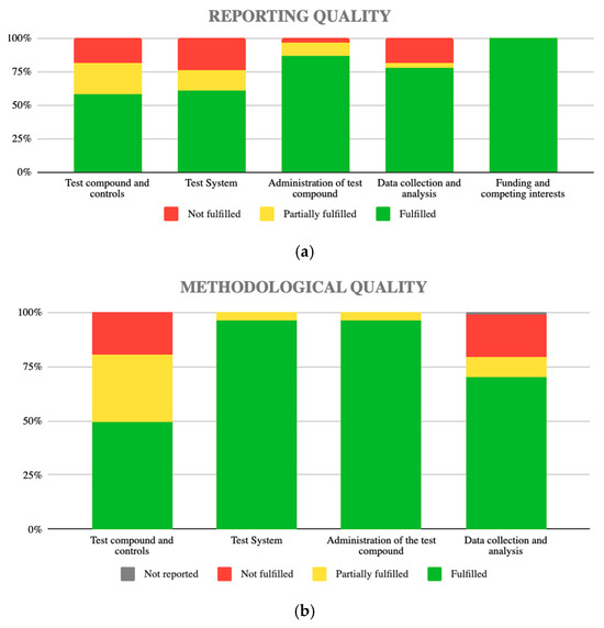
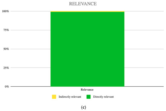
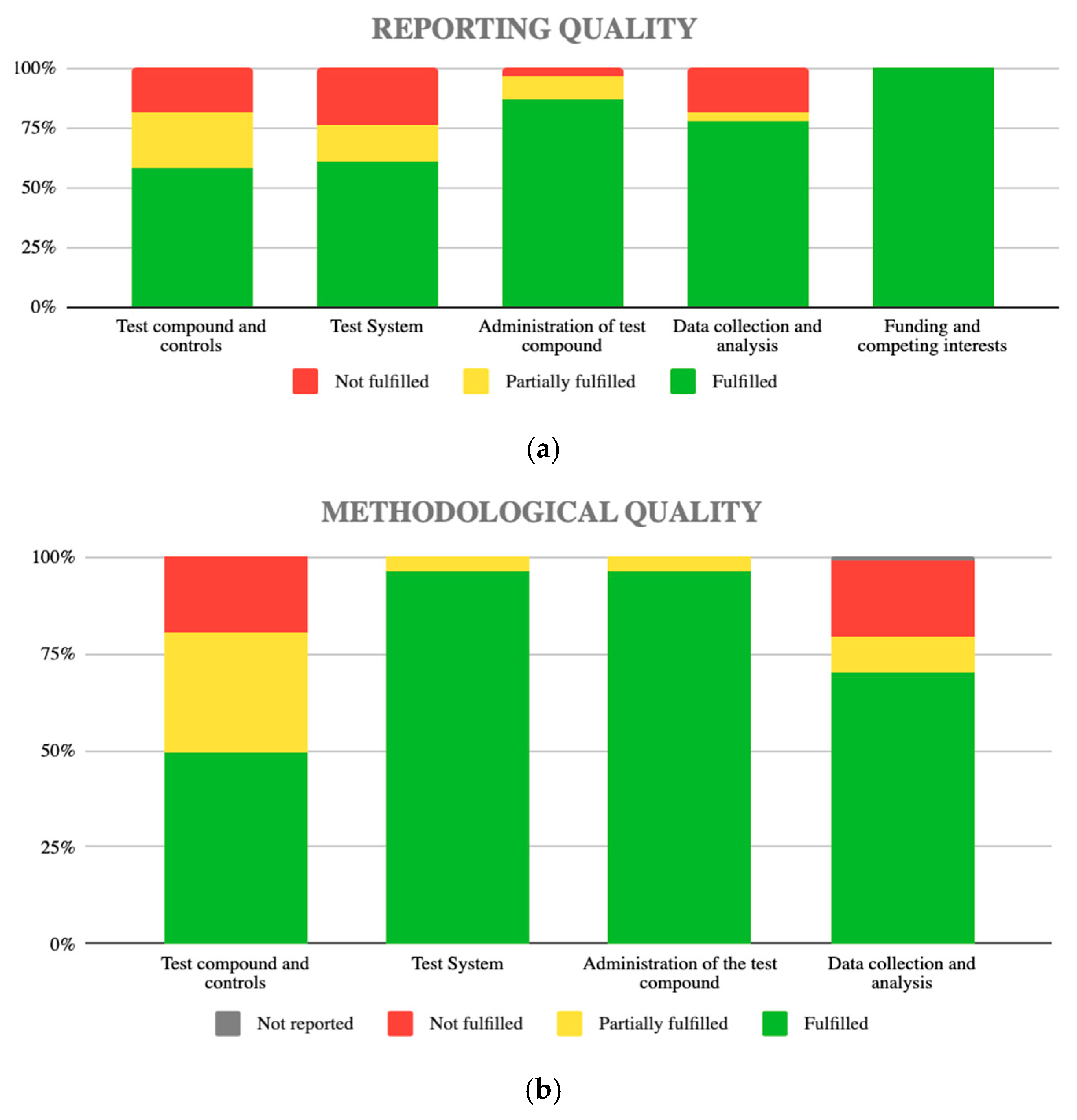
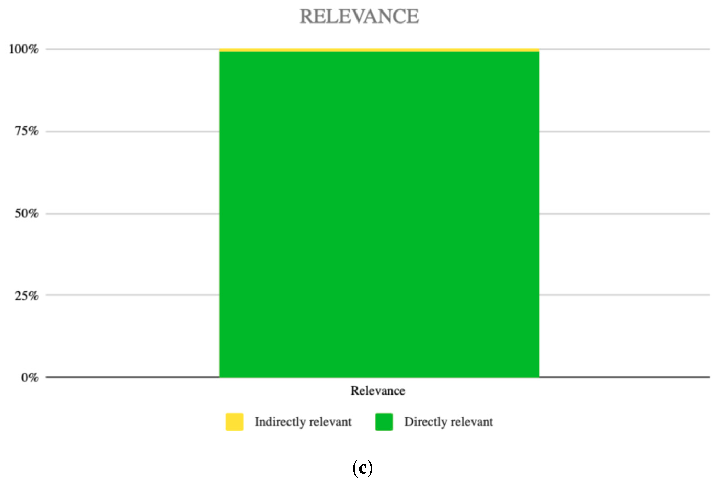
The risk-of-bias assessment is presented in Figure 2, which summarizes the evaluation of the included studies across three key domains: reporting quality (Figure 2a), methodological quality (Figure 2b), and relevance to the review objective (Figure 2c). Although most studies aligned with the review objective and satisfied several methodological criteria, considerable variability was evident across these domains. Specifically, frequent limitations in the description and implementation of experimental controls and test compounds were identified, potentially compromising internal validity. Further variability was observed in the standardization and characterization of experimental models. In contrast, domains related to intervention administration and the disclosure of funding and competing interests were generally adequately addressed. Together, these findings indicate that most studies exhibited satisfactory methodological quality and a low overall risk of bias, although relevant inconsistencies remain in essential criteria, particularly in the application and description of experimental controls. These recurring inconsistencies necessitate cautious interpretation of the results.
Figure 2.
The risk of bias of the selected articles was assessed using the SciRAP in vitro tool, version 1.0: (a) Reporting quality across the included studies, presented as the proportion of criteria fulfilled within each reporting domain; (b) Methodological quality across the included studies, illustrating the proportion of studies meeting the predefined methodological criteria in each domain; (c) Overall relevance of the included studies to the objective of the review.
4. Discussion
Natural antimicrobial peptides have garnered increasing attention as therapeutic and adjuvant agents in oral diseases associated with biofilms, such as caries, periodontal diseases, and endodontic infections. Their selective cytotoxicity against microorganisms and low toxicity to mammalian cells contribute to a favorable biocompatibility profile. This review of the past five years evaluated 12 natural antimicrobial peptides, including those produced by bacteria, humans, and animals. Among the natural AMPs assessed, nisin was the most extensively studied, followed by LL-37 and other AMPs, which are discussed in subsequent sections.
Nisin
Nisin is a natural cationic antimicrobial peptide belonging to the class I bacteriocin group, produced by the bacterium Lactococcus lactis. Its primary application is as a potent food preservative in various products, including dairy and processed meats [16]. Nisin exhibits two principal mechanisms of action: pore formation in the bacterial membrane and inhibition of cell wall synthesis. Additionally, nisin has been reported to penetrate multiple layers of biofilm, although it does not achieve complete eradication [17,18]. These characteristics underscore nisin’s potential as a biotherapeutic agent for human and animal health [16]. In this review, nisin was the most frequently investigated antimicrobial peptide, emphasizing its prominence and therapeutic potential in the context of oral infections. Most studies included nisin as an active agent against pathogens associated with both dental caries and endodontic infections.
For caries prevention and management, nisin has been evaluated alone or in combination with other antimicrobial strategies. In one study, nisin was evaluated in combination with gallic acid to target S. mutans. Nisin alone did not demonstrate antibacterial activity, whereas the addition of gallic acid significantly enhanced activity against S. mutans, exhibiting bacteriostatic properties [19]. Another study compared the antibiofilm and antibacterial effects of nisin with those of chlorhexidine. Nisin exhibited inhibitory, bactericidal, and antibiofilm effects against S. mutans, although its efficacy was lower than that of chlorhexidine [20]. The antimicrobial action of nisin was also investigated when incorporated into various dental materials, either alone or in combination with other antimicrobial agents. Incorporation of antimicrobial peptides into dental adhesives as bioactive components has been widely studied to reduce deterioration of composite resin restoration margins and the formation of white spots. This review identified five articles that tested the incorporation of nisin into dental adhesives to enhance antibacterial activity. In all studies, adhesives containing nisin significantly inhibited bacterial growth and acid production without compromising adhesion or polymerization properties [21,22,23,24,25]. In the restorative context, nisin was also added to high-viscosity glass ionomer cement, resulting in improved antimicrobial activity against S. mutans and increased surface microhardness. These findings suggest that modifying dental materials with nisin is a promising approach to enhance the antibacterial efficacy of restorations and prevent recurrent caries [26].
In a study on periodontal diseases, Enigk et al. [27] evaluated the efficacy of nisin against 36 bacterial strains associated with oral infections. Nisin demonstrated significant activity against various microbial groups, including early colonizers, aggregating bacteria, and late colonizers of the oral biofilm. Additionally, nisin exhibited thermal stability, maintaining activity at temperatures above physiological levels (up to 50 °C), and demonstrated tolerance to acidic pH, suggesting robustness under non-physiological experimental and processing conditions.
In a study comparing nisin with chlorhexidine and amoxicillin as root canal irrigants in endodontic treatment [28], nisin at concentrations ≥ 1 mg/mL was found to be more effective than chlorhexidine and amoxicillin based on in vitro time–kill assays after 45 min of exposure. Pinheiro et al. [11] also evaluated nisin as a root canal irrigant, assessing its action alone and in combination with sodium hypochlorite and EDTA. Sodium hypochlorite and EDTA were effective in reducing mature biofilms, but the addition of nisin did not significantly enhance their activity. Notably, these investigations were conducted under experimental conditions using irrigation protocols that differ from standard clinical endodontic practice, which typically employs multistep regimens with various antiseptic agents. In regenerative endodontic procedures, nisin was investigated as an intracanal medication in combination with triple antibiotic paste (TAP). The addition of nisin improved TAP activity after short exposure, but prolonged treatment for 7 days, either alone or in combination, was most effective in eliminating biofilm. These findings suggest that nisin has potential as a short-term adjuvant treatment in the root canal to optimize antibiotic effectiveness [12].
Recent strategies have focused on developing systems that enable prolonged nisin release to enhance its clinical efficacy. A biocompatible PVA-alginate hydrogel incorporating nisin demonstrated flexibility, swelling capacity, absence of hemolytic activity, effective antimicrobial activity, and low toxicity. Such biomaterials are notable for their biocompatibility, bioactivity, mechanical integrity, and long-term stability, which are essential for therapeutic applications in clinical settings [29]. The combination of nisin with licorice polyphenols showed high efficacy in eliminating pre-formed E. faecalis biofilms [30]. Additionally, solid lipid nanoparticles loaded with nisin (SLN-Nisin) were significantly more effective than free nisin against Treponema denticola and in reducing the viability of oral carcinoma cells, indicating that encapsulated nisin has potential for medical application, particularly in the treatment of periodontal disease and oral cancer [31]. However, as these results were obtained under in vitro conditions, they should be interpreted with caution because they do not reflect the complexity of the in vivo oral environment.
LL-37
Five articles included in this review assessed the antimicrobial properties of LL-37 against oral pathogens. LL-37, the only human cathelicidin antimicrobial peptide, is derived from the precursor hCAP18 and demonstrates broad-spectrum antibacterial activity against Gram-positive and Gram-negative bacteria, as well as fungi and viruses [32]. This peptide is widely expressed in epithelial cells, immune cells, mesenchymal stem cells, and various tissues, and is also present in biological fluids such as saliva, sweat, breast milk, and semen. Its antimicrobial mechanisms include pore formation in bacterial membranes and neutralization of lipopolysaccharide (LPS), while its cellular effects influence phagocytosis, differentiation, and apoptosis [32,33,34]. LL-37 exhibits complex, microenvironment-dependent immunomodulatory effects, promoting pro-inflammatory responses through chemotaxis of mast cells, monocytes, T lymphocytes, and neutrophils. It also exerts anti-inflammatory effects by attenuating IFN-γ, TNF-α, IL-4, and IL-12 responses and by reducing LPS-induced cytokine production by interfering with the TLR-4 receptor. Additionally, LL-37 regulates angiogenesis and wound healing, and is considered a promising therapeutic strategy for tissue regeneration due to its capacity to promote neovascularization and recruit mesenchymal stem cells. These properties have generated increasing research interest in their application for periodontal and bone regeneration [34].
The involvement of LL-37 in periodontal diseases has been extensively studied, with elevated LL-37 levels consistently reported in patients with periodontitis compared to healthy controls. There is a significant correlation between LL-37 concentrations, clinical parameters, and the presence of periodontopathogens, suggesting that elevated local levels may represent a host defense response to microbiological changes associated with disease [33]. Evidence from one study indicates that LL-37 overexpression markedly reduces the burden of Porphyromonas gingivalis in keratinocytes, which is essential for the management of periodontitis and related systemic conditions. Autophagy serves as the primary mechanism by which LL-37 facilitates the clearance of viable intracellular P. gingivalis, rather than through direct bactericidal activity, which may be constrained by the pathogen’s low susceptibility or inherent resistance to the peptide [32]. In contrast, Enigk et al. [27] did not detect antimicrobial effects of LL-37 against periodontopathogenic bacteria. Nevertheless, LL-37 demonstrated additional biological functions, such as anti-inflammatory properties and protection against periodontal bone resorption.
Two studies investigated the effects of LL-37 in combination with antibiotics. Pereira et al. [13] reported that LL-37 combined with triple antibiotic paste (TAP) exhibited the highest antibacterial activity against a biofilm model of endodontic infection, while TAP alone and the isolated peptide did not demonstrate significant antimicrobial effects. Similarly, Wuersching et al. [14] observed enhanced antimicrobial efficacy when LL-37 was combined with antibiotics, including amoxicillin, clindamycin, and metronidazole, particularly against biofilms with elevated antibiotic tolerance. Another study assessed the antimicrobial activity of LL-37 against strict and facultative anaerobic bacteria in both planktonic and biofilm states [15]. The results indicated that antimicrobial activity against biofilms was reduced compared to planktonic cells, likely due to structural barriers, such as extracellular DNA and the polymeric matrix, that hinder antimicrobial peptide penetration. However, the residual antimicrobial effect observed suggests that LL-37 may also inhibit bacterial adhesion and disrupt quorum sensing, supporting its potential as a therapeutic agent for oral diseases associated with biofilms.
Other AMPs
Salivary antimicrobial peptides, including Histatin-5, Cystatin C, and Chromogranin A, have been investigated for their capacity to induce structural damage in Enterococcus faecalis and Streptococcus mutans, which are implicated in endodontic infections and dental caries, respectively [35]. These peptides possess strong antimicrobial and antifungal properties and play a significant role in wound healing and oral cavity defense. Blancas et al. [35] reported that these human salivary peptides disrupt bacterial cell walls and interact with bacterial DNA, leading to cell death.
Lactoferrin is an antimicrobial peptide found in mucous secretions, including milk, lacrimal fluid, and saliva, and is produced by epithelial cells. Its antibacterial activity is primarily due to its ability to sequester iron from microorganisms, maintaining the lactoferrin-iron bond even at low pH values (4.5–5.0) generated by bacterial metabolism. As a result, lactoferrin inhibits the adhesion of S. mutans and suppresses the growth of both Gram-positive and Gram-negative bacteria associated with periodontal disease [27,36]. Lactoferricin, a smaller peptide generated from the digestion of lactoferrin, also exhibits antimicrobial and immunomodulatory properties. Owing to its reduced size, lactoferricin interacts more efficiently with bacterial membranes. Research has examined human lactoferricin against strict and facultative anaerobic bacteria implicated in caries and periodontitis, evaluating its effects on both planktonic growth and multispecies biofilm formation. Although lactoferricin inhibited planktonic bacterial growth at all concentrations, its impact on biofilms was limited, suggesting it may not be effective for therapeutic use against oral biofilms [15]. Furthermore, lactoferricin has been investigated in combination with antibiotics to address antibiotic resistance in oral infections. The combination of lactoferricin with antibiotics such as amoxicillin and clindamycin demonstrated synergistic effects, enhancing activity against strict and facultative aerobic biofilms [14].
Lysozyme is a natural antimicrobial peptide found in secretions such as saliva and tears. It catalyzes the cleavage of the bond between N-acetylmuramic acid and N-acetylglucosamine in bacterial cell walls, leading to bacterial lysis and reducing the pathogenicity of both Gram-positive and Gram-negative bacteria. The combination of hydroxyapatite with the enzymes lysozyme, lactoferrin, and lactoperoxidase has been proposed as an alternative strategy for controlling S. mutans in dental caries [36]. Furthermore, the development of a lysozyme-based adhesive system has enabled homogeneous peptide release to the oral mucosa without rapid dissolution, thereby preserving biological activity and providing potential for the treatment or prevention of oral bacterial infections [37].
Regarding natural antimicrobial peptides of non-human origin, this review evaluated the antimicrobial activity of five peptides: Protamine, Melittin, DCD-1L, Parasin-1, and AmyI-1-18 (a rice-derived peptide). Most of these peptides were tested in association with dental materials. Protamine is a cationic AMP rich in arginine, derived from fish such as salmon. It was tested in association with an adhesive system against S. mutans. The authors concluded that the addition of Protamine decreased the viability of S. mutans. Therefore, the authors observed that Protamine has the potential to be incorporated into oral hygiene products [38].
One study investigated the antibacterial properties of the rice-derived peptide AmyI-1-18 against two bacteria associated with periodontal disease. Against Porphyromonas gingivalis, the AMP exerted bacteriostatic activity without disrupting the cell membrane. This is because the anti-P. gingivalis property does not depend on membrane destabilization, but rather on the inhibition of protein translation and synthesis. Regarding its effect on Fusobacterium nucleatum, AmyI-1-18 disrupted the membrane, exhibiting bactericidal activity. According to the authors, this occurred because the outer membrane of F. nucleatum has a stronger negative charge than that of P. gingivalis. Therefore, the cationic characteristic of AmyI-1-18 allowed it to act more effectively [39].
A separate study assessed the antibiofilm efficacy of antimicrobial sonodynamic therapy (aSDT) mediated by nanoparticles and the natural antimicrobial peptide DCD-1L against resistant S. mutans cells. Distinct from most antimicrobial peptides (AMPs), DCD-1L is an anionic peptide. The primary antimicrobial effect of aSDT relies on the generation of reactive oxygen species, which can be induced by AMPs. DCD-1L-mediated aSDT effectively inactivated both biofilms and resistant S. mutans cells, thereby reducing the pathogenic potential of the bacteria [40]. Enigk et al. [27] investigated the activity of several antimicrobial peptides, including Melittin and Parasin-1. Melittin inhibited the growth of both Gram-positive and Gram-negative periodontal bacteria, whereas Parasin-1 showed no antibacterial activity against any of the tested bacteria. In a related study, Melittin was incorporated into a poly(lactic-co-glycolic acid) (PLGA) polymer, which promoted apatite formation and demonstrated antibacterial activity against Staphylococcus aureus, indicating its potential to enhance osseointegration and prevent implant-associated infections [41].
Limitations and Future Perspectives
While this review underscores the promising antimicrobial and antibiofilm potential of natural antimicrobial peptides, its conclusions are inherently limited by the exclusive reliance on in vitro evidence. Although these models provide valuable mechanistic insights, they do not fully recapitulate the biological, immunological, and ecological complexity of oral infections, particularly those involving polymicrobial biofilms. Notably, only five studies evaluated the efficacy of AMPs in multispecies biofilm models [11,12,13,14,15]. In these complex systems, peptides such as nisin, LL-37, and lactoferricin generally exhibited reduced antibiofilm activity when used alone, while improved outcomes were primarily observed when they were combined with antibiotic-based strategies, supporting a predominantly adjuvant role. Given that oral infections are inherently polymicrobial, the predominance of monospecies biofilm models, mainly composed of Streptococcus mutans and Enterococcus faecalis, may therefore lead to an overestimation of antimicrobial efficacy under clinically relevant conditions, underscoring the need for standardized multispecies models that better replicate the oral cavity environment.
A key limitation identified in this review is the lack of methodological homogeneity across studies, particularly in the types of analyses employed and the techniques used to obtain antimicrobial peptides. This variability hindered direct comparisons of results and limited the ability to perform robust, integrative analyses. Moreover, critical translational challenges, including peptide stability, bioavailability, susceptibility to proteolytic degradation, and scalability, remain insufficiently addressed, highlighting the need for standardized preclinical platforms, such as controlled-release dental materials and in vivo models incorporating salivary flow and mechanical forces, alongside well-designed clinical studies to substantiate the therapeutic relevance of these findings.
5. Conclusions
Overall, natural antimicrobial peptides of both human and animal origin have demonstrated efficacy in reducing bacteria involved in oral infectious processes. Streptococcus mutans and Enterococcus faecalis were the most frequently investigated species. Additionally, these peptides have been evaluated in various forms of application, including as solutions, incorporated into dental materials and polymers, and used in sonodynamic antimicrobial therapy. Among the most studied natural AMPs in laboratory research over the past five years, nisin and LL-37 are prominent. These peptides are closest to clinical translation because of their consistent preclinical efficacy and advanced application strategies. In contrast, peptides such as melittin, parasin-1, lactoferricin, histatin-5, and AmyI-1-18 have been less frequently investigated in the past five years and therefore have more limited recent evidence. Despite encouraging in vitro results, long-term clinical studies are required to assess the persistence of antimicrobial peptides in the oral environment and their sustained efficacy against biofilm-associated challenges.
Supplementary Materials
The following supporting information can be downloaded at: https://www.mdpi.com/article/10.3390/j9010002/s1, Table S1. Search Strategy Applied in Electronic Databases; Table S2. Most Frequently Evaluated Natural Antimicrobial Peptides in Oral Applications; Table S3. Summary of Included Studies.
Author Contributions
Conceptualization, A.C.C.P., M.C.S.-M. and E.T.P.; methodology, A.C.C.P., A.C.B.P., T.F.H.M. and M.C.S.-M.; data curation, A.C.C.P., A.C.B.P. and T.F.H.M.; writing—original draft preparation, A.C.C.P. and T.F.H.M.; writing—review and editing, M.C.S.-M. and E.T.P. All authors have read and agreed to the published version of the manuscript.
Funding
This research was funded by the São Paulo Research Foundation (FAPESP 2019/12908-3) and by the Coordination for the Improvement of Higher Education Personnel Foundation (CAPES, finance code 001).
Institutional Review Board Statement
Not applicable.
Informed Consent Statement
Not applicable.
Data Availability Statement
All data needed to evaluate and reproduce the results are contained in the main manuscript.
Acknowledgments
This work utilized Grammarly Pro (Grammarly Inc., San Francisco, CA, USA), an AI-powered grammar checker, to enhance language clarity and accuracy.
Conflicts of Interest
The authors declare no conflicts of interest.
References
- Tokajuk, J.; Deptula, P.; Piktel, E.; Daniluk, T.; Chmielewska, S.; Wollny, T.; Wolak, P.; Fiedoruk, K.; Bucki, R. Cathelicidin LL-37 in Health and Diseases of the Oral Cavity. Biomedicines 2022, 10, 1086. [Google Scholar] [CrossRef] [Scilit]
- Niu, J.Y.; Yin, I.X.; Wu, W.K.K.; Li, Q.L.; Mei, M.L.; Chu, C.H. Antimicrobial peptides for the prevention and treatment of dental caries: A concise review. Arch. Oral Biol. 2021, 122, 105022. [Google Scholar] [CrossRef] [Scilit]
- Anderson, A.C.; von Ohle, C.; Frese, C.; Boutin, S.; Bridson, C.; Schoilew, K.; Peikert, S.A.; Hellwig, E.; Pelz, K.; Wittmer, A.; et al. The oral microbiota is a reservoir for antimicrobial resistance: Resistome and phenotypic resistance characteristics of oral biofilm in health, caries, and periodontitis. Ann. Clin. Microbiol. Antimicrob. 2023, 22, 37. [Google Scholar] [CrossRef] [Scilit] [PubMed]
- Pinheiro, E.T.; Karygianni, L.; Candeiro, G.T.M.; Vilela, B.G.; Dantas, L.O.; Pereira, A.C.C.; Gomes, B.P.F.A.; Attin, T.; Thurnheer, T.; Russo, G. Metatranscriptome and Resistome of the Endodontic Microbiome. J. Endod. 2024, 50, 1059–1072. [Google Scholar] [CrossRef] [Scilit]
- Pepperney, A.; Chikindas, M.L. Antibacterial Peptides: Opportunities for the Prevention and Treatment of Dental Caries. Probiotics Antimicrob. Proteins 2011, 3, 68. [Google Scholar] [CrossRef] [Scilit]
- Kumar, R.; Ali, S.A.; Singh, S.K.; Bhushan, V.; Mathur, M.; Jamwal, S.; Mohanty, A.K.; Kaushik, J.K.; Kumar, S. Antimicrobial Peptides in Farm Animals: An Updated Review on Its Diversity, Function, Modes of Action and Therapeutic Prospects. Vet. Sci. 2020, 7, 206. [Google Scholar] [CrossRef] [Scilit] [PubMed]
- Mai, S.; Mauger, M.T.; Niu, L.N.; Barnes, J.B.; Kao, S.; Bergeron, B.E.; Ling, J.Q.; Tay, F.R. Potential applications of antimicrobial peptides and their mimics in combating caries and pulpal infections. Acta Biomater. 2017, 49, 16–35. [Google Scholar] [CrossRef] [Scilit]
- Hardan, L.; Chedid, J.C.A.; Bourgi, R.; Cuevas-Suárez, C.E.; Lukomska-Szymanska, M.; Tosco, V.; Monjarás-Ávila, A.J.; Jabra, M.; Salloum-Yared, F.; Kharouf, N.; et al. Peptides in Dentistry: A Scoping Review. Bioengineering 2023, 10, 214. [Google Scholar] [CrossRef] [Scilit] [PubMed]
- Silveira, P.S.P.; Siqueira, J.O. Better to be in agreement than in bad company: A critical analysis of many kappa-like tests. Behav. Res. Methods 2023, 55, 3326–3347. [Google Scholar] [CrossRef] [Scilit]
- Roth, N.; Zilliacus, J.; Beronius, A. Development of the SciRAP approach for evaluating the reliability and relevance of in vitro toxicity data. Front. Toxicol. 2021, 3, 746430. [Google Scholar] [CrossRef] [Scilit]
- Pinheiro, E.T.; Karygianni, L.; Attin, T.; Thurnheer, T. Antibacterial Effect of Sodium Hypochlorite and EDTA in Combination with High-Purity Nisin on an Endodontic-like Biofilm Model. Antibiotics 2021, 10, 1141. [Google Scholar] [CrossRef] [Scilit] [PubMed]
- Pereira, A.C.C.; Aguiar, A.P.S.; Barbosa, V.L.; Régis, J.R.; Miyazima, E.M.; Araujo, L.M.P.; Dantas, L.O.; Mayer, M.P.A.; Andrade, F.B.; Karygianni, L.; et al. Enhancing Antibiotic Efficacy in Regenerative Endodontics by Improving Biofilm Susceptibility. J. Endod. 2024, 50, 962–965. [Google Scholar] [CrossRef] [Scilit] [PubMed]
- Pereira, A.C.C.; Aguiar, A.P.S.; Araujo, L.M.P.; Dantas, L.O.; Mayer, M.P.A.; Karygianni, L.; Thurnheer, T.; Pinheiro, E.T. Antibiofilm Activity of LL-37 Peptide and D-Amino Acids Associated with Antibiotics Used in Regenerative Endodontics on an Ex Vivo Multispecies Biofilm Model. Life 2022, 12, 1686. [Google Scholar] [CrossRef] [Scilit] [PubMed]
- Wuersching, S.N.; Huth, K.C.; Hickel, R.; Kollmuss, M. Targeting antibiotic tolerance in anaerobic biofilms associated with oral diseases: Human antimicrobial peptides LL-37 and lactoferricin enhance the antibiotic efficacy of amoxicillin, clindamycin and metronidazole. Anaerobe 2021, 71, 102439. [Google Scholar] [CrossRef] [Scilit]
- Wuersching, S.N.; Huth, K.C.; Hickel, R.; Kollmuss, M. Inhibitory effect of LL-37 and human lactoferricin on growth and biofilm formation of anaerobes associated with oral diseases. Anaerobe 2021, 67, 102301. [Google Scholar] [CrossRef] [Scilit]
- Field, D.; Fernandez de Ullivarri, M.; Ross, R.P.; Hill, C. After a century of Nisin research—Where are we now? FEMS Microbiol. Rev. 2023, 47, fuad023. [Google Scholar] [CrossRef] [Scilit]
- Davison, W.M.; Pitts, B.; Stewart, P.S. Spatial and temporal patterns of biocide action against Staphylococcus epidermidis biofilms. Antimicrob. Agents Chemother. 2010, 54, 2920–2927. [Google Scholar] [CrossRef] [Scilit]
- Okuda, K.; Zendo, T.; Sugimoto, S.; Iwase, T.; Tajima, A.; Yamada, S.; Sonomoto, K.; Mizunoe, Y. Effects of bacteriocins on methicillin-resistant Staphylococcus aureus biofilm. Antimicrob. Agents Chemother. 2013, 57, 5572–5579. [Google Scholar] [CrossRef] [Scilit]
- Jiamboonsri, P.; Kanchanadumkerng, P. Influence of Gallic Acid and Thai Culinary Essential Oils on Antibacterial Activity of Nisin against Streptococcus mutans. Adv. Pharmacol. Pharm. Sci. 2021, 2021, 5539459. [Google Scholar] [CrossRef] [Scilit]
- Mojarad, F.; Alikhani, M.Y.; Daneshyar, F.; Abedi Firouzjaei, D. An investigation of the antibiofilm and antibacterial effects of Ganoderma lucidum and Nisin compared with chlorhexidine mouthwash on Streptococcus mutans. J. Babol Univ. Med. Sci. 2025, 27, e37. [Google Scholar] [CrossRef]
- Zhao, M.; Qu, Y.; Liu, J.; Mai, S.; Gu, L. A universal adhesive incorporating antimicrobial peptide Nisin: Effects on Streptococcus mutans and saliva-derived multispecies biofilms. Odontology 2020, 108, 376–385. [Google Scholar] [CrossRef] [Scilit]
- Chanachai, S.; Chaichana, W.; Insee, K.; Benjakul, S.; Aupaphong, V.; Panpisut, P. Physical/Mechanical and Antibacterial Properties of Orthodontic Adhesives Containing Calcium Phosphate and Nisin. J. Funct. Biomater. 2021, 12, 73. [Google Scholar] [CrossRef] [Scilit] [PubMed]
- Campos, R.P.; Mailart, M.C.; Matuda, A.G.N.; Yui, K.C.K.; Chagas, G.S.; Rocha, M.B.; Tay, F.R.; Pucci, C.R. Effect of Nisin-based pretreatment solution on dentin bond strength, antibacterial property, and MMP activity of the adhesive interface. J. Mech. Behav. Biomed. Mater. 2024, 152, 106407. [Google Scholar] [CrossRef] [Scilit]
- Demirsoy, K.; Buyuk, S.K.; Kaplan, M.H.; Kokbas, U.; Abay, F.; Ozen, C.; Akkaya, A. The effect of antimicrobial peptide-added adhesive resins on shear bond strength and the adhesive remnant index of orthodontic brackets. BMC Oral Health 2024, 24, 822. [Google Scholar] [CrossRef] [Scilit]
- Kaplan, M.H.; Balkaya, H.; Kökbaş, U.; Akkaya, A.; Özmen, P.; Demirbuga, S.; Demirsoy, K.K. Evaluation of Nisin-added adhesive resins: Antimicrobial properties, dentin bond strength, degree of monomer conversion and structural/chemical characterization. Odontology 2025, 114, 1–12. [Google Scholar] [CrossRef] [Scilit]
- Hegde, D.; Suprabha, B.S.; Ginjupalli, K.; Suman, E.; Natarajan, S.; Shenoy, R.; Rao, A. Addition of Nisin to high-viscosity glass-ionomer cement: A comparative in vitro study on antibacterial and physical properties. Eur. Arch. Paediatr. Dent. 2024, 25, 523–532. [Google Scholar] [CrossRef] [Scilit]
- Enigk, K.; Jentsch, H.; Rodloff, A.C.; Eschrich, K.; Stingua, C.-S. Activity of five antimicrobial peptides against periodontal as well as non-periodontal pathogenic strains. J. Oral Microbiol. 2020, 12, 1829405. [Google Scholar] [CrossRef] [Scilit] [PubMed]
- Khan, M.; Samant, P.S.; Chauhan, R.; Khan, R.; Siddiqui, S.; Sachan, S. Comparative Evaluation of Effectiveness of Nisin, Amoxicillin/Clavulanic Acid (Augmentin) and Chlorhexidine on E. faecalis as an Intracanal Irrigant: An In-vitro Study. J. Pharm. Bioallied Sci. 2023, 15, S1317–S1320. [Google Scholar] [CrossRef] [Scilit]
- Chandran, M.R.; Usha, R. Bio evaluation of lantibiotic Nisin loaded polyvinyl alcohol–sodium alginate crosslinked hydrogel as novel dental temporary filling material. J. Pure Appl. Microbiol. 2023, 17, 2514–2524. [Google Scholar] [CrossRef] [Scilit]
- Grenier, D.; Marcoux, E.; Azelmat, J.; Ben Lagha, A.; Gauthier, P. Biocompatible combinations of Nisin and licorice polyphenols exert synergistic bactericidal effects against Enterococcus faecalis and inhibit NF-κB activation in monocytes. AMB Express 2020, 10, 120. [Google Scholar] [CrossRef] [Scilit]
- Radaic, A.; Malone, E.; Kamarajan, P.; Kapila, Y.L. Solid Lipid Nanoparticles Loaded with Nisin (SLN-Nisin) are More Effective Than Free Nisin as Antimicrobial, Antibiofilm, and Anticancer Agents. J. Biomed. Nanotechnol. 2022, 18, 1227–1235. [Google Scholar] [CrossRef] [Scilit] [PubMed]
- Yang, X.; Niu, L.; Pan, Y.; Feng, X.; Liu, J.; Guo, Y.; Pan, C.; Geng, F.; Tang, X. LL-37-Induced Autophagy Contributed to the Elimination of Live Porphyromonas gingivalis Internalized in Keratinocytes. Front. Cell Infect. Microbiol. 2020, 10, 561761. [Google Scholar] [CrossRef] [Scilit] [PubMed]
- Jourdain, M.L.; Velard, F.; Pierrard, L.; Sergheraert, J.; Gangloff, S.C.; Braux, J. Cationic antimicrobial peptides and periodontal physiopathology: A systematic review. J. Periodontal Res. 2019, 54, 589–600. [Google Scholar] [CrossRef] [Scilit]
- Chinipardaz, Z.; Zhong, J.M.; Yang, S. Regulation of LL-37 in Bone and Periodontium Regeneration. Life 2022, 12, 1533. [Google Scholar] [CrossRef] [Scilit]
- Blancas, B.; Lanzagorta, M.L. Study of the ultrastructure of Enterococcus faecalis and Streptococcus mutans incubated with salivary antimicrobial peptides. Clin. Exp. Dent. Res. 2021, 7, 365–375. [Google Scholar] [CrossRef] [Scilit]
- Pinheiro, S.R.L.; da Silva, C.C.; da Silva, L.A.; Cicotti, M.P. Antimicrobial Capacity of a Hydroxyapatite-Lysozyme-Lactoferrin-Lactoperoxidase Combination Against Streptococcus mutans for the Treatment of Dentinal Caries. Indian J. Dent. Res. 2020, 31, 916–920. [Google Scholar] [CrossRef] [Scilit]
- Edmans, J.G.; Murdoch, C.; Santocildes-Romero, M.E.; Hatton, P.V.; Colley, H.E.; Spain, S.G. Incorporation of lysozyme into a mucoadhesive electrospun patch for rapid protein delivery to the oral mucosa. Mater. Sci. Eng. C 2020, 112, 110917. [Google Scholar] [CrossRef] [Scilit] [PubMed]
- Fujiki, M.; Honda, M. The investigation of synergistic activity of protamine with conventional antimicrobial agents against oral bacteria. Biochem. Biophys. Res. Commun. 2020, 523, 561–566. [Google Scholar] [CrossRef] [Scilit]
- Matsugishi, A.; Aoki-Nonaka, Y.; Yokoji-Takeuchi, M.; Yamada-Hara, M.; Mikami, Y.; Hayatsu, M.; Terao, Y.; Domon, H.; Taniguchi, M.; Takahashi, N.; et al. Rice peptide with amino acid substitution inhibits biofilm formation by Porphyromonas gingivalis and Fusobacterium nucleatum. Arch. Oral Biol. 2021, 121, 104956. [Google Scholar] [CrossRef] [Scilit]
- Pourhajibagher, M.; Parker, S.; Pourakbari, B.; Valian, N.K.; Raoofian, R.; Bahador, A. Enhancement of hypericin nanoparticle-mediated sonoinduced disruption of biofilm and persister cells of Streptococcus mutans by dermcidin-derived peptide DCD-1L. Photodiagnosis Photodyn. Ther. 2023, 41, 103308. [Google Scholar] [CrossRef] [Scilit]
- Noothongkaew, S.; Ariyachaokun, K.; Phuttakhaw, A.; Phongsaphatcharamon, T. Synthesis and tunable properties of double-walled and single-walled zirconia nanotubes: Enhancing bioactivity for bone and dental implants with controlled local delivery of antimicrobial peptides. Mater. Chem. Phys. 2026, 348, 131582. [Google Scholar] [CrossRef] [Scilit]
Disclaimer/Publisher’s Note: The statements, opinions and data contained in all publications are solely those of the individual author(s) and contributor(s) and not of MDPI and/or the editor(s). MDPI and/or the editor(s) disclaim responsibility for any injury to people or property resulting from any ideas, methods, instructions or products referred to in the content. |
© 2026 by the authors. Licensee MDPI, Basel, Switzerland. This article is an open access article distributed under the terms and conditions of the Creative Commons Attribution (CC BY) license.