Rapid Electrophoretic Staining and Destaining of Polyacrylamide Gels


Abstract
1. Introduction
2. Experimental Design
2.1 Materials and Methods
2.1.1. Gel Electrophoresis
2.1.2. Conventional CBB Staining and Destaining
2.1.3. Quantification of CBB-Stained Bands
2.2 Experimental Setup
- A semi-dry transfer unit: The semi-dry transfer unit used in this study had an anode and cathode made of stainless and platinum-coated titanium, respectively (Bio craft, Tokyo, Japan, Cat. No.: BE-320). The carbon electrode may absorb CBB and affect the general use for western blotting.
- High current power supply: MP-3AP (Major science, Saratoga, CA, USA) was used in this study.
- Filter paper was cut to the size of a PAGE gel. In this study, 0.9 mm thick filter paper (ATTO, Tokyo, Japan, Cat. No.: CB-09A) was used.
- The cathode staining solution: 20% (v/v) ethanol, 10% (v/v) acetic acid, 0.1 M glycine, and 0.08% (w/v) CBB-R 250.
- The anode solution: 20% (v/v) ethanol, 10% (v/v) acetic acid, and 0.1 M glycine.
- The destaining solution: 20% (v/v) ethanol and 5% (v/v) acetic acid.
3. Procedure
3.1. Fixing Protein Bands by Boiling. Time for Completion: ~5 min
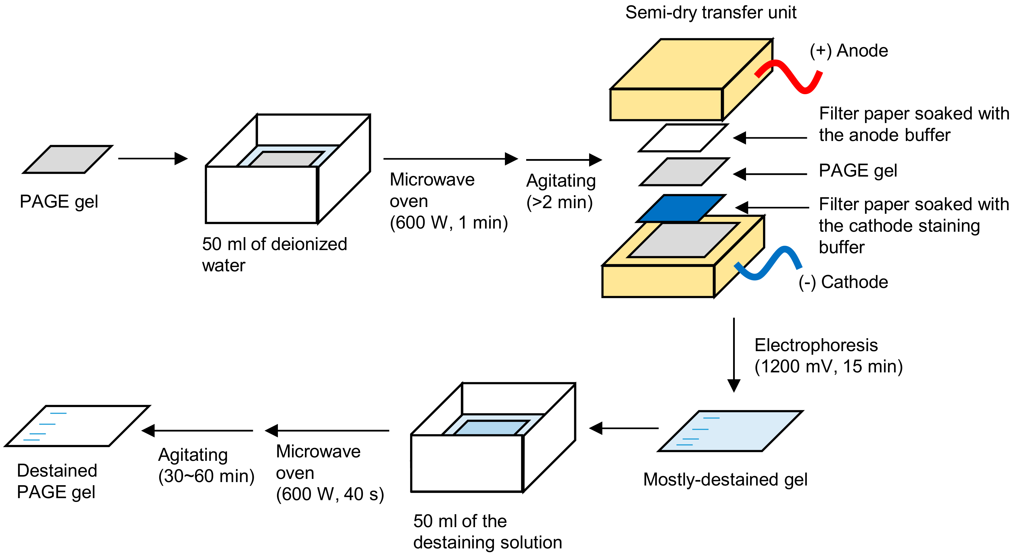
- Immerse a PAGE gel into 50 mL of deionized water in a plastic container, such as a ZipLoc® container (S. C. Johnson, Racine, WI, USA, Cat. No.: small square, 156 × 156 × 57 mm).
- The lid of the container should be opened to release the pressure from the boiled water.
- Heat the solution in a microwave oven at 600 W for 60 s to boil the solution and fix the proteins. Extend the heating time if the solution did not boil.
- Agitate it for more than 2 min to remove SDS and cool by adding water.
3.2. Electrophoretic-Staining/Destaining. Time for Completion: ~20 min
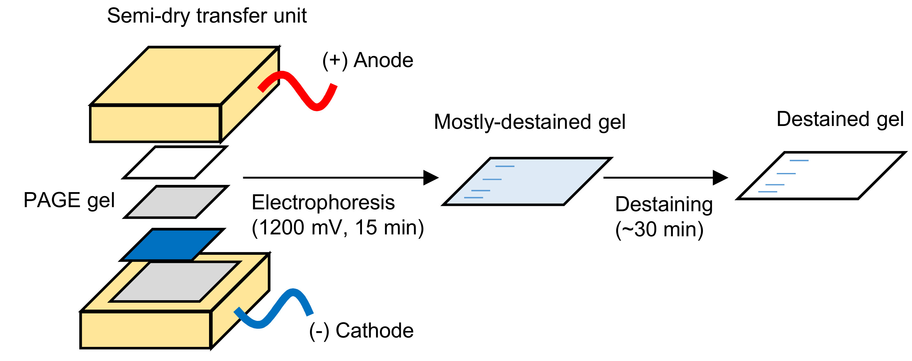
- Soak the filter paper in the cathode staining solution and spread the solution on the cathode of the semi-dry transfer unit with the filter paper, avoiding air bubbles at the interface.
- Place the soaked filter paper on the cathode.
- Immerse the fixed PAGE gel in the anode solution for a few seconds and place it on the filter paper. Avoid air bubbles between the filter paper and the gel.
- Soak a filter paper in the anode solution and place it on the PAGE gel.
- Spread the anode solution on the anode to avoid air bubbles.
- Place the anode on the filter paper.
- Place a weight of ~1 kg on the semi-dry transfer unit for close contact between the filter paper and electrodes.
- Run at a constant current of 1200 mA for 15 min by using a high current power supply. A higher current generates heat, resulting in bending of the gel and uneven destaining.
- If the CBB remained in the gel, change the arrangement of gel and the filter paper and run for an extra ~5 min.
- Wash the semi-dry transfer unit and cool down the electrode before the next staining.
3.3. Secondary Destaining. Time for Completion: 30–60 min
- Heat the gel in the destaining solution in a microwave oven for 40 s and subsequently agitate at room temperature (~25 °C) for 30–60 min for complete destaining. It takes 1–2 h without heating.
- OPTIONAL STEP: If the background level of CBB in the gel is high, replace the destaining solution containing 0.0001% (w/v) CBB R-250 to avoid excess destaining.
4. Expected Results and Discussion
5. Conclusions
6. Reagents Setup
Acknowledgments
Conflicts of Interest
References
- Laemmli, U.K. Cleavage of structural proteins during the assembly of the head of bacteriophage T4. Nature 1970, 227, 680–685. [Google Scholar] [CrossRef] [Scilit] [PubMed]
- Meyer, T.S.; Lamberts, B.L. Use of coomassie brilliant blue R250 for the electrophoresis of microgram quantities of parotid saliva proteins on acrylamide-gel strips. Biochim. Biophys. Acta 1965, 107, 144–145. [Google Scholar] [CrossRef] [Scilit]
- Merril, C.R.; Goldman, D.; Sedman, S.A.; Ebert, M.H. Ultrasensitive stain for proteins in polyacrylamide gels shows regional variation in cerebrospinal fluid proteins. Science 1981, 211, 1437–1438. [Google Scholar] [CrossRef] [Scilit] [PubMed]
- Weber, K.; Osborn, M. The reliability of molecular weight determinations sulfate-polyacrylamide gel electrophoresis. J. Biol. Chem. 1969, 244, 4406–4412. [Google Scholar] [PubMed]
- Sambrook, J.; Fritsch, E.F.; Maniatis, T. Molecular Cloning: A Laboratory Manual, 2nd ed.; CSH Laboratory Press: Cold Spring Harbor, NY, USA, 1989. [Google Scholar]
- Hervieu, G. A quick and safe method for destaining Coomassie-blue-stained protein gels. Tech. Tips Online 1997, 2, 94–96. [Google Scholar] [CrossRef] [Scilit]
- Kurien, B.T.; Scofield, R.H. Heat mediated quick Coomassie blue protein staining and destaining of SDS-PAGE gels. Indian J. Biochem. Biophys. 1998, 35, 385–389. [Google Scholar] [PubMed]
- Kurien, B.T.; Scofield, R.H. Accelerated coomassie blue staining and destaining of SDS-PAGE gels with application of heat. Methods Mol. Biol. 2012, 869, 471–479. [Google Scholar] [CrossRef] [Scilit] [PubMed]
- Fairbanks, G.; Steck, T.L.; Wallach, D.F. Electrophoretic analysis of the major polypeptides of the human erythrocyte membrane. Biochemistry 1971, 10, 2606–2617. [Google Scholar] [CrossRef] [Scilit] [PubMed]
- Wong, C.; Sridhara, S.; Bardwell, J.C.; Jakob, U. Heating greatly speeds Coomassie blue staining and destaining. Biotechniques 2000, 28, 426–432. [Google Scholar] [PubMed]
- Dong, W.H.; Wang, T.Y.; Wang, F.; Zhang, J.H. Simple, time-saving dye staining of proteins for sodium dodecyl sulfate-polyacrylamide gel electrophoresis using coomassie blue. PLoS ONE 2011, 6, 6–8. [Google Scholar] [CrossRef] [Scilit] [PubMed]
- Lawrence, A.-M.; Besir, H.U.S. Staining of proteins in gels with Coomassie G-250 without organic solvent and acetic acid. J. Vis. Exp. 2009, 2–4. [Google Scholar] [CrossRef] [Scilit] [PubMed]
- Kang, D.; Gho, Y.S.; Suh, M.; Kang, C. Highly sensitive and fast protein detection with Coomassie brilliant blue in sodium dodecyl sulfate-polyacrylamide gel electrophoresis. Bull. Korean Chem. Soc. 2002, 23, 1511–1512. [Google Scholar] [CrossRef] [Scilit]
- Dyballa, N.; Metzger, S. Fast and sensitive colloidal coomassie G-250 staining for proteins in polyacrylamide gels. J. Vis. Exp. 2009, 2–5. [Google Scholar] [CrossRef] [Scilit] [PubMed]
- Wenk, M.R.; Fernandis, A.Z. A Manual for Biochemistry Protocols; World Scientific: Singapore, 2007; Volume 3. [Google Scholar]
- Schneider, C.A.; Rasband, W.S.; Eliceiri, K.W. NIH Image to ImageJ: 25 years of image analysis. Nat. Methods 2012, 9, 671–675. [Google Scholar] [CrossRef] [Scilit] [PubMed]
- Chial, H.J.; Thompson, H.B.; Splittgerber, A.G. A spectral study of the charge forms of Coomassie blue G. Anal. Biochem. 1993, 209, 258–266. [Google Scholar] [CrossRef] [Scilit] [PubMed]
- Motojima, F.; Makio, T.; Aoki, K.; Makino, Y.; Kuwajima, K.; Yoshida, M. Hydrophilic residues at the apical domain of GroEL contribute to GroES binding but attenuate polypeptide binding. Biochem. Biophys. Res. Commun. 2000, 267, 842–849. [Google Scholar] [CrossRef] [Scilit] [PubMed]
© 2018 by the author. Licensee MDPI, Basel, Switzerland. This article is an open access article distributed under the terms and conditions of the Creative Commons Attribution (CC BY) license (http://creativecommons.org/licenses/by/4.0/).
Share and Cite
Motojima, F. Rapid Electrophoretic Staining and Destaining of Polyacrylamide Gels. Methods Protoc. 2018, 1, 13. https://doi.org/10.3390/mps1020013
Motojima F. Rapid Electrophoretic Staining and Destaining of Polyacrylamide Gels. Methods and Protocols. 2018; 1(2):13. https://doi.org/10.3390/mps1020013
Chicago/Turabian StyleMotojima, Fumihiro. 2018. "Rapid Electrophoretic Staining and Destaining of Polyacrylamide Gels" Methods and Protocols 1, no. 2: 13. https://doi.org/10.3390/mps1020013
APA StyleMotojima, F. (2018). Rapid Electrophoretic Staining and Destaining of Polyacrylamide Gels. Methods and Protocols, 1(2), 13. https://doi.org/10.3390/mps1020013