

Severe Myocardial Involvement and Persistent Supraventricular Arrhythmia in a Premature Infant Due to Enterovirus Infection: Case Report and Literature Review
 , ,
, ,  , , , and
Abstract
1. Introduction
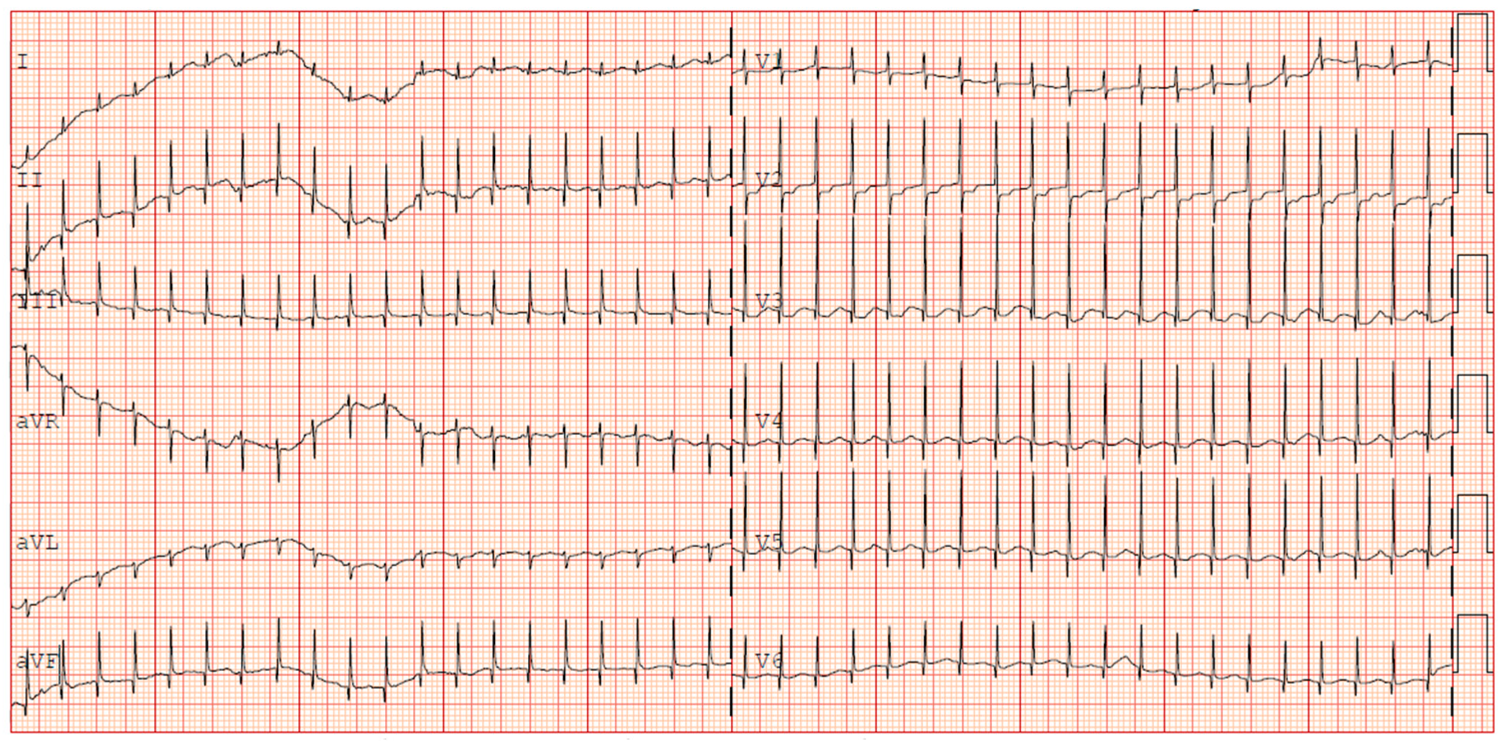
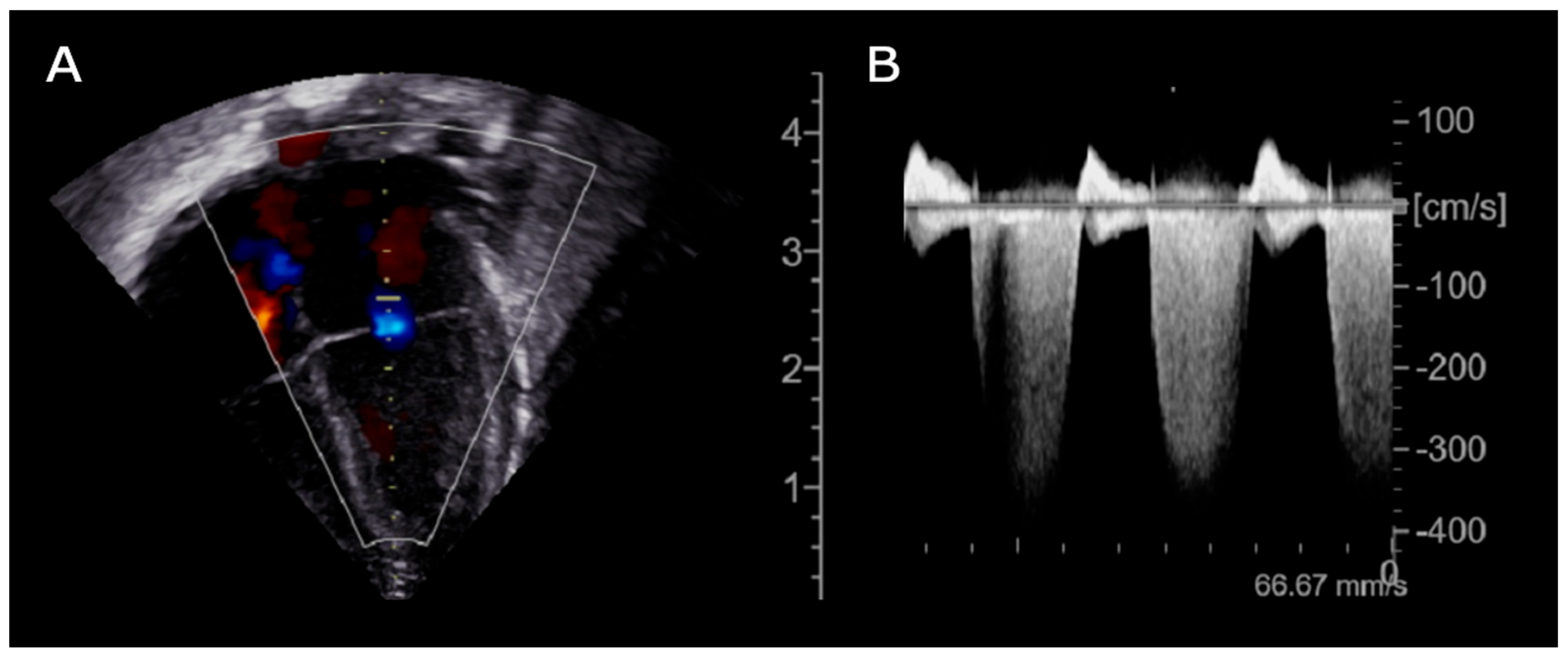
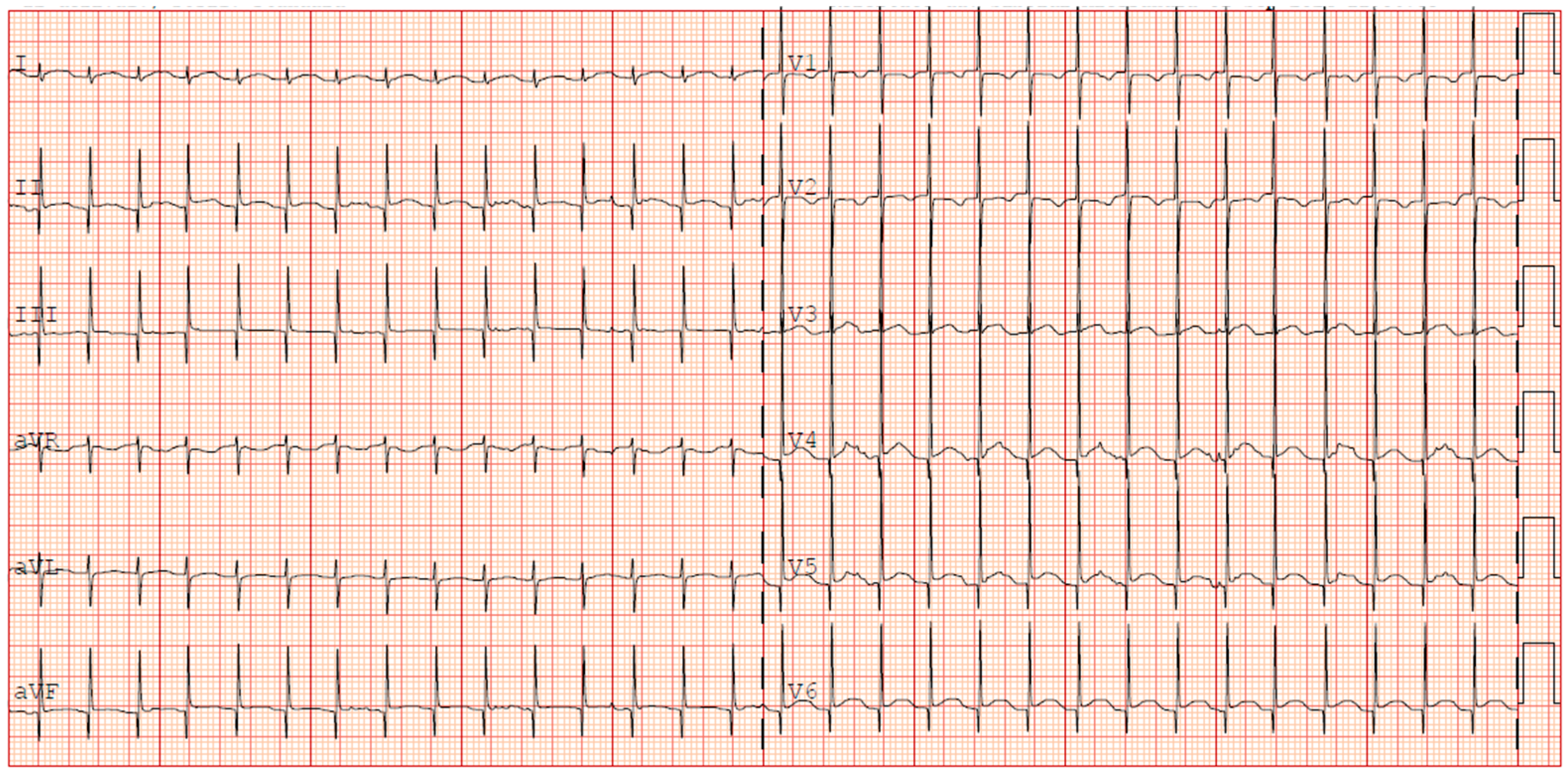
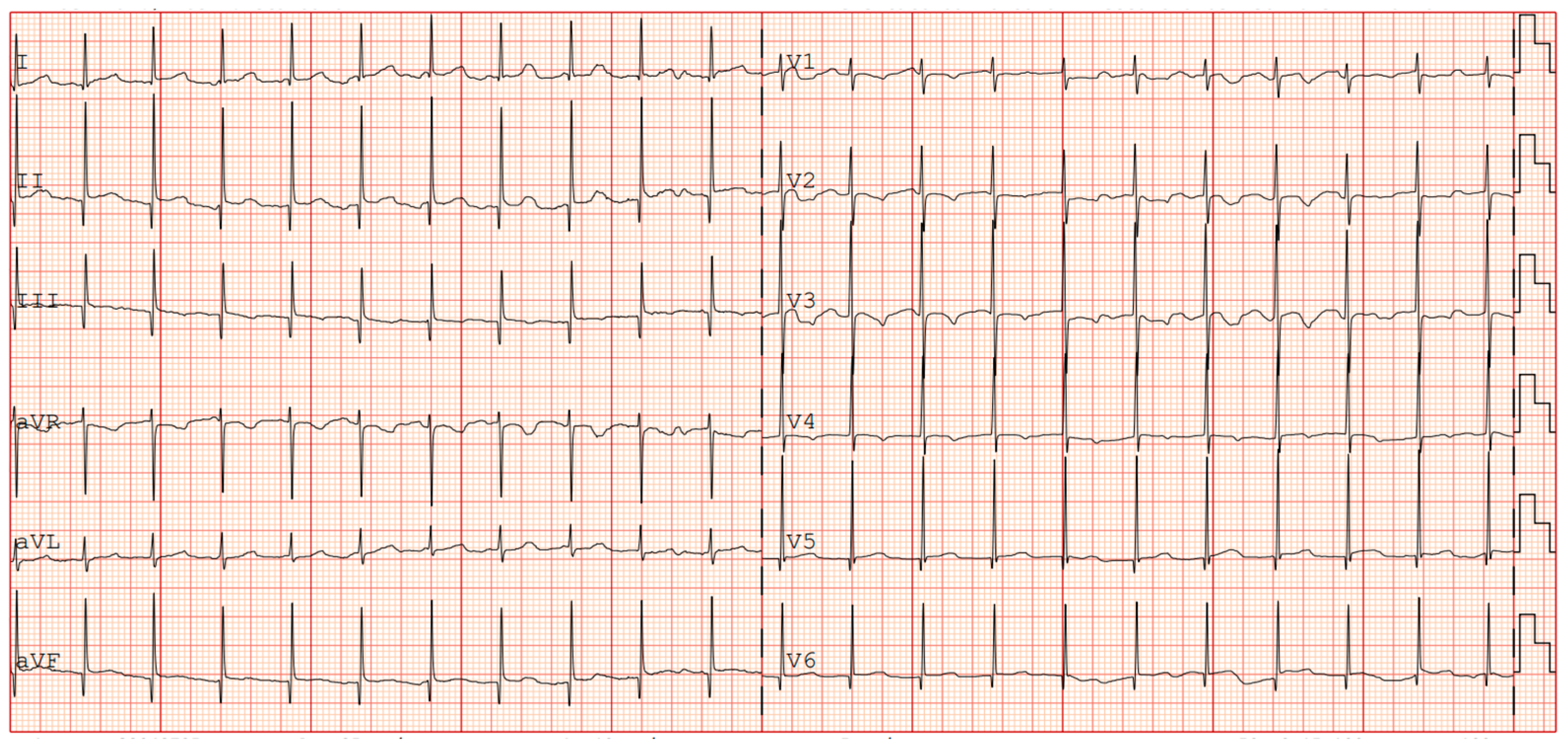
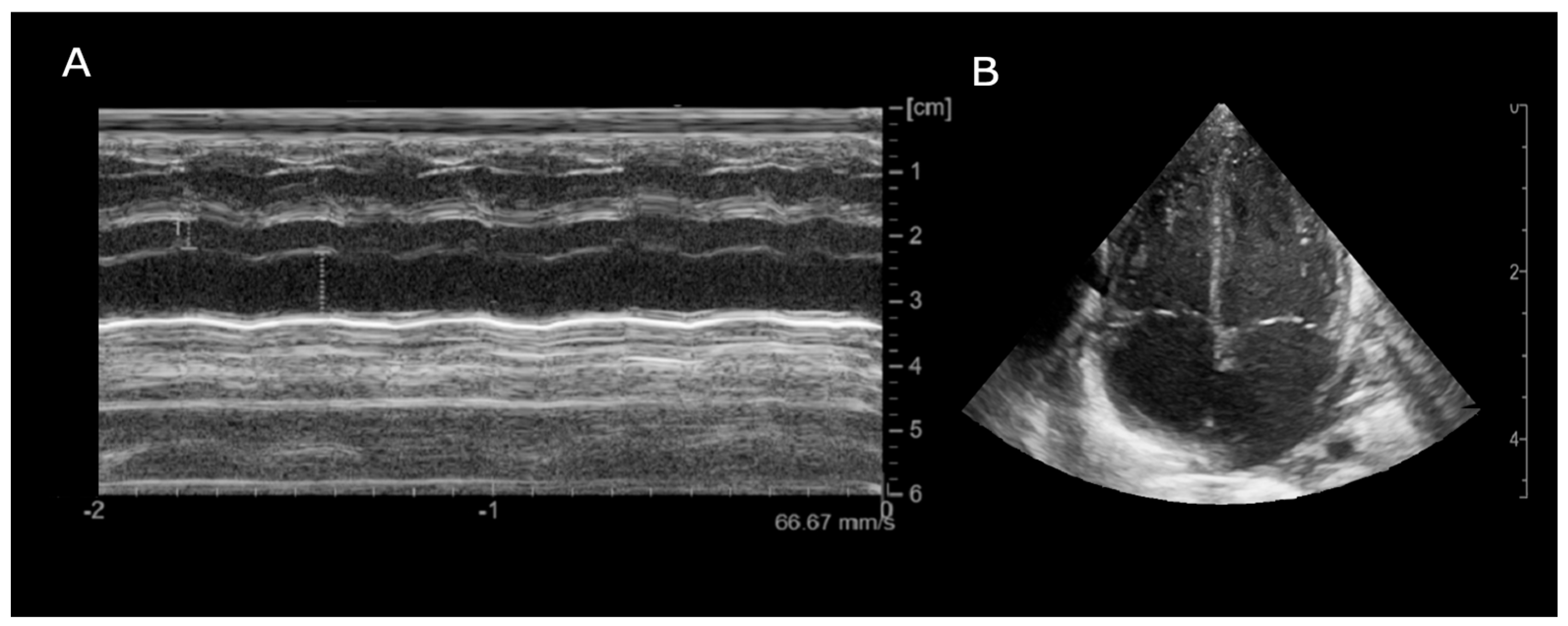
2. Case Report
3. Discussion

| Pt. | Type of Arrhythmia | GW | BW (g) | Onset (DOL) | Diagnosis Method (Virus) | Antiarrhythmic Treatment | Follow Up (months) | Ref. | Pt. Nationality |
|---|---|---|---|---|---|---|---|---|---|
| 1 | AF—SVT | 36 | 3700 | 1 | Stools (CoxsackieB2) | Electric Cardioversion (2J) + Digoxin until 6 months | AW (18) | [25] | USA |
| 2 | Intermittent JT | 36 | N/A | 6 | CSF (Enterovirus) | Electric Cardioversion + Amiodarone + Propranolol (→ Sotalol) + Digoxin | AW (1) | [1] | USA |
| 3 | Complete AVB, intermittent JT | 30 | N/A | 30 | Blood (Enterovirus) | Atropine + Digoxin | Recurrence AF (1) LV dilatation (3) | ||
| 4 | AF | 27 | N/A | 39 | CSF (Enterovirus) | Adenosine → Digoxin + Electric Cardioversion → Amiodarone + Propanolol | Moderate bi-atrial dilation, no arrhythmia (5) | ||
| 5 | PVC, ventricular bigeminy | 34 + 5 | 2460 | 9 | CSF, blood, stools (Enterovirus CVB5) | BB | No follow-up data reported | [26] | Italy |
| 6 | PAC, PVC, SVT | 35 | 3035 | 2 | CSF (CoxsackieB1) | Not reported | AW (no timing reported) | [3] | USA |
| 7 | SVT, ST depression | 38 | 1770 | 38 | Stools (ECHO 11) | Digoxin | No follow-up data reported | [27] | Australia |
| 8 | SVT, ST depression | 34 | 1870 | 25 | Stools (ECHO 11) | Digoxin | No follow-up data reported | ||
| 9 | SVT | 26 | 846 | 31 | Stools (ECHO 11) | Digoxin | No follow-up data reported | ||
| 10 | SVT, JET, EAT, VT, PAC, PVC, ST abnormalities | 37 + 6 | 3365 | 7 | Stools, blood (Enterovirus) | Adenosine + Cardioversion + BB | AW (8) | [28] | Republic of Korea |
| 11 | SVT | 38 | 3400 | 11 | Blood (Enterovirus) | Adenosine + Amiodarone+ Propranolol | No follow-up data reported | [29] | Croatia |
| 12 | JET | 37 | 2200 | 11 | Blood (ECHO 6) | Adenosine + Electric cardioversion + Digoxin + Amiodarone + Carvedilol | AW (3) | [30] | Germany |
| 13 | AVR, PVC | Term | 3400 | 1 | Blood, Antibodies (Coxsackie B4) | Antiarrhythmic (not specified) | AW (36) | [31] | Japan |
| 14 | SVT | 27 + 6 | 760 | 29 | Blood | Adenosine, Propranolol, Flecainide (→ Digoxin), Amiodarone | AW (7) | Our case | Italy |
4. Conclusions
Author Contributions
Funding
Institutional Review Board Statement
Informed Consent Statement
Data Availability Statement
Acknowledgments
Conflicts of Interest
Abbreviations
| NICU | Neonatal Intensive Care Unit |
| DOL | Days of Life |
| VT | Ventricular Tachycardia |
| SVT | Supraventricular Tachycardia |
| ECG | Electrocardiogram |
| ECMO | Extracorporeal Membrane Oxygenation |
| NAs | Neonatal Arrhythmias |
| AV | Atrioventricular |
| VF | Ventricular Fibrillation |
| ELBW | Extremely Low Birth Weight |
| LBW | Low Birth Weight |
| AVRT | Atrioventricular Reentrant Tachycardia |
| WPW | Wolff–Parkinson–White syndrome |
| AVNRT | Atrioventricular Nodal Reentrant Tachycardia |
| PJRT | Permanent Junctional Reciprocating Tachycardia |
| JET | Junctional Ectopic Tachycardia |
| GW | Gestational Weeks |
| FGR | Fetal Growth Restriction |
| FiO2 | Fraction of Inspired Oxygen |
| BID | Bis in Die |
| PDA | Patent Ductus Arteriosus |
| CPAP | Continuous Positive Airway Pressure |
| LOS | Late Onset Sepsis |
| CSF | Cerebrospinal Fluid |
| NT-proBNP | N-terminal pro B-type Natriuretic Peptide |
| AST | Aspartate Aminotransferase |
| ALT | Alanine Aminotransferase |
| CRP | C-Reactive Protein |
| MRI | Magnetic Resonance Imaging |
| IVH | Intraventricular Hemorrhage |
| EAT | Ectopic Atrial Tachycardia |
| EF | Ejection Fraction |
References
- Simpson, K.E.; Hulse, E.; Carlson, K. Atrial Tachyarrhythmias in Neonatal Enterovirus Myocarditis. Pediatr. Cardiol. 2009, 30, 827–830. [Google Scholar] [CrossRef] [PubMed]
- Zhang, M.; Wang, H.; Tang, J.; He, Y.; Xiong, T.; Li, W.; Qu, Y.; Mu, D. Clinical characteristics of severe neonatal enterovirus infection: A systematic review. BMC Pediatr. 2021, 21, 127. [Google Scholar] [CrossRef]
- Lu, J.C.; Koay, K.W.; Ramers, C.B.; Milazzo, A.S.; Carolina, N. Neonate with Coxsackie B1 Infection, Cardiomyopathy and Arrhythmias. J. Natl. Med. Assoc. 2005, 97, 1028–1030. [Google Scholar] [PubMed]
- Madden, K.; Thiagarajan, R.R.; Rycus, P.T.; Rajagopal, S.K. Survival of neonates with enteroviral myocarditis requiring extracorporeal membrane oxygenation. Pediatr. Crit. Care Med. 2011, 12, 314–318. [Google Scholar] [CrossRef]
- Chaudhry-Waterman, N.; Nashed, L.; Chidester, R.; Nalewanski, A.; Bastawrous, D.; Busch, H.; Jeong, H.; Baker, R.; Donnelly, K.; Cohen, M. A Prospective Evaluation of Arrhythmias in a Large Tertiary Neonatal Intensive Care Unit. Pediatr. Cardiol. 2023, 44, 1319–1326. [Google Scholar] [CrossRef]
- Jaeggi, E.; Öhman, A. Fetal and Neonatal Arrhythmias. Clin. Perinatol. 2016, 43, 99–112. [Google Scholar] [CrossRef] [PubMed]
- Bruder, D.; Weber, R.; Gass, M.; Balmer, C.; Cavigelli-Brunner, A. Antiarrhythmic Medication in Neonates and Infants with Supraventricular Tachycardia. Pediatr. Cardiol. 2022, 43, 1311–1318. [Google Scholar] [CrossRef]
- Kang, K.T.; Potts, J.E.; Radbill, A.E.; La Page, M.J.; Papagiannis, J.; Garnreiter, J.M.; Kubus, P.; Kantoch, M.J.; Von Bergen, N.H.; Fournier, A.; et al. Permanent junctional reciprocating tachycardia in children: A multicenter experience. Heart Rhythm. 2014, 11, 1426–1432. [Google Scholar] [CrossRef]
- Aydoğan, S.; Fettah, N.D.; Tuğcu, A.U.; Koyuncu, E.; Yoldaş, T.; Zenciroğlu, A. Supraventricular tachycardia after respiratory syncytial virus infection in a newborn. Bayl. Univ. Med. Cent. Proc. 2022, 35, 705–706. [Google Scholar] [CrossRef]
- Radaelli, M.; Keller, C.P.T.L.; Franca, H.; Mehrotra, K. Mycoplasma myocarditis presenting with sustained SVT and acute heart failure without signs of myocardiocytolysis and extra-cardiac disease. Clin. Case Rep. 2024, 12, e8851. [Google Scholar] [CrossRef]
- Muehlenbachs, A.; Bhatnagar, J.; Zaki, S.R. Tissue tropism, pathology and pathogenesis of enterovirus infection. J. Pathol. 2015, 235, 217–228. [Google Scholar] [CrossRef] [PubMed]
- Gaaloul, I.; Riabi, S.; Evans, M.; Hunter, T.; Huber, S.; Aouni, M. Postmortem diagnosis of infectious heart diseases: A mystifying cause of Sudden Infant Death. Forensic Sci. Int. 2016, 262, 166–172. [Google Scholar] [CrossRef] [PubMed]
- Neagu, O.; Rodríguez, A.F.; Callon, D.; Andréoletti, L.; Cohen, M.C. Myocarditis Presenting as Sudden Death in Infants and Children: A Single Centre Analysis by ESGFOR Study Group. Pediatr. Dev. Pathol. 2021, 24, 327–336. [Google Scholar] [CrossRef] [PubMed]
- Grangeot-Keros, L.; Broyer, M.; Briand, E.; Gut, J.P.; Turkoglü, S.; Chretien, P.; Emilie, D.; Dussaix, E.; Lazizi, Y.; Dehan, M. Enterovirus in sudden unexpected deaths in infants. Pediatr. Infect. Dis. J. 1996, 15, 123–128. [Google Scholar] [CrossRef]
- Laitinen, O.H.; Svedin, E.; Kapell, S.; Nurminen, A.; Hytönen, V.P.; Flodström-Tullberg, M. Enteroviral proteases: Structure, host interactions and pathogenicity: Pathogenicity of enteroviral proteases. Rev. Med. Virol. 2016, 26, 251–267. [Google Scholar] [CrossRef]
- Venteo, L.; Bourlet, T.; Renois, F.; Douche-Aourik, F.; Mosnier, J.-F.; Lorain De La Grand Maison, G.; Pluot, M.; Pozzetto, B.; Andreoletti, L. Enterovirus-related activation of the cardiomyocyte mitochondrial apoptotic pathway in patients with acute myocarditis. Eur. Heart J. 2010, 31, 728–736. [Google Scholar] [CrossRef]
- Bouin, A.; Gretteau, P.-A.; Wehbe, M.; Renois, F.; N’Guyen, Y.; Lévêque, N.; Vu, M.N.; Tracy, S.; Chapman, N.M.; Bruneval, P.; et al. Enterovirus Persistence in Cardiac Cells of Patients With Idiopathic Dilated Cardiomyopathy Is Linked to 5’ Terminal Genomic RNA-Deleted Viral Populations With Viral-Encoded Proteinase Activities. Circulation 2019, 139, 2326–2338. [Google Scholar] [CrossRef]
- Aretz, H.T.; Billingham, M.E.; Edwards, W.D.; Factor, S.M.; Fallon, J.T.; Fenoglio, J.J.; Olsen, E.G.; Schoen, F.J. Myocarditis. A histopathologic definition and classification. Am. J. Cardiovasc. Pathol. 1987, 1, 3–14. [Google Scholar]
- Law, Y.M.; Lal, A.K.; Chen, S.; Čiháková, D.; Cooper, L.T.; Deshpande, S.; Godown, J.; Grosse-Wortmann, L.; Robinson, J.D.; Towbin, J.A.; et al. Diagnosis and Management of Myocarditis in Children: A Scientific Statement from the American Heart Association. Circulation 2021, 144, 6. [Google Scholar] [CrossRef]
- Batra, A.S.; Silka, M.J.; Borquez, A.; Cuneo, B.; Dechert, B.; Jaeggi, E.; Kannankeril, P.J.; Tabulov, C.; Tisdale, J.E.; Wolfe, D.; et al. Pharmacological Management of Cardiac Arrhythmias in the Fetal and Neonatal Periods: A Scientific Statement From the American Heart Association. Circulation 2024, 149, e937–e952. [Google Scholar] [CrossRef]
- Lim, Y.T.; Kim, Y.H.; Kwon, J.E. Effective Control of Supraventricular Tachycardia in Neonates May Requires Combination Pharmacologic Therapy. JCM 2022, 11, 3279. [Google Scholar] [CrossRef] [PubMed]
- Aljohani, O.A.; Herrick, N.L.; Borquez, A.A.; Shepard, S.; Wieler, M.E.; Perry, J.C.; Williams, M.R. Antiarrhythmic Treatment Duration and Tachycardia Recurrence in Infants with Supraventricular Tachycardia. Pediatr. Cardiol. 2021, 42, 716–720. [Google Scholar] [CrossRef] [PubMed]
- Mecarini, F.; Comitini, F.; Bardanzellu, F.; Neroni, P.; Fanos, V. Neonatal supraventricular tachycardia and necrotizing enterocolitis: Case report and literature review. Ital. J. Pediatr. 2020, 46, 117. [Google Scholar] [CrossRef] [PubMed]
- Freund, M.W.; Kleinveld, G.; Krediet, T.G.; Van Loon, A.M.; Verboon-Maciolek, M.A. Prognosis for neonates with enterovirus myocarditis. Arch. Dis. Child.—Fetal Neonatal Ed. 2010, 95, F206–F212. [Google Scholar] [CrossRef]
- Shah, S.S.; Hellenbrand, W.E.; Gallagher, P.G. Atrial Flutter Complicating Neonatal Coxsackie B2 Myocarditis. Pediatr. Cardiol. 1998, 19, 185–186. [Google Scholar] [CrossRef]
- Giampetruzzi, S.; Sirico, D.; Mainini, N.; Meneghelli, M.; Valerio, E.; Salvadori, S.; Di Salvo, G. Neonatal Enterovirus-Associated Myocarditis in Dizygotic Twins: Myocardial Longitudinal Strain Pattern Analysis. Children 2024, 11, 506. [Google Scholar] [CrossRef]
- Drew, J.H. Echo 11 virus outbreak in a nursery associated with myocarditis. J. Paediatr. Child. Health 1973, 9, 90–95. [Google Scholar] [CrossRef]
- Lee, S.R.; Ko, S.Y.; Yoon, S.Y.; Lee, Y.K.; Shin, S.M. Early Detection and Successful Treatment of Vertically Transmitted Fulminant Enteroviral Infection Associated with Various Forms of Arrhythmia and Severe Hepatitis with Coagulopathy. Pediatr. Infect. Vaccine 2019, 26, 199. [Google Scholar] [CrossRef]
- Banjac, L.; Nikcević, D.; Vujosević, D.; Raonić, J.; Banjac, G. Tachycardia in a newborn with enterovirus infection. Acta Clin. Croat. 2014, 53, 102–106. [Google Scholar]
- Weickmann, J.; Gebauer, R.A.; Paech, C. Junctional ectopic tachycardia in neonatal enterovirus myocarditis. Clin. Case Rep. 2020, 8, 987–990. [Google Scholar] [CrossRef]
- Yokota, Y.; Wada, M. Accelerated ventricular rhythm associated with myocarditis in a neonate. Pediatr. Cardiol. 1989, 10, 178. [Google Scholar] [CrossRef] [PubMed]
Disclaimer/Publisher’s Note: The statements, opinions and data contained in all publications are solely those of the individual author(s) and contributor(s) and not of MDPI and/or the editor(s). MDPI and/or the editor(s) disclaim responsibility for any injury to people or property resulting from any ideas, methods, instructions or products referred to in the content. |
© 2025 by the authors. Licensee MDPI, Basel, Switzerland. This article is an open access article distributed under the terms and conditions of the Creative Commons Attribution (CC BY) license (https://creativecommons.org/licenses/by/4.0/).
Share and Cite
Montobbio, C.; Conte, A.; Calandrino, A.; Pepe, A.; Vinci, F.; Siboldi, A.; Formigari, R.; Ramenghi, L.A. Severe Myocardial Involvement and Persistent Supraventricular Arrhythmia in a Premature Infant Due to Enterovirus Infection: Case Report and Literature Review. J. Cardiovasc. Dev. Dis. 2025, 12, 228. https://doi.org/10.3390/jcdd12060228
Montobbio C, Conte A, Calandrino A, Pepe A, Vinci F, Siboldi A, Formigari R, Ramenghi LA. Severe Myocardial Involvement and Persistent Supraventricular Arrhythmia in a Premature Infant Due to Enterovirus Infection: Case Report and Literature Review. Journal of Cardiovascular Development and Disease. 2025; 12(6):228. https://doi.org/10.3390/jcdd12060228
Chicago/Turabian StyleMontobbio, Carolina, Alessio Conte, Andrea Calandrino, Alessia Pepe, Francesco Vinci, Alessandra Siboldi, Roberto Formigari, and Luca Antonio Ramenghi. 2025. "Severe Myocardial Involvement and Persistent Supraventricular Arrhythmia in a Premature Infant Due to Enterovirus Infection: Case Report and Literature Review" Journal of Cardiovascular Development and Disease 12, no. 6: 228. https://doi.org/10.3390/jcdd12060228
APA StyleMontobbio, C., Conte, A., Calandrino, A., Pepe, A., Vinci, F., Siboldi, A., Formigari, R., & Ramenghi, L. A. (2025). Severe Myocardial Involvement and Persistent Supraventricular Arrhythmia in a Premature Infant Due to Enterovirus Infection: Case Report and Literature Review. Journal of Cardiovascular Development and Disease, 12(6), 228. https://doi.org/10.3390/jcdd12060228