

Advancements and Challenges in Sucralose Determination: A Comparative Review of Chromatographic, Electrochemical, and Spectrophotometric Methods

 , ,
, ,  ,
, 

Abstract
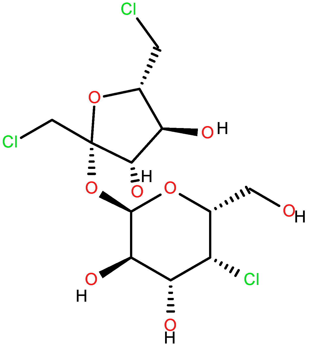
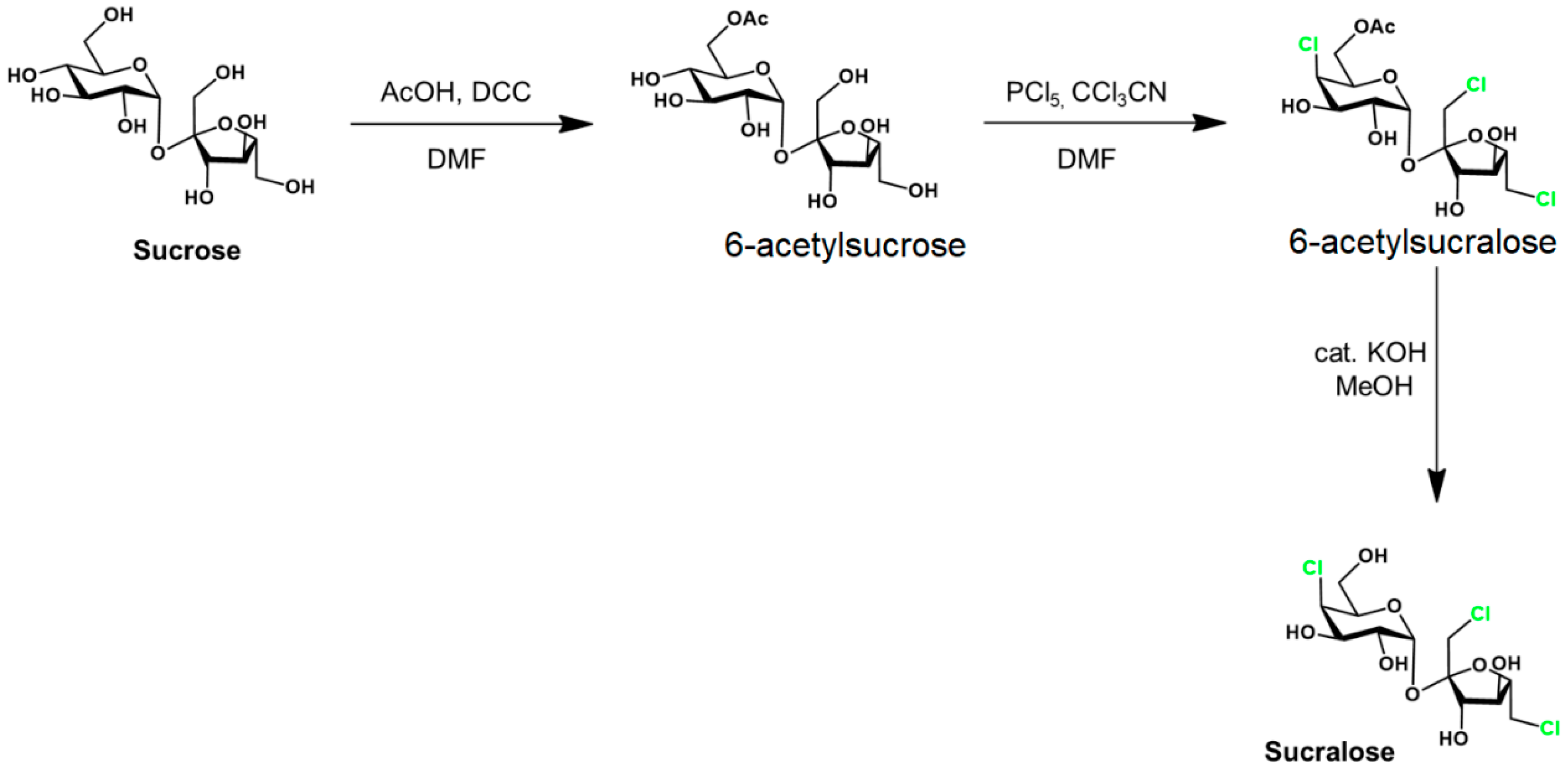
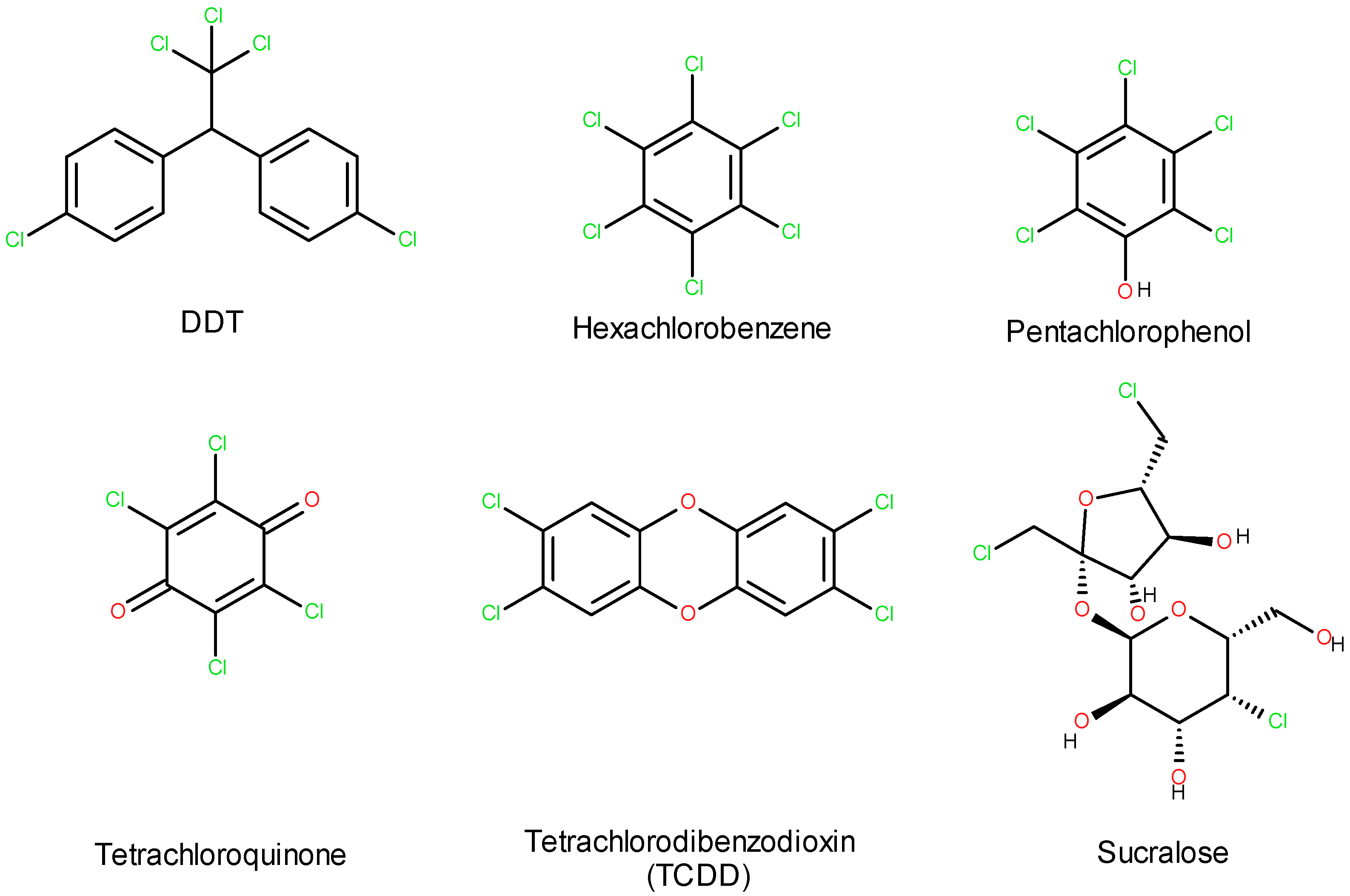
1. Introduction
2. Materials and Methods
3. Sucralose Determination Methods
3.1. Spectrophotometric Methods
3.2. Chromatographic Methods
3.3. Electrophoretic and Electrochemical Methods
3.4. Comparative Overview of Chromatographic and Electrochemical Approaches for Sucralose Determination
4. Conclusions
Author Contributions
Funding
Institutional Review Board Statement
Informed Consent Statement
Data Availability Statement
Acknowledgments
Conflicts of Interest
References
- Debras, C.; Chazelas, E.; Sellem, L.; Porcher, R.; Druesne-Pecollo, N.; Esseddik, Y.; de Edelenyi, F.S.; Agaësse, C.; De Sa, A.; Lutchia, R.; et al. Artificial sweeteners and risk of cardiovascular diseases: Results from the prospective NutriNet-Santé cohort. BMJ 2022, 378, e071204. [Google Scholar] [CrossRef] [PubMed]
- Juárez, G.; Sanz-Novo, M.; Alonso, J.L.; Alonso, E.R.; León, I. Rotational Spectrum and Conformational Analysis of Perillartine: Insights into the Structure–Sweetness Relationship. Molecules 2022, 27, 1924. [Google Scholar] [CrossRef] [PubMed]
- Xiao, Y.; Xiao, L.; Li, M.; Liu, S.; Wang, Y.; Huang, L.; Liu, S.; Jiang, T.; Zhou, L.; Li, Y. Perillartine protects against metabolic associated fatty liver in high-fat diet-induced obese mice. Food Funct. 2022, 14, 961–977. [Google Scholar] [CrossRef]
- Xiao, Y.; Jia, M.; Jiang, T.; Zhang, C.; Qi, X.; Sun, Y.; Gao, J.; Zhou, L.; Li, Y. Dietary supplementation with perillartine ameliorates lipid metabolism disorder induced by a high-fat diet in broiler chickens. Biochem. Biophys. Res. Commun. 2022, 625, 66–74. [Google Scholar] [CrossRef]
- del Pozo, S.; Gómez-Martínez, S.; Díaz, L.E.; Nova, E.; Urrialde, R.; Marcos, A. Potential Effects of Sucralose and Saccharin on Gut Microbiota: A Review. Nutrients 2022, 14, 1682. [Google Scholar] [CrossRef]
- The Science Snail. Available online: https://www.sciencesnail.com/science/the-synthesis-of-sucralose-from-sucrose (accessed on 10 March 2025).
- Méndez-García, L.A.; Bueno-Hernández, N.; Cid-Soto, M.A.; De León, K.L.; Mendoza-Martínez, V.M.; Espinosa-Flores, A.J.; Carrero-Aguirre, M.; Esquivel-Velázquez, M.; León-Hernández, M.; Viurcos-Sanabria, R.; et al. Ten-Week Sucralose Consumption Induces Gut Dysbiosis and Altered Glucose and Insulin Levels in Healthy Young Adults. Microorganisms 2022, 10, 434. [Google Scholar] [CrossRef]
- Huang, H.; Liu, S.; Peng, Z.; Wang, B.; Zhan, S.; Huang, S.; Li, W.; Liu, D.; Yang, X.; Zhu, Y.; et al. Comparative effects of different sugar substitutes: Mogroside V, stevioside, sucralose, and erythritol on intestinal health in a type 2 diabetes mellitus mouse. Food Funct. 2025, 16, 2108–2123. [Google Scholar] [CrossRef]
- Ashwell, M.; Gibson, S.; Bellisle, F.; Buttriss, J.; Drewnowski, A.; Fantino, M.; Gallagher, A.M.; de Graaf, K.; Goscinny, S.; Hardman, C.A.; et al. Expert consensus on low-calorie sweeteners: Facts, research gaps and suggested actions. Nutr. Res. Rev. 2020, 33, 145–154. [Google Scholar] [CrossRef]
- Plaza-Diaz, J.; Pastor-Villaescusa, B.; Rueda-Robles, A.; Abadia-Molina, F.; Ruiz-Ojeda, F.J. Plausible Biological Interactions of Low- and Non-Calorie Sweeteners with the Intestinal Microbiota: An Update of Recent Studies. Nutrients 2020, 12, 1153. [Google Scholar] [CrossRef]
- Singh, A.S.; Singh, S.; Begum, R.F.; Vijayan, S.; Vellapandian, C. Unveiling the profound influence of sucralose on metabolism and its role in shaping obesity trends. Front. Nutr. 2024, 11, 1387646. [Google Scholar] [CrossRef]
- Aguayo-Guerrero, J.A.; Méndez-García, L.A.; Manjarrez-Reyna, A.N.; Esquivel-Velázquez, M.; León-Cabrera, S.; Meléndez, G.; Zambrano, E.; Ramos-Martínez, E.; Fragoso, J.M.; Briones-Garduño, J.C.; et al. Newborns from Mothers Who Intensely Consumed Sucralose during Pregnancy Are Heavier and Exhibit Markers of Metabolic Alteration and Low-Grade Systemic Inflammation: A Cross-Sectional, Prospective Study. Biomedicines 2023, 11, 650. [Google Scholar] [CrossRef] [PubMed]
- Stampe, S.; Leth-Møller, M.; Greibe, E.; Hoffmann-Lücke, E.; Pedersen, M.; Ovesen, P. Artificial Sweeteners in Breast Milk: A Clinical Investigation with a Kinetic Perspective. Nutrients 2022, 14, 2635. [Google Scholar] [CrossRef] [PubMed]
- Yang, Y.; Liu, Z.; Zheng, H.; Zhu, S.; Zhang, K.; Li, X.; Ma, X.; Dietrich, A.M. Sucralose, a persistent artificial sweetener in the urban water cycle: Insights into occurrence, chlorinated byproducts formation, and human exposure. J. Environ. Chem. Eng. 2021, 9, 105293. [Google Scholar] [CrossRef]
- Colín-García, K.; Elizalde-Velázquez, G.A.; Gómez-Oliván, L.M.; Islas-Flores, H.; García-Medina, S.; Galar-Martínez, M. Acute exposure to environmentally relevant concentrations of sucralose disrupts embryonic development and leads to an oxidative stress response in Danio rerio. Sci. Total. Environ. 2022, 829, 154689. [Google Scholar] [CrossRef]
- Mattoli, L.; Fodaroni, G.; Proietti, G.; Flamini, E.; Paoli, B.; Massa, L.; Ferrara, G.C.; Giovagnoni, E.; Gianni, M. Biodegradability of dietary supplements: Advanced analytical methods to study the environmental fate of artificial sweeteners and dyes. J. Pharm. Biomed. Anal. 2024, 255, 116575. [Google Scholar] [CrossRef]
- Tilg, H.; Adolph, T.E. Sucralose and Erythritol—Not Too Sweet. N. Engl. J. Med. 2023, 389, 859–861. [Google Scholar] [CrossRef]
- Schiffman, S.S.; Scholl, E.H.; Furey, T.S.; Nagle, H.T. Toxicological and pharmacokinetic properties of sucralose-6-acetate and its parent sucralose: In vitro screening assays. J. Toxicol. Environ. Health Part B 2023, 26, 307–341. [Google Scholar] [CrossRef]
- Dietrich, A.M.; Pang, Z.; Zheng, H.; Ma, X. Mini Review: Will Artificial Sweeteners Discharged to the Aqueous Environment Unintentionally “Sweeten” the Taste of Tap Water? Chem. Eng. Adv. 2021, 6, 100100. [Google Scholar] [CrossRef]
- Bornemann, V.; Werness, S.C.; Buslinger, L.; Schiffman, S.S. Intestinal Metabolism and Bioaccumulation of Sucralose In Adipose Tissue In The Rat. J. Toxicol. Environ. Health Part A 2018, 81, 913–923. [Google Scholar] [CrossRef]
- Suez, J.; Cohen, Y.; Valdés-Mas, R.; Mor, U.; Dori-Bachash, M.; Federici, S.; Zmora, N.; Leshem, A.; Heinemann, M.; Linevsky, R.; et al. Personalized microbiome-driven effects of non-nutritive sweeteners on human glucose tolerance. Cell 2022, 185, 3307–3328.e19. [Google Scholar] [CrossRef]
- Hosseini, A.; Barlow, G.M.; Leite, G.; Rashid, M.; Parodi, G.; Wang, J.; Morales, W.; Weitsman, S.; Rezaie, A.; Pimentel, M.; et al. Consuming artificial sweeteners may alter the structure and function of duodenal microbial communities. iScience 2023, 26, 108530. [Google Scholar] [CrossRef] [PubMed]
- Jarmakiewicz-Czaja, S.; Sokal-Dembowska, A.; Filip, R. Effects of Selected Food Additives on the Gut Microbiome and Metabolic Dysfunction-Associated Steatotic Liver Disease (MASLD). Medicina 2025, 61, 192. [Google Scholar] [CrossRef] [PubMed]
- Harpaz, D.; Yeo, L.P.; Cecchini, F.; Koon, T.H.P.; Kushmaro, A.; Tok, A.I.Y.; Marks, R.S.; Eltzov, E. Measuring Artificial Sweeteners Toxicity Using a Bioluminescent Bacterial Panel. Molecules 2018, 23, 2454. [Google Scholar] [CrossRef]
- Ahmad, S.Y.; Friel, J.K.; Mackay, D.S. Effect of sucralose and aspartame on glucose metabolism and gut hormones. Nutr. Rev. 2020, 78, 725–746. [Google Scholar] [CrossRef]
- Zani, F.; Blagih, J.; Gruber, T.; Buck, M.D.; Jones, N.; Hennequart, M.; Newell, C.L.; Pilley, S.E.; Soro-Barrio, P.; Kelly, G.; et al. The dietary sweetener sucralose is a negative modulator of T cell-mediated responses. Nature 2023, 615, 705–711. [Google Scholar] [CrossRef]
- Risdon, S.; Battault, S.; Romo-Romo, A.; Roustit, M.; Briand, L.; Meyer, G.; Almeda-Valdes, P.; Walther, G. Sucralose and Cardiometabolic Health: Current Understanding from Receptors to Clinical Investigations. Adv. Nutr. Int. Rev. J. 2021, 12, 1500–1513. [Google Scholar] [CrossRef]
- Zhai, Y.; Bai, D.; Yang, H.; Li, X.; Zhu, D.; Cao, X.; Ma, H.; Zheng, X. Hazardous Effects of Sucralose and Its Disinfection Byproducts Identified From an E. coli Whole-Cell Array Analysis. Front. Environ. Sci. 2021, 9, 724685. [Google Scholar] [CrossRef]
- dos Santos, C.A.; Pedrosa, D.S.; Santos, M.D.S. Adoçantes Sintéticos e Seus Riscos à Saúde. Rev. Ciên. Saúde 2023, 27, 122. (In Portuguese) [Google Scholar] [CrossRef]
- Aguayo-Guerrero, J.A.; Méndez-García, L.A.; Solleiro-Villavicencio, H.; Viurcos-Sanabria, R.; Escobedo, G. Sucralose: From Sweet Success to Metabolic Controversies—Unraveling the Global Health Implications of a Pervasive Non-Caloric Artificial Sweetener. Life 2024, 14, 323. [Google Scholar] [CrossRef]
- Blenkley, E.; Suckling, J.; Morse, S.; Murphy, R.; Raats, M.; Astley, S.; Halford, J.C.G.; Harrold, J.A.; Le-Bail, A.; Koukouna, E.; et al. Environmental life cycle assessment of production of the non-nutritive sweetener sucralose (E955) derived from cane sugar produced in the United States of America: The SWEET project. Int. J. Life Cycle Assess. 2023, 28, 1689–1704. [Google Scholar] [CrossRef]
- Wiklund, A.-K.E.; Guo, X.; Gorokhova, E. Cardiotoxic and neurobehavioral effects of sucralose and acesulfame in Daphnia: Toward understanding ecological impacts of artificial sweeteners. Comp. Biochem. Physiol. Part C Toxicol. Pharmacol. 2023, 273, 109733. [Google Scholar] [CrossRef] [PubMed]
- Young, N.; Welch, J.; Hill, T.; Sees, M.; Beazley, M.; Heider, E.C. Longitudinal Analysis of Sucralose at a Water Treatment Wetland. Environments 2022, 9, 111. [Google Scholar] [CrossRef]
- Islam, M.; Thompson, K.; Dickenson, E.; Quiñones, O.; Steinle-Darling, E.; Westerhoff, P. Sucralose and Predicted De Facto Wastewater Reuse Levels Correlate with PFAS Levels in Surface Waters. Environ. Sci. Technol. Lett. 2023, 10, 431–438. [Google Scholar] [CrossRef]
- Whitall, D.; Curtis, M.; Mason, A. Use of sucralose and caffeine as tracers of human waste in a coral reef ecosystem. Reg. Stud. Mar. Sci. 2021, 44, 101740. [Google Scholar] [CrossRef]
- Serville-Tertullien, M.; McDermott, K.; Majury, A.; Liang, T.; Sultana, T.; Metcalfe, C.D. Sucralose and caffeine as chemical indicators of domestic wastewater contamination in the Laurentian Great Lakes Basin. Environ. Monit. Assess. 2024, 196, 1085. [Google Scholar] [CrossRef]
- Westmoreland, A.G.; Schafer, T.B.; Breland, K.E.; Beard, A.R.; Osborne, T.Z. Sucralose (C12H19Cl3O8) impact on microbial activity in estuarine and freshwater marsh soils. Environ. Monit. Assess. 2024, 196, 451. [Google Scholar] [CrossRef]
- Kerberová, V.; Gargošová, H.Z.; Čáslavský, J. Occurrence and ecotoxicity of selected artificial sweeteners in the Brno city waste water. Int. J. Environ. Sci. Technol. 2021, 19, 9055–9066. [Google Scholar] [CrossRef]
- Luo, J.; Zhang, Q.; Cao, M.; Wu, L.; Cao, J.; Fang, F.; Li, C.; Xue, Z.; Feng, Q. Ecotoxicity and environmental fates of newly recognized contaminants-artificial sweeteners: A review. Sci. Total. Environ. 2019, 653, 1149–1160. [Google Scholar] [CrossRef]
- Lewis, K.; Tzilivakis, J. Review and synthesis of data on the potential environmental impact of artificial sweeteners. EFSA Support. Publ. 2021, 18, 6918E. [Google Scholar] [CrossRef]
- Vásquez-Reyes, S.; Velázquez-Villegas, L.A.; Vargas-Castillo, A.; Noriega, L.G.; Torres, N.; Tovar, A.R. Dietary bioactive compounds as modulators of mitochondrial function. J. Nutr. Biochem. 2021, 96, 108768. [Google Scholar] [CrossRef]
- Silva, L.B.A.R.; Pinheiro-Castro, N.; Novaes, G.M.; Pascoal, G.d.F.L.; Ong, T.P. Bioactive food compounds, epigenetics and chronic disease prevention: Focus on early-life interventions with polyphenols. Food Res. Int. 2019, 125, 108646. [Google Scholar] [CrossRef] [PubMed]
- Samtiya, M.; Aluko, R.E.; Dhewa, T.; Moreno-Rojas, J. Potential Health Benefits of Plant Food-Derived Bioactive Components: An Overview. Foods 2021, 10, 839. [Google Scholar] [CrossRef] [PubMed]
- Banwo, K.; Olojede, A.O.; Adesulu-Dahunsi, A.T.; Verma, D.K.; Thakur, M.; Tripathy, S.; Singh, S.; Patel, A.R.; Gupta, A.K.; Aguilar, C.N.; et al. Functional importance of bioactive compounds of foods with Potential Health Benefits: A review on recent trends. Food Biosci. 2021, 43, 101320. [Google Scholar] [CrossRef]
- Câmara, J.S.; Albuquerque, B.R.; Aguiar, J.; Corrêa, R.C.G.; Gonçalves, J.L.; Granato, D.; Pereira, J.A.M.; Barros, L.; Ferreira, I.C.F.R. Food Bioactive Compounds and Emerging Techniques for Their Extraction: Polyphenols as a Case Study. Foods 2020, 10, 37. [Google Scholar] [CrossRef]
- Vignesh, A.; Amal, T.C.; Sarvalingam, A.; Vasanth, K. A review on the influence of nutraceuticals and functional foods on health. Food Chem. Adv. 2024, 5, 100749. [Google Scholar] [CrossRef]
- Kris-Etherton, P.M.; Hecker, K.D.; Bonanome, A.; Coval, S.M.; Binkoski, A.E.; Hilpert, K.F.; Griel, A.E.; Etherton, T.D. Bioactive compounds in foods: Their role in the prevention of cardiovascular disease and cancer. Am. J. Med. 2002, 113, 71–88. [Google Scholar] [CrossRef]
- Sorrenti, V.; Burò, I.; Consoli, V.; Vanella, L. Recent Advances in Health Benefits of Bioactive Compounds from Food Wastes and By-Products: Biochemical Aspects. Int. J. Mol. Sci. 2023, 24, 2019. [Google Scholar] [CrossRef]
- Knezovic, Z.; Jurcevic Zidar, B.; Pribisalic, A.; Luetic, S.; Jurcic, K.; Knezovic, N.; Sutlovic, D. Artificial Sweeteners in Food Products: Concentration Analysis, Label Practices and Cumulative Intake Assessment in Croatia. Nutrients 2025, 17, 1110. [Google Scholar] [CrossRef]
- Sharma, V.; Devkota, L.; Kishore, N.; Dhital, S. Understanding the interplay between dietary fiber, polyphenols, and digestive enzymes. Food Hydrocoll. 2025, 166, 111310. [Google Scholar] [CrossRef]
- Kris-Etherton, P.; Lefevre, M.; Beecher, G.; Gross, M.D.; Keen, C.; Etherton, T. Bioactive compounds in nutrition and health-research methodologies for establishing biological function: The Antioxidant and Anti-inflammatory Effects of Flavonoids on Atherosclerosis. Annu. Rev. Nutr. 2004, 24, 511–538. [Google Scholar] [CrossRef]
- Kussmann, M.; Cunha, D.H.A.; Berciano, S. Bioactive compounds for human and planetary health. Front. Nutr. 2023, 10, 1193848. [Google Scholar] [CrossRef] [PubMed]
- Brizuela, A.B.; Raschi, A.B.; Castillo, M.V.; Leyton, P.; Romano, E.; Brandán, S.A. Theoretical structural and vibrational properties of the artificial sweetener sucralose. Comput. Theor. Chem. 2013, 1008, 52–60. [Google Scholar] [CrossRef]
- Simončič, M.; Lukšič, M. Mechanistic differences in the effects of sucrose and sucralose on the phase stability of lysozyme solutions. J. Mol. Liq. 2020, 326, 115245. [Google Scholar] [CrossRef]
- Idris, M.; Rao, V.J.; Middha, D.; Shukla, S.K.; Baggi, T.R.R. Determination of Sucralose by Controlled UV Photodegradation Followed by UV Spectrophotometry. J. AOAC Int. 2013, 96, 603–606. [Google Scholar] [CrossRef]
- Voss, S.; Newman, E.; Miller-Schulze, J.P. Quantification of sucralose in groundwater well drinking water by silylation derivatization and gas chromatography-mass spectrometry. Anal. Methods 2019, 11, 2790–2799. [Google Scholar] [CrossRef]
- Youssef, R.M.; Korany, M.A.; Khamis, E.F.; Mahgoub, H.; Kamal, M.F. Kinetic spectrophotometric methods for the determination of artificial sweetener (sucralose) in tablets. Drug Test. Anal. 2010, 3, 214–220. [Google Scholar] [CrossRef]
- Wang, Y.-T.; Li, B.; Xu, X.-J.; Ren, H.-B.; Yin, J.-Y.; Zhu, H.; Zhang, Y.-H. FTIR spectroscopy coupled with machine learning approaches as a rapid tool for identification and quantification of artificial sweeteners. Food Chem. 2020, 303, 125404. [Google Scholar] [CrossRef]
- Palanisamy, A.; Veerappan, V. Impact of pH and Sucralose on the Non-Covalent Interaction of Ovalbumin: FT-IR Analysis. Spectroscopy 2023, 38, 23–28. [Google Scholar] [CrossRef]
- Guven, B.; Durakli-Velioglu, S.; Boyaci, I.H. Rapid identification of some sweeteners and sugars by attenuated total reflectance-fourier transform infrared (atr-ftir), near-infrared (nir) and raman spectroscopy. GIDA/J. Food 2019, 44, 274–290. [Google Scholar] [CrossRef]
- de Oliveira, D.N.; de Menezes, M.; Catharino, R.R. Thermal degradation of sucralose: A combination of analytical methods to determine stability and chlorinated byproducts. Sci. Rep. 2015, 5, 9598. [Google Scholar] [CrossRef]
- Schlappack, T.; Kappacher, C.; Demetz, M.; Jakschitz, T.; Bonn, G.K.; Huck, C.W.; Rainer, M. Ambient mass spectrometry and near-infrared spectroscopy—A direct comparison of methods for the quantification of sucralose in e-liquids. Anal. Methods 2023, 15, 2448–2455. [Google Scholar] [CrossRef] [PubMed]
- Nojiri, S.; Taguchi, N.; Oishi, M.; Suzuki, S. Determination of sugar alcohols in confectioneries by high-performance liquid chromatography after nitrobenzoylation. J. Chromatogr. A 2000, 893, 195–200. [Google Scholar] [CrossRef] [PubMed]
- Nojiri, S.; Nakazato, M.; Kasuya, Y.; Takano, I.; Oishi, M.; Yasuda, K.; Suzuki, S. Determination of Sucralose in Foods by HPLC Using Pre-column Derivatization. J. Food Hyg. Soc. Jpn. (Shokuhin Eiseigaku Zasshi) 2002, 43, 289–294. [Google Scholar] [CrossRef] [PubMed]
- Zygler, A.; Wasik, A.; Namieśnik, J. Analytical methodologies for determination of artificial sweeteners in foodstuffs. TrAC Trends Anal. Chem. 2009, 28, 1082–1102. [Google Scholar] [CrossRef]
- Akyüz, E.; Başkan, K.S.; Tütem, E.; Apak, R. High performance liquid chromatographic method with post-column detection for quantification of reducing sugars in foods. J. Chromatogr. A 2021, 1660, 462664. [Google Scholar] [CrossRef]
- Pereira da Costa, M.; Corte-Junior, C.A. Chromatographic Methods for the Determination of Carbohydrates and Organic Acids in Foods of Animal Origin. Comp. Rev. Food. Sci. Food Saf. 2015, 14, 586–600. [Google Scholar] [CrossRef]
- Qiu, W.; Wang, Z.; Nie, W.; Guo, Y.; Huang, L. GC–MS Determination of Sucralose in Splenda. Chromatographia 2007, 66, 935–939. [Google Scholar] [CrossRef]
- Farhadi, A.; Keshavarzian, A.; Holmes, E.W.; Fields, J.; Zhang, L.; Banan, A. Gas chromatographic method for detection of urinary sucralose: Application to the assessment of intestinal permeability. J. Chromatogr. B 2002, 784, 145–154. [Google Scholar] [CrossRef]
- Greibe, E.; Leth-Møller, M.; Stampe, S.; Ovesen, P.; Pedersen, M.; Hoffmann-Lücke, E. Development and validation of an LC–MS/MS method for the quantification of artificial sweeteners in human matrices. Biomed. Chromatogr. 2022, 36, e5350. [Google Scholar] [CrossRef]
- Ferrer, I.; Zweigenbaum, J.A.; Thurman, E.M. Analytical Methodologies for the Detection of Sucralose in Water. Anal. Chem. 2013, 85, 9581–9587. [Google Scholar] [CrossRef]
- Chang, C.-S.; Yeh, T.S. Detection of 10 sweeteners in various foods by liquid chromatography/tandem mass spectrometry. J. Food Drug Anal. 2014, 22, 318–328. [Google Scholar] [CrossRef] [PubMed]
- Moldoveanu, S.C. A liquid chromatography-tandem mass spectrometry method for the analysis of sucralose and five glycoside sweeteners. Sep. Sci. Plus 2023, 6, 230068. [Google Scholar] [CrossRef]
- Loos, R.; Gawlik, B.M.; Boettcher, K.; Locoro, G.; Contini, S.; Bidoglio, G. Sucralose screening in European surface waters using a solid-phase extraction-liquid chromatography–triple quadrupole mass spectrometry method. J. Chromatogr. A 2009, 1216, 1126–1131. [Google Scholar] [CrossRef] [PubMed]
- Batchu, S.R.; Quinete, N.; Panditi, V.R.; Gardinali, P.R. Online solid phase extraction liquid chromatography tandem mass spectrometry (SPE-LC-MS/MS) method for the determination of sucralose in reclaimed and drinking waters and its photo degradation in natural waters from South Florida. BMC Chem. 2013, 7, 141. [Google Scholar] [CrossRef]
- Neves, N.; Ribeiro, N.; Esteves, C.; Barros, P. Determination of sweeteners in wine by liquid chromatography coupled with mass spectrometry (LC/MS). Cienc. Tec. Vitivinic. 2021, 36, 32–44. [Google Scholar] [CrossRef]
- Rong, S.; Shao, N.; Zou, P.; Zhu, D.; Zhang, C.; Zhu, X. Optimization and validation of an analytical method for the determination of fifteen sweeteners in diabetic foods by HPLC–MS/MS. Microchem. J. 2025, 209, 112803. [Google Scholar] [CrossRef]
- Johns, P.; Dowlati, L. Determination of Acesulfame and Sucralose in Oral Electrolyte Maintenance Solution by Liquid Chromatography. J. AOAC Int. 2003, 86, 79–85. [Google Scholar] [CrossRef]
- Yan, W.; Wang, N.; Zhang, P.; Zhang, J.; Wu, S.; Zhu, Y. Simultaneous determination of sucralose and related compounds by high-performance liquid chromatography with evaporative light scattering detection. Food Chem. 2016, 204, 358–364. [Google Scholar] [CrossRef]
- Wu, M.; Qian, Y.; Boyd, J.M.; Hrudey, S.E.; Le, X.C.; Li, X.-F. Direct large volume injection ultra-high performance liquid chromatography-tandem mass spectrometry determination of artificial sweeteners sucralose and acesulfame in well water. J. Chromatogr. A 2014, 1359, 156–161. [Google Scholar] [CrossRef]
- Ma, K.; Li, X.; Zhang, Y.; Liu, F. Determining High-Intensity Sweeteners in White Spirits Using an Ultrahigh Performance Liquid Chromatograph with a Photo-Diode Array Detector and Charged Aerosol Detector. Molecules 2019, 25, 40. [Google Scholar] [CrossRef]
- Diviš, P.; Jurečková, Z.; Vespalcová, M.; Pořízka, J.; Punčochářová, L. Simultaneous determination of sweeteners and preservatives in beverages by HPLC-DAD-ELSD. Potravinarstvo Slovak J. Food Sci. 2020, 14, 881–886. [Google Scholar] [CrossRef]
- Shen, X.; Wu, P.; Schäfer, C.G.; Guo, J.; Wang, C. Ultrafast assembly of nanoparticles to form smart polymeric photonic crystal films: A new platform for quick detection of solution compositions. Nanoscale 2018, 11, 1253–1261. [Google Scholar] [CrossRef]
- Hanko, V.P.; Rohrer, J.S. Determination of Sucralose in Splenda and a Sugar-Free Beverage Using High-Performance Anion-Exchange Chromatography with Pulsed Amperometric Detection. J. Agric. Food Chem. 2004, 52, 4375–4379. [Google Scholar] [CrossRef] [PubMed]
- Li, S. Optimization of Determination of Sucralose in Drink by HPLC. J. Econ. Sci. Res. 2019, 2, 50–54. [Google Scholar] [CrossRef]
- Sandoval-González, A.; Álvarez-Gallegos, A.; Hernández, J.A.; Silva-Martínez, S. Degradation of sucralose present in Splenda® sweetener by TiO2 photocatalysis assisted with photo-Fenton. Rev. Mex. Ing. Quim. 2020, 20, 215–228. [Google Scholar] [CrossRef]
- Sarotra, P.; Dutta, U.; Gupta, H.; Kartha, K.P.R.; Kochhar, R.; Prakash, A.; Sarma, P.; Shah, J.; Medhi, B. Simultaneous determination of lactulose, sucrose, sucralose, and mannitol using high-performance liquid chromatography-refractive index to estimate intestinal permeability in patients with active ulcerative colitis. Indian J. Pharmacol. 2022, 54, 270–277. [Google Scholar] [CrossRef]
- Abdelbarey, N.M.; Mabrouk, M.M.; Kamal, M.F. HPLC/ELSD determination and validation of alpha lipoic acid and sucralose in bulk and in their pharmaceutical dosage forms. J. Adv. Med. Pharm. Res. 2021, 2, 39–47. [Google Scholar] [CrossRef]
- Morlock, G.; Vega-Herrera, M. Two new derivatization reagents for planar chromatographic quantification of sucralose in dietetic products. JPC—J. Planar Chromatogr.—Mod. TLC 2007, 20, 411–417. [Google Scholar] [CrossRef]
- Morlock, G.E.; Schuele, L.; Grashorn, S. Development of a quantitative high-performance thin-layer chromatographic method for sucralose in sewage effluent, surface water, and drinking water. J. Chromatogr. A 2011, 1218, 2745–2753. [Google Scholar] [CrossRef]
- Zeng, C.; Liang, J.; Chen, R.; Chen, Z.; Xu, Z. Determination of Sucralose in Food by Solid Phase Extraction and High-Performance Liquid Chromatography-Tandem Mass Spectrometry. China Food Addit. 2023, 34, 27. [Google Scholar] [CrossRef]
- Wang, Y.-H. Determination of Sucralose Content in Cookies by High Performance Liquid Chromatography. J. Food Saf. Qual. 2021, 12, 985–989. [Google Scholar]
- de Sousa, R.C.S.; de Fatima Gomides, M.; Costa, K.; Cunha, M.R.R.; de Oliveira Almeida, M.; Custódio, F.B.; Gloria, M.B.A. Optimization and Validation of an Analytical Method for the Determination of Sweeteners in Beverages by HPLC-ELSD. Food Anal. Methods 2023, 17, 207–225. [Google Scholar] [CrossRef]
- Hellwig, M. Formation of Chlorinated Carbohydrate Degradation Products and Amino Acids during Heating of Sucralose in Model Systems and Food. J. Agric. Food Chem. 2024, 72, 26441–26450. [Google Scholar] [CrossRef]
- Soyseven, M.; Sezgin, B.; Arli, G. The development and validation of a novel, green, sustainable and eco-friendly HPLC-ELSD method approach for the simultaneous determination of seven artificial sweeteners in various food products: An assessment of the greenness profile of the developed method with an analytical eco-scale, NEMI, GAPI and AGREE. Microchem. J. 2023, 193, 109225. [Google Scholar] [CrossRef]
- Gvozdić, E.; Bujagić, I.M.; Đurkić, T.; Grujić, S. Artificial sweeteners acesulfame and sucralose: From wastewater constituents to groundwater contaminants. In Proceedings of the 37th International Congress on Process Industry, Belgrade, Serbia, 29–31 May 2024. [Google Scholar] [CrossRef]
- Naik, R.; Pradhan, S.R.; Sobhana, P.P.; Shakappa, D. Examining the Artificial Sweeteners in Commonly Consumed Beverages, Chewing Gums, Chocolates, and Mouthwashes using HPLC and TLC Methodology. Indian J. Public Health Res. Dev. 2024, 15, 294–300. [Google Scholar] [CrossRef]
- Zheng, Z.; Xiao, Y.; Ma, L.; Lyu, W.; Peng, H.; Wang, X.; Ren, Y.; Li, J. Low Dose of Sucralose Alter Gut Microbiome in Mice. Front. Nutr. 2022, 9, 848392. [Google Scholar] [CrossRef]
- Chakravartti, S.P.; Jann, K.; Veit, R.; Liu, H.; Yunker, A.G.; Angelo, B.; Monterosso, J.R.; Xiang, A.H.; Kullmann, S.; Page, K.A. Non-caloric sweetener effects on brain appetite regulation in individuals across varying body weights. Nat. Metab. 2025, 7, 574–585. [Google Scholar] [CrossRef]
- de Diniz, J.A.; Pedreira, M.E.d.O.; Moore, S.R.; Scholar, U.I.; Dala-Paula, B.M. Artificial sweeteners: Regulation in Brazil, technological implications in food production and health. Rev. Uninga 2023, 59, eUJ4280. [Google Scholar] [CrossRef]
- Martínez, V.; Lee, D.; Alyami, I.; Zimila, H.; Bautista, F.; Fuentes, A.; López, M.J.; Valencia, G.; Quanrud, D.; Arnold, R.G.; et al. Trace organic compounds and photosensitizing activity in Salvadoran surface and tap water sources: A first look. Environ. Pollut. 2025, 367, 125622. [Google Scholar] [CrossRef]
- Čović, M.; Zjalić, M.; Mihajlović, L.; Pap, M.; Wagner, J.; Mandić, D.; Debeljak, Ž.; Heffer, M. Sucralose Targets the Insulin Signaling Pathway in the SH-SY5Y Neuroblastoma Cell Line. Metabolites 2023, 13, 817. [Google Scholar] [CrossRef]
- Nicoluci, Í.G.; da Silva, B.S.; Braga, P.A.d.C.; Bragotto, A.P.A. Simultaneous determination of nine high-intensity sweeteners in liquid and powder tabletop sweeteners. Food Addit. Contam. Part A 2023, 40, 1298–1306. [Google Scholar] [CrossRef] [PubMed]
- Noronha, I.F.P.C. Determinação de Edulcorantes e Constituintes Inorgânicos em Adoçantes de Mesa. Master’s Thesis, UFMS, Belo Horizonte, Brazil, 2019; 111p. (In Portuguese). [Google Scholar]
- Agullo, V.; Garcia-Viguera, C.; Dominguez-Perles, R. The Use of Alternative Sweeteners (Sucralose and Stevia) in Healthy Soft-Drink Beverages Enhances the Bioavailability of Polyphenols, Relative to the Classical Caloric Sucrose. Food Chem. 2022, 370, 131051. [Google Scholar] [CrossRef] [PubMed]
- Changbin, L.; Yan, H.; Na, Y.; Zhao, W. Study of the Influence of QuEChERS Extraction on the Determination of Sucralose in Soy Sauce. China Food Addit. 2022, 33, 190. [Google Scholar] [CrossRef]
- Wang, B.; Li, M.; Zhang, F.; Zhang, Y. Determination of Sucralose Content in Canned Food by Solid-Phase Extraction-High Performance Liquid Chromatography. J. Food Saf. Qual. 2019, 10, 2391–2396. [Google Scholar]
- Huang, Y.; Law, J.C.-F.; Wang, Y.; Deng, Y.; Liu, L.; Zhang, Y.; Ding, J.; Yang, Y.; Leung, K.S.-Y.; Zhang, T. Sucralose biodegradation and enriched degrading consortia revealed by combining Illumina and Nanopore sequencing. Chem. Eng. J. 2023, 461, 141766. [Google Scholar] [CrossRef]
- Tkach, V.; Kushnir, M.; Oliveira, S.; Ivanushko, Y.; Ahafonova, O.; Kornet, M.; Zavhorodnii, M.; Brazhko, O.; Stratiichuk, N.; Yagodynets, P. The Theoretical Description for Sucralose Electrochemical Determination on Poly(9-Triphenylphosphazo)acridine), Doped by Novel Quinolinic Aminoacids. Appl. J. Environ. Eng. Sci. 2021, 7, 144–150. [Google Scholar] [CrossRef]
- Tkach, V.; Kushnir, M.V.; de Oliveira, S.C.; Kryvetskyi, V.V.; Kryvetska, I.I.; Kryvetsky, I.V.; Banul, B.Y.; Honchar, T.V.; Diychuk, V.V.; Ivanushko, Y.G.; et al. The Theoretical Description for CoO(OH)-Assisted The Theoretical Description for CoO(OH)-Assisted Electrochemical Determination of Sucralose and Perillartine in Beverages. Lett. Appl. NanoBioSci. 2024, 13, 117. [Google Scholar] [CrossRef]
- Elmore, S.A.; Rehg, J.E.; Schoeb, T.R.; Everitt, J.I.; Bolon, B. Pathologists’ perspective on the study design, analysis, and interpretation of proliferative lesions in a lifetime rodent carcinogenicity bioassay of sucralose. Food Chem. Toxicol. 2024, 188, 114524. [Google Scholar] [CrossRef]
- Prasanth, S.S.; Unnikrishnan, S.; Ananya, V.M.; Sreelekha, P.P.; Afra, P.; Sudeep, M.M.; Anjoom, N. Analytical methods to estimate the amount of additives in confectioneries. Glob. J. Res. Anal. 2024, 50–52. [Google Scholar] [CrossRef]
- Stroka, J.; Dossi, N.; Anklam, E. Determination of the artificial sweetener Sucralose® by capillary electrophoresis. Food Addit. Contam. 2003, 20, 524–527. [Google Scholar] [CrossRef]
- McCourt, J.; Stroka, J.; Anklam, E. Experimental design-based development and single laboratory validation of a capillary zone electrophoresis method for the determination of the artificial sweetener sucralose in food matrices. Anal. Bioanal. Chem. 2005, 382, 1269–1278. [Google Scholar] [CrossRef] [PubMed]
- Nikolelis, D.P.; Pantoulias, S. A minisensor for the rapid screening of sucralose based on surface-stabilized bilayer lipid membranes. Biosens. Bioelectron. 2000, 15, 439–444. [Google Scholar] [CrossRef] [PubMed]
- Ayyappa, B.; Kanchi, S.; Sabela, M.I.; Bisetty, K. Separation of Sucralose in Food Samples using Amines as Background Electrolyte Supported with DFT Calculations. Curr. Anal. Chem. 2021, 17, 989–1002. [Google Scholar] [CrossRef]
- Tkach, V.V.; Kushnir, M.V.; de OIiveira, S.C.; Briosa e Gala, H.; Luganska, O.V.; Lukanova, S.M.; Yagodynets’, P.I. The Mathematical Description for Sucralose Electrochemical Detection on the Novel Acridinic Derivative. Appl. J. Environ. Eng. Sci. 2018, 4, 141–148. [Google Scholar] [CrossRef]
- Bathinapatla, A.; Kanchi, S.; Sabela, M.I.; Ling, Y.C.; Bisetty, K.; Inamuddin. Experimental and Computational Studies of a Laccase Immobilized ZnONPs/GO-Based Electrochemical Enzymatic Biosensor for the Detection of Sucralose in Food Samples. Food Anal. Methods 2020, 13, 2014–2027. [Google Scholar] [CrossRef]
- Khmeliar, I.; Kushnir, L.; Tkach, V. Study of Sucralose Content in Carbonated Drinks of Different Manufacturers. Sci. Issue Ternopil Volodymyr Hnatiuk Natl. Pedagog. Univ. Ser. Biol. 2025, 84, 21–29. [Google Scholar] [CrossRef]

| Presence of Analytes | Objects | Methodology | Concentrations | Reference |
|---|---|---|---|---|
| Sucralose, its degradation products, maltodextrin, and binders | Commercial sucralose samples which have undergone photodegradation | UV/Vis | Over 0.2 g/L | [55] |
| Sucralose | Pharmaceutical formulations | UV/Vis kinetic determination | 4–16 μg/mL | [57] |
| 10–30 μg/mL | ||||
| Sucralose, sodium cyclamate, sodium aspartame, sodium saccharin, and aspartame | Beverages (diet teas) | FTIR | NQ | [58] |
| Sucralose and ovalbumin degradation products | Chicken egg ovalbumin samples | IR | NQ | [58] |
| Sucrose, fructose, galactose, glucose, acesulfame K, neotame, saccharin, rebaudioside A, and sucralose | Commercial sweetener samples | ATR, FTIR, NIR, Raman | (Qualitative analysis) NQ | [60] |
| Sucralose alongside its thermal degradation products | Commercial sweetener samples in normal and heated form | FTIR, HRMS | NQ | [61] |
| Sucralose | Electronic liquids | AMS, NIR | 0.02–0.22% w/v | [62] |
| Sucralose, meso-erythritol, xylitol, d-glucitol, d-mannitol, maltitol, and parachinit | Commercial sweetener samples and dietetic beverages | UV/Vis | 10–250 μg/mL | [63] |
| Sucralose | Liquid and solid alimentary products | UV/Vis, refractometry, MS | 1–50 μg/mL | [64,65] |
| Analytes | Objects | Methodology | Concentrations | Reference |
|---|---|---|---|---|
| Sucralose | Splenda commercial sweetener | GC-MS | 0.005–0.06 mg/mL | [67] |
| Sucralose | Well water | GC-MS | Over 21.8 ng/L | [68] |
| Sucralose, lactulose, mannitol | Urine | GC-MS | 0.2–40 g/L | |
| Acesulfame K, cyclamate, saccharin, sucralose | Biological liquids | LC-MS/MS | 10–500 ng/L | [70] |
| Sucralose (deprotonated and sodium adduct) | Drinking and wastewater | LC/Q-TOF-MS LC-MS/MS | Over 15 ng/L | [71] |
| Acesulfame K, aspartame, cyclamate, dulcin, glycyrrhizic acid, neotame, neohesperidin dihydrochalcone, saccharin, sucralose, stevioside | Beverages | LC-MS/MS | 10–500 ng/L | [72] |
| Sucralose, mogroside V, neohesperidin, dihydrochalcone, rebaudioside A, stevioside, glycyrrhyzic acid | Foods, beverages, and dietary supplements | LC-MS/MS | Over 10 ng/mL | [73] |
| Sucralose | River water | SPE, ESI, Triple/quadruple LC-MS/MS | Over 10 ng/mL | [74] |
| Sucralose | River water | SPE-LC-MS/MS | 8.5–2500 ng/L | [75] |
| Acesulfame K, aspartame, cyclamate Na, saccharin, stevioside, sucralose | Wine | LC/MS | Over 0.022 mg/L | [76] |
| Erythritol, xylitol, sorbitol, maltitol, acesulfame-K, saccharin-Na, sucralose, aspartame, cyclamate, alitame, NHDC, advantame, stevioside, neotame, perillartine | Diabetic foods | HPLC/MS/MS | Over 260 ng/L | [77] |
| Acesulfame-K, sucralose | Foods and beverages | LC-UV | Over 32 mg/L | [78] |
| Sucralose and 6-acetylsucralose among 9 chlorinated carbohydrates | Foods, beverages, and commercial samples | HPLC-ELSD, LC-MS, NMR | 6–600 μg/mL | [79] |
| Sucralose, aspartame | Well water | HPLC-MS/MS | Over 5 ng/L for sucralose | [80] |
| Acesulfame, alitame, aspartame, dulcin, neotame, neohesperidine dihydrochalcone, saccharin, sodium cyclamate, sucralose | Chinese white spirits | UHPLC/PDA/CAD | 0.5–50 μg/g | [81] |
| Acesulfame K, saccharin, sucralose, aspartame, steviol glycosides, benzoic acid, sorbic acid | Alcoholic and soft beverages | HPLC-DAD-ELSD | Over 31.4 mg/L | [82] |
| Sucralose, dextrose, maltodextrin | Splenda®, beverages | HPAE, PAD | 0.01–40 μM | [84] |
| Sucralose | Beverages | HPLC, RID | 20–400 mg/L | [85] |
| Sucralose among degradation products, dextrose, maltodextrin | Splenda®, sugar-free beverages | HPLC/MS | NQ | [86] |
| Sugar, lactulose, sucralose, mannitol | Human urine | HPLC | Over 50.908 mg/L for sucralose | [87] |
| Lipoic acid and sucralose | Pharmaceutical formulations, bulk | HPLC/ELSD | Over 0.320 ppm | [88] |
| Sucralose | Dietetic products | HPTLC/UV | Over 4 ng/L, depending on adsorption band chosen | [89] |
| Sucralose | Drinking water | HPLC/UV | Over 100 ng/L | [90] |
| Sucralose | Beverages | HPLC-MS/MS | 10–500 ng/mL | [91] |
| Sucralose | Cookies | HPLC | Over 0.2 mg/kg | [92] |
| Acesulfame-K, aspartame, cyclamic acid, neotame, saccharin, sucralose | Beverages (soft and powdered drinks) | HPLC-ELSD | Over 1.2 μg/ml | [93] |
| Sucralose among its degradation products | Model and food samples | HPLC-TOP-MS | NQ | [94] |
| Sucralose, acesulfame-K, cyclamate, aspartame, alitame, neohesperydine dihydrochalcone, neotame | Soft drinks, energy drinks, and candy | HPLC-ELSD | Over 1.96 µg/mL (DL) 6.53 µg/mL (QL) | [95] |
| Sucralose, acesulfame-K | River water | LC-MS | 1.836–4.766 µg/L | [96] |
| Acesulfame K, sucralose, saccharin, aspartame | Food, beverages, and chewing gums | HPLC, TLC | 83 to 93 mg/100 mL (beverages) 82 to 155 mg/100 mL (chewing gums) | [97] |
| Atrazine, benzotriazole, bisphenol A, caffeine, iopromide, sucralose, TCPP. Propylparaben, carbamazepine, dexamethasone, diphenhydramine, fluoxetine, gemfibrozil, hydrochlorothiazide, hydrocortisone, prednisone, sulfamethoxazole, trimethoprim | Surface and tap water | LC-MS/MS | Over 94 ng/L | [101] |
| Sucralose, L-DOPA, insulin | Biological environments | HPLC | NQ | [102] |
| Sucralose, acesulfame K, aspartame, advantame, sodium cyclamate, neotame, saccharin, stevioside, rebaudioside | Beverages | UHPLC/MS-MS | 19.4 ± 7.9 mg/mL | [103] |
| Sucralose and stevia | Beverages | HPLC-DAD/MS | NQ | [105] |
| Sucralose | Soy sauce | HPLC-DR, QuEChERS | 20–1000 mg/L | [106] |
| Sucralose | Deproteinized food samples | SPE-HPLC-ELSD | 20.26–405.2 μg/mL | [107] |
| Sucralose alongside its degradation products | Bacterial consortia | UYPLC-QTOF-MS | NQ | [108] |
Disclaimer/Publisher’s Note: The statements, opinions and data contained in all publications are solely those of the individual author(s) and contributor(s) and not of MDPI and/or the editor(s). MDPI and/or the editor(s) disclaim responsibility for any injury to people or property resulting from any ideas, methods, instructions or products referred to in the content. |
© 2025 by the authors. Licensee MDPI, Basel, Switzerland. This article is an open access article distributed under the terms and conditions of the Creative Commons Attribution (CC BY) license (https://creativecommons.org/licenses/by/4.0/).
Share and Cite
Tkach, V.V.; Morozova, T.V.; de Mascarenhas Gaivão, I.O.; Ivanushko, Y.G.; da Paiva Martins, J.I.F.; Barros, A.N. Advancements and Challenges in Sucralose Determination: A Comparative Review of Chromatographic, Electrochemical, and Spectrophotometric Methods. Foods 2025, 14, 1267. https://doi.org/10.3390/foods14071267
Tkach VV, Morozova TV, de Mascarenhas Gaivão IO, Ivanushko YG, da Paiva Martins JIF, Barros AN. Advancements and Challenges in Sucralose Determination: A Comparative Review of Chromatographic, Electrochemical, and Spectrophotometric Methods. Foods. 2025; 14(7):1267. https://doi.org/10.3390/foods14071267
Chicago/Turabian StyleTkach, Volodymyr V., Tetiana V. Morozova, Isabel O’Neill de Mascarenhas Gaivão, Yana G. Ivanushko, José Inácio Ferrão da Paiva Martins, and Ana Novo Barros. 2025. "Advancements and Challenges in Sucralose Determination: A Comparative Review of Chromatographic, Electrochemical, and Spectrophotometric Methods" Foods 14, no. 7: 1267. https://doi.org/10.3390/foods14071267
APA StyleTkach, V. V., Morozova, T. V., de Mascarenhas Gaivão, I. O., Ivanushko, Y. G., da Paiva Martins, J. I. F., & Barros, A. N. (2025). Advancements and Challenges in Sucralose Determination: A Comparative Review of Chromatographic, Electrochemical, and Spectrophotometric Methods. Foods, 14(7), 1267. https://doi.org/10.3390/foods14071267