Next Issue
Volume 10, November
Previous Issue
Volume 10, September


Dent. J., Volume 10, Issue 10 (October 2022) – 20 articles

Cover Story (view full-size image): 
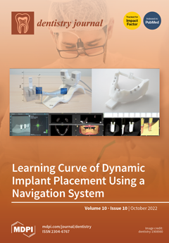
Dynamic navigation systems offer an excellent opportunity to safely treat complex and severe patient cases with dental implants. This new technique can reduce the risk of injury to important anatomical structures, such as nerves, vessels, or soft tissue. There are anatomical situations in which the established surgical procedures reach their limits, and sufficient surgical experience is required to ensure optimal implant placement. In this way, dynamic navigation offers help in the first surgical procedures for young dentists. For this reason, younger professionals should learn to use navigation systems in their surgical training or during their studies to be able to offer the entire repertoire of dental implant surgery. View this paper
  • Issues are regarded as officially published after their release is announced to the table of contents alert mailing list.
  • You may sign up for e-mail alerts to receive table of contents of newly released issues.
  • PDF is the official format for papers published in both, html and pdf forms. To view the papers in pdf format, click on the "PDF Full-text" link, and use the free Adobe Reader to open them.
Order results
Result details
Section
Select all
Export citation of selected articles as:
10 pages, 1795 KiB  
Article
Comparative Evaluation of Two Bracket Systems’ Bond Strength: Conventional and Self-Ligating
by Aurel-Claudiu Vartolomei, Dana-Valentina Ghiga, Dan-Cosmin Serbanoiu, Marioara Moldovan, Stanca Cuc, Mariana Pacurar and Maria Cristina Figueiredo Pollmann
Dent. J. 2022, 10(10), 196; https://doi.org/10.3390/dj10100196 - 21 Oct 2022
Cited by 2 | Viewed by 1644
Abstract
Adhesion remains a key element in dentistry, whether approached in prosthetics, odontology, or orthodontics. It is a continuously researched aspect, as improved materials and adhesive methods keep emerging in the market. No orthodontic treatment can be effective without the proper adhesion strength of [...] Read more.
Adhesion remains a key element in dentistry, whether approached in prosthetics, odontology, or orthodontics. It is a continuously researched aspect, as improved materials and adhesive methods keep emerging in the market. No orthodontic treatment can be effective without the proper adhesion strength of the bonded elements on the teeth. The objective of this research, in the broad context of self-ligating versus conventional brackets, was to compare active and passive self-ligating systems with a conventional one by conducting an in vitro study on human-extracted premolars. Shear bond strength tests were executed by means of an advanced materials-testing machine that generated maximum load and tensile strength values. The data obtained underwent statistical analysis with a statistical threshold of p < 0.05. The results regarding the statistical significance were acquired when comparing the passive self-ligating system with the active self-ligating and conventional systems (load-at-maximum-load mean 204.9, SD 91.09, and p < 0.05). In this study, the passive self-ligating bracket system appears to present increased shear bond strength. Full article
(This article belongs to the Special Issue Dentistry Journal: 10th Anniversary)
Show Figures

Figure 1

8 pages, 1271 KiB  
Review
Calculus as a Risk Factor for Periodontal Disease: Narrative Review on Treatment Indications When the Response to Scaling and Root Planing Is Inadequate
by Stephen K. Harrel, Charles M. Cobb, Lee N. Sheldon, Michael P. Rethman and John S. Sottosanti
Dent. J. 2022, 10(10), 195; https://doi.org/10.3390/dj10100195 - 20 Oct 2022
Cited by 3 | Viewed by 3387
Abstract
Background: Based on the 2018 classification of periodontal disease, a series of articles have been published describing the decision points of periodontal therapy and how the findings collected at those decision points can be used as guidelines for periodontal therapy. The articles are [...] Read more.
Background: Based on the 2018 classification of periodontal disease, a series of articles have been published describing the decision points of periodontal therapy and how the findings collected at those decision points can be used as guidelines for periodontal therapy. The articles are reviewed with a focus on the finding of inadequate calculus removal at the decision points and how that finding impacts treatment outcomes. Methods: A narrative review of the literature discussing the influence of calculus on inflammation was performed and the effects of inadequate removal of calculus during periodontal therapy were analyzed in light of the 2018 classification of periodontal disease, the decision points of periodontal therapy, and the guidelines of periodontal therapy. Results: The published literature supports that calculus is a major risk factor for periodontal inflammation. Recent studies indicate that the pathologic risk of calculus goes beyond the retention of biofilm and may represent a different pathophysiologic pathway for periodontal disease separate from the direct action of biofilm. The inadequate removal of calculus is a factor in the failure of periodontal therapy. Conclusions: The inadequate removal of calculus plays an important role in the frequent failure of non-surgical periodontal therapy to eliminate inflammation. Full article
(This article belongs to the Special Issue Dentistry Journal: 10th Anniversary)
Show Figures

Figure 1

10 pages, 1323 KiB  
Article
Effectiveness of 38% Silver Diamine Fluoride in Reducing Dentine Hypersensitivity on Exposed Root Surface in Older Chinese Adults: Study Protocol for a Randomised Double-Blind Study
by Alice Kit Ying Chan, Manisha Tamrakar, Chloe Meng Jiang, Yiu Cheung Tsang, Katherine Chiu Man Leung and Chun Hung Chu
Dent. J. 2022, 10(10), 194; https://doi.org/10.3390/dj10100194 - 19 Oct 2022
Cited by 5 | Viewed by 1720
Abstract
Background: Dentine hypersensitivity on an exposed root surface induces pain, affects daily oral hygiene practice, limits dietary choices and negatively affects quality of life. Silver diamine fluoride is marketed in the United States as a desensitising agent, but well-designed clinical trials are limited. [...] Read more.
Background: Dentine hypersensitivity on an exposed root surface induces pain, affects daily oral hygiene practice, limits dietary choices and negatively affects quality of life. Silver diamine fluoride is marketed in the United States as a desensitising agent, but well-designed clinical trials are limited. This study evaluates the anti-hypersensitivity effect of silver diamine fluoride on hypersensitive teeth due to an exposed root surface in older Chinese adults. Methods/design: We will conduct a randomised double-blind clinical trial with a sample size of at least 148 Chinese older adults aged 65 or above who have dentine hypersensitivity due to an exposed root surface. We will collect written consent before the study. A trained examiner will examine the participants’ teeth with a blast of compressed air from a 3-in-1 syringe. Those adults who report a self-perceived sensitivity score (SS) (0 to 10) of 8 or more on at least one tooth with an exposed root surface will be recruited. The recruited older adults will be randomly allocated into two groups using a block randomisation of six. Group 1 participants will receive the application of 38% silver diamine fluoride solution every 4 weeks. Group 2 participants will receive the application of 5% potassium nitrate solution every 4 weeks. Dietary advice, oral hygiene instruction and fluoride toothpaste at 1450 ppm will be provided to participants in both groups. The same trained examiner will perform follow-up examinations for the participants and determine the dentine hypersensitivity in SS of the most hypersensitive tooth (with the highest pre-treatment SS) immediately after the intervention and at 4-week and 8-week intervals. Discussion: There is no consensus on the standard of care for a professionally applied desensitising agent in older adults. This trial will provide evidence for clinicians to devise an effective dental care plan for older adults with dentine hypersensitivity. Trial registration: NCT05392868 Registered on 22 May 2022. Full article
(This article belongs to the Special Issue Preventive Dental Care, Chairside and Beyond)
Show Figures

Figure 1

14 pages, 976 KiB  
Review
Cannabidiol in Dentistry: A Scoping Review
by Carla David, Alejandro Elizalde-Hernández, Andressa S. Barboza, Gabriela C. Cardoso, Mateus B. F. Santos and Rafael R. Moraes
Dent. J. 2022, 10(10), 193; https://doi.org/10.3390/dj10100193 - 17 Oct 2022
Cited by 6 | Viewed by 7251
Abstract
Cannabidiol (CBD) has been gaining increased attention in contemporary society but seems to have been little explored in dentistry. This scoping review mapped the scientific and technological scenarios related to the use of CBD in dentistry. Peer-reviewed publications were searched in five international [...] Read more.
Cannabidiol (CBD) has been gaining increased attention in contemporary society but seems to have been little explored in dentistry. This scoping review mapped the scientific and technological scenarios related to the use of CBD in dentistry. Peer-reviewed publications were searched in five international databases, patents were searched in five technological platforms. In total, 11 articles and 13 patents involving CBD in dentistry-related applications were included. The countries contributing to most articles were Brazil (27.3%) and USA (18.2%). The studies involved experiments on animals (63.6%) and/or using bacteria or cells (36.4%), and no clinical study was found. Three different applications of CBD were observed: periodontal therapy (45.4%), aid for bone regeneration (27.3%), and general use in oral therapies (27.3%). Patent inventors were based in China (53.8%) or USA (46.2%). The patent claims were mainly compositions for oral care, tooth whitening, injury repair, antifungal, anti-inflammatory, and analgesic effects. A total of 76.9% of the patents were filed in association with a company. In general, research suggests that CBD has promising biological properties for applications in dentistry, whereas patents indicate that the current interest of industry relies on compositions for oral care. There appears to be extensive room available for research and technological applications of CBD in dentistry. Full article
(This article belongs to the Special Issue Dentistry Journal: 10th Anniversary)
Show Figures

Figure 1

17 pages, 1912 KiB  
Article
Two Gingival Cell Lines Response to Different Dental Implant Abutment Materials: An In Vitro Study
by Muataz A. Osman, Evgeny Kushnerev, Rasha A. Alamoush, Kevin. G. Seymour and Julian M. Yates
Dent. J. 2022, 10(10), 192; https://doi.org/10.3390/dj10100192 - 17 Oct 2022
Cited by 2 | Viewed by 1887
Abstract
Objectives: This study aimed to investigate the response of human gingival fibroblasts (HGFB) and human gingival keratinocytes (HGKC) towards different dental implant abutment materials. Methods: Five materials were investigated: (1) titanium (Ti), (2) titanium nitride (TiN), (3) cobalt-chromium (CoCr), (4) zirconia [...] Read more.
Objectives: This study aimed to investigate the response of human gingival fibroblasts (HGFB) and human gingival keratinocytes (HGKC) towards different dental implant abutment materials. Methods: Five materials were investigated: (1) titanium (Ti), (2) titanium nitride (TiN), (3) cobalt-chromium (CoCr), (4) zirconia (ZrO2), and (5) modified polyether ether ketone (m-PEEK). Both cell lines were cultured, expanded, and seeded in accordance with the protocol of their supplier. Cell proliferation and cytotoxicity were evaluated at days 1, 3, 5, and 10 using colourimetric viability and cytotoxicity assays. Data were analysed via two-way ANOVA, one-way ANOVA, and Tukey’s post hoc test (p < 0.05 for all tests). Results: There was a statistically significant difference in cell proliferation of HGKC and HGFB cells in contact with different abutment materials at different time points, with no significant interaction between different materials. There was a significant effect on cell proliferation and cytotoxicity with different exposure times (p < 0.0001) for each material. Cell proliferation rates were comparable for both cell lines at the beginning of the study, however, HGFB showed higher proliferation rates for all materials at day 10 with better proliferation activities with ZrO and m-PEEK (40.27%) and (48.38%) respectively. HGKC showed significant interactions (p < 0.0001) in cytotoxicity between different materials. Conclusion: The present in vitro assessment investigated the biocompatibility of different abutment materials with soft tissue cells (HGFB and HGKC). The findings suggest that m-PEEK and TiN are biologically compatible materials with human cells that represent the soft tissue and can be considered as alternative implant abutment materials to Ti and ZrO2, especially when the aesthetic is of concern. Full article
(This article belongs to the Special Issue Dentistry Journal: 10th Anniversary)
Show Figures

Figure 1

13 pages, 2997 KiB  
Article
Effect of Whitening Toothpaste on Surface Roughness and Colour Alteration of Artificially Extrinsic Stained Human Enamel: In Vitro Study
by Sarat Suriyasangpetch, Pimduean Sivavong, Boondarick Niyatiwatchanchai, Thanaphum Osathanon, Puliwan Gorwong, Chawalid Pianmee and Dusit Nantanapiboon
Dent. J. 2022, 10(10), 191; https://doi.org/10.3390/dj10100191 - 13 Oct 2022
Cited by 9 | Viewed by 3420
Abstract
Objective: The aim of this study was to compare five toothpastes in terms of alteration of surface roughness and colour of red-wine-stained human enamel over time after brushing simulation. Methods: Stained specimens were randomly divided into five groups (n = 8): Oral-B [...] Read more.
Objective: The aim of this study was to compare five toothpastes in terms of alteration of surface roughness and colour of red-wine-stained human enamel over time after brushing simulation. Methods: Stained specimens were randomly divided into five groups (n = 8): Oral-B Gum and Enamel (C), ZACT Stain Fighter (ZW), Colgate Optic White Volcanic Mineral (CW), Oral-B 3D White Luxe Fresh Breath (3DW), and Thepthai (TW). The colour and surface roughness of the specimens were measured after brushing simulation for four durations; two weeks, one month, six months, and twelve months. Abrasive particles in toothpaste were inspected under a scanning electron microscope. Results: Surface roughness was increased in the specimens that underwent brushing simulation in all groups (p < 0.05). ZW (6.33 ± 0.98 μm) exerted the most changes in surface roughness for all durations. Other groups showed similar surface roughness at each time point. ΔE00 and ΔL were increased in all groups until twelve months; however, there were no significant differences between C (ΔE00 = 30.17 ± 4.16, ΔL = 38.19 ± 4.34), CW (ΔE00 = 24.25 ± 10.52, ΔL = 31.12 ± 11.00), and TW (ΔE00 = 29.00 ± 3.96, ΔL = 36.68 ± 4.53) at any time period. Moreover, ZW (ΔE00 = 45.6 ± 8.01, ΔL = 53.03 ± 6.71) and 3DW (ΔE00 = 43.6 ± 7.33, ΔL = 51.03 ± 6.47) exhibited a substantial change and were statistically differed from the other groups after four-weeks. Various characteristics of abrasive particles were inspected under SEM. Conclusion: All five toothpastes increased the surface roughness altered the colour of red wine-stained human enamel over time. Full article
Show Figures

Graphical abstract

10 pages, 245 KiB  
Article
Knowledge and Perceptions of Molar Incisor Hypomineralisation among General Dental Practitioners, Paediatric Dentists, and Other Dental Specialists in Indonesia
by Enrita Dian, Sarworini Bagio Budiardjo, Aghareed Ghanim, Lisa Rinanda Amir and Diah Ayu Maharani
Dent. J. 2022, 10(10), 190; https://doi.org/10.3390/dj10100190 - 12 Oct 2022
Cited by 1 | Viewed by 1622
Abstract
Molar incisor hypomineralisation (MIH) is a qualitative, dental enamel hypomineralisation defect affecting one to four first permanent molars, characterised by the present of asymmetries demarcated opacities, and is prevalent worldwide. An early diagnosis of MIH is crucial, to prevent further complications including the [...] Read more.
Molar incisor hypomineralisation (MIH) is a qualitative, dental enamel hypomineralisation defect affecting one to four first permanent molars, characterised by the present of asymmetries demarcated opacities, and is prevalent worldwide. An early diagnosis of MIH is crucial, to prevent further complications including the development of dental caries, hypersensitivity, pulp inflammation, and pain. Therefore, a thorough understanding of MIH is of clinical importance. This cross-sectional study aimed to evaluate the knowledge and perception of MIH among general dental practitioners (GD), paediatric dentists (PD), and other dental specialists (DS) in Indonesia using a self-administered questionnaire. Chi-square tests and Kruskal–Wallis tests were employed to analyse the data. A total of 266 dental practitioners—112 GD, 84 PD, and 60 DS—were included in this study and completed the online questionnaire. There were significant differences in the overall knowledge scores between GD, PD, and DS (p < 0.001). Specifically, the different between the tested groups were observed in the knowledge of caries pattern related to MIH and the aetiology of MIH. The majority of PD (83.3%) can differentiate the MIH-related caries pattern from the classic caries pattern. Moreover, the confident level in diagnosing and treating MIH teeth were higher in PD compared to GD and DS (p = 0.000). The majority of dental practitioners in all groups agreed on the necessity to obtain continuing education on MIH including the aetiology, diagnosis, and its treatment to improve their knowledge and confidence in MIH clinical management. Full article
(This article belongs to the Special Issue Dentistry Journal: 10th Anniversary)
10 pages, 711 KiB  
Article
An Analysis of Different Techniques Used to Seal Post-Extractive Sites—A Preliminary Report
by Paolo Pesce, Eitan Mijiritsky, Luigi Canullo, Maria Menini, Vito Carlo Alberto Caponio, Andrea Grassi, Luca Gobbato and Domenico Baldi
Dent. J. 2022, 10(10), 189; https://doi.org/10.3390/dj10100189 - 9 Oct 2022
Cited by 6 | Viewed by 1822
Abstract
Background: Bone grafting in post-extractive site improves tissue regeneration. Soft tissue sealing of the grafted post-extractive alveolus is supposed to limit microbiological contamination from the oral cavity and to stabilize the coagulum. Several techniques are presented in the literature to reach this goal [...] Read more.
Background: Bone grafting in post-extractive site improves tissue regeneration. Soft tissue sealing of the grafted post-extractive alveolus is supposed to limit microbiological contamination from the oral cavity and to stabilize the coagulum. Several techniques are presented in the literature to reach this goal using different heterologous matrices or autogenous grafts. In addition, recently, a technique based on the use of granulation tissue in the post-extractive alveolus has been proposed. Aim: To compare the effect of different graft sealing approaches in post-extractive sites by qualitatively evaluating their healing process. Materials and Methods: This retrospective investigation included 30 patients requiring post-extractive site regeneration in the aesthetic area. Post-extractive sites were regenerated using a bovine bone matrix and patients were divided into three groups (10 patients in each group) according to the material used to seal the alveolar socket. In the UD group, the granulation tissue was used to seal the defect; in the PC group, epithelial-connective soft tissue graft was used, and in the COLL group, a collagen-based membrane was employed. Images of the post-extractive sites at different follow-up periods (2 and 12 weeks) were taken and the healing process was blindly evaluated by two independent practitioners. The Healing Index (HI) by Landry, Turnbull and Howley was used to assess the quality of the healing process. The combination of presence/absence of five clinical criteria defines an HI ranging from 1 (very poor) to 5 (excellent). Patients’ clinical-pathological variables were recorded. One-way ANOVA was used to explore the dependence of HI on the different socket preservation protocols. Results: Based on clinical-pathological characteristics of the included patients, there were no statistically significant differences among the different sealing techniques. At the 2-week follow-up appointment, HI did not differ among the socket preservation protocols evaluated. Moreover, smoking status and reason for extraction did not influence the HI among the three groups (two-way ANOVA p-value = 0.686, p-value = 0.248 respectively). At the 2-week follow-up appointment, HI was significantly different among the socket preservation protocols investigated. Specifically, the group undergoing collagen-based socket preservation procedure reported the highest HI, compared to the other two techniques (COLL mean 4.60 ± 0.5; PC mean 3.5 ± 1.2; UD mean 3.4 ± 0.5, one-way ANOVA p-value 0.006). Conclusions: The use of collagen porcine membranes may represent a suitable option to improve the patient healing process in grafted post-extractive sites together with reducing the surgical intervention time compared to alternative sealing techniques. Full article
(This article belongs to the Special Issue Oral Implantology and Rehabilitation)
Show Figures

Figure 1

11 pages, 569 KiB  
Review
Challenges and Opportunities for Dental Education from COVID-19
by Bolei Li, Lei Cheng and Haohao Wang
Dent. J. 2022, 10(10), 188; https://doi.org/10.3390/dj10100188 - 6 Oct 2022
Cited by 6 | Viewed by 2854
Abstract
With the ongoing COVID-19 pandemic, dental education has been profoundly affected by this crisis. First of all, COVID-19 brought physical and psychological health problems to dental students and educators. In addition, both non-clinical teaching and clinical-based training experienced challenges, ranging from fully online [...] Read more.
With the ongoing COVID-19 pandemic, dental education has been profoundly affected by this crisis. First of all, COVID-19 brought physical and psychological health problems to dental students and educators. In addition, both non-clinical teaching and clinical-based training experienced challenges, ranging from fully online educational content to limited dental training, students’ research was delayed in achieving project milestones and there was hesitancy in respect of the COVID-19 vaccine. On the other hand, the COVID-19 pandemic has increased the demand for teledentistry and dental emergency treatment, and brought awareness of the advantages and high-speed development of distance education. This review aims to present these challenges and opportunities for dental education, and suggest how dental institutions should prepare for the future demand for dental education. Full article
Show Figures

Figure 1

11 pages, 2498 KiB  
Article
Learning Curve and Comparison of Dynamic Implant Placement Accuracy Using a Navigation System in Young Professionals
by Johannes Spille, Eva Helmstetter, Paul Kübel, Jan-Tobias Weitkamp, Juliane Wagner, Henning Wieker, Hendrik Naujokat, Christian Flörke, Jörg Wiltfang and Aydin Gülses
Dent. J. 2022, 10(10), 187; https://doi.org/10.3390/dj10100187 - 2 Oct 2022
Cited by 7 | Viewed by 1991
Abstract
The aim of the current study was to evaluate the learning curve and accuracy of implant placement by young professionals using a dynamic computer-assisted surgical system for dental implant placement. Ten students tried to place eight implants with a dynamic surgical system in [...] Read more.
The aim of the current study was to evaluate the learning curve and accuracy of implant placement by young professionals using a dynamic computer-assisted surgical system for dental implant placement. Ten students tried to place eight implants with a dynamic surgical system in predefined positions on two consecutive weekends, resulting in 160 implant placements in total. Postoperatively, the positions of the implants were scanned with an intraoral scanner and compared for deviations at the entry point, the apex, as well as angular deviations to the master model. The mean values of all measurements improved; statistical significance was found for the changes in the angle as well as for the position of the implants to the apex (p < 0.001). Furthermore, the young professionals indicated subjective improvement in handling the dynamic surgery system. Navigated surgical dental implant placement can be learned quickly and can support young professionals in everyday clinical practice, especially in difficult anatomic situations. Full article
(This article belongs to the Special Issue Dentistry Journal: 10th Anniversary)
Show Figures

Figure 1

9 pages, 2006 KiB  
Article
Evaluation of the Efficacy of CPP-ACP Remineralizing Mousse in MIH White and Yellow Opacities—In Vitro Vickers Microhardness Analysis
by Inês Cardoso-Martins, Sofia Arantes-Oliveira, Ana Coelho, Sofia Pessanha and Paula F. Marques
Dent. J. 2022, 10(10), 186; https://doi.org/10.3390/dj10100186 - 2 Oct 2022
Cited by 3 | Viewed by 2591
Abstract
Remineralization of tooth enamel can be partially achieved by the application of a casein phosphopeptides and amorphous phosphate (CPP-ACP) complex. However, evidence to support its effectiveness in Molar-incisor-hypomineralization (MIH)-affected teeth is scarce. The study’s aim is to evaluate the efficacy of CPP-ACP mousse [...] Read more.
Remineralization of tooth enamel can be partially achieved by the application of a casein phosphopeptides and amorphous phosphate (CPP-ACP) complex. However, evidence to support its effectiveness in Molar-incisor-hypomineralization (MIH)-affected teeth is scarce. The study’s aim is to evaluate the efficacy of CPP-ACP mousse in remineralizing MIH-affected enamel using a Vickers microhardness test. Two groups of enamel opacities of hypomineralized permanent teeth, white (group A) and yellow (group B) lesions (n = 14), went through a 28-day treatment protocol with GC Tooth Mousse. Before and after treatment, microhardness was measured in three different areas of each tooth (hypomineralized, transition, and outside the hypomineralized area). Data were analyzed using parametric and non-parametric tests with a significance of p < 0.05. The mean microhardness values increased in the hypomineralized and transition areas in both groups after the treatment protocol (in group A, 105.38 ± 11.70 to 158.26 ± 37.34; 123.04 ± 22.84 to 156.33 ± 35.70; in group B, 108.63 ± 14.66 to 143.06 ± 22.81; 132.55 ± 20.66 to 146.00 ± 12.88) and the differences pre/post-treatment were statistically significant within each group (p < 0.001 for both groups). Between groups, there was a statistically significant difference in the same areas (hypomineralized: p = 0.003; transition: p = 0.008) with a higher improvement in enamel hardness in group A. Topical application of CPP-ACP showed an increase in the physical strength of the hypomineralized and transition areas of MIH-affected enamel, likely due to an increase in mineral content. Full article
(This article belongs to the Special Issue Studies on Dental Enamel)
Show Figures

Figure 1

10 pages, 577 KiB  
Article
Caries Experience and Treatment Needs in Urban and Rural Environments in School-Age Children from Three Provinces of Ecuador: A Cross-Sectional Study
by Eleonor María Vélez-León, Alberto Albaladejo-Martínez, Katherine Cuenca-León, Liliana Encalada-Verdugo, Ana Armas-Vega and María Melo
Dent. J. 2022, 10(10), 185; https://doi.org/10.3390/dj10100185 - 1 Oct 2022
Cited by 3 | Viewed by 3223
Abstract
In Ecuador, national epidemiological surveys have not been updated; however, some regional studies in the northern areas of the country still report a high prevalence of dental caries. The aim of this study was to determine the experience, severity, and need for treatment [...] Read more.
In Ecuador, national epidemiological surveys have not been updated; however, some regional studies in the northern areas of the country still report a high prevalence of dental caries. The aim of this study was to determine the experience, severity, and need for treatment of dental caries in school children aged 6 to 12 years in urban and rural settings in three provinces of southern Ecuador. This cross-sectional, relational study examined 1938 schoolchildren in the provinces of Azuay, Cañar, and Morona Santiago. The survey instruments were based mainly on the WHO manual Methods of Oral Health Surveys (dmft) for primary and permanent dentition (DMFT), as well as the prevalence, severity, and Significant Caries Index (SCI). The parametric Student’s t-test was used to compare two groups, and the Spearman’s Rho and Tau-c Kendall correlation coefficients were used to associate the categorical variables. Results: The prevalence of caries in the primary dentition was 78% and 89.2% in the permanent dentition. The dmft (M = 4.12, SD = 2.86) and DMFT (M = 3.62: SD = 3.07) placed the general group in a moderate caries index. The need for treatment was 90.68% in the primary dentition, while it was 87.99% in the permanent dentition. Caries severity in both dentitions was high (M = 7.74; SD = 3.42). Conclusions. Alarming indicators of caries experience and the need for treatment were observed in the population studied. Full article
(This article belongs to the Special Issue Preventive Dentistry and Dental Public Health)
Show Figures

Figure 1

12 pages, 1016 KiB  
Review
Oral Microbiome and Dental Caries Development
by Josie Shizhen Zhang, Chun-Hung Chu and Ollie Yiru Yu
Dent. J. 2022, 10(10), 184; https://doi.org/10.3390/dj10100184 - 30 Sep 2022
Cited by 23 | Viewed by 9171
Abstract
Dental caries remains the most prevalent oral disease worldwide. The development of dental caries is highly associated with the microbiota in the oral cavity. Microbiological research of dental caries has been conducted for over a century, with conventional culture-based methods and targeted molecular [...] Read more.
Dental caries remains the most prevalent oral disease worldwide. The development of dental caries is highly associated with the microbiota in the oral cavity. Microbiological research of dental caries has been conducted for over a century, with conventional culture-based methods and targeted molecular methods being used in order to identify the microorganisms related to dental caries. These methods’ major limitation is that they can identify only part of the culturable microorganisms in the oral cavity. Introducing sequencing-based technology and bioinformatics analysis has boosted oral microbiome research and greatly expanded the understanding of complex oral microbiology. With the continuing revolution of molecular technologies and the accumulated sequence data of the oral microbiome, researchers have realized that microbial composition alone may be insufficient to uncover the relationship between caries and the microbiome. Most updated evidence has coupled metagenomics with transcriptomics and metabolomics techniques in order to comprehensively understand the microbial contribution to dental caries. Therefore, the objective of this article is to give an overview of the research of the oral microbiome and the development of dental caries. This article reviews the classical concepts of the microbiological aspect of dental caries and updates the knowledge of caries microbiology with the results of current studies on the oral microbiome. This paper also provides an update on the caries etiological theory, the microorganisms related to caries development, and the shifts in the microbiome in dental caries development. Full article
(This article belongs to the Special Issue Updates and Highlights in Cariology)
Show Figures

Figure 1

10 pages, 249 KiB  
Article
Awareness and Attitudes of Dental Students toward Older Adults in Indonesia
by Anton Rahardjo, Fakhira Hanna Safira Firdaus, Peter Andreas, Yuniardini Septorini Wimardhani and Diah Ayu Maharani
Dent. J. 2022, 10(10), 183; https://doi.org/10.3390/dj10100183 - 29 Sep 2022
Cited by 2 | Viewed by 1729
Abstract
In this study, we aimed to analyse the awareness and attitudes of dental students (DS) toward older adults (OAs) in Indonesia. Compromised oral health (OH) among OAs is a significant public health problem that is a global health burden. Furthermore, appropriate education can [...] Read more.
In this study, we aimed to analyse the awareness and attitudes of dental students (DS) toward older adults (OAs) in Indonesia. Compromised oral health (OH) among OAs is a significant public health problem that is a global health burden. Furthermore, appropriate education can provide DS with information about strategic and efficient treatments for OAs. An online invitation was provided to every dental student in Indonesia in the third to sixth year of study to answer a web-based questionnaire as a part of a survey on awareness and attitudes (14 questions) towards OAs. From 1288 valid forms submitted, it was evident that most students (87%) expressed a positive attitude toward OAs. Female students showed a more positive attitude compared to males, with an odds ratio of 1.539 (p = 0.036), and students who had a connection to an older adult demonstrated a more positive attitude than those who did not (OR = 2.076; p < 0.001). The majority of the DS (98%) were conscious of the importance of geriatrics in dentistry, with female students showing more awareness than their male counterparts (OR = 2.553; p = 0.033). Positive awareness and attitudes must be accompanied by knowledge so that DS can offer appropriate and effective oral care to geriatric patients. Full article
(This article belongs to the Special Issue Dentistry Journal: 10th Anniversary)
10 pages, 5294 KiB  
Case Report
Peripheral Osteoma of the Mandibular Condyle—Case Series
by Ioannis Tilaveridis, Theodora Katopodi, Panagiotis Karakostas, Gregory Venetis, Ioannis Dimitrakopoulos, Stavros Tilaveridis, Sofia Tilaveridou and Katerina Zarampouka
Dent. J. 2022, 10(10), 182; https://doi.org/10.3390/dj10100182 - 29 Sep 2022
Cited by 2 | Viewed by 3216
Abstract
The purpose of this article is to present four new cases of peripheral osteoma of the mandibular condyle and the literature review. A retrospective study of files from our Department of Oral and Maxillofacial Surgery over the last 6 years revealed four cases [...] Read more.
The purpose of this article is to present four new cases of peripheral osteoma of the mandibular condyle and the literature review. A retrospective study of files from our Department of Oral and Maxillofacial Surgery over the last 6 years revealed four cases of peripheral osteomas located in the area of the mandibular condyle. Diagnostic procedure included clinical, radiographic, and histologic criteria. Gardner’s syndrome was excluded from patient history and clinical evaluation. One patient had only an aesthetic disturbance, with facial swelling, and the other three patients presented disturbances of the mandibular function, including deviation during mouth opening along with malocclusion. Three of the patients were male and one was female; all were of middle age (45–65 years old). The proposed surgical treatment was accepted by half of the patients, while the remaining half declined the operation after a confirmation of the diagnosis. Peripheral osteomas of the maxillofacial region are uncommon, and some cases with multiple osteomas are related to Gardner’s syndrome. An osteoma of the mandibular condyle is very rare and surgical treatment is challenging for the surgeon with regards to the approach selection and the related complications. In the two cases that accepted the proposed surgical treatment, no recurrence and no complication was observed. Full article
(This article belongs to the Special Issue Dentistry Journal: 10th Anniversary)
Show Figures

Figure 1

8 pages, 1088 KiB  
Article
Comparison in Terms of Accuracy between DLP and LCD Printing Technology for Dental Model Printing
by Ioannis A. Tsolakis, William Papaioannou, Erofili Papadopoulou, Maria Dalampira and Apostolos I. Tsolakis
Dent. J. 2022, 10(10), 181; https://doi.org/10.3390/dj10100181 - 28 Sep 2022
Cited by 23 | Viewed by 3534
Abstract
Background: The aim of this study is to evaluate the accuracy of a Liquid Crystal Display (LCD) 3D printer compared to a Direct Light Processing (DLP) 3D printer for dental model printing. Methods: Two different printers in terms of 3D printing technology were [...] Read more.
Background: The aim of this study is to evaluate the accuracy of a Liquid Crystal Display (LCD) 3D printer compared to a Direct Light Processing (DLP) 3D printer for dental model printing. Methods: Two different printers in terms of 3D printing technology were used in this study. One was a DLP 3D printer and one an LCD 3D printer. The accuracy of the printers was evaluated in terms of trueness and precision. Ten STL reference files were used for this study. For trueness, each STL file was printed once with each 3D printer. For precision, one randomly chosen STL file was printed 10 times with each 3D printer. Afterward, the models were scanned with a model scanner, and reverse engineering software was used for the STL comparisons. Results: In terms of trueness, the comparison between the LCD 3D printer and DLP 3D printer was statistically significant, with a p-value = 0.004. For precision, the comparison between the LCD 3D printer and the DLP 3D printer was statistically significant, with a p-value = 0.011. Conclusions: The DLP 3D printer is more accurate in terms of dental model printing than the LCD 3D printer. However, both DLP and LCD printers can accurately be used to print dental models for the fabrication of orthodontic appliances. Full article
(This article belongs to the Special Issue Dentistry Journal: 10th Anniversary)
Show Figures

Figure 1

14 pages, 290 KiB  
Article
Effects of Contact Sports on Temporomandibular Disorders: An Observational Study
by Vito Crincoli, Corrado De Biase, Angela Pia Cazzolla, Alessandra Campobasso, Mario Dioguardi, Maria Grazia Piancino, Luigi Mattia, Domenico Ribatti and Mariasevera Di Comite
Dent. J. 2022, 10(10), 180; https://doi.org/10.3390/dj10100180 - 27 Sep 2022
Cited by 2 | Viewed by 1627
Abstract
The study investigated the prevalence of temporomandibular disorders in 100 competitive athletes in contact sports, equally grouped by the practiced game: Soccer (SoG), Rugby (RG), American Football (AFG), Boxing (BoG), Basketball (BaG), compared to a randomly control group of 20 non-athletes (CG). Symptoms [...] Read more.
The study investigated the prevalence of temporomandibular disorders in 100 competitive athletes in contact sports, equally grouped by the practiced game: Soccer (SoG), Rugby (RG), American Football (AFG), Boxing (BoG), Basketball (BaG), compared to a randomly control group of 20 non-athletes (CG). Symptoms and signs were examined according to the standardized Diagnostic Criteria for Temporomandibular Disorders through a questionnaire and clinical evaluation. Arthralgia showed significant differences between RG and CG and between AFG and CG (p < 0.05). Study groups reported masticatory muscle pain during function, neck and shoulder pain more frequently than CG, except for BoG. Closing click was significantly more present in study groups than CG, while crepitation was significantly higher only in RG and AFG. The deviation was wider in SoG, RG and AFG compared to CG (p < 0.05). Tukey’s multiple comparisons test showed a statistically significant reduction in right laterotrusion in RG vs. CG (p < 0.05); the comparison showed a decrease in right laterotrusion in RG vs. SoG and BoG (p < 0.05), a decrease in endfeel in RG vs. CG, BaG and AFG (p < 0.05). The data seem to support a relationship between the prevalence of TMD symptoms and signs in competitive athletes in contact sports, especially in RG and AFG compared to CG. Full article
(This article belongs to the Section Restorative Dentistry and Traumatology)
11 pages, 7574 KiB  
Article
Comparing Distal-Jet with Dental Anchorage to Distal-Jet with Skeletal Anchorage: A Prospective Parallel Cohort Study
by Federica Altieri, Martina Mezio, Rosanna Guarnieri and Michele Cassetta
Dent. J. 2022, 10(10), 179; https://doi.org/10.3390/dj10100179 - 27 Sep 2022
Cited by 7 | Viewed by 3276
Abstract
The use of traditional intra-oral devices in maxillary molar distalization is not without undesirable consequences. The aim of the present study was to compare the miniscrew-supported distal-jet appliance to a traditional distal-jet appliance by evaluating the amount of upper first molar distalization and [...] Read more.
The use of traditional intra-oral devices in maxillary molar distalization is not without undesirable consequences. The aim of the present study was to compare the miniscrew-supported distal-jet appliance to a traditional distal-jet appliance by evaluating the amount of upper first molar distalization and the dentoalveolar side effects. Data of 600 subjects visited at the orthodontic unit of Sapienza University of Rom were analyzed. Only 46 patients met the inclusion criteria and were selected and treated. Subjects were assigned randomly to receive treatment either with miniscrew-supported distal-jet appliance (Group A) or with a traditional distal-jet appliance (Group B). In Group A, miniscrews were inserted using a computer-guided surgical guide. The amount of upper first molar distalization and the dentoalveolar side effects were assessed both on the digital casts and on the lateral cephalometric radiograph at the end of the distalization phase. A descriptive statistical analysis that included the mean values and the standard deviation was conducted to evaluate the molar distalization and the dentoalveolar effects in two groups. Intergroup differences were determined using the Student’s t-test. The significance was set at p ≤ 0.05. In Group A, greater maxillary first molar distalization and a spontaneous distalization of the first premolars and a palatal inclination of central incisors were observed. By contrast, in Group B, the first premolars tipped mesially and a proclination of the maxillary central incisors was observed. In both groups, the transverse widths of the dental arch increased while a greater tendency of first premolar extrusion and of maxillary first molar rotation was observed in Group B. The skeletal anchorage device achieved greater first molar distalization and did not cause dento-alveolar side effects. Full article
(This article belongs to the Special Issue Orthodontics and New Technologies)
Show Figures

Figure 1

8 pages, 233 KiB  
Article
Assessment of Biochemical Parameters of the Oral Fluid before and after Using Office Teeth Whitening Systems
by Elena A. Ryskina, Frida N. Gilmiyarova, Oksana A. Magsumova, Mikhail A. Postnikov, Tatiana A. Lobaeva and Dmitry D. Zhdanov
Dent. J. 2022, 10(10), 178; https://doi.org/10.3390/dj10100178 - 21 Sep 2022
Cited by 1 | Viewed by 1684
Abstract
One of the most important functions of the oral fluid is to maintain oral homeostasis. In-office teeth whitening systems are able to change the mineral metabolism and the activity of a number of enzymes in the oral fluid, but there are conflicting data [...] Read more.
One of the most important functions of the oral fluid is to maintain oral homeostasis. In-office teeth whitening systems are able to change the mineral metabolism and the activity of a number of enzymes in the oral fluid, but there are conflicting data in publications about this. The aim of this study was to compare the effect of Opalescense Boost, ZOOM Advance POWER, and ZOOM Phillips White Speed, which contain different percentages of hydrogen peroxide, on the performance of oral fluid. After the procedure of whitening teeth with the studied in-office systems, the concentration of calcium in the oral fluid increased, and the activity of alkaline phosphatase decreased. Calcium levels returned to baseline values after 30 days, and alkaline phosphatase activity returned after 14 days. There was no significant difference in the changes in calcium concentration and alkaline phosphatase activity between different tooth whitening systems. Chemical teeth whitening with the Opalescense Boost system caused the largest change in the activity of superoxide dismutase in the oral fluid compared to the ZOOM Advance POWER and ZOOM Phillips White Speed photocatalytic teeth whitening systems. An increase in the activity of superoxide dismutase by +75.5% was shown immediately after the procedure of teeth whitening with the Opalescense Boost system, which indicated an increase in the power of antioxidant defense mechanisms. To assess the effectiveness and safety of using various whitening systems, it is possible to study the dynamics of the activity of superoxide dismutase, which reflects the processes of antioxidant protection of the oral cavity. Full article
(This article belongs to the Special Issue Preventive Dental Care, Chairside and Beyond)
13 pages, 404 KiB  
Review
Effects of Multibracket Orthodontic Treatment versus Clear Aligners on Periodontal Health: An Integrative Review
by Aaron Jacob David Partouche, Filipe Castro, Ana Sofia Baptista, Liliana Gavinha Costa, Juliana Campos Hasse Fernandes and Gustavo Vicentis de Oliveira Fernandes
Dent. J. 2022, 10(10), 177; https://doi.org/10.3390/dj10100177 - 21 Sep 2022
Cited by 6 | Viewed by 2553
Abstract
Objective: This integrative review aimed to identify studies comparing the periodontal health in patients wearing multibracket orthodontic appliances and clear aligners. Materials and methods: An integrative literature search was performed through different databases, PubMed/Medline, PMC, and the Cochrane Library. This work [...] Read more.
Objective: This integrative review aimed to identify studies comparing the periodontal health in patients wearing multibracket orthodontic appliances and clear aligners. Materials and methods: An integrative literature search was performed through different databases, PubMed/Medline, PMC, and the Cochrane Library. This work was submitted to a search strategy following the PICO method and included the focus question: “Could the chosen orthodontic appliance change significantly the oral hygiene of the patient, impairing the periodontal health?” This work included analytical and controlled studies on humans published between 2005 and 2020, in the English language, establishing a comparison of the periodontal status in patients undergoing orthodontic multibracket and clear aligners therapies. The main periodontal indexes assessed were plaque index (PI), pocket depth (PD), gingival index (GI), and bleeding on probing (BoP). Results: The electronic research displayed 386 articles on PMC, 106 on PubMed, and 40 on the Cochrane Library. After removal, just 25 articles were selected for full-text screening, but just eight studies were eligible for this integrative review. It was enumerated that 204 patients were treated with aligners and 294 with multibracket orthodontic appliances, mainly elastomeric ligated brackets. Only the plaque index displayed a significant difference between the two groups and general data obtained showed a better control for periodontal health in the clear aligners. Limitations such as age, malocclusion severity, therapeutic choice, and different time measure was observed. In addition, the oral hygiene instruction and follow-up by a professional were different, and the role of malocclusion was not present in the studies. Conclusions: Within the limitations of this study, better results for periodontal health were found in the clear aligners. Therefore, more studies are necessary to affirm that aligners are synonymous with better gingival conditions in comparison with multibracket appliances. Other variables such as oral hygiene instructions, motivation, and supportive treatment tend to be more prevalent than the type of appliance itself in the periodontal evaluation. Full article
(This article belongs to the Special Issue Clinical Oral Implants and Periodontal Research)
Show Figures

Figure 1

Previous Issue
Next Issue
Back to TopTop