Strategies for Suppression and Compensation of Signal Loss in Ptychography

 , , , , , and
Abstract
1. Introduction
2. Methods
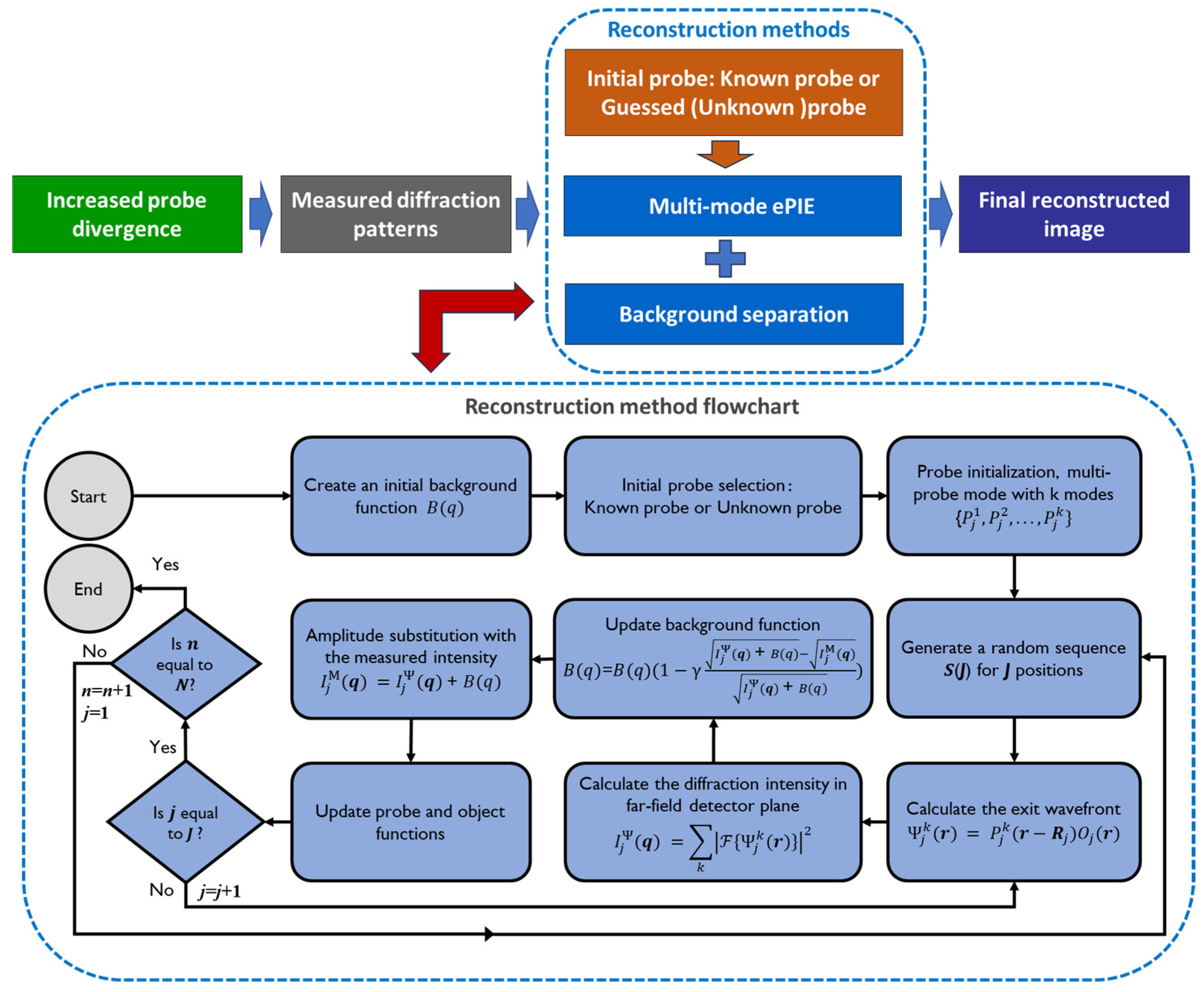
2.1. Partial Recovery of Lost Signals—Using a Known or Near-Accurate Probe
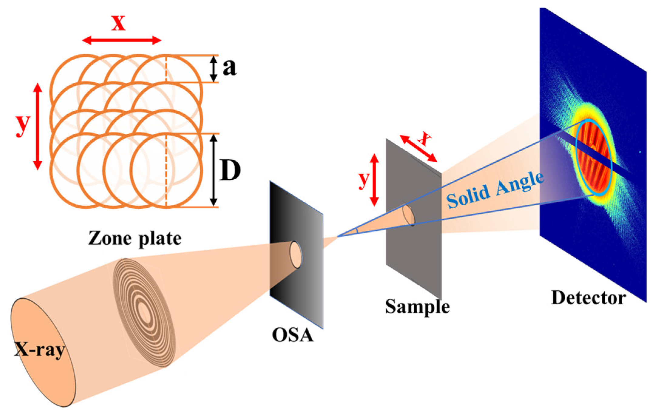
2.2. Suppression of Signal Loss Effects—Probe Divergence
3. Results
3.1. Strategy 1: Utilizing a Known Probe
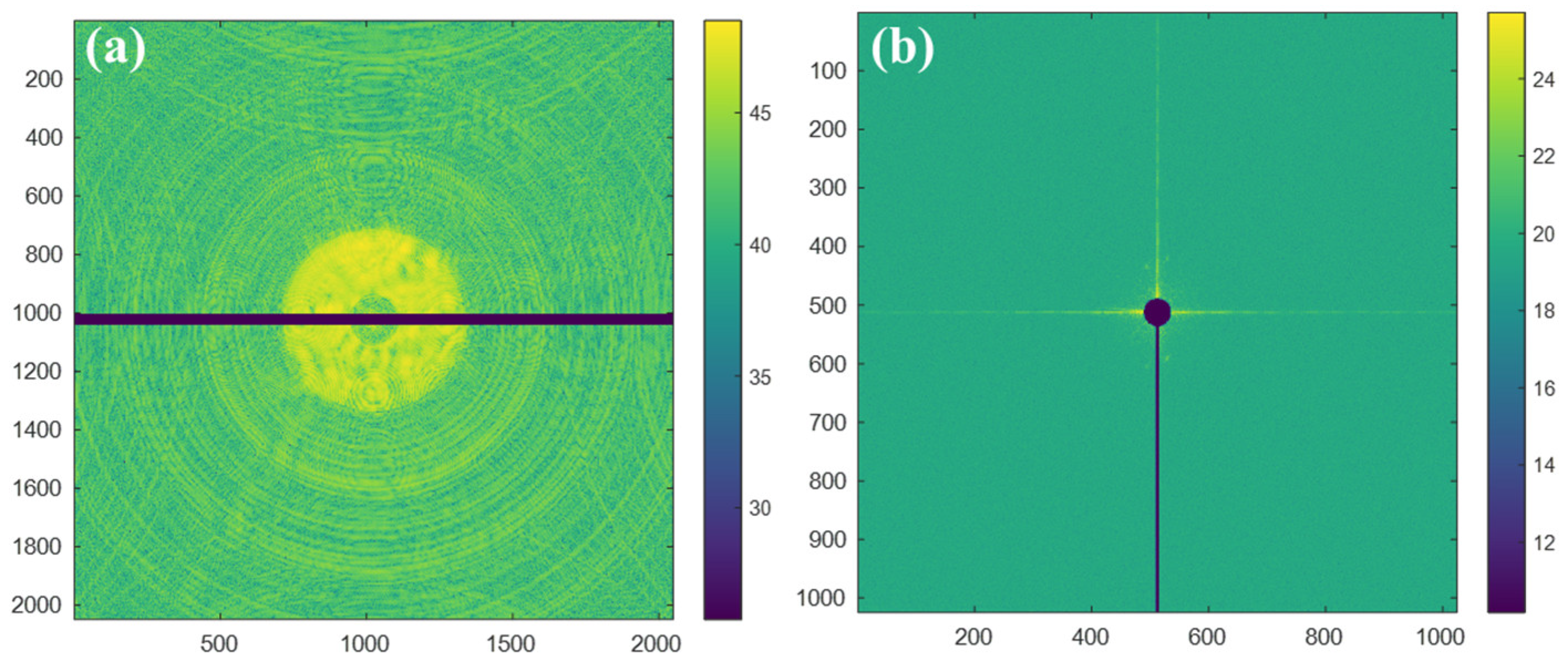
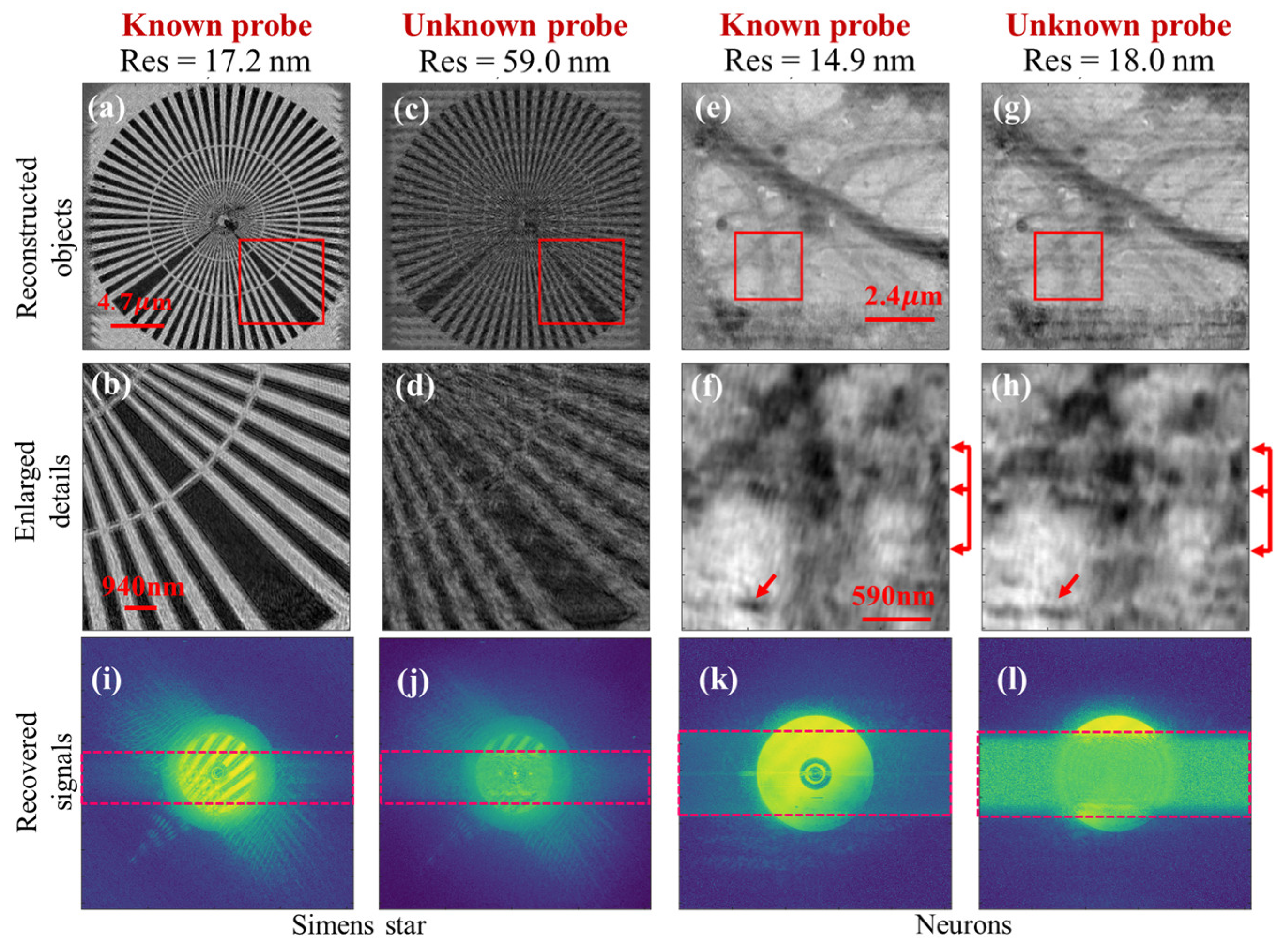
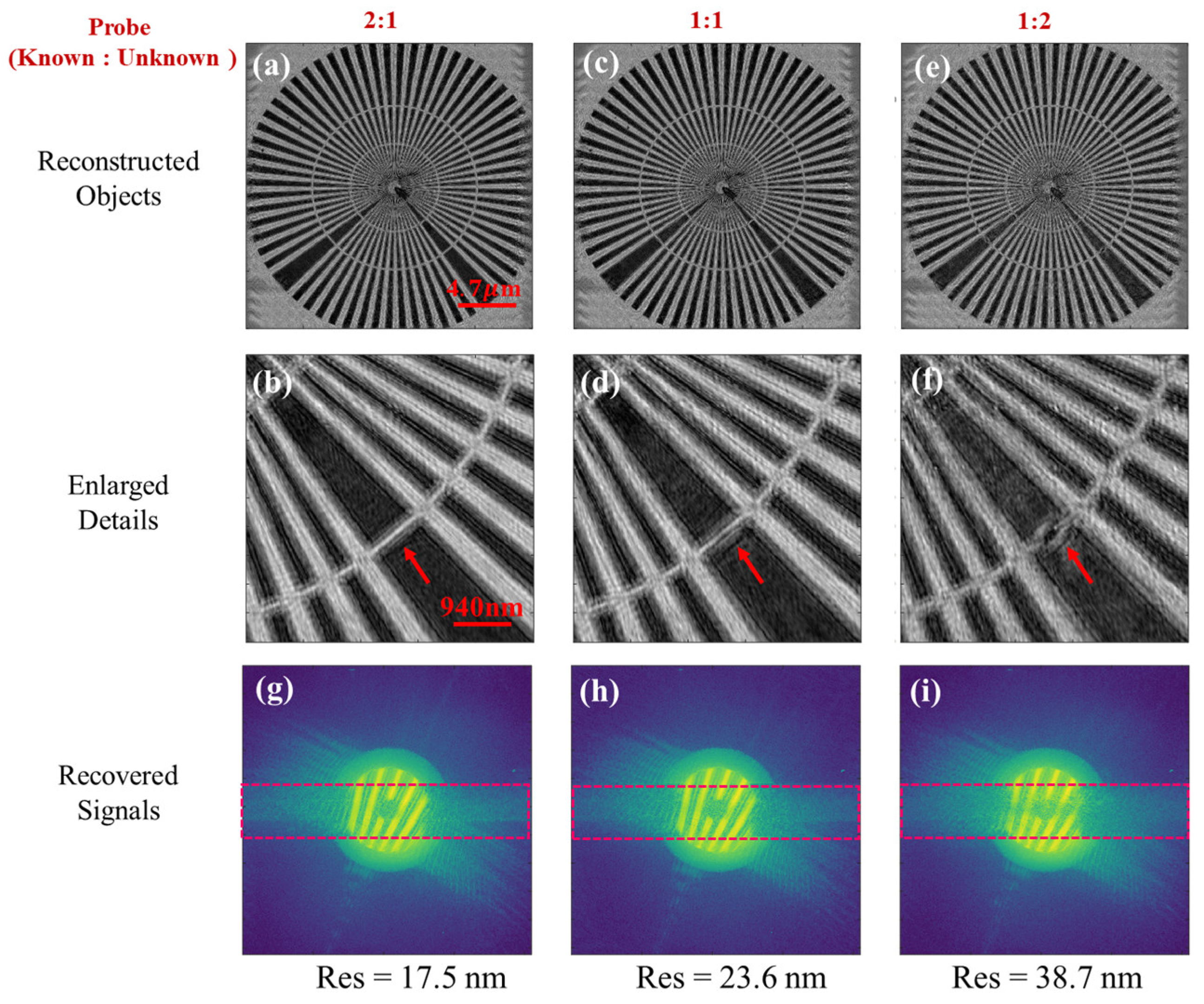
3.1.1. Signal Loss in a Strip-Shaped Gap Region
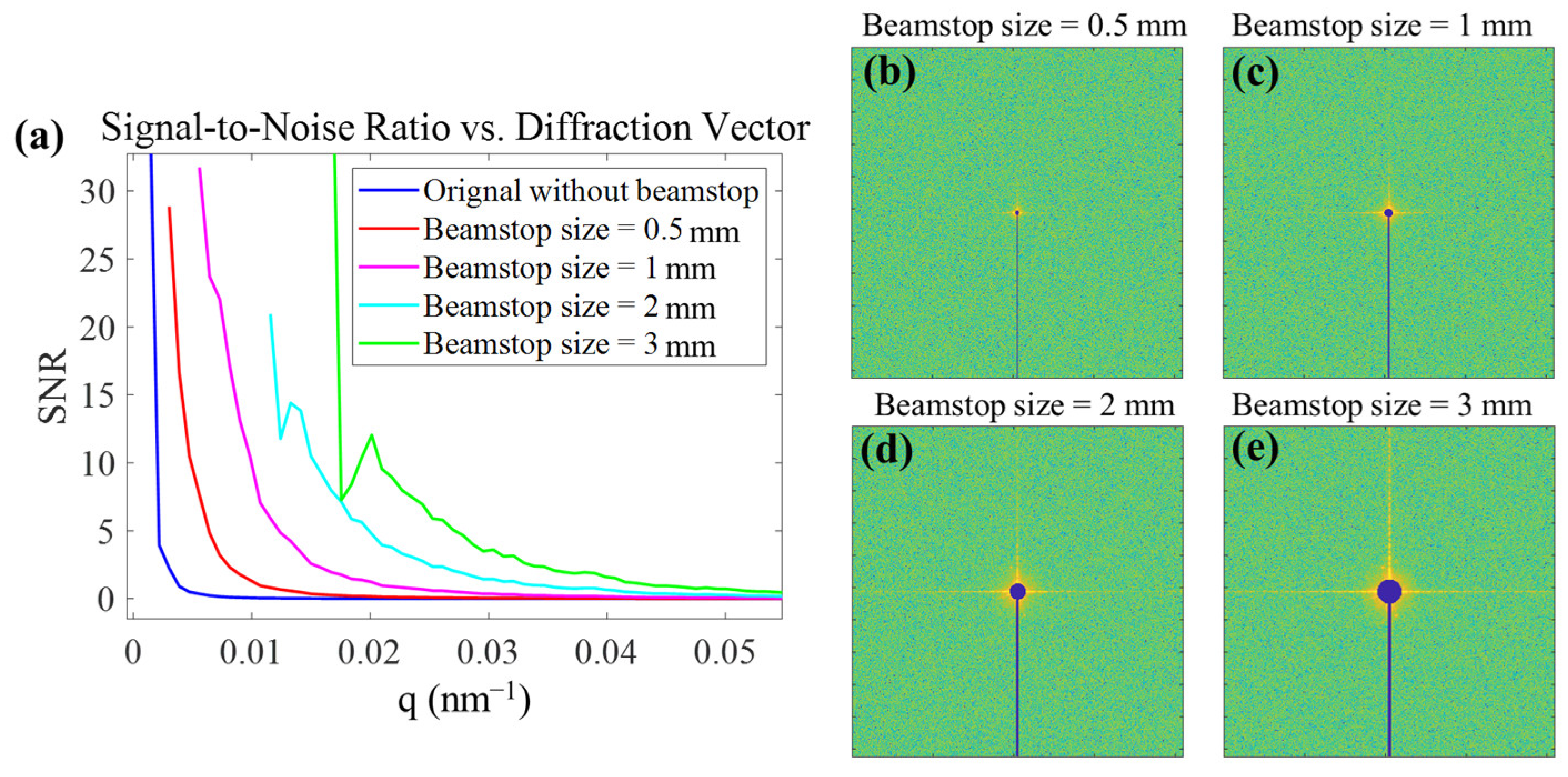
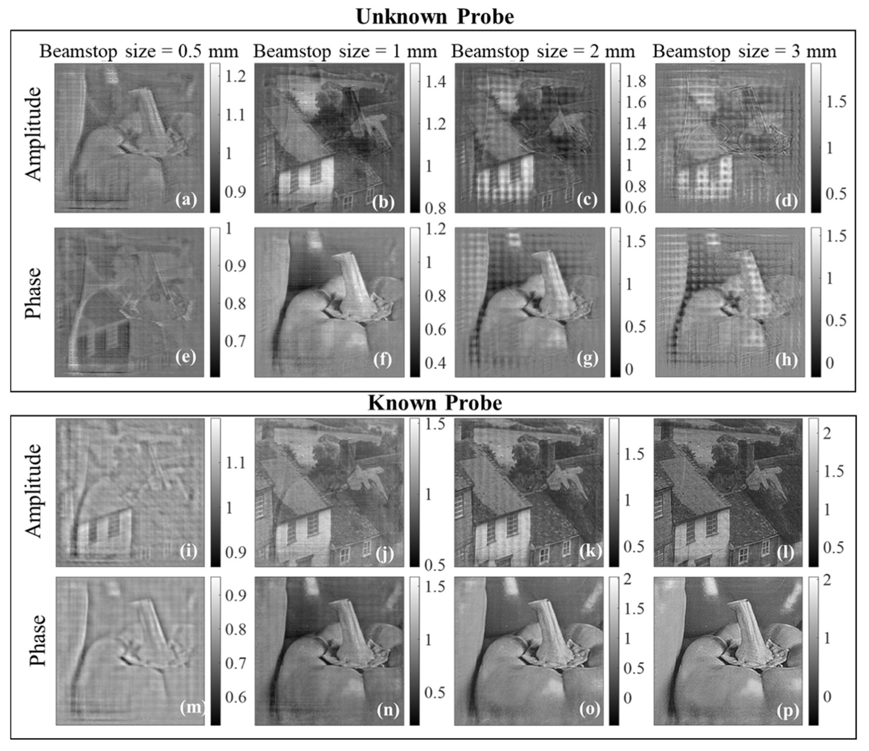
3.1.2. Signal Loss in Central Beamstop Region
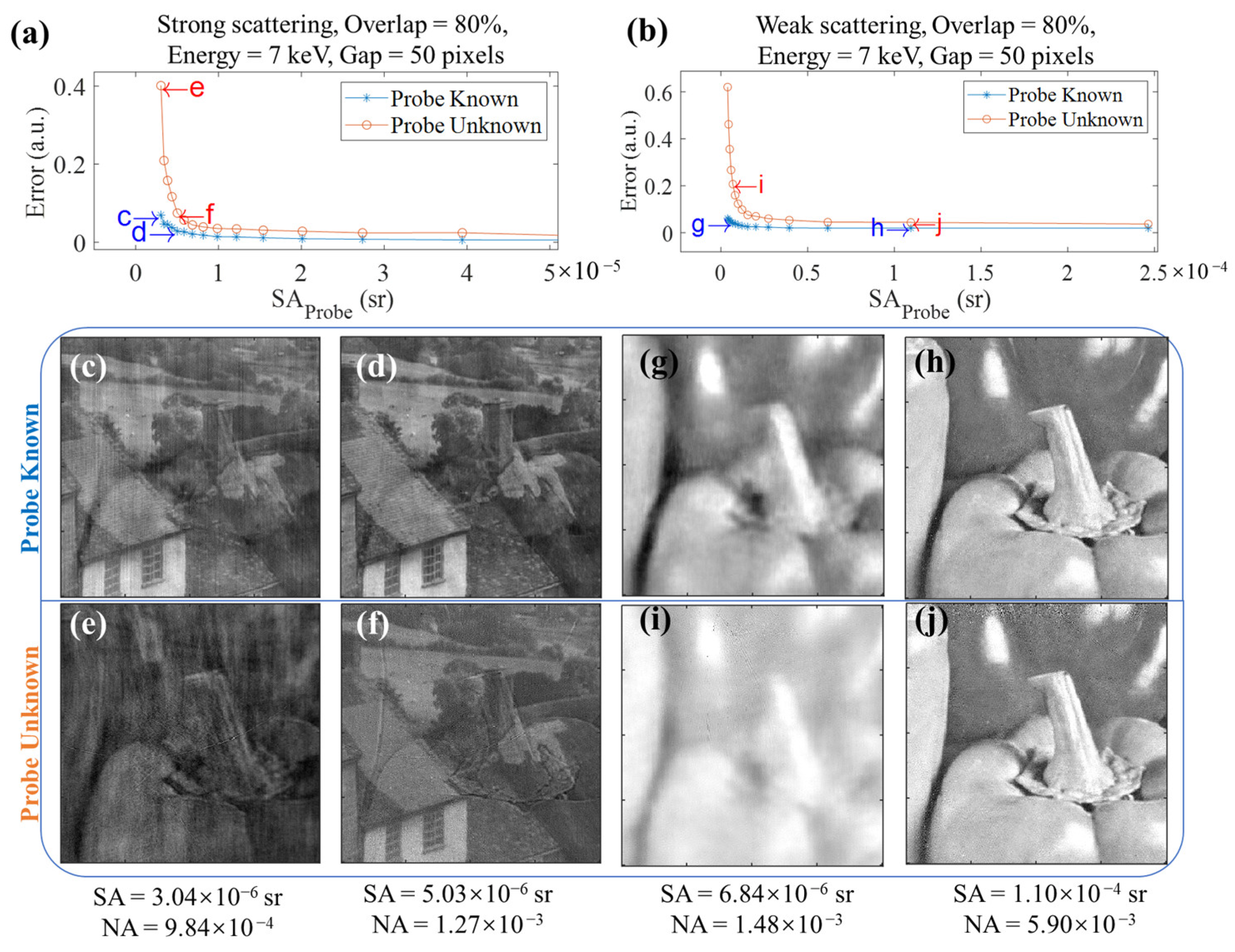
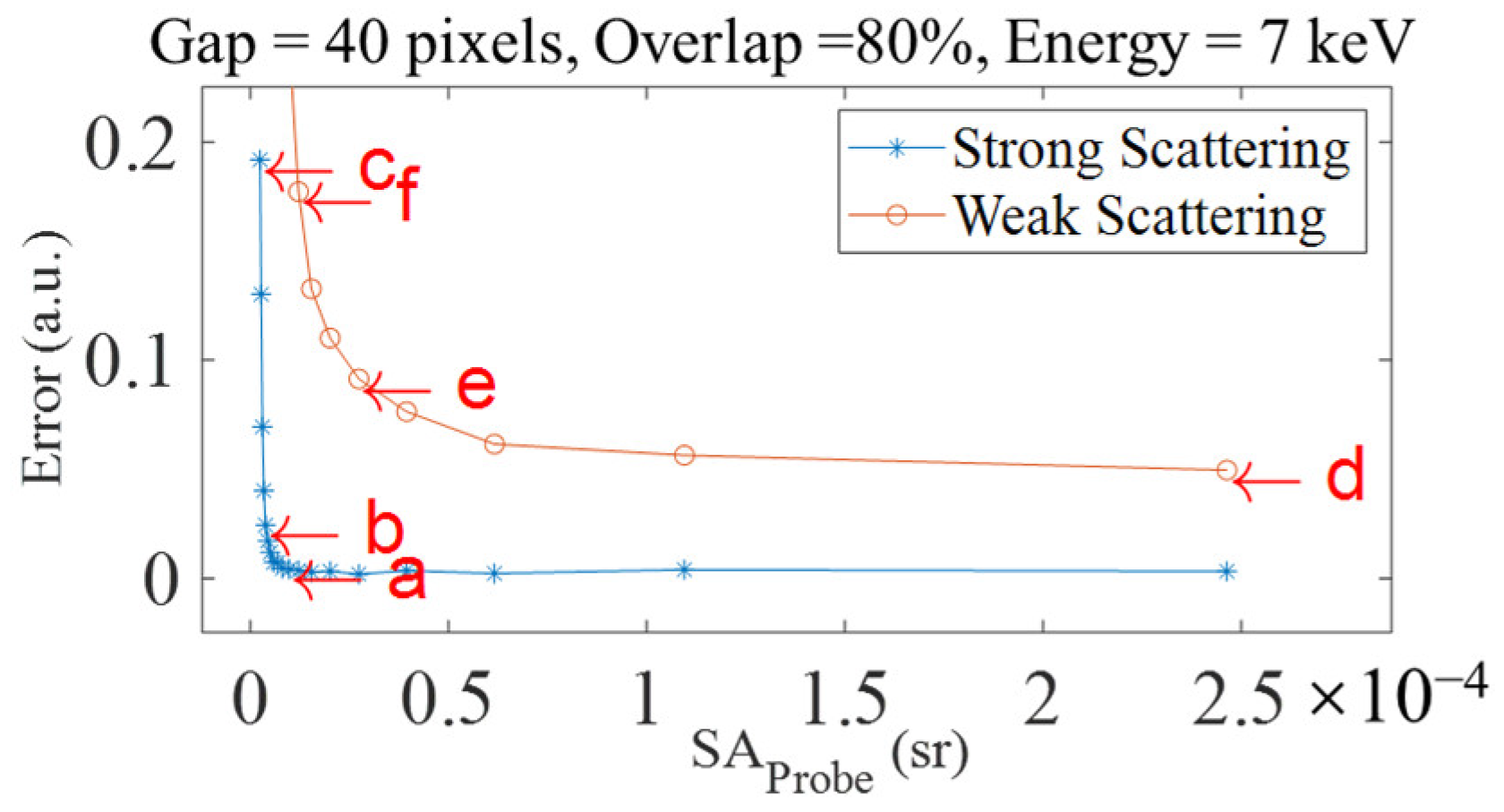
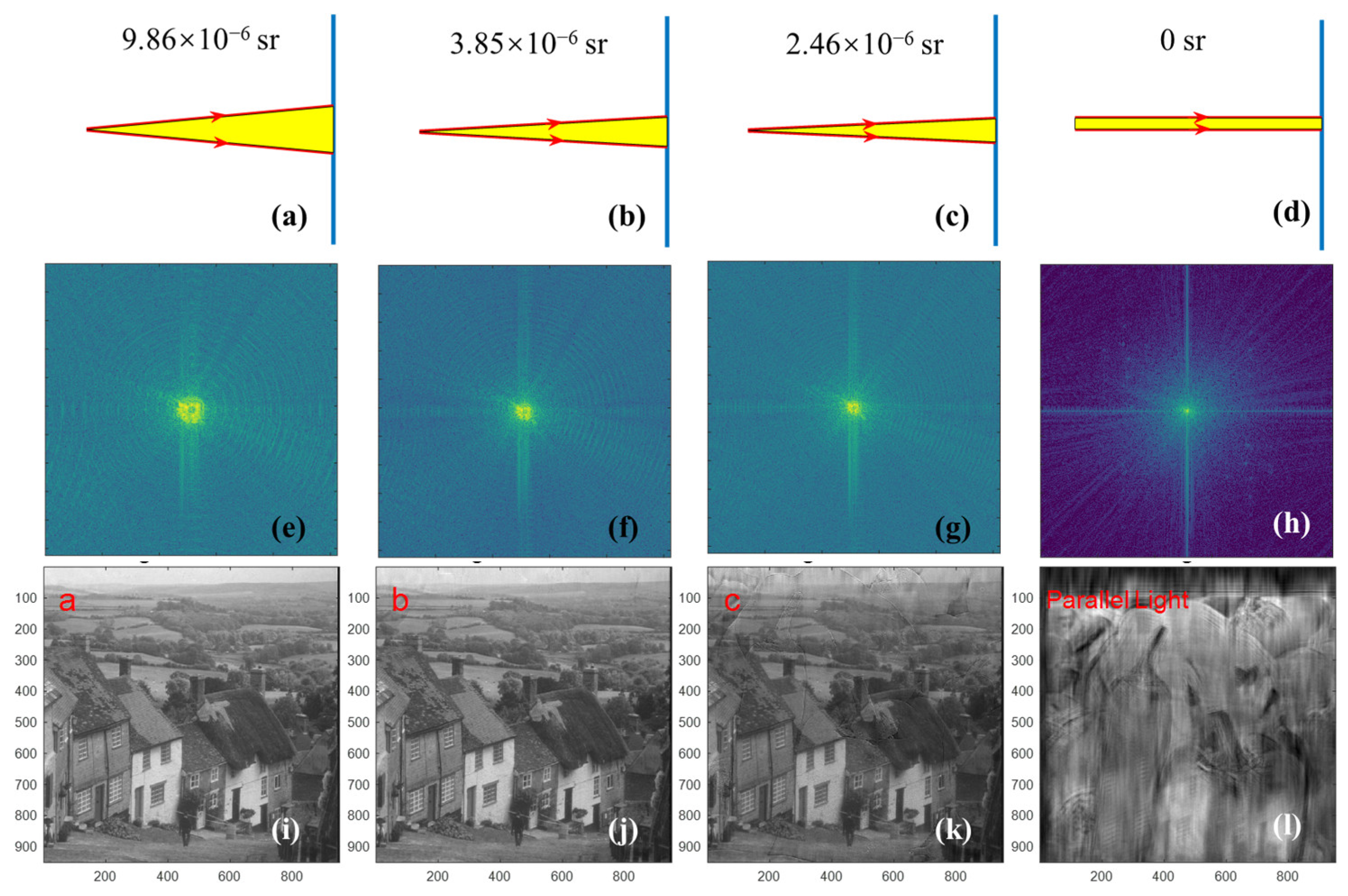
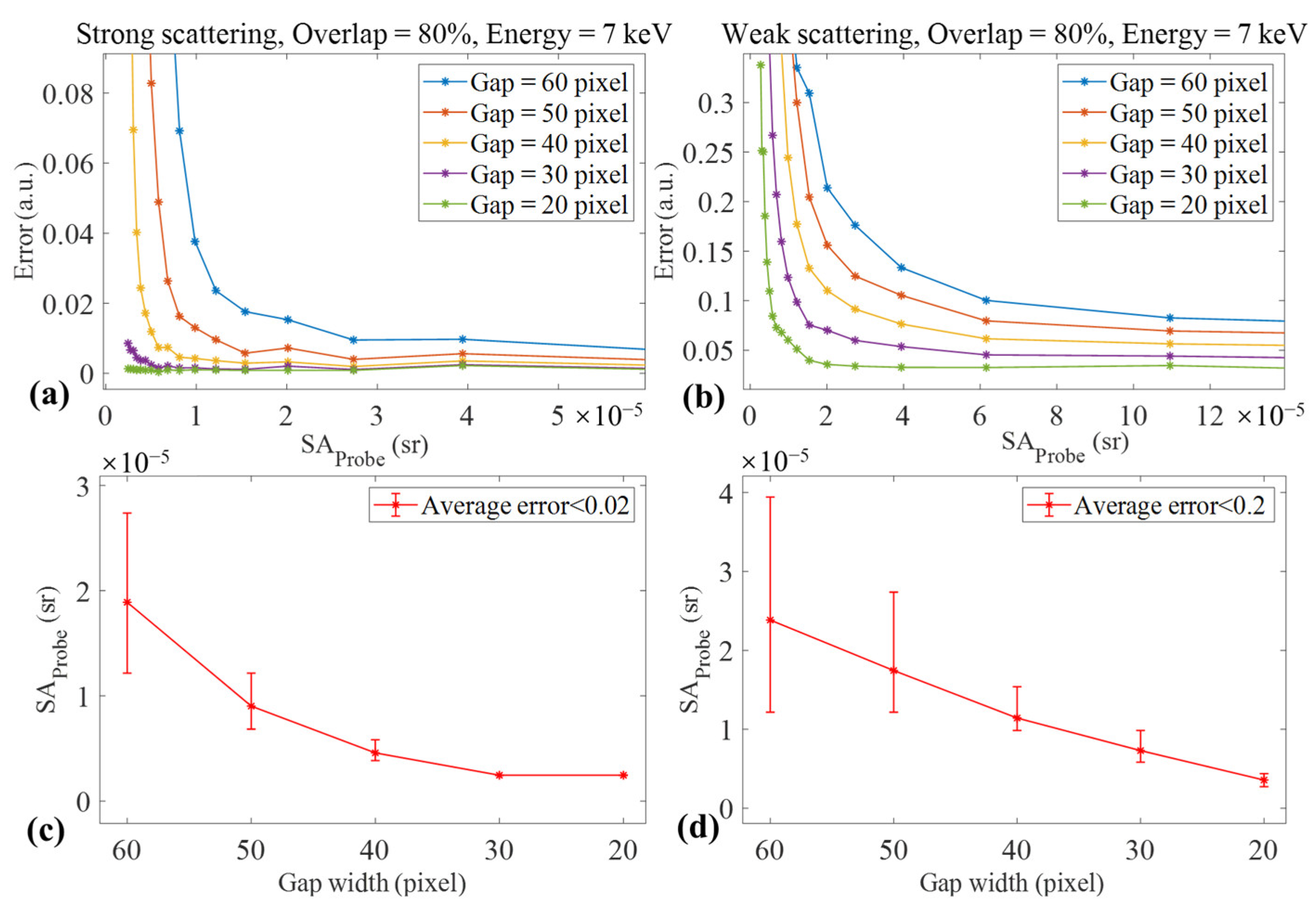
3.2. Strategy 2: Increasing the Divergence of Incident Light
3.2.1. Simulation Results
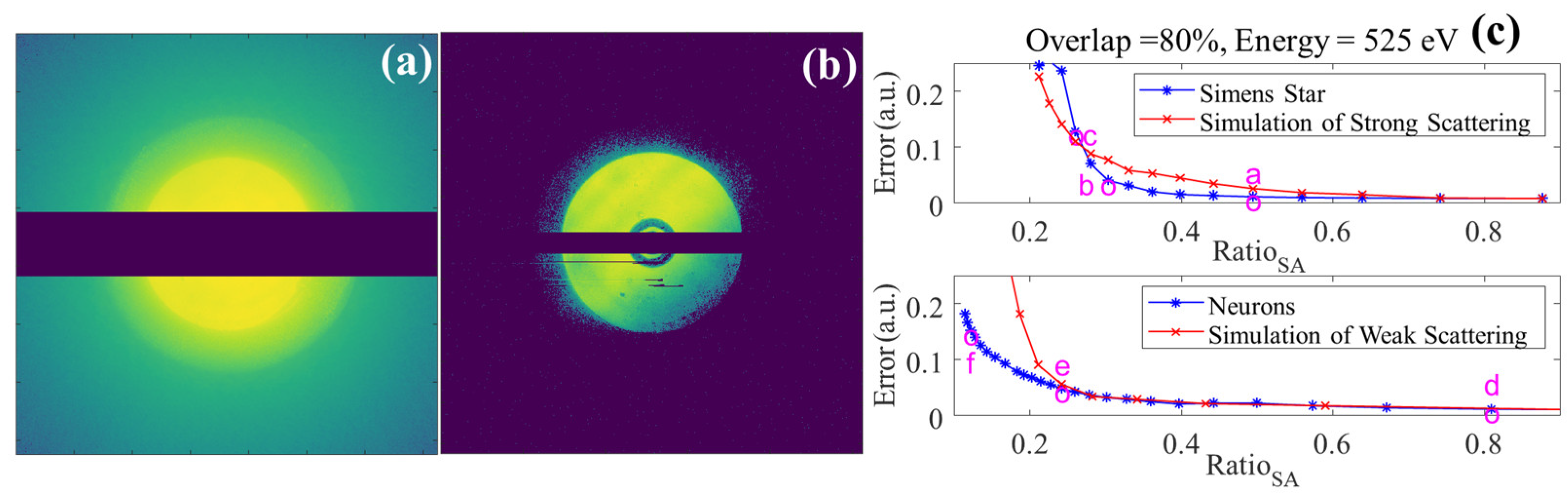
3.2.2. Experimental Results
4. Discussion
5. Conclusions
Supplementary Materials
Author Contributions
Funding
Institutional Review Board Statement
Informed Consent Statement
Data Availability Statement
Acknowledgments
Conflicts of Interest
References
- Bosch, C.; Aidukas, T.; Holler, M.; Pacureanu, A.; Müller, E.; Peddie, C.J.; Zhang, Y.; Cook, P.; Collinson, L.; Bunk, O.; et al. Non-Destructive X-Ray Tomography of Brain Tissue Ultrastructure. bioRxiv 2024. [Google Scholar] [CrossRef]
- Csernica, P.M.; Kalirai, S.S.; Gent, W.E.; Lim, K.; Yu, Y.-S.; Liu, Y.; Ahn, S.-J.; Kaeli, E.; Xu, X.; Stone, K.H.; et al. Persistent and Partially Mobile Oxygen Vacancies in Li-Rich Layered Oxides. Nat. Energy 2021, 6, 642–652. [Google Scholar] [CrossRef]
- Deng, J.; Lo, Y.H.; Gallagher-Jones, M.; Chen, S.; Pryor, A.; Jin, Q.; Hong, Y.P.; Nashed, Y.S.G.; Vogt, S.; Miao, J.; et al. Correlative 3D X-Ray Fluorescence and Ptychographic Tomography of Frozen-Hydrated Green Algae. Sci. Adv. 2018, 4, eaau4548. [Google Scholar] [CrossRef] [PubMed]
- Dierolf, M.; Menzel, A.; Thibault, P.; Schneider, P.; Kewish, C.M.; Wepf, R.; Bunk, O.; Pfeiffer, F. Ptychographic X-Ray Computed Tomography at the Nanoscale. Nature 2010, 467, 436–439. [Google Scholar] [CrossRef]
- Donnelly, C.; Guizar-Sicairos, M.; Scagnoli, V.; Gliga, S.; Holler, M.; Raabe, J.; Heyderman, L.J. Three-Dimensional Magnetization Structures Revealed with X-Ray Vector Nanotomography. Nature 2017, 547, 328–331. [Google Scholar] [CrossRef]
- Lo, Y.H.; Zhou, J.; Rana, A.; Morrill, D.; Gentry, C.; Enders, B.; Yu, Y.-S.; Sun, C.-Y.; Shapiro, D.A.; Falcone, R.W.; et al. X-Ray Linear Dichroic Ptychography. Proc. Natl. Acad. Sci. USA 2021, 118, e2019068118. [Google Scholar] [CrossRef]
- Guizar-Sicairos, M.; Johnson, I.; Diaz, A.; Holler, M.; Karvinen, P.; Stadler, H.-C.; Dinapoli, R.; Bunk, O.; Menzel, A. High-Throughput Ptychography Using Eiger: Scanning X-Ray Nano-Imaging of Extended Regions. Opt. Express 2014, 22, 14859–14870. [Google Scholar] [CrossRef] [PubMed]
- Jørgensen, P.S.; Besley, L.; Slyamov, A.M.; Diaz, A.; Guizar-Sicairos, M.; Odstrčil, M.; Holler, M.; Silvestre, C.; Chang, B.; Detlefs, C.; et al. Hard X-Ray Grazing-Incidence Ptychography: Large Field-of-View Nanostructure Imaging with Ultra-High Surface Sensitivity. Optica 2024, 11, 197. [Google Scholar] [CrossRef]
- Holler, M.; Diaz, A.; Guizar-Sicairos, M.; Karvinen, P.; Färm, E.; Härkönen, E.; Ritala, M.; Menzel, A.; Raabe, J.; Bunk, O. X-Ray Ptychographic Computed Tomography at 16 Nm Isotropic 3D Resolution. Sci. Rep. 2014, 4, 3857. [Google Scholar] [CrossRef]
- Deng, J.; Preissner, C.; Klug, J.A.; Mashrafi, S.; Roehrig, C.; Jiang, Y.; Yao, Y.; Wojcik, M.; Wyman, M.D.; Vine, D.; et al. The Velociprobe: An Ultrafast Hard X-Ray Nanoprobe for High-Resolution Ptychographic Imaging. Rev. Sci. Instrum. 2019, 90, 083701. [Google Scholar] [CrossRef]
- Yao, Y.; Jiang, Y.; Klug, J.A.; Wojcik, M.; Maxey, E.R.; Sirica, N.S.; Roehrig, C.; Cai, Z.; Vogt, S.; Lai, B.; et al. Multi-Beam X-Ray Ptychography for High-Throughput Coherent Diffraction Imaging. Sci. Rep. 2020, 10, 19550. [Google Scholar] [CrossRef] [PubMed]
- Rana, A.; Liao, C.-T.; Iacocca, E.; Zou, J.; Pham, M.; Lu, X.; Subramanian, E.-E.C.; Lo, Y.H.; Ryan, S.A.; Bevis, C.S.; et al. Three-Dimensional Topological Magnetic Monopoles and Their Interactions in a Ferromagnetic Meta-Lattice. Nat. Nanotechnol. 2023, 18, 227–232. [Google Scholar] [CrossRef]
- Deng, J.; Hong, Y.P.; Chen, S.; Nashed, Y.S.G.; Peterka, T.; Levi, A.J.F.; Damoulakis, J.; Saha, S.; Eiles, T.; Jacobsen, C. Nanoscale X-Ray Imaging of Circuit Features without Wafer Etching. Phys. Rev. B 2017, 95, 104111. [Google Scholar] [CrossRef] [PubMed]
- Gorkhover, T.; Rupp, D. Microchip Minutiae Imaged Using X-Ray Bursts. Nature 2024, 632, 36–38. [Google Scholar] [CrossRef]
- Miao, J. Computational Microscopy with Coherent Diffractive Imaging and Ptychography. Nature 2025, 637, 281–295. [Google Scholar] [CrossRef] [PubMed]
- Reinhardt, J.; Hoppe, R.; Hofmann, G.; Damsgaard, C.D.; Patommel, J.; Baumbach, C.; Baier, S.; Rochet, A.; Grunwaldt, J.-D.; Falkenberg, G.; et al. Beamstop-Based Low-Background Ptychography to Image Weakly Scattering Objects. Ultramicroscopy 2017, 173, 52–57. [Google Scholar] [CrossRef]
- Schropp, A.; Schroer, C.G. Dose Requirements for Resolving a given Feature in an Object by Coherent X-Ray Diffraction Imaging. New J. Phys. 2010, 12, 035016. [Google Scholar] [CrossRef]
- Granados, M.; Ajdin, B.; Wand, M.; Theobalt, C.; Seidel, H.-P.; Lensch, H.P.A. Optimal HDR Reconstruction with Linear Digital Cameras. In Proceedings of the 2010 IEEE Computer Society Conference on Computer Vision and Pattern Recognition, San Francisco, CA, USA, 13–18 June 2010; pp. 215–222. [Google Scholar]
- Dierolf, M.; Thibault, P.; Menzel, A.; Kewish, C.M.; Jefimovs, K.; Schlichting, I.; König, K.V.; Bunk, O.; Pfeiffer, F. Ptychographic Coherent Diffractive Imaging of Weakly Scattering Specimens. New J. Phys. 2010, 12, 035017. [Google Scholar] [CrossRef]
- Henrich, B.; Bergamaschi, A.; Broennimann, C.; Dinapoli, R.; Eikenberry, E.F.; Johnson, I.; Kobas, M.; Kraft, P.; Mozzanica, A.; Schmitt, B. PILATUS: A Single Photon Counting Pixel Detector for X-Ray Applications. Nucl. Instrum. Methods Phys. Res. Sect. A Accel. Spectrometers Detect. Assoc. Equip. 2009, 607, 247–249. [Google Scholar] [CrossRef]
- Shukla, A.; Wright, J.; Henningsson, A.; Stieglitz, H.; Colegrove, E.; Besley, L.; Baur, C.; De Angelis, S.; Stuckelberger, M.; Poulsen, H.F.; et al. Grain Boundary Strain Localization in a CdTe Solar Cell Revealed by Scanning 3D X-Ray Diffraction Microscopy. J. Mater. Chem. A 2024, 12, 16793–16802. [Google Scholar] [CrossRef]
- Takahashi, Y.; Suzuki, A.; Furutaku, S.; Yamauchi, K.; Kohmura, Y.; Ishikawa, T. High-Resolution and High-Sensitivity Phase-Contrast Imaging by Focused Hard x-Ray Ptychography with a Spatial Filter. Appl. Phys. Lett. 2013, 102, 094102. [Google Scholar] [CrossRef]
- Thibault, P.; Dierolf, M.; Menzel, A.; Bunk, O.; David, C.; Pfeiffer, F. High-Resolution Scanning X-Ray Diffraction Microscopy. Science 2008, 321, 379–382. [Google Scholar] [CrossRef]
- Rose, M.; Dzhigaev, D.; Senkbeil, T.; von Gundlach, A.R.; Stuhr, S.; Rumancev, C.; Besedin, I.; Skopintsev, P.; Viefhaus, J.; Rosenhahn, A.; et al. High-Dynamic-Range Water Window Ptychography. J. Phys. Conf. Ser. 2017, 849, 012027. [Google Scholar] [CrossRef]
- Takahashi, Y.; Suzuki, A.; Zettsu, N.; Kohmura, Y.; Yamauchi, K.; Ishikawa, T. Multiscale Element Mapping of Buried Structures by Ptychographic X-Ray Diffraction Microscopy Using Anomalous Scattering. Appl. Phys. Lett. 2011, 99, 131905. [Google Scholar] [CrossRef]
- Guizar-Sicairos, M.; Holler, M.; Diaz, A.; Vila-Comamala, J.; Bunk, O.; Menzel, A. Role of the Illumination Spatial-Frequency Spectrum for Ptychography. Phys. Rev. B 2012, 86, 100103. [Google Scholar] [CrossRef]
- Holler, M.; Guizar-Sicairos, M.; Tsai, E.H.R.; Dinapoli, R.; Müller, E.; Bunk, O.; Raabe, J.; Aeppli, G. High-Resolution Non-Destructive Three-Dimensional Imaging of Integrated Circuits. Nature 2017, 543, 402–406. [Google Scholar] [CrossRef]
- Vila-Comamala, J.; Diaz, A.; Guizar-Sicairos, M.; Mantion, A.; Kewish, C.M.; Menzel, A.; Bunk, O.; David, C. Characterization of High-Resolution Diffractive X-Ray Optics by Ptychographic Coherent Diffractive Imaging. Opt. Express 2011, 19, 21333. [Google Scholar] [CrossRef]
- Shapiro, D.A.; Yu, Y.-S.; Tyliszczak, T.; Cabana, J.; Celestre, R.; Chao, W.; Kaznatcheev, K.; Kilcoyne, A.L.D.; Maia, F.; Marchesini, S.; et al. Chemical Composition Mapping with Nanometre Resolution by Soft X-Ray Microscopy. Nat. Photon 2014, 8, 765–769. [Google Scholar] [CrossRef]
- Aidukas, T.; Phillips, N.W.; Diaz, A.; Poghosyan, E.; Müller, E.; Levi, A.F.J.; Aeppli, G.; Guizar-Sicairos, M.; Holler, M. High-Performance 4-nm-Resolution X-Ray Tomography Using Burst Ptychography. Nature 2024, 632, 81–88. [Google Scholar] [CrossRef]
- Atakul-Özdemir, A.; Warren, X.; Martin, P.G.; Guizar-Sicairos, M.; Holler, M.; Marone, F.; Martínez-Pérez, C.; Donoghue, P.C.J. X-Ray Nanotomography and Electron Backscatter Diffraction Demonstrate the Crystalline, Heterogeneous and Impermeable Nature of Conodont White Matter. R. Soc. Open Sci. 2021, 8, 202013. [Google Scholar] [CrossRef]
- Jiang, Y.; Deng, J.; Yao, Y.; Klug, J.A.; Mashrafi, S.; Roehrig, C.; Preissner, C.; Marin, F.S.; Cai, Z.; Lai, B.; et al. Achieving High Spatial Resolution in a Large Field-of-View Using Lensless X-Ray Imaging. Appl. Phys. Lett. 2021, 119, 124101. [Google Scholar] [CrossRef]
- Weber, C.C.; De Angelis, S.; Meinert, R.; Appel, C.; Holler, M.; Guizar-Sicairos, M.; Gubler, L.; Büchi, F.N. Microporous Transport Layers Facilitating Low Iridium Loadings in Polymer Electrolyte Water Electrolysis. EES. Catal. 2024, 2, 585–602. [Google Scholar] [CrossRef]
- Dejkameh, A.; Nebling, R.; Locans, U.; Kim, H.-S.; Mochi, I.; Ekinci, Y. Recovery of Spatial Frequencies in Coherent Diffraction Imaging in the Presence of a Central Obscuration. Ultramicroscopy 2024, 258, 113912. [Google Scholar] [CrossRef]
- Thibault, P.; Menzel, A. Reconstructing state mixtures from diffraction measurements. Nature 2023, 494, 68–71. [Google Scholar] [CrossRef] [PubMed]
- Xing, Z.; Xu, Z.; Sun, T.; Zhang, X.; Tai, R. Research on Signal-Noise Separation Algorithm of Ptychography. Acta Opt. Sin. 2021, 41, 2234001. [Google Scholar] [CrossRef]
- Qin, Z.; Xu, Z.; Li, R.; Liu, H.; Liu, S.; Wen, Q.; Chen, X.; Zhang, X.; Tai, R. Initial Probe Function Construction in Ptychography Based on Zone-Plate Optics. Appl. Opt. 2023, 62, 3542. [Google Scholar] [CrossRef]
- Quiney, H.M.; Peele, A.G.; Cai, Z.; Paterson, D.; Nugent, K.A. Diffractive Imaging of Highly Focused X-Ray Fields. Nat. Phys. 2006, 2, 101–104. [Google Scholar] [CrossRef]
- Zhang, W.; Dresselhaus, J.L.; Fleckenstein, H.; Prasciolu, M.; Zakharova, M.; Ivanov, N.; Li, C.; Yefanov, O.; Li, T.; Egorov, D.; et al. Fast and Efficient Hard X-Ray Projection Imaging below 10 nm Resolution. Opt. Express 2024, 32, 30879. [Google Scholar] [CrossRef]
- Cherukara, M.J.; Zhou, T.; Nashed, Y.; Enfedaque, P.; Hexemer, A.; Harder, R.J.; Holt, M.V. Real-Time Sparse-Sampled Ptychographic Imaging Through Deep Neural Networks. arXiv 2020, arXiv:2004.08247. [Google Scholar] [CrossRef]
- Chavez, T.; Roberts, E.J.; Zwart, P.H.; Hexemer, A. A Comparison of Deep-Learning-Based Inpainting Techniques for Experimental X-Ray Scattering. J. Appl. Crystallogr. 2022, 55, 1277–1288. [Google Scholar] [CrossRef]
- Hoidn, O.; Mishra, A.A.; Mehta, A. Physics Constrained Unsupervised Deep Learning for Rapid, High Resolution Scanning Coherent Diffraction Reconstruction. Sci. Rep. 2023, 13, 22789. [Google Scholar] [CrossRef] [PubMed]


Disclaimer/Publisher’s Note: The statements, opinions and data contained in all publications are solely those of the individual author(s) and contributor(s) and not of MDPI and/or the editor(s). MDPI and/or the editor(s) disclaim responsibility for any injury to people or property resulting from any ideas, methods, instructions or products referred to in the content. |
© 2025 by the authors. Licensee MDPI, Basel, Switzerland. This article is an open access article distributed under the terms and conditions of the Creative Commons Attribution (CC BY) license (https://creativecommons.org/licenses/by/4.0/).
Share and Cite
Li, R.; Xu, Z.; Chen, S.; Wu, S.; Zhang, Y.; Zhang, X.; Tai, R. Strategies for Suppression and Compensation of Signal Loss in Ptychography. Photonics 2025, 12, 636. https://doi.org/10.3390/photonics12070636
Li R, Xu Z, Chen S, Wu S, Zhang Y, Zhang X, Tai R. Strategies for Suppression and Compensation of Signal Loss in Ptychography. Photonics. 2025; 12(7):636. https://doi.org/10.3390/photonics12070636
Chicago/Turabian StyleLi, Ruoru, Zijian Xu, Sheng Chen, Shuhan Wu, Yingling Zhang, Xiangzhi Zhang, and Renzhong Tai. 2025. "Strategies for Suppression and Compensation of Signal Loss in Ptychography" Photonics 12, no. 7: 636. https://doi.org/10.3390/photonics12070636
APA StyleLi, R., Xu, Z., Chen, S., Wu, S., Zhang, Y., Zhang, X., & Tai, R. (2025). Strategies for Suppression and Compensation of Signal Loss in Ptychography. Photonics, 12(7), 636. https://doi.org/10.3390/photonics12070636