Article

Green Synthesis of Zinc Oxide Nanoparticles Using Aqueous Extracts of Hibiscus cannabinus L.: Wastewater Purification and Antibacterial Activity

1
Institute of Bast Fiber Crops, Chinese Academy of Agricultural Sciences, Changsha 410205, China
2
College of Life Sciences, Shangrao Normal University, Shangrao 334001, China
*
Author to whom correspondence should be addressed.
Separations 2023, 10(9), 466; https://doi.org/10.3390/separations10090466
Submission received: 26 July 2023 / Revised: 11 August 2023 / Accepted: 14 August 2023 / Published: 24 August 2023
(This article belongs to the Special Issue Advances in Chromatographic Analysis of Bioactive Compounds)

Abstract

:
The green preparation of metal oxide nanoparticles is an environmentally friendly method, which could reduce the use of toxic solvents and their impact on the environment. The purpose of this study is to investigate the green synthesis of zinc oxide (ZnO) nanoparticles using extracts of Hibiscus cannabinus leaves and to evaluate their potential applications in environmental remediation. In this work, ZnO nanoparticles were successfully prepared and thoroughly characterized using UV–vis, Fourier transform infrared analysis (FTIR), X-ray diffraction (XRD), transmission electron microscope (TEM) analysis, and scanning electron microscope (SEM) with energy dispersive x-ray analysis (EDAX). As a result, the synthesized ZnO nanoparticles showed a good adsorption capacity for Congo red (CR), and satisfactory antioxidant and antibacterial activities. They exhibited good adsorption and removal abilities for CR in aqueous solutions. With the conditions optimized, the adsorption kinetics and isotherms were fitted to the pseudo-second-order model and the Langmuir model. The ZnO nanoparticles could also effectively scavenge 2-2-diphenyl-1-picrylhydrazyl (DPPH) and 2,2′-azino-di(3-ethylbenzthiazoline sulphonate) (ABTS) radicals, and appeared to inhibit the growth of Escherichia coli and Staphylococcus aureus bacteria. Based on the identified adsorption capacity, the green synthesized ZnO nanoparticles demonstrated their potential to be used in the removal of dyeing wastewater and in the further purification of water due to their antioxidant activity and antibacterial activity.

1. Introduction

With the rapid advancement of printing and dyeing technologies, synthetic dyes have been extensively utilized in various industries over the past few decades. Consequently, a significant amount of printing and dyeing wastewater is discharged into water bodies, constituting a substantial portion of industrial wastewater discharge [1]. This dyeing wastewater poses a severe threat in terms of water pollution due to its intense color, high toxicity, elevated organic pollutant content, and poor biodegradability of organic compounds [2]. Multiple approaches have been introduced for the treatment of dye wastewater, including physical methods (such as adsorption and membrane technology), chemical methods (including electrochemistry and advanced oxidation), and biological methods. Among these, physical methods are commonly used to treat water-soluble dyes due to their simplicity, cost-effectiveness, and notable decolorization efficacy [3].
Nanomaterials possess remarkable physicochemical properties and biocompatibility, rendering them highly valuable in the fields of biomedical, environmental, and energy research [4]. Zinc oxide (ZnO) nanoparticles are a new type of inorganic nanomaterial with various special properties, such as photocatalytic, antioxidant, antibacterial, and antifungal activities, which gives them significant value to various research fields [5]. Traditional synthesis methods for ZnO nanoparticles include the hydrothermal, sol–gel, chemical precipitation, and spray pyrolysis techniques [6]. Meanwhile, in recent years, green synthesis technology using plant extracts has become increasingly popular due to its simplicity, low costs, safety, non-toxicity, abundant raw materials, and environmentally friendly characteristics [7]. Plant extracts are readily available, inexpensive, and often contain significant amounts of reducing and capping agents, ensuring their suitability for large-scale production [8]. Dangana et al. prepared ZnO nanoparticles using extracts obtained from the leaves of Chaya Cnidoscolus aconitifolius and used them as nano fertilizers on Sorghum bicolour plants [9]. Elshafie et al. carried out the green synthesis of ZnO nanoparticles using Moringa oleifera leaves [10]. Rini et al. reported a green method of synthesizing ZnO nanoparticles using pineapple-peel extract and evaluated their antibacterial activity [11]. Moreover, almond shells, sugar cane bagasse, eggshells, Viscum album leaves, peppermint tea dregs, marine brown algae, and many plant extracts have been used for the synthesis of ZnO nanoparticles [12,13,14,15]. Hence, the green synthesis approach to obtaining ZnO nanoparticles has significant potential for further exploration and development.
Hibiscus cannabinus L., commonly referred to as kenaf, is an annual herbaceous plant belonging to the Malvaceae family [16]. It has a wide distribution across regions such as India, China, and Thailand. Hibiscus cannabinus serves as an important industrial fiber crop, finding applications in various fields such as textiles, animal feed, papermaking, and construction. This plant contains numerous bioactive components including polyphenols, alkaloids, tannins, and saponins [17]. Consequently, Hibiscus cannabinus exhibits medicinal properties with potential anti-tumor, antioxidant, anti-inflammatory, anti-hypertensive, and anti-proliferative activities [18]. Apart from their primary use as a bast fiber, the leaves of Hibiscus cannabinus could offer an additional source for the production of nanomaterials.
This article focused on the synthesis of ZnO nanoparticles using extracts of Hibiscus cannabinus L. leaves and investigated their ability to adsorb organic dyes (Figure 1). The crystalline properties, elemental composition, and structure of the synthesized nanoparticles were analyzed using various characterization techniques, including X-ray diffraction (XRD), UV–Vis spectroscopy, Fourier transform infrared (FTIR) spectroscopy, transmission electron microscopy (TEM), scanning electron microscopy (SEM), and energy-dispersive X-ray analysis (EDAX). The presence in dyeing wastewater of azo dye compounds, which are extensively utilized synthetic dyes, poses a significant threat to human health and aquatic organisms due to these compounds’ carcinogenic properties [19]. Moreover, the aromatic structure of azo dyes presents challenges in terms of their degradation, leading to prolonged environmental pollution [20]. Exposure to azo dyes can result in various ailments such as edema, allergies, skin irritation, and respiratory issues [21]. To assess the efficacy of removing contaminants, the adsorption of Congo red (CR) dye was evaluated using synthesized ZnO nanoparticles. To enhance our understanding of their efficacy in environmental remediation, the antioxidant potential of the ZnO nanoparticles synthesized using the green method was assessed using the 2-2-diphenyl-1-picrylhydrazyl (DPPH) and 2,2′-azino-di(3-ethylbenzthiazoline sulpho-nate) (ABTS) radical scavenging assays. Additionally, the antibacterial properties of these nanoparticles were further examined against Escherichia coli (E. coli) and Staphylococcus aureus (S. aureus) bacteria.

2. Materials and Methods

2.1. Materials

All the chemicals and reagents used in this study were of analytical grade, purchased from Macklin Inc. (Shanghai, China), and used without treatment. Hibiscus cannabinus leaves were collected from the Innovation experimental base of the Institute of Bast Fiber Crops, Chinese Academy of Agricultural Sciences, Changsha, China, in 2023. The samples were dried, smashed, and stored at 4 °C for further use.

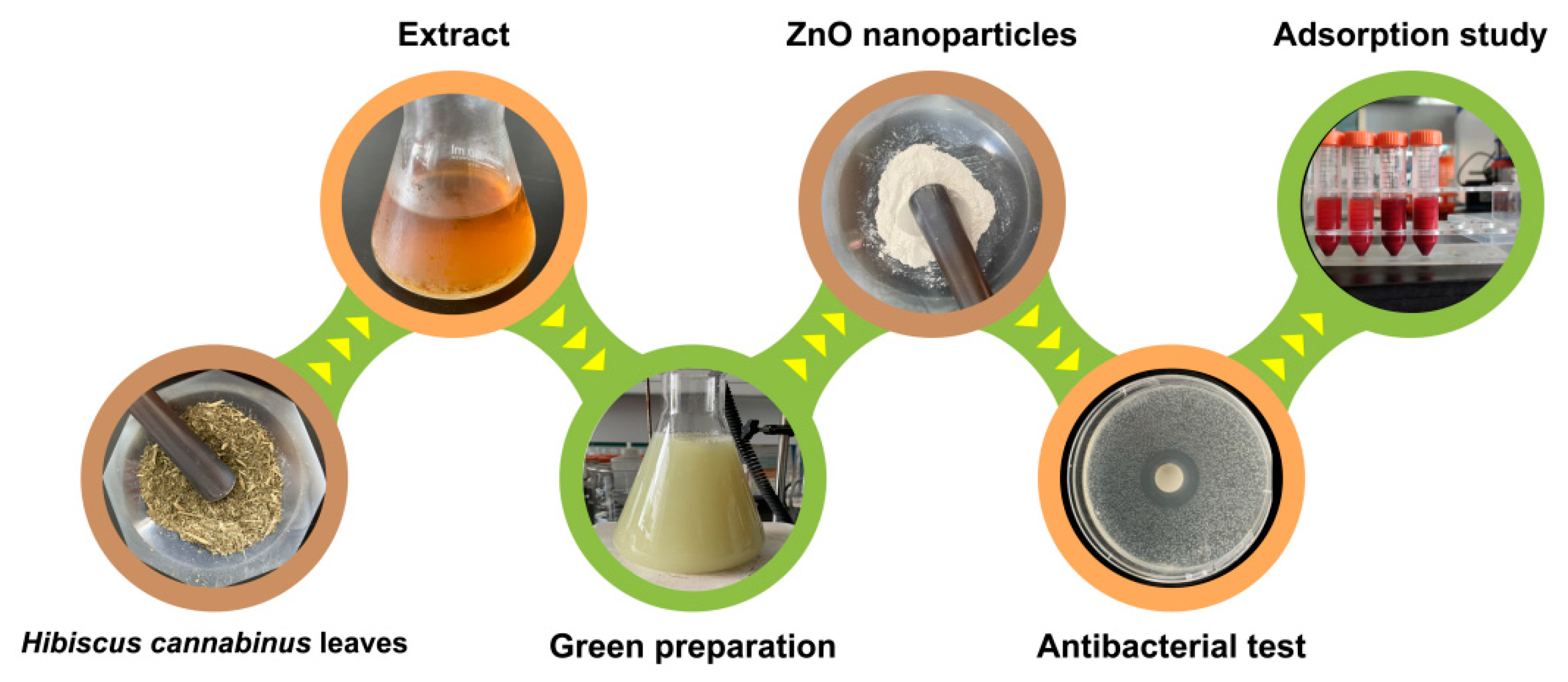
2.2. Green Preparation of ZnO Nanoparticles

First, 10 g of Hibiscus cannabinus leaves was weighed and transferred in a 250 mL round-bottom flask containing 100 mL of water at a ratio of 1:10 (g/mL). The mixture was then extracted under heating at 90 °C for 4 h. After extraction and cooling to room temperature, the extraction solution was centrifuged at 3000 rpm for 3 min. The supernatant was stored at 4 °C.
For the green preparation of the ZnO nanoparticles, 20 mL of Hibiscus cannabinus leaf extract was added to 50 mL of 0.02 mol/L zinc sulfate solution in a conical flask and incubated under magnetic stirring for 1.5 h. Then, some 0.2 mol/L sodium hydroxide solution was slowly added to the mixture to adjust the pH to 8. The mixture was magnetically stirred for 3.5 h. After the reaction, the products were centrifuged at 4000 rpm for 5 min. The supernatant was discarded, and the precipitates were washed twice with water and dried at 70 °C.
For the gas chromatography–mass spectrometer (GC–MS) analysis, 10 g of Hibiscus cannabinus leaves was weighed and transferred to a 250 mL round-bottom flask containing 100 mL of ethanol at a ratio of 1:10 (g/mL). The mixture was then extracted under heating at 80 °C for 4 h. After extraction and cooling to room temperature, the solution was centrifuged at 3000 rpm for 3 min. The supernatant was concentrated via reduced pressure distillation, filtrated using a 0.45 μm membrane, and subjected to GC–MS analysis.

2.3. Characterizations

The green-synthesized ZnO nanoparticles were characterized using a SEM (Apreo 2 Thermo Scientific, Waltham, MA, USA) equipped with an X-ray fluorescence spectrometer Oxford INCA X-max 80 for EDAX, TEM (JEM-2100F, JEOL, Tokyo, Japan), and XRD (Rigaku SmartLab 9, Tokyo, Japan) using Cu-Kα radiation at 40 kV and 40 mA, and UV spectroscopy was undertaken using a Shimadzu UV-2700 absorption spectrophotometer (Kyoto, Japan) and FT-IR (IRSpirit, Shimadzu, Kyoto, Japan) in the range of 400–4000 cm−1.
The GC–MS analytical method was used to detect the components in the ethanol extract of Hibiscus cannabinus leaves. The analysis was carried out on a 7890B GC equipped with a 5977A MSD (Agilent Technologies, Santa Clara, CA, USA) under the following conditions: 1 μL of the sample was injected in the HP-5MS (5% phenyl methyl siloxane) capillary column (30 m × 250 μm × 0.25 μm); helium was used as the carrier gas at a constant flow of 1.2 mL/min; and the temperatures of the injector and MSD were set to 250 °C and 280 °C, respectively. The column was first maintained at 60 °C for 2 min; then, the temperature increased to 280 °C at the rate of 5 °C/min, and it was finally maintained for 9 min. The MS parameters were operated at 70 eV and scanned from 40 to 600 amu. The chemical information was identified via their mass spectral data using the standard NIST library 2011 during the GC–MS analysis [22].

2.4. Adsorption of Dyes

First, 0.5 g of the ZnO nanoparticles was added to 50 mL of the aqueous solution of the CR dye at different initial concentrations in a conical flask. The solution was shaken at 200 rpm on an oscillator at 25 °C to reach adsorption equilibrium. A portion of the solution was taken at various time intervals and filtered using a 0.45 μm membrane. The solution was then monitored using a UV spectrophotometer (UV2700 UV–Vis Spectrophotometer (Shimadzu, Kyoto, Japan)) at 510 nm. The adsorption capacity was calculated according to the following formula [23]:
q t = ( C 0 C t ) × V w
where qt is the adsorbed amount of the sample at a certain time, C0 is the initial concentration of the sample, Ct is the concentration of the sample at a certain time, V is the volume of the solution, and w is the weight of the used adsorbent.

2.5. Antioxidant Activity

The antioxidant activity of ZnO nanoparticles was analyzed via DPPH and ABTS free radical scavenging assays. The procedures were slightly changed according to the report by Hu et al. [24]. Different weights of ZnO nanoparticles and 3.0 mL of 0.1 mmol/L DPPH radical solution were mixed and shaken on an oscillator for 20 min in the dark. After that, the solution was filtrated with a 0.45 μm membrane and the absorbance was measured at 517 nm using a UV spectrophotometer. The scavenging rate of each sample was calculated using the following formula:
S c a v e n g i n g   r a t e   ( % ) = 1 A s A 0 × 100 %
where A0 is the absorbance of the control group and As is the absorbance of the sample.
For the ABTS assay, the same volume of 2.45 mmol/L potassium persulfate solution and 7 mmol/L ABTS solution was mixed and incubated in the dark for 18 h [25]. The ABTS stock solution was diluted to obtain an absorbance between 0.7 and 0.72 at 734 nm before use. Different amounts of ZnO nanoparticles and 3.0 mL of the ABTS radical solution were mixed and shaken on an oscillator for 20 min in the dark. The solution was filtrated using a 0.45 μm membrane after the reaction and the absorbance was measured at 734 nm. The scavenging rate of each sample was calculated according to Formula (1).

2.6. Antibacterial Activity

The antibacterial activity of the ZnO nanoparticles was evaluated using the disc diffusion method [26]. Escherichia coli and Staphylococcus aureus bacteria were grown in an LB broth containing 5  g/L of beef extract, 10  g/L of peptones, 20  g/L of agar, and 5 g/L of NaCl, and they were diluted to 1 × 10⁻⁶ CFU prior to use. Then, 0.2g of the ZnO nanoparticles was weighed and pressed into a tablet using a tablet press machine. The sterilized filter paper was immersed in different concentrations of the extract for further use. Then, 100 µL of the bacterial suspensions was evenly spread on an agar plate, and the ZnO nanoparticle tablet (Φ = 13 mm) or the sterilized filter paper (Φ = 6 mm) was carefully placed on the plate. The plates were incubated at 37 °C for 24 h, and the diameter of the inhibition zone was measured.
The minimal inhibitory concentration of the prepared ZnO nanoparticles was determined according to the micro-dilution broth method [27]. In brief, 180 µL of the nutrient broth and different weights of the ZnO nanoparticles were mixed in each well of a sterile 96-well plate, followed by the addition of 20 μL of the bacterial solution (1 × 10⁻⁶ CFU). The 96-well plate was sealed and placed on a shaker at 37 °C for cultivation. The absorbance at 600 nm (OD600) of each well of the plate was measured using a microplate reader (Epoch, BioTek Instruments Inc., Winooski, VT, USA) at predetermined time intervals, the growth curve was plotted based on the obtained data. The nutrient broth without a sample was used as a negative control. The lowest concentration of ZnO nanoparticles inhibiting the growth of bacteria was defined as the minimal inhibitory concentration value.

3. Results and Discussion

3.1. Characterizations of ZnO Nanoparticles

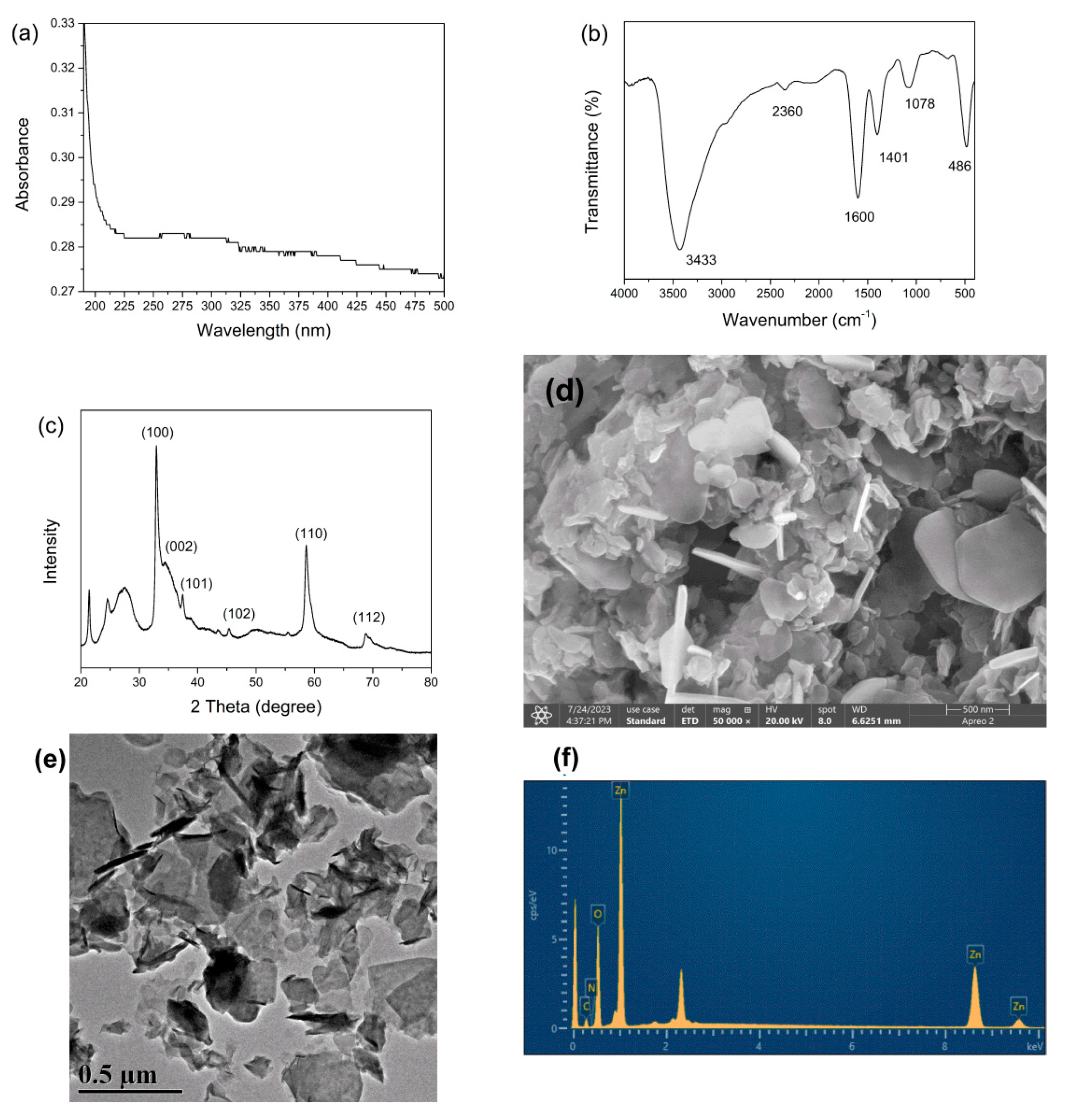
3.1.1. UV–Vis Spectroscopic Analysis

After preparation, the ZnO nanoparticles were thoroughly characterized (Figure 1). The UV–Vis absorption spectra of the ZnO nanoparticles and the Hibiscus cannabinus leaf extracts were observed in the range of 190 to 500 nm (Figure 2a). For the ZnO nanoparticles, a weak absorption peak was observed around 270 nm. Compared to the reported data, many of the prepared ZnO nanoparticles showed adsorption at a wavelength around 370 nm; there is a blue (short wavelength) shift of the peak in this study [28,29]. The spectrum of the Hibiscus cannabinus leaf extract showed adsorptions at 265 nm and 325 nm. This might be because of the existence of extract molecules, since many compounds in extract show adsorption around 254 nm. The highest peak at 265 nm could explain the shift in the spectrum of the ZnO nanoparticles.

3.1.2. FTIR Analysis

The FTIR spectrum of the ZnO nanoparticles is shown in the range 400–4000 cm−1 in Figure 2b. The prepared ZnO nanoparticles showed the band at 3433 cm−1 as O-H stretching and N-H stretching, the band at 2360 cm−1 as strong (O=C=O) stretching carbon dioxide, the band at 1600 cm−1 as the chemisorbed and/or physisorbed water on the particle surface, the band at 1401 cm−1 as the angular deformation of C-H, and the band at 1078 cm−1 as being ascribed to the C-O ether bond of the glucose ring of starch [30]. Moreover, the intense peak at 486 cm−1 was attributed to the ZnO stretching frequency of Zn-O bonds [31]. These peaks showed the typical groups of ZnO nanoparticles and many groups from the extract.

3.1.3. XRD Analysis

The XRD pattern of the synthesized ZnO nanoparticles was recorded in the range of 20° to 80° of 2θ. As shown in Figure 2c, the ZnO nanoparticles showed peaks assigned to the (100), (002), (101), (102), (110), and (112) planes [32]. Unlike many of the reported data, these peaks were hidden by several wide peaks, which might have come from the amorphous materials [33]. This might be due to the presence of natural compounds during the preparation procedures.

3.1.4. Morphology Analysis

SEM and TEM were used to analyze the morphology of the ZnO nanoparticles. As shown in Figure 2d, the SEM image shows that there was an aggregation of ZnO nanoparticles, and most of the ZnO nanoparticles demonstrated flake shapes at the nanoscale. This kind of aggregation can also be observed in some other reports, and it does not affect the related applications of ZnO nanoparticles [31,34]. Like the SEM image, the shapes of the ZnO nanoparticles observed in TEM were not uniform (Figure 2e). Based on our calculations, the average size of the products was around 70 nm (Supplementary File, Figure S1).

3.1.5. Elemental Composition Analysis

The elemental composition of the ZnO nanoparticles (C, N, O, and Zn) was analyzed via EDAX analysis (Figure 2e). As a result, 22.54 At% of C, 46.30 At% of O, 30.88 At% of Zn, and 0.29 At% of N were detected. Except for the composition of ZnO (Zn and O), the high contents of carbon and oxygen exhibited the existence of organic natural active compounds from the natural extract. The ZnO sample was made up of only Zn and O elements, with no other impurities, which was consistent with the XRD pattern.

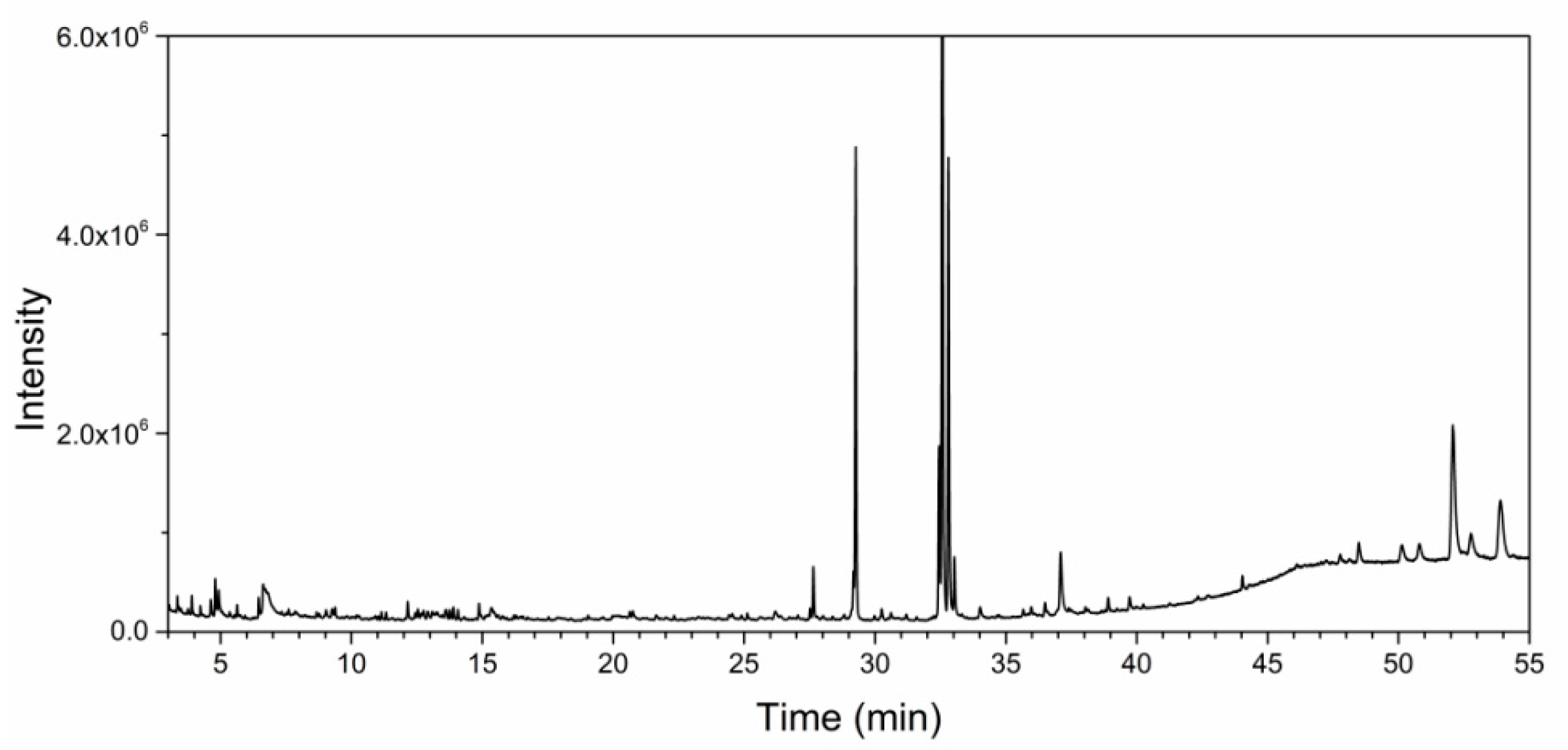
3.2. GC–MS Analysis of Hibiscus cannabinus Leaf Extracts

The GC–MS analysis of Hibiscus cannabinus leaves extract was performed, and the results were depicted in Figure 3. The chemical composition of the extract plays a crucial role in the green synthesis of nanoparticles, as various components can act as stabilizing, functional, and capping agents [35]. As a result, about thirteen phytochemicals were identified in Hibiscus cannabinus leaf aqueous extracts, as presented in Table 1. Among them, methyl linolenate (33.3%), methyl hexadecanoate (13.1%), phytol (14.6%), and β-sitosterol (11.5%) were main components in the aqueous extract; they have been reported to exhibit diverse biological properties [36,37,38]. These phytochemicals were the active molecules in the formation of the ZnO nanoparticles [39]. They were also found to exist in the characterizations.

3.3. Impact of Different Parameters on the Adsorption of the CR Dye

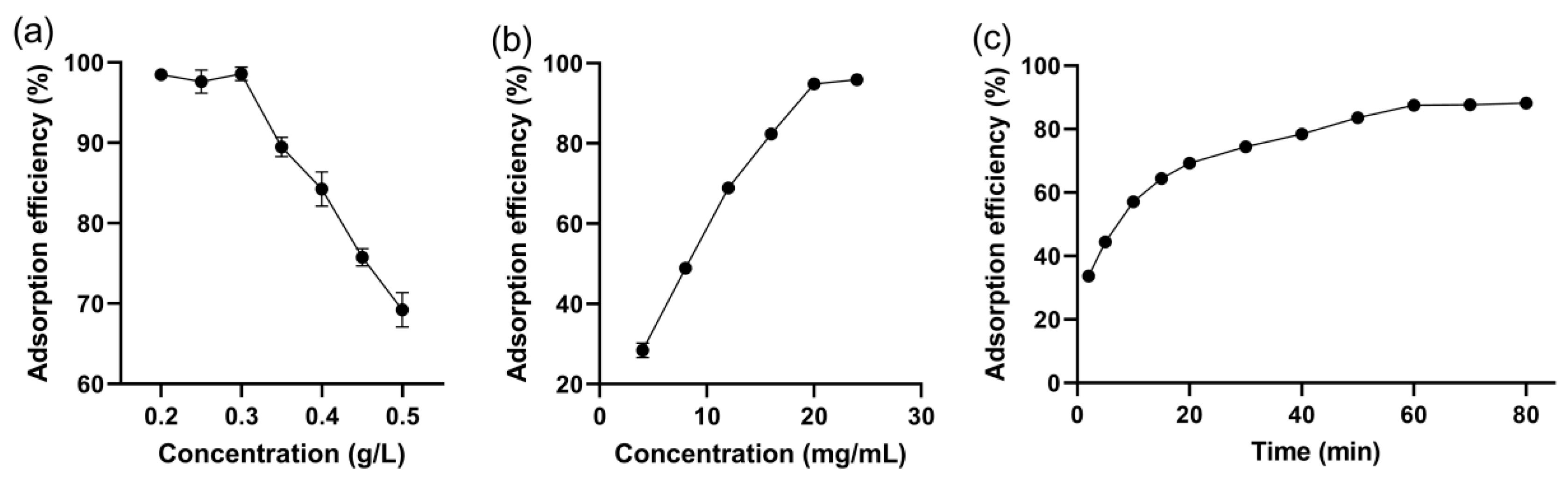
3.3.1. Effect of the Initial Dye Concentration

Figure 4a illustrates the adsorption efficiency of various dye concentrations (ranging from 0.2 to 0.5 g/L). It can be observed that, as the initial concentration of the dye solution increases, the adsorption efficiency of CR gradually diminishes. However, when the initial concentration of CR is lower than 0.3 g/L, the adsorption efficiency is maintained at the highest level. As the concentration of CR continually increases, the limited adsorption sites eventually reach saturation [40].

3.3.2. Effect of Adsorbent Dosage

The impact of the adsorbent dosage on the CR adsorption efficiency was investigated by varying the ZnO nanoparticle dosages from 4 mg/mL to 24 mg/mL, as depicted in Figure 4b. It was observed that, as the adsorbent dosage increased, the adsorption efficiency of CR also increased, and it eventually reached a plateau when the adsorbent dosage exceeded 20 mg/mL. This is mainly because more active sites for CR are available with the increase in the adsorbent dosage [23]. In this study, an adsorbent dosage of 20 mg/mL was selected as the optimum concentration.

3.3.3. Effect of Contact Time

The influence of time on the adsorption of CR by the ZnO nanoparticles was investigated, and the findings are presented in Figure 4c. It is evident that the adsorption efficiency increased and reached a plateau after 60 min. This suggests that the adsorption process needs time and, after 60 minutes, the vacant sites become saturated, resulting in an unchanged adsorption efficiency. Therefore, we recommend performing the adsorption of CR for a duration of 60 min.

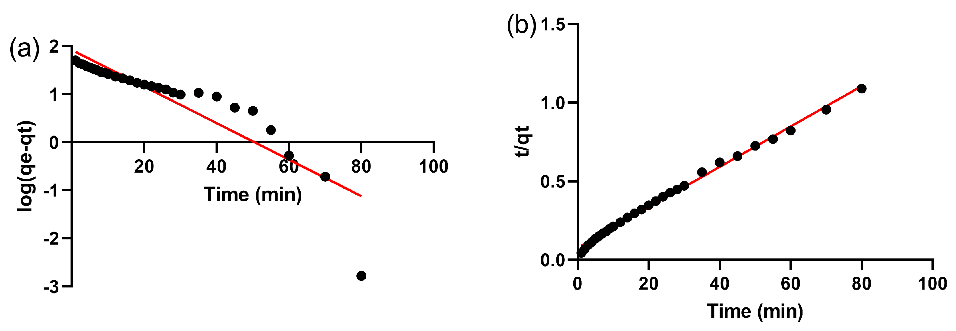
3.4. Adsorption Kinetics

The adsorption kinetics were analyzed using the pseudo-first-order and pseudo-second-order kinetic models. The pseudo-first-order model can be expressed as the following equation [41]:
log q e q t = log q e K 1 t
where qt is the adsorption capacity at a certain time, qe is the adsorption capacity in equilibrium, and K1 is the rate constant of the pseudo-first-order model. Moreover, the pseudo-second-order model can be expressed as the following equation:
t q t = 1 K 2 q e 2 + t q e
where K2 is the rate constant for the pseudo-second-order model. The adsorption data were fitted and the correlation coefficients of the two models were compared (Figure 5). When the correlation coefficient approaches 1, it indicates that the corresponding model is more appropriate for explaining the adsorption mechanism. In this study, the correlation coefficients of the pseudo-first-order and pseudo-second-order models were found to be 0.8051 and 0.9957, respectively. The pseudo-second-order model exhibited a higher correlation coefficient compared to the pseudo-first-order model. These results strongly suggest that the adsorption behavior of CR on the ZnO nanoparticles follows a chemical adsorption process [42]. A similar kinetic process was observed in the studies conducted by Arab and Marahel regarding ZnO nanoparticles [42,43]. The results suggest that the adsorption behavior of CR on ZnO nanoparticles is a chemical adsorption process.

3.5. Adsorption Isotherm

The isotherm study was conducted using the Langmuir and Freundlich isotherms. The linear equation for Langmuir and Freundlich can be expressed as [44]:
C e q e = C e q m + 1 K l q m
log q e = log K f + 1 n log C e
where Ce is the concentration of the sample in equilibrium, qm is the maximum adsorption capacity, Kl is the Langmuir coefficient, Kf is the Freundlich coefficient, and 1/n is the adsorption intensity. The adsorption data were fitted and the correlation coefficients of the two models were compared, as illustrated in Figure 6. The obtained correlation coefficients for the Langmuir and Freundlich isotherms were 0.9929 and 0.8642, respectively. It is worth noting that the correlation coefficient for the Langmuir model was significantly higher, indicating that the adsorption of CR on ZnO nanoparticles follows a monolayer sorption mechanism with a uniform distribution of active sites on the surface [45].

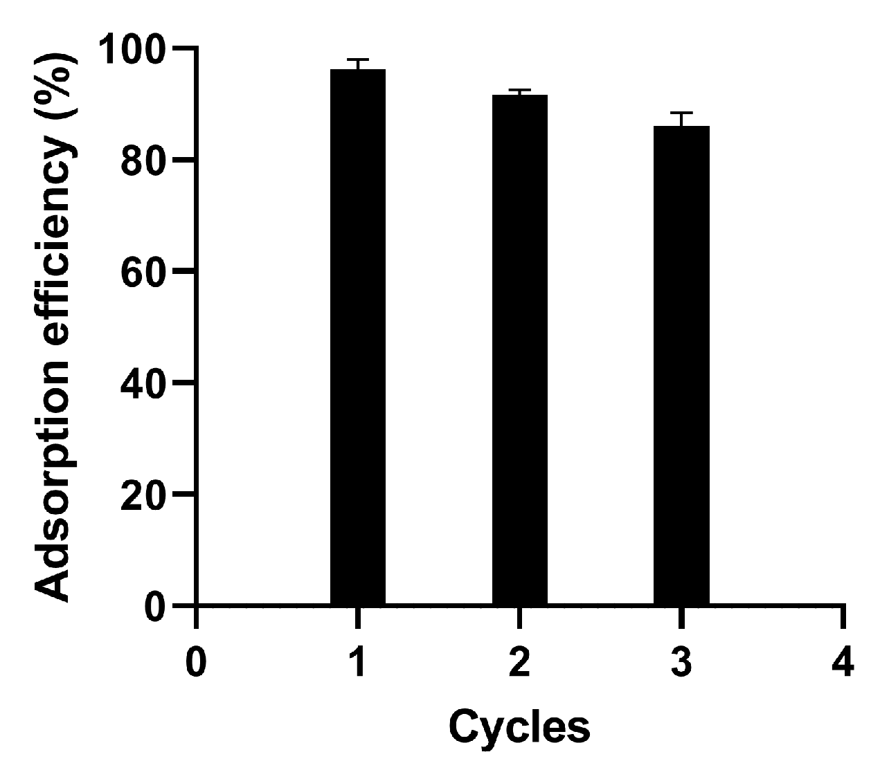
3.6. Reusability Study

After the adsorption of CR, the ZnO nanoparticles were refreshed with ethanol and water. Subsequently, the materials were subjected to a new round of CR adsorption to evaluate their reusability. As shown in Figure 7, the adsorption efficiency gradually decreases over multiple cycles, ranging from 96.2% to 86.1%. This decline in the adsorption efficiency can be attributed to material loss during repeated centrifugation and dispersion, as well as the incomplete desorption of the dye molecules [23]. Nevertheless, the results indicate that the prepared ZnO nanoparticles can be reused for a minimum of three cycles for the effective adsorption and removal of CR.

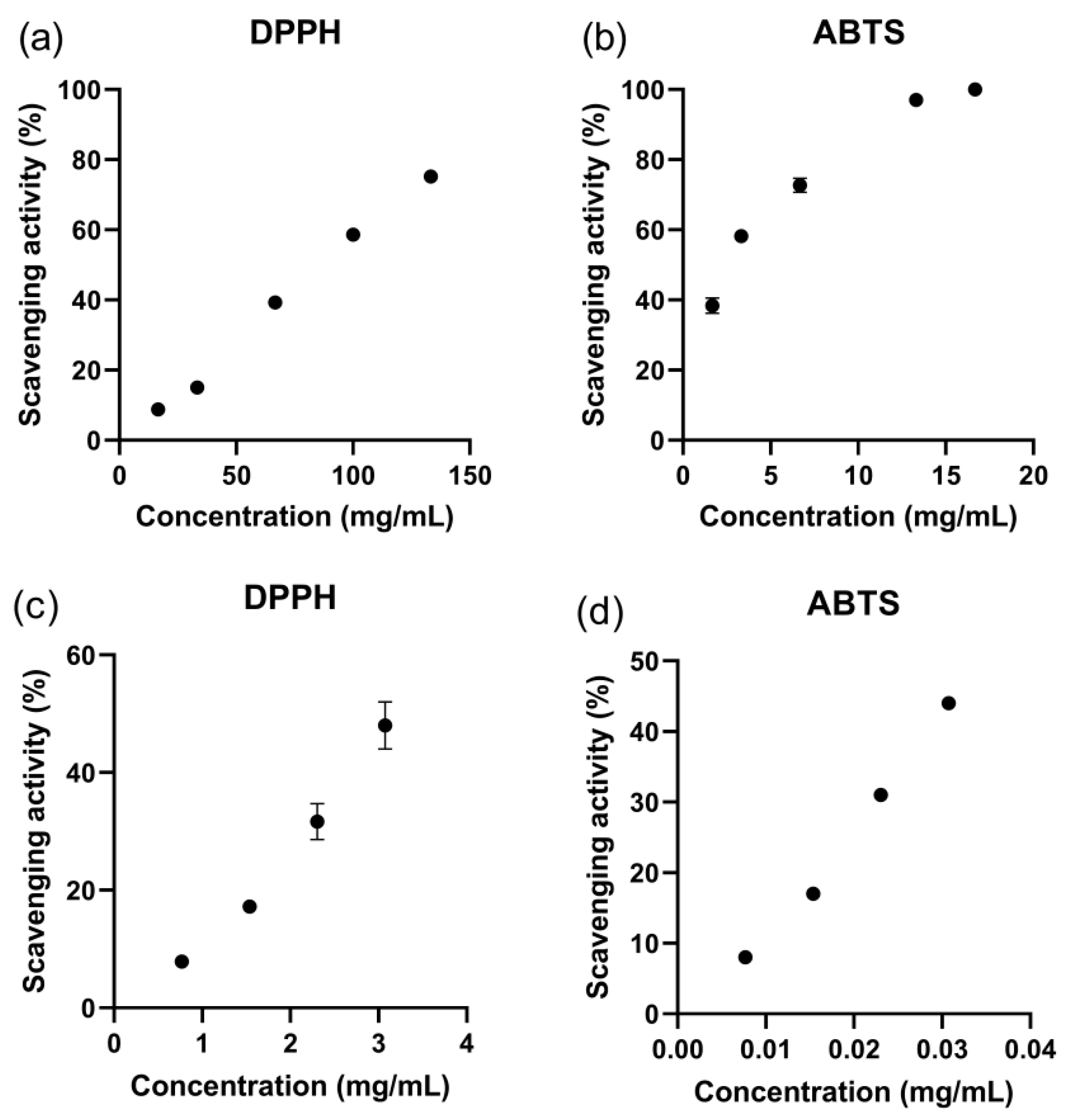
3.7. Antioxidant Activity of ZnO Nanoparticles

Oxidative stress is a kind of chemical reaction that occurs in the body and can lead to the formation of free radicals, which are unstable molecules that can attack lipids, proteins, and DNA, damaging their functions. Antioxidants can protect the body from oxidative stress by scavenging free radicals. ABTS was the utilized hydrogen atom transfer method, and DPPH was the electron transfer method. These were the two typical methods used in this study to evaluate antioxidant activities via colorimetric reactions [46]. DPPH is a relatively stable nitrogen-centered free radical that can accept an electron to become a stable diamagnetic molecule [47]. Meanwhile, ABTS can determine the total antioxidant activity of a hydrogen-donating ability [48]. Assessments of the in vitro antioxidant activities of ZnO nanoparticles and Hibiscus cannabinus leaf extracts were carried out to determine their pharmacological activities (Figure 8). Figure 8a,b display the antioxidant activity of the ZnO nanoparticles against DPPH and ABTS. Ascorbic acid was used as a standard in this process. The scavenging activity of the as-synthesized nanoparticles at different concentrations ranged from 8.8 to 75.2% and 38.4 to 100% against DPPH and ABTS, respectively. As a comparison, the scavenging activity of ascorbic acid at 0.25 mg/mL was 21.9% for DPPH and 52.7% for ABTS. Although the antioxidant activity of the ZnO nanoparticles was less than that of standard ascorbic acid, they still exhibited a dose-dependent trend and the values are in a promising range. Moreover, the ZnO nanoparticles showed a better scavenging ability for ABTS than for DPPH. The maximum rate of free-radical scavenging (97.0%) was reached when the concentration of the ZnO nanoparticles was 16.7 mg/mL. As shown in Figure 8c,d, the antioxidant activity of the Hibiscus cannabinus leaf extract was also exhibited. The extract showed good scavenging abilities, and the scavenging rate for ABTS was much better than that for DPPH. The same characteristics might indicate that the antioxidant activity of the ZnO nanoparticles was derived from the extract. Sharma reported the ZnO nanoparticles expressed an IC50 value of DPPH free-radical-scavenging activity of 34.22 ± 2.52 μg/mL [30]. As many reports show the particles’ antioxidant activity against DPPH, it should be noted that, in this study, the antioxidant activity against ABTS of ZnO nanoparticles prepared using Hibiscus cannabinus leaves was much better [49,50,51].

3.8. Antibacterial Activity of ZnO Nanoparticles

The disc diffusion test was used to study the antibacterial activity of the synthesized ZnO nanoparticles. The results showed that apparent inhibition zones were found with diameters around 27.0  mm, according to bacterial strains of E. coli and S. aureus (Figure 9a,b). The inhibition zones for the two bacterial strains of Hibiscus cannabinus leaf extracts were also tested. As Figure 9c–f show, the extract showed moderate inhibition. The inhibition diameters of the extract were 12.34  mm for E. coli and 9.74  mm for S. aureus. However, when the concentrations were reduced to 15 mg/mL, there was no inhibition, and the MIC values for both were 30 mg/mL. Based on these results, the growth curve of the two bacterial strains was also monitored by measuring the absorbance. As shown in Figure 10, the growth of the control group followed the typical S-shaped bacterial growth rule. With the increasing addition of ZnO nanoparticles into the bacteria, the growth of the bacteria was slowed down. When the concentration of the ZnO nanoparticles reached 0.45 mg/mL, the sample did not show significant growth. This indicates that the treatment with ZnO nanoparticles could delay the growth rate of the strain and even inhibit growth at 0.45 mg/mL, which can be confirmed as the minimal inhibitory concentration. This phenomenon is consistent with the inhibition effects on pathogenic bacteria of other prepared ZnO nanoparticles [30,52,53,54,55]. The well-proven antibacterial activities of ZnO nanoparticles ensure the material’s wider applications and give them more functions in environmental remediation.

4. Conclusions

This study presents a green synthesis approach for the preparation of ZnO nanoparticles, utilizing an extract derived from Hibiscus cannabinus leaves. The synthesized ZnO nanoparticles were characterized using UV–Vis, FTIR, XRD, TEM, and SEM techniques, which confirmed their morphology and structure in accordance with previously reported data. The prepared nanoparticles were effectively employed for the adsorption of Congo red in an aqueous solution. The factors influencing the adsorption efficiency were investigated and optimized. The adsorption kinetics were suitably described by the pseudo-second-order model, while the adsorption isotherm followed the Langmuir model. Notably, the as-synthesized materials also exhibited antioxidant and antibacterial properties. The DPPH scavenging activity was found to be moderate, whereas the ABTS scavenging activity was remarkable, displaying a 97.0% radical scavenging rate at a concentration of 16.7 mg/mL. The ZnO nanoparticles also demonstrated strong antibacterial activity against both E. coli and S. aureus. In conclusion, the environmentally friendly synthesis method employed in this study enabled the production of ZnO nanoparticles that possess excellent adsorption capabilities, as well as antioxidant and antibacterial activities, and which thus have significant potential for use in environmental remediation applications.

Supplementary Materials

The following supporting information can be downloaded at: https://www.mdpi.com/article/10.3390/separations10090466/s1, Figure S1: The size distribution of ZnO nanoparticles.

Author Contributions

Data curation, X.Y.; formal analysis, X.C. and C.C.; investigation, S.Y.; methodology, L.L.; project administration, S.H.; writing—original draft, X.Y.; writing—review and editing, S.H. All authors have read and agreed to the published version of the manuscript.

Funding

This work was supported by the Natural Science Foundation of Changsha, China (Grant No. kq2202323), the Agriculture Research System of China (CARS-19-E22), the Central Public-Interest Scientific Institution Basal Research Fund (1610242023005), the Hunan Provincial Major Science and Technology Project (2021NK1010-3), and the Chinese Agricultural Science and Technology Innovation Project (No. ASTIP-IBFC05).

Institutional Review Board Statement

Not applicable.

Informed Consent Statement

Not applicable.

Data Availability Statement

The data presented in this study are available on request from the corresponding author. The data are not publicly available due to the security requirements of the institute of the corresponding author.

Conflicts of Interest

The authors declare no conflict of interest.

References

  1. Majeed, K.; Ambreen, J.; Khan, S.A.; Muhammad, S.; Shah, A.A.; Bhatti, M.A.; Batool, S.S.; Farooq, M.; Shah Bukhari, S.N.; Chandio, A.D.; et al. Effective Removal of Methylene Blue by Mn3O4/NiO Nanocomposite under Visible Light. Separations 2023, 10, 200. [Google Scholar] [CrossRef]
  2. Iqbal, M.; Hanif, M.A.; Rashid, U.; Jilani, M.I.; Alharthi, F.A.; Kazerooni, E.A. Optimization and Kinetic Study of Treating Dye-Contaminated Wastewater Using Bio-Composite Synthesized from Natural Waste. Separations 2023, 10, 386. [Google Scholar] [CrossRef]
  3. Ahmad, A.; Kamaruddin, M.A.; Hps, A.K.; Yahya, E.B.; Muhammad, S.; Rizal, S.; Ahmad, M.I.; Surya, I.; Abdullah, C.K. Recent Advances in Nanocellulose Aerogels for Efficient Heavy Metal and Dye Removal. Gels 2023, 9, 416. [Google Scholar] [CrossRef]
  4. Xue, X.; Cheng, R.; Shi, L.; Ma, Z.; Zheng, X. Nanomaterials for Water Pollution Monitoring and Remediation. Environ. Chem. Lett. 2017, 15, 23–27. [Google Scholar] [CrossRef]
  5. Zelekew, O.A.; Haitosa, H.H.; Chen, X.; Wu, Y.-N. Recent Progress on Plant Extract-Mediated Biosynthesis of ZnO-Based Nanocatalysts for Environmental Remediation: Challenges and Future Outlooks. Adv. Colloid Interface Sci. 2023, 317, 102931. [Google Scholar] [CrossRef]
  6. Chauhan, A.K.; Kataria, N.; Garg, V.K. Green Fabrication of ZnO Nanoparticles Using Eucalyptus spp. Leaves Extract and Their Application in Wastewater Remediation. Chemosphere 2020, 247, 125803. [Google Scholar] [CrossRef]
  7. Alharthi, M.N.; Ismail, I.; Bellucci, S.; Salam, M.A. Green Synthesis of Zinc Oxide Nanoparticles by Ziziphus Jujuba Leaves Extract: Environmental Application, Kinetic and Thermodynamic Studies. J. Phys. Chem. Solids 2021, 158, 110237. [Google Scholar] [CrossRef]
  8. Batra, V.; Kaur, I.; Pathania, D.; Sonu; Chaudhary, V. Efficient Dye Degradation Strategies Using Green Synthesized ZnO-Based Nanoplatforms: A Review. Appl. Surf. Sci. Adv. 2022, 11, 100314. [Google Scholar] [CrossRef]
  9. Dangana, R.S.; George, R.C.; Shittu, U.O.; Agboola, F.K. Facile Biosynthesis, Characterisation and Biotechnological Application of ZnO Nanoparticles Mediated by Leaves of Cnidoscolus Aconitifolius. Artif. Cells Nanomed. Biotechnol. 2023, 51, 309–317. [Google Scholar] [CrossRef]
  10. Elshafie, H.S.; Osman, A.; El-Saber, M.M.; Camele, I.; Abbas, E. Antifungal Activity of Green and Chemically Synthesized ZnO Nanoparticles against Alternaria Citri, the Causal Agent Citrus Black Rot. Plant Pathol. J. 2023, 39, 265–274. [Google Scholar] [CrossRef]
  11. Rini, A.; Linda, T.; Hamzah, Y.; Umar, L.; Sari, M.; Rati, Y. Antibacterial Activity of Green Synthesized ZnO Nano-Flower Using Pineapple Peel Extract. Adv. Nat. Sci.-Nanosci. Nanotechnol. 2023, 14, 025008. [Google Scholar] [CrossRef]
  12. Abid, S.; Munir, R.; Sayed, M.; Nadeem, R.; Muneer, A.; Zahid, M.; Yaseen, M.; Siddiqua, U.; Noreen, S. Synthesis of Zinc Oxide, Ferric, Cu Nano Particles by Almond Shells, Sugar Cane Bagasse, Eggshells, and Their Application as Catalyst for Dye Reactive Red 195 (RR 195) Removal. Catal. Surv. Asia 2023, 1–17. [Google Scholar] [CrossRef]
  13. Loh, X.; Ooi, Z.; Teoh, Y.; Shuit, S. Synthesis and Characterization of Zinc Oxide Nanoparticles Using Peppermint Tea (Mentha Piperita) Dregs Extract and Their Photocatalytic Performance. Environ. Prog. Sustain. Energy 2023, e14202. [Google Scholar] [CrossRef]
  14. Jalali, T.; Ghanavati, F.; Osfouri, S. Green Synthesis of ZnO Nanoparticles Using Marine Brown Algae (Cystoseira) Extract Comprising Sol-Gel, and Combustion Techniques Based on Dye-Sensitized Solar Cells Application. Int. J. Mod. Phys. B 2023, 2450178. [Google Scholar] [CrossRef]
  15. Mushtaq, W.; Ishtiaq, M.; Maqbool, M.; Mazhar, M.W.; Casini, R.; Abd-ElGawad, A.M.; Elansary, H.O. Green Synthesis of Zinc Oxide Nanoparticles Using Viscum Album Extracts: Unveiling Bioactive Compounds, Antibacterial Potential, and Antioxidant Activities. Plants 2023, 12, 2130. [Google Scholar] [CrossRef]
  16. Al-Mamun, M.; Rafii, M.Y.; Misran, A.B.; Berahim, Z.; Ahmad, Z.; Khan, M.M.H.; Oladosu, Y.; Arolu, F. Kenaf (Hibiscus cannabinus L.): A Promising Fiber Crop with Potential for Genetic Improvement Utilizing Both Conventional and Molecular Approaches. J. Nat. Fibers 2023, 20, 2145410. [Google Scholar] [CrossRef]
  17. Al-Mamun, M.; Rafii, M.Y.; Misran, A.B.; Berahim, Z.; Ahmad, Z.; Khan, M.M.H.; Oladosu, Y. Heterosis and Combining Ability Estimate on Yield and Yield-Related Traits in a Half Diallel Crosses of Kenaf (Hibiscus cannabinus L.) in Malaysia. J. Nat. Fibers 2023, 20, 2192541. [Google Scholar] [CrossRef]
  18. Ayadi, R.; Hanana, M.; Mzid, R.; Hamrouni, L.; Khouja, M.L.; Salhi Hanachi, A. Hibiscus cannabinus L.—Kenaf: A Review Paper. J. Nat. Fibers 2017, 14, 466–484. [Google Scholar] [CrossRef]
  19. Liu, H.; Ying, Q.; Li, C.; Norra, S.; Lichtfouse, E. Enhanced Removal of Antimony in Dyeing Wastewater by Mixing Fe3O4 with Manganese Sand Filter Material. Water Environ. Res. 2020, 92, 1208–1213. [Google Scholar] [CrossRef]
  20. Nho, S.W.; Cui, X.; Kweon, O.; Jin, J.; Chen, H.; Moon, M.S.; Kim, S.-J.; Cerniglia, C.E. Phylogenetically Diverse Bacteria Isolated from Tattoo Inks, an Azo Dye-Rich Environment, Decolorize a Wide Range of Azo Dyes. Ann. Microbiol. 2021, 71, 35. [Google Scholar] [CrossRef]
  21. Alshehrei, F. Role of Microorganisms in Biodegradation of Food Additive Azo Dyes: A Review. Afr. J. Biotechnol. 2020, 19, 799–805. [Google Scholar] [CrossRef]
  22. Sharmila, G.; Thirumarimurugan, M.; Muthukumaran, C. Green Synthesis of ZnO Nanoparticles Using Tecoma Castanifolia Leaf Extract: Characterization and Evaluation of Its Antioxidant, Bactericidal and Anticancer Activities. Microchem. J. 2019, 145, 578–587. [Google Scholar] [CrossRef]
  23. Zhang, M.; Chang, L.; Zhao, Y.; Yu, Z. Fabrication of Zinc Oxide/Polypyrrole Nanocomposites for Brilliant Green Removal from Aqueous Phase. Arab. J. Sci. Eng. 2019, 44, 111–121. [Google Scholar] [CrossRef]
  24. Hu, Y.; Liang, P.; Wang, Z.; Jiang, C.; Zeng, Q.; Shen, C.; Wu, Y.; Liu, L.; Yi, Y.; Zhu, H.; et al. Explore the Effect of the Structure-Activity Relationship and Dose-Effect Relationship on the Antioxidant Activity of Licorice Flavonoids. J. Mol. Struct. 2023, 1292, 136101. [Google Scholar] [CrossRef]
  25. Zhang, L.; Liu, L.; Xiao, A.; Huang, S.; Li, D. Screening and Analysis of Xanthine Oxidase Inhibitors in Jute Leaves and Their Protective Effects against Hydrogen Peroxide-Induced Oxidative Stress in Cells. Open Chem. 2020, 18, 1481–1494. [Google Scholar] [CrossRef]
  26. Bourhia, M.; Bouothmany, K.; Bakrim, H.; Hadrach, S.; Salamatullah, A.M.; Alzahrani, A.; Khalil Alyahya, H.; Albadr, N.A.; Gmouh, S.; Laglaoui, A.; et al. Chemical Profiling, Antioxidant, Antiproliferative, and Antibacterial Potentials of Chemically Characterized Extract of Citrullus colocynthis L. Seeds. Separations 2021, 8, 114. [Google Scholar] [CrossRef]
  27. Agarwal, H.; Nakara, A.; Menon, S.; Shanmugam, V. Eco-Friendly Synthesis of Zinc Oxide Nanoparticles Using Cinnamomum Tamala Leaf Extract and Its Promising Effect towards the Antibacterial Activity. J. Drug Deliv. Sci. Technol. 2019, 53, 101212. [Google Scholar] [CrossRef]
  28. Mallikarjunaswamy, C.; Lakshmi Ranganatha, V.; Ramu, R.; Udayabhanu; Nagaraju, G. Facile Microwave-Assisted Green Synthesis of ZnO Nanoparticles: Application to Photodegradation, Antibacterial and Antioxidant. J. Mater. Sci. Mater. Electron. 2020, 31, 1004–1021. [Google Scholar] [CrossRef]
  29. Abdelbaky, A.S.; Abd El-Mageed, T.A.; Babalghith, A.O.; Selim, S.; Mohamed, A.M.H.A. Green Synthesis and Characterization of ZnO Nanoparticles Using Pelargonium odoratissimum (L.) Aqueous Leaf Extract and Their Antioxidant, Antibacterial and Anti-Inflammatory Activities. Antioxidants 2022, 11, 1444. [Google Scholar] [CrossRef]
  30. Sharma, A.; Nagraik, R.; Venkidasamy, B.; Khan, A.; Dulta, K.; Kumar Chauhan, P.; Kumar, D.; Shin, D.-S. In Vitro Antidiabetic, Antioxidant, Antimicrobial, and Cytotoxic Activity of Murraya Koenigii Leaf Extract Intercedes ZnO Nanoparticles. Luminescence 2023, 38, 1139–1148. [Google Scholar] [CrossRef]
  31. Saad Algarni, T.; Abduh, N.A.Y.; Al Kahtani, A.; Aouissi, A. Photocatalytic Degradation of Some Dyes under Solar Light Irradiation Using ZnO Nanoparticles Synthesized from Rosmarinus Officinalis Extract. Green Chem. Lett. Rev. 2022, 15, 460–473. [Google Scholar] [CrossRef]
  32. Li, W.; You, Q.; Zhang, J.; Li, W.; Xu, H. Green Synthesis of Antibacterial LFL-ZnO Using L. Plantarum Fermentation Liquid Assisted by Ultrasound-Microwave. J. Alloys Compd. 2023, 947, 169697. [Google Scholar] [CrossRef]
  33. Agarwal, H.; Menon, S.; Shanmugam, V.K. Functionalization of Zinc Oxide Nanoparticles Using Mucuna Pruriens and Its Antibacterial Activity. Surf. Interfaces 2020, 19, 100521. [Google Scholar] [CrossRef]
  34. Fakhari, S.; Jamzad, M.; Kabiri Fard, H. Green Synthesis of Zinc Oxide Nanoparticles: A Comparison. Green Chem. Lett. Rev. 2019, 12, 19–24. [Google Scholar] [CrossRef]
  35. Hasan, I.M.A.; Tawfik, A.R.; Assaf, F.H. GC/MS Screening of Buckthorn Phytochemicals and Their Use to Synthesize ZnO Nanoparticles for Photocatalytic Degradation of Malachite Green Dye in Water. Water Sci. Technol. 2021, 85, 664–684. [Google Scholar] [CrossRef]
  36. Ko, G.-A.; Shrestha, S.; Kim Cho, S. Sageretia Thea Fruit Extracts Rich in Methyl Linoleate and Methyl Linolenate Downregulate Melanogenesis via the Akt/GSK3β Signaling Pathway. Nutr. Res. Pract. 2018, 12, 3–12. [Google Scholar] [CrossRef]
  37. Pejin, B.; Savic, A.; Sokovic, M.; Glamoclija, J.; Ciric, A.; Nikolic, M.; Radotic, K.; Mojovic, M. Further In Vitro Evaluation of Antiradical and Antimicrobial Activities of Phytol. Nat. Prod. Res. 2014, 28, 372–376. [Google Scholar] [CrossRef]
  38. Saeidnia, S.; Manayi, A.; Gohari, A.R.; Abdollahi, M. The Story of Beta-Sitosterol- A Review. Eur. J. Med. Plants 2014, 4, 590–609. [Google Scholar] [CrossRef]
  39. Vijayakumar, S.; Divya, M.; Vaseeharan, B.; Ranjan, S.; Kalaiselvi, V.; Dasgupta, N.; Chen, J.; Durán-Lara, E.F. Biogenic Preparation and Characterization of ZnO Nanoparticles from Natural Polysaccharide Azadirachta indica L. (Neem Gum) and Its Clinical Implications. J. Clust. Sci. 2021, 32, 983–993. [Google Scholar] [CrossRef]
  40. Nayak, A.; Sahoo, J.K.; Sahoo, S.K.; Sahu, D. Removal of Congo Red Dye from Aqueous Solution Using Zinc Oxide Nanoparticles Synthesised from Ocimum sanctum (Tulsi Leaf): A Green Approach. Int. J. Environ. Anal. Chem. 2022, 102, 7889–7910. [Google Scholar] [CrossRef]
  41. Ahmad, S.Z.N.; Salleh, W.N.W.; Yusof, N.; Mohd Yusop, M.Z.; Hamdan, R.; Awang, N.A.; Ismail, N.H.; Rosman, N.; Sazali, N.; Ismail, A.F. Pb(II) Removal and Its Adsorption from Aqueous Solution Using Zinc Oxide/Graphene Oxide Composite. Chem. Eng. Commun. 2021, 208, 646–660. [Google Scholar] [CrossRef]
  42. Arab, C.; El Kurdi, R.; Patra, D. Effect of PH on the Removal of Anionic and Cationic Dyes Using Zinc Curcumin Oxide Nanoparticles as Adsorbent. Mater. Chem. Phys. 2022, 277, 125504. [Google Scholar] [CrossRef]
  43. Marahel, F.; Ghaedi, M.; Ansari, A. Zinc Oxide Nanoparticles Loaded on Activated Carbon and Its Application for Adsorption Removal of Uric Acid. Synth. React. Inorg. Met.-Org. Nano-Met. Chem. 2015, 45, 1387–1395. [Google Scholar] [CrossRef]
  44. Eleryan, A.; Aigbe, U.O.; Ukhurebor, K.E.; Onyancha, R.B.; Hassaan, M.A.; Elkatory, M.R.; Ragab, S.; Osibote, O.A.; Kusuma, H.S.; El Nemr, A. Adsorption of Direct Blue 106 Dye Using Zinc Oxide Nanoparticles Prepared via Green Synthesis Technique. Environ. Sci. Pollut. Res. 2023, 30, 69666–69682. [Google Scholar] [CrossRef]
  45. Phan, D.-N.; Rebia, R.A.; Saito, Y.; Kharaghani, D.; Khatri, M.; Tanaka, T.; Lee, H.; Kim, I.-S. Zinc Oxide Nanoparticles Attached to Polyacrylonitrile Nanofibers with Hinokitiol as Gluing Agent for Synergistic Antibacterial Activities and Effective Dye Removal. J. Ind. Eng. Chem. 2020, 85, 258–268. [Google Scholar] [CrossRef]
  46. Zare, M.; Namratha, K.; Byrappa, K.; Surendra, D.M.; Yallappa, S.; Hungund, B. Surfactant Assisted Solvothermal Synthesis of ZnO Nanoparticles and Study of Their Antimicrobial and Antioxidant Properties. J. Mater. Sci. Technol. 2018, 34, 1035–1043. [Google Scholar] [CrossRef]
  47. Ansari, A.; Ali, A.; Khan, N.; Saad Umar, M.; Owais, M.; Shamsuzzaman. Synthesis of Steroidal Dihydropyrazole Derivatives Using Green ZnO NPs and Evaluation of Their Anticancer and Antioxidant Activity. Steroids 2022, 188, 109113. [Google Scholar] [CrossRef]
  48. Krishnan, B.R.; Ramesh, M.; Selvakumar, M.; Karthick, S.; Sasikumar, A.; Geerthi, D.V.; Senthilkumar, N. A Facile Green Approach of Cone-like ZnO NSs Synthesized Via Jatropha Gossypifolia Leaves Extract for Photocatalytic and Biological Activity. J. Inorg. Organomet. Polym. Mater. 2020, 30, 4441–4451. [Google Scholar] [CrossRef]
  49. Velsankar, K.; Venkatesan, A.; Muthumari, P.; Suganya, S.; Mohandoss, S.; Sudhahar, S. Green Inspired Synthesis of ZnO Nanoparticles and Its Characterizations with Biofilm, Antioxidant, Anti-Inflammatory, and Anti-Diabetic Activities. J. Mol. Struct. 2022, 1255, 132420. [Google Scholar] [CrossRef]
  50. Ganesan, V.; Hariram, M.; Vivekanandhan, S.; Muthuramkumar, S. Periconium sp. (Endophytic Fungi) Extract Mediated Sol-Gel Synthesis of ZnO Nanoparticles for Antimicrobial and Antioxidant Applications. Mater. Sci. Semicond. Process. 2020, 105, 104739. [Google Scholar] [CrossRef]
  51. Rahman, A.; Harunsani, M.H.; Tan, A.L.; Ahmad, N.; Khan, M.M. Antioxidant and Antibacterial Studies of Phytogenic Fabricated ZnO Using Aqueous Leaf Extract of Ziziphus mauritiana Lam. Chem. Pap. 2021, 75, 3295–3308. [Google Scholar] [CrossRef]
  52. Vieira, I.R.S.; da Silva, A.A.; da Silva, B.D.; Neto, L.T.; Tessaro, L.; Furtado, C.R.G.; de Sousa, A.M.F.; Carvalho, N.M.F.; Conte-Junior, C.A. Eco-Friendly Synthesis of ZnO Nanomaterial from Green Tea Extract: Photocatalytic, Antibacterial and Antioxidant Potential. Biomass Convers. Biorefin. 2023, 1–15. [Google Scholar] [CrossRef]
  53. Ali, A.H. Experimental Investigations on Effects of ZnO NPS and Annona Muricata Extract for in Vitro and in Vivo Antibacterial Activity. Mater. Today Proc. 2022, 57, 527–530. [Google Scholar] [CrossRef]
  54. Hameed, H.; Waheed, A.; Sharif, M.S.; Saleem, M.; Afreen, A.; Tariq, M.; Kamal, A.; Al-onazi, W.A.; Al Farraj, D.A.; Ahmad, S.; et al. Green Synthesis of Zinc Oxide (ZnO) Nanoparticles from Green Algae and Their Assessment in Various Biological Applications. Micromachines 2023, 14, 928. [Google Scholar] [CrossRef]
  55. Dhandapani, K.V.; Anbumani, D.; Gandhi, A.D.; Annamalai, P.; Muthuvenkatachalam, B.S.; Kavitha, P.; Ranganathan, B. Green Route for the Synthesis of Zinc Oxide Nanoparticles from Melia azedarach Leaf Extract and Evaluation of Their Antioxidant and Antibacterial Activities. Biocatal. Agric. Biotechnol. 2020, 24, 101517. [Google Scholar] [CrossRef]
Figure 1. Diagram of the ZnO nanoparticles synthesized using green methods with Hibiscus cannabinus leaf extracts.
Separations 10 00466 g001
Figure 2. (a) UV–Vis spectrum, (b) FTIR spectrum, (c) XRD pattern, (d) SEM image, (e) TEM image, and (f) EDAX spectra of ZnO nanoparticles.
Separations 10 00466 g002
Figure 3. GC–MS spectrum of the Hibiscus cannabinus leaf extract.
Separations 10 00466 g003
Figure 4. Effects of (a) the initial dye concentration, (b) the adsorbent dosage, and (c) the contact time on the adsorption efficiency of CR.
Separations 10 00466 g004
Figure 5. (a) Pseudo-first-order kinetics and (b) Pseudo-second-order kinetics for ZnO nanoparticles.
Separations 10 00466 g005
Figure 6. (a) Langmuir isotherm and (b) Freundlich isotherm for ZnO nanoparticles.
Separations 10 00466 g006
Figure 7. The reusability of the ZnO nanoparticles in the adsorption of CR.
Separations 10 00466 g007
Figure 8. Antioxidant activities of ZnO nanoparticles for (a) DPPH free radicals and (b) ABTS free radicals; antioxidant activities of the Hibiscus cannabinus leaf extract for (c) DPPH free radicals and (d) ABTS free radicals.
Separations 10 00466 g008
Figure 9. The inhibition zones of ZnO nanoparticles and Hibiscus cannabinus leaf extracts on (a) E. coli and (b) S. aureus; Hibiscus cannabinus leaf extract at 30 mg/mL on (c) E. coli and (d) S. aureus; and Hibiscus cannabinus leaf extract at 15 mg/mL on (e) E. coli and (f) S. aureus.
Separations 10 00466 g009
Figure 10. The growth curves of (a) E. coli and (b) S. aureus in the presence of different concentrations of ZnO nanoparticles.
Separations 10 00466 g010
Table 1. The phytochemicals identified in the Hibiscus cannabinus leaf extract.
Rt (min)MwMolecular FormulaMolecular Name
4.79584.0C4H4O2Butenolide
4.95488.1C4H8O2Acetaldol
6.79992.0C3H8O3Glycerin
12.156198.2C14H30Tetradecane
27.660268.3C18H36OHexahydrofarnesyl acetone
29.289270.3C17H34O2Methyl hexadecanoate
32.438294.3C19H34O2Methyl linoleate
32.597292.2C19H32O2Methyl linolenate
32.820296.3C20H40OPhytol
33.068438.8C29H58O2Methyl montanate
37.121281.3C18H35NOOleamide
52.122414.4C29H50Oβ-Sitosterol
53.961426.4C30H50OAmyrin
Disclaimer/Publisher’s Note: The statements, opinions and data contained in all publications are solely those of the individual author(s) and contributor(s) and not of MDPI and/or the editor(s). MDPI and/or the editor(s) disclaim responsibility for any injury to people or property resulting from any ideas, methods, instructions or products referred to in the content.

