Next Issue
Volume 9, December
Previous Issue
Volume 9, June


Dermatopathology, Volume 9, Issue 3 (September 2022) – 13 articles

Cover Story (view full-size image): 
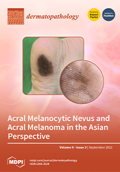
Melanocytic neoplasms in Asia are characterized by a predominance of acral melanocytic nevus and acral melanoma, which occur frequently on the sole and nails. Anatomic mapping shows that acral melanomas occur mostly on the weight-bearing portion of the sole, whereas acral melanocytic nevi on the arch area of the sole. Acral melanomas appear as large irregular black patches on the heel. Dermoscopy is a useful diagnostic tool for the differential diagnosis and reveals parallel ridge pattern on acral melanoma with a parallel furrow pattern on the acral melanocytic nevus. Histopathologic examination shows lentiginous proliferation of atypical melanocytes in the epidermal basal layer in the very early stages of acral lentiginous melanoma. Immunohistochemical staining such as HMB45 and Melan-A highlights prominent dendritic processes of melanoma cells in the epidermis. View this paper
  • Issues are regarded as officially published after their release is announced to the table of contents alert mailing list.
  • You may sign up for e-mail alerts to receive table of contents of newly released issues.
  • PDF is the official format for papers published in both, html and pdf forms. To view the papers in pdf format, click on the "PDF Full-text" link, and use the free Adobe Reader to open them.
Order results
Result details
Section
Select all
Export citation of selected articles as:
14 pages, 6255 KiB  
Article
Cutaneous Involvement of Extranodal NK/T Cell Lymphoma, Nasal Type, a Clinical and Histopathological Mimicker of Various Skin Diseases
by Preeyawat Ngamdamrongkiat, Sanya Sukpanichnant, Manasmon Chairatchaneeboon, Archrob Khuhapinant and Panitta Sitthinamsuwan
Dermatopathology 2022, 9(3), 307-320; https://doi.org/10.3390/dermatopathology9030037 - 09 Sep 2022
Cited by 1 | Viewed by 2299
Abstract
Background: Extranodal NK/T cell lymphoma, nasal type (ENK/T) with cutaneous involvement has various histopathological findings and diverse clinical manifestations. Methods: A retrospective study of cutaneous involvement of ENK/T lymphoma between 2006 and 2018 was conducted. Results: Twenty-two cases were eligible for this study. [...] Read more.
Background: Extranodal NK/T cell lymphoma, nasal type (ENK/T) with cutaneous involvement has various histopathological findings and diverse clinical manifestations. Methods: A retrospective study of cutaneous involvement of ENK/T lymphoma between 2006 and 2018 was conducted. Results: Twenty-two cases were eligible for this study. Twelve cases could be proven as secondary cutaneous involvement by ENK/T lymphoma, while the remaining could not be confirmed as primary cutaneous ENK/T lymphoma. The histopathological patterns included dermal and subcutaneous nodular infiltration pattern in 11/22 cases (50%), lobular panniculitis pattern in 6/22 cases (27.3%), interface dermatitis pattern in 4/22 cases (18.2%), and granulomatous dermatitis pattern in 1/22 case (4.5%). The median follow-up was 18.3 months. Overall, the one-year and five-year survival rates were 31.3% and 13.3%, respectively. Conclusions: A variety of histopathological patterns of cutaneous involvement by ENK/T lymphoma should be differentiated from other cutaneous lymphomas, dermatitis, and infection. When atypical medium or large-sized lymphoid cells are encountered within skin lesions, pathologists should realize these lesions can be ENK/T lymphoma, especially in cases with coexisting tumor necrosis or angioinvasion. A complete evaluation of the upper aerodigestive tract is mandatory to identify the occult primary site of ENK/T lymphoma before establishing primary cutaneous ENK/T lymphoma. Full article
(This article belongs to the Special Issue Dermatopathology in Asia)
Show Figures

Figure 1

3 pages, 381 KiB  
Obituary
In Memoriam—Martin C. Mihm, Jr.
by Thomas Kupper, Adriano Piris, Daniela Kroshinsky and Gürkan Kaya
Dermatopathology 2022, 9(3), 304-306; https://doi.org/10.3390/dermatopathology9030036 - 08 Sep 2022
Viewed by 1530
Abstract
Martin C [...] Full article
Show Figures

Figure 1

12 pages, 3794 KiB  
Review
Acral Melanocytic Neoplasms: A Comprehensive Review of Acral Nevus and Acral Melanoma in Asian Perspective
by Sanghyun Park and Sook-Jung Yun
Dermatopathology 2022, 9(3), 292-303; https://doi.org/10.3390/dermatopathology9030035 - 19 Aug 2022
Cited by 5 | Viewed by 7198
Abstract
Acral melanocytic neoplasms, including acral melanocytic nevus and acral melanoma, are common melanocytic lesions in Asian populations. Both lesions occur on the volar surface of the hands and feet, and on nail units. Acral melanocytic nevi occur on the arch area of the [...] Read more.
Acral melanocytic neoplasms, including acral melanocytic nevus and acral melanoma, are common melanocytic lesions in Asian populations. Both lesions occur on the volar surface of the hands and feet, and on nail units. Acral melanocytic nevi occur on the arch area of the sole, whereas acral melanomas frequently occur on weight-bearing areas of the sole, and on the fingernails. Therefore, the development of acral melanoma may be associated with chronic pressure, physical stress, or trauma. Dermoscopy is a useful adjunctive diagnostic tool for differential diagnosis. Acral melanocytic nevus is characterized by a parallel furrow pattern, whereas acral melanoma has a parallel ridge pattern. Genetic alterations are also different between the two types of lesion. BRAF and NRAS mutations are common in acral melanocytic nevus, whereas acral melanoma shows lower rates of KIT, NF1, BRAF, and NRAS mutations and remarkable copy number variations in genes such as CCND1, CDK4, hTERT, PAK1, and GAB2. Sentinel lymph node biopsy is important for staging and prognosis. Contemporary treatments for melanoma include targeted therapy for mutations and immunotherapy, such as anti-PD1 inhibitors. Full article
(This article belongs to the Special Issue Dermatopathology in Asia)
Show Figures

Figure 1

5 pages, 8766 KiB  
Case Report
A Case of IgG and IgA Anti-Laminin-332 Antibody-Positive Mucous Membrane Pemphigoid with IgG and IgA Anti-Envoplakin and Anti-Periplakin Antibodies
by Yoshiaki Matsushima, Masako Kitano, Daisuke Hayashi, Hiroyuki Goto, Mako Mine, Takeshi Yokoe, Makoto Kondo, Koji Habe, Yuji Toiyama, Takashi Hashimoto, Daisuke Tsuruta, Kazuhiko Takeuchi and Keiichi Yamanaka
Dermatopathology 2022, 9(3), 287-291; https://doi.org/10.3390/dermatopathology9030034 - 08 Aug 2022
Cited by 1 | Viewed by 2146
Abstract
A 76-year-old Japanese man presented with a 6-year history of a sore throat. He was treated at several clinics without any improvement before being referred to us. Physical examination revealed widespread erosions and ulcers from the palate to the larynx. Approximately 25 × [...] Read more.
A 76-year-old Japanese man presented with a 6-year history of a sore throat. He was treated at several clinics without any improvement before being referred to us. Physical examination revealed widespread erosions and ulcers from the palate to the larynx. Approximately 25 × 15 mm in size, erosive lesions were present on the retroauricular regions, forearms, and glans penis. Pseudomembranous conjunctivitis was also observed. The skin biopsy revealed a partial cleft formation below the epidermis, suggesting subepidermal bullous disease. Immuno-serological tests were negative for anti-desmoglein 1 (Dsg1), anti-Dsg3, anti-BP180, and anti-BP230 antibodies by ELISAs. A whole-body examination revealed gastric cancer. The possibility of mucous membrane pemphigoid (MMP) or paraneoplastic pemphigus (PNP) was considered. Indirect immunofluorescence using rat bladders showed positive IgG reactivity with cell surfaces on the transitional epithelia. Immunoblotting using recombinant proteins of laminin-332 showed both IgG and IgA reactivities with laminin-α3, and immunoblotting using normal human epidermal extract showed double-positive reactivities with envoplakin and periplakin for both IgG and IgA antibodies. Based on the clinical and histopathological features and results of various immuno-serological tests, our case was diagnosed as anti-laminin-332-type MMP with serological findings of PNP. Twenty days after laparoscopic gastrectomy, treatment with oral methylprednisolone 32 mg/day was initiated, and mucosal and skin lesions improved. Full article
Show Figures

Figure 1

5 pages, 7637 KiB  
Case Report
Blisters and Milia around the Peritoneal Dialysis Catheter: A Case of Localized Bullous Pemphigoid
by Andrea Michelerio and Carlo Tomasini
Dermatopathology 2022, 9(3), 282-286; https://doi.org/10.3390/dermatopathology9030033 - 04 Aug 2022
Cited by 1 | Viewed by 2756
Abstract
We report on the appearance of multiple tense blisters surrounding the exit site of a Tenckhoff catheter in a 79-year-old woman with end-stage renal disease in peritoneal dialysis. The differential diagnoses included a contact allergic or irritative dermatitis to peritoneal dialysis catheter material [...] Read more.
We report on the appearance of multiple tense blisters surrounding the exit site of a Tenckhoff catheter in a 79-year-old woman with end-stage renal disease in peritoneal dialysis. The differential diagnoses included a contact allergic or irritative dermatitis to peritoneal dialysis catheter material and antiseptic agents, bacterial infection, and herpes virus infection, but milia were a clue for a subepidermal blistering disease and lead to appropriate investigations. The laboratory findings, the histopathological examination and the direct immunofluorescence assay confirmed the diagnosis of localized bullous pemphigoid. The disorder typically occurs in elderly people and may be related to drugs, hematological malignancies or neurological conditions but it can also be a complication of hemodialysis or peritoneal dialysis. Full article
(This article belongs to the Special Issue In Memory of Raffaele Gianotti)
Show Figures

Figure 1

5 pages, 2358 KiB  
Case Report
Genital Folliculosebaceous Cystic Hamartoma: A Case Report and Concise Review of the Literature
by Maged Daruish and Mona Abdel-Halim Ibrahim
Dermatopathology 2022, 9(3), 277-281; https://doi.org/10.3390/dermatopathology9030032 - 01 Aug 2022
Cited by 2 | Viewed by 2203
Abstract
Folliculosebaceous cystic hamartoma (FSCH) is an uncommon hamartoma that usually presents on the central face area of adults as an asymptomatic, solitary dome-shaped or pedunculated papule. We report a case of a 35-year-old female who presented with six-months history of skin lesions on [...] Read more.
Folliculosebaceous cystic hamartoma (FSCH) is an uncommon hamartoma that usually presents on the central face area of adults as an asymptomatic, solitary dome-shaped or pedunculated papule. We report a case of a 35-year-old female who presented with six-months history of skin lesions on her labia majora. Histological findings included cystically dilated hair follicles with branching epithelial strands and interconnecting sebaceous gland consistent with the diagnosis of FSCH. The genital variant of FSCH was first described in 1998 and since then only six cases have been reported in the literature. We aim to increase awareness of this rare presentation due to the significant psychological implications and the risk of misdiagnosis. Full article
Show Figures

Figure 1

6 pages, 1943 KiB  
Case Report
Large Lobular Capillary Hemangioma Associated with Ingrown Toenail: Histopathological Features and Case Report
by Antonio Córdoba-Fernández, María Dolores Jiménez-Cristino and Victoria Eugenia Córdoba-Jiménez
Dermatopathology 2022, 9(3), 271-276; https://doi.org/10.3390/dermatopathology9030031 - 25 Jul 2022
Viewed by 3165
Abstract
Lobular capillary hemangioma (LCH-PG) is a type of pyogenic granuloma characterized by proliferating blood vessels that resemble conventional granulation tissue. Granulation tissue is very often seen in association with ingrown toenails. Despite the close relationship between both entities, LCH-PG shows clinically different behaviors, [...] Read more.
Lobular capillary hemangioma (LCH-PG) is a type of pyogenic granuloma characterized by proliferating blood vessels that resemble conventional granulation tissue. Granulation tissue is very often seen in association with ingrown toenails. Despite the close relationship between both entities, LCH-PG shows clinically different behaviors, such as rapid growth and frequent recurrence. Currently, it is unknown exactly how the different etiological factors contribute to the formation of differences between entities. We present a case of a large LCH-PG associated with chronic onychocryptosis in a 26-year-old man. Histopathological features included extensive signs of ulceration, hyperkeratosis, and patchy epidermal acanthosis with the presence of fibrous septa with lobular areas beneath the ulcerative area. The presence of stroma with a marked proliferation of blood vessels with wall thickening and mixed-type inflammatory changes was also characteristic. In advanced stages of onychocryptosis, as presented here, conventional granulation tissue or pyogenic granuloma can be clinically difficult to distinguish from other benign or malignant neoplasms. Histological examination is mandatory, and excisional biopsy can provide a definitive diagnosis. Full article
Show Figures

Figure 1

13 pages, 3519 KiB  
Case Report
Kaposi Sarcoma in Afghanistan: A Case Series from a Tertiary Referral Center
by Alyssa D. Higgins, Richard J. Dunn, Omer Malikzai, Mirwais Ahmadzai, Jerad M. Gardner, Benjamin K. Stoff and Josette R. McMichael
Dermatopathology 2022, 9(3), 258-270; https://doi.org/10.3390/dermatopathology9030030 - 15 Jul 2022
Viewed by 2765
Abstract
Kaposi sarcoma is a vascular endothelial neoplasm caused by human herpesvirus 8. Although it is a well-studied disease, little is known about the specific characteristics or epidemiology of Kaposi sarcoma in Afghanistan. The data consist primarily of anecdotal reports and epidemiological studies extrapolated [...] Read more.
Kaposi sarcoma is a vascular endothelial neoplasm caused by human herpesvirus 8. Although it is a well-studied disease, little is known about the specific characteristics or epidemiology of Kaposi sarcoma in Afghanistan. The data consist primarily of anecdotal reports and epidemiological studies extrapolated from neighboring countries. In this case series, we summarize existing data about Kaposi sarcoma in Afghanistan and present seven histologically confirmed cases with associated clinical features to shed light on the characteristics of Kaposi sarcoma in this unique geographic setting. Full article
Show Figures

Figure 1

7 pages, 2687 KiB  
Systematic Review
Histopathology of Hidradenitis Suppurativa: A Systematic Review
by Shane David Basil Smith, Ginette A. Okoye and Olayemi Sokumbi
Dermatopathology 2022, 9(3), 251-257; https://doi.org/10.3390/dermatopathology9030029 - 14 Jul 2022
Cited by 7 | Viewed by 4631
Abstract
Hidradenitis suppurativa (HS) is a chronic inflammatory scarring disease felt to be related to occlusion of the hair follicle unit in intertriginous areas. We perform a systematic review on HS histopathology to evaluate current knowledge and discuss future directions. PubMed and Scopus databases [...] Read more.
Hidradenitis suppurativa (HS) is a chronic inflammatory scarring disease felt to be related to occlusion of the hair follicle unit in intertriginous areas. We perform a systematic review on HS histopathology to evaluate current knowledge and discuss future directions. PubMed and Scopus databases were searched for relevant articles published from January 1985 to January 2021 that discussed the pathology of HS. Additional articles were identified by hand-searching, which entailed manually scanning selected journals. A total of 355 citations were identified in the primary search within the main databases. Two hundred and seventy-nine articles were excluded after a review of titles, abstracts, and duplicates. Sixty-one studies did not meet the inclusion criteria or were found to be duplicates, resulting in a total of 15 articles for analysis. Three articles were hand-searched. This comprehensive systematic review of the histopathology of HS confirms a high prevalence of follicular occlusion, follicular hyperkeratosis, and hyperplasia of the follicular epithelium. These findings support the central role of follicular occlusion in the development and progression of HS while providing a potential path to directing therapeutics against follicular occlusion. Full article
Show Figures

Figure 1

7 pages, 3068 KiB  
Case Report
Pityriasis Lichenoides et Varioliformis Acuta as a Complication of COVID-19 Infection
by Henriette De La Garza, Elie Saliba, Monica Rosales Santillan, Candice Brem and Neelam A. Vashi
Dermatopathology 2022, 9(3), 244-250; https://doi.org/10.3390/dermatopathology9030028 - 05 Jul 2022
Cited by 2 | Viewed by 7221
Abstract
Pityriasis lichenoides is an acute and/or chronic skin disease associated with recurrent erythematous papules that self-resolve. While its etiology is unknown, preceding viral infection may play a role. We present an atypical case of a 40-year-old woman with pityriasis lichenoides et varioliformis acuta [...] Read more.
Pityriasis lichenoides is an acute and/or chronic skin disease associated with recurrent erythematous papules that self-resolve. While its etiology is unknown, preceding viral infection may play a role. We present an atypical case of a 40-year-old woman with pityriasis lichenoides et varioliformis acuta as a complication of a COVID-19 infection. Full article
(This article belongs to the Special Issue Dermatopathology and COVID-19)
Show Figures

Figure 1

32 pages, 2487 KiB  
Review
The Impact of COVID-19 Pandemic on Dermatological Conditions: A Novel, Comprehensive Review
by Gehan A. Pendlebury, Peter Oro, William Haynes, Drew Merideth, Samantha Bartling and Michelle A. Bongiorno
Dermatopathology 2022, 9(3), 212-243; https://doi.org/10.3390/dermatopathology9030027 - 29 Jun 2022
Cited by 6 | Viewed by 8956
Abstract
Background: The earliest cases of Severe Acute Respiratory Syndrome Coronavirus 2 (SARS-CoV-2) emerged in Wuhan, China, in December 2019. Since the declaration as a pandemic on 11 March 2020, further dermatological conditions continue to be documented. We herein present a novel literature review [...] Read more.
Background: The earliest cases of Severe Acute Respiratory Syndrome Coronavirus 2 (SARS-CoV-2) emerged in Wuhan, China, in December 2019. Since the declaration as a pandemic on 11 March 2020, further dermatological conditions continue to be documented. We herein present a novel literature review of dermatological manifestations associated with the Coronavirus Disease 2019 (COVID-19) pandemic. To date, this literature review is the first broad-spectrum examination that analyzes a range of dermatological manifestations related to the COVID-19 pandemic: infection, vaccinations, personal protective equipment (PPE), and psychosocial factors. Methods: A detailed literature search was conducted using key terms for cutaneous manifestations associated with the scope of this review. The search retrieved 2199 articles. Results: The COVID-19 pandemic has triggered a significant range of dermatologic sequela. Etiologies of lesions continue to be investigated. Proposed mechanisms include inflammatory response to spike protein, vitamin D deficiency, ACE2 receptor activation, androgen levels, and increased psychological stress. One prominent mechanism describes viral spike protein invasion into the dermis by binding to the angiotensin-converting enzyme 2 (ACE-2) receptors in keratinocytes, with a secondary immunological response. Conclusions: Dermatologists play an integral role in the proper diagnosis and treatment of COVID-related lesions. Early treatment regimens and timely prophylaxis have been shown to safely reduce infection-related dermatological sequelae. Additional investigations and data collection can reduce disease burden and improve overall prognosis. Full article
(This article belongs to the Special Issue Dermatopathology and COVID-19)
Show Figures

Figure 1

5 pages, 3156 KiB  
Case Report
Cocaine/Levamisole-Induced, Skin-Limited ANCA-Associated Vasculitis with Pyoderma Gangrenosum-like Presentation
by Mirjana Urosevic-Maiwald, Jan-Hendrik B. Hardenberg, Jürg Hafner and Marie-Charlotte Brüggen
Dermatopathology 2022, 9(3), 207-211; https://doi.org/10.3390/dermatopathology9030026 - 29 Jun 2022
Viewed by 2687
Abstract
The use of levamisole as the most frequent adulterant of cocaine has merged in previously unknown toxicities, notably a disease entity called cocaine/levamisole-associated autoimmune syndrome (CLAAS). Clinically, CLAAS can manifest with diverse cutaneous and extracutaneous features sharing common laboratory findings (neutropenia, autoantibody patterns). [...] Read more.
The use of levamisole as the most frequent adulterant of cocaine has merged in previously unknown toxicities, notably a disease entity called cocaine/levamisole-associated autoimmune syndrome (CLAAS). Clinically, CLAAS can manifest with diverse cutaneous and extracutaneous features sharing common laboratory findings (neutropenia, autoantibody patterns). We report the case of a cocaine-abusing female patient with relapsing episodes of painful ulcers, worsening and expanding over a three-year period. The case exhibited all features of a drug-induced, skin-limited, ANCA-associated vasculitis, evolving over time to PG-like findings. In both disease stages, the patient responded well to the cessation of cocaine exposure and systemic glucocorticosteroids. This case demonstrates the continuous nature of cutaneous CLAAS manifestations in a single patient. CLAAS has become a major public health issue in the at-risk group of cocaine users, and clinicians should be alert of this condition when treating cocaine users presenting with single or multiple skin ulcerations. Full article
(This article belongs to the Section Clinico-Pathological Correlation in Dermatopathology)
Show Figures

Figure 1

4 pages, 810 KiB  
Case Report
Pseudo-Pemphigoid Gestationis Eruption Following SARS-CoV-2 Vaccination with mRNA Vaccine
by Caroline de Lorenzi, Gürkan Kaya and Laurence Toutous Trellu
Dermatopathology 2022, 9(3), 203-206; https://doi.org/10.3390/dermatopathology9030025 - 24 Jun 2022
Cited by 1 | Viewed by 2619
Abstract
Auto-immune reactions, including auto-immune bullous disease, have been reported following SARS-CoV-2 virus vaccination. Few cases of bullous pemphigoid are described, but there has been no case of pemphigoid gestationis. We report the first case here. Full article
(This article belongs to the Special Issue Dermatopathology and COVID-19)
Show Figures

Figure 1

Previous Issue
Next Issue
Back to TopTop