


Abstract
In laryngeal squamous cell carcinoma, hypoxia plays a major role in growth, spread, and treatment resistance. HIF-1α, VEGF, and GLUT1 are three linked biomarkers that show different aspects of this response. Low oxygen can be sensed by HIF-1α, and it follows up by activating genes that help with the survival of the tumour cells. VEGF helps to form new blood vessels, while GLUT1 increases the uptake of glucose and maintains the production of energy even when there is a lack of oxygen. Tissue and serum studies have reported higher levels of these biomarkers in malignant than in benign or normal samples, together with associations with adverse clinicopathological features. However, their diagnostic and prognostic utility remains exploratory because clinically validated thresholds, diagnostic performance measures, and independent prospective validation remain limited. Combined assessment of these biomarkers may provide complementary information on hypoxic signalling, angiogenesis and metabolic adaptation, although its diagnostic or prognostic superiority over individual biomarkers has not yet been established. However, serum measurements can be influenced by pre-analytical and systemic factors. So, in order for these biomarkers to advance enough to be used in clinical laryngology, large multicenter studies and a standardized set of testing methods will be necessary.
1. Introduction
Laryngeal diseases can range from benign vocal fold lesions such as nodules, polyps, and papillomas, to malignancies, most importantly invasive laryngeal squamous cell carcinoma (LSCC). LSCC is a key environment for hypoxia-related biomarkers, as it affects swallowing, voice, and airway protection mechanisms. As such, the chosen treatment must provide adequate oncological control while preserving laryngeal functions. Laryngeal cancer is one of the most important malignancies of the upper aerodigestive tract, with squamous cell carcinoma accounting for almost 95% of laryngeal tumours [1]. Current diagnostic procedures still rely mainly on symptoms, endoscopy, imaging, histopathology, and tumour, node, metastasis (TNM) staging. However, these methods cannot fully apprehend tumour biology or provide predictions as to which lesions could behave aggressively. This is why molecular biomarkers are being explored as possible tools to provide earlier detection, risk stratification, and better prognosis in LSCC [2]. Benign laryngeal lesions are important comparison groups as they help with differentiating cancer-related molecular changes from inflammation, trauma, or epithelial hyperplasia. It has also been reported in serum studies that high concentrations of hypoxia-inducible factor 1-alpha (HIF-1α/HIF1A), glucose transporter type 1 (GLUT1), and vascular endothelial growth factor (VEGF) may be suggestive of cancer when diagnosing patients with laryngeal lesions [3]. Hypoxia is important as it is not just insufficient oxygen but also a biological signal that influences the behaviour of tumours. In solid tumours, the lack of oxygen stabilizes HIF-1α, which is known to regulate genes that are involved in adaptation, angiogenesis, and metabolism [4]. HIF-1α has also been linked with GLUT1 and VEGF in LSCC, which connects hypoxia with the uptake of glucose, glycolytic metabolism, and angiogenesis [5]. Another study on laryngeal carcinoma concluded that HIF-1α may stimulate GLUT1 and VEGF expression, which can in turn support tumour angiogenesis, invasion, and metastasis [6]. Clinically, this matters as hypoxic head and neck cancers are often aggressive and show resistance to radiotherapy or chemotherapy [7]. In head and neck squamous cell carcinoma (HNSCC), hypoxia can be observed to increase radioresistance. This makes oxygen-related markers even more important not only in diagnosis, but also in predicting the response to treatment [8].
This review discusses serum HIF-1α, VEGF, and GLUT1 and their use as hypoxia-related biomarkers in laryngology, particularly in laryngeal squamous cell carcinoma. These three molecules were selected as they represent an interlinked aspect of the hypoxic tumour response. HIF-1α acts as a central transcription factor that is sensitive to oxygen. VEGF shows angiogenic signalling that is caused by hypoxia, while GLUT1 is an indicator of metabolic adaptation through increased glucose transport and glycolysis. It has been reported that serum and tissue concentrations of HIF-1α, VEGF, and GLUT1 have been higher in laryngeal carcinoma than in benign laryngeal lesions and controls. These findings support their potential usefulness during diagnosis [3]. Also discussed in this review are tissue-based studies that link HIF-1α and GLUT1 expression with tumour behaviour and prognosis [5]. Material suggesting that HIF-1α may regulate GLUT1 and VEGF so that it can promote angiogenesis, invasion, and metastasis [6] is also reviewed. Overall, this review combines biochemical mechanisms, methods of detection, and clinical relevance to determine whether these biomarkers can improve diagnosis and therapeutic stratification in laryngeal disease. A structured literature search was performed, with the study identification and selection process documented using a PRISMA flow diagram (Figure 1). A comprehensive search of the PubMed and PubMed Central (PMC) databases provided 1127 records. After the removal of duplicates (n = 33), non-medical or retracted publications (n = 47), and other reasons (n = 18), 1029 records were further screened. Following abstract screening, 388 reports were sought for full-text retrieval, out of which 367 were assessed for eligibility. After the exclusion of studies due to an unrelated cancer type (n = 46), lack of assessment of relevant biomarkers (n = 97), lack of relevant serum, tissue, or clinical outcome data (n = 66), and insufficient methodological information (n = 27), 131 studies were included in the final review.
Figure 1.
Preferred Reporting Items for Systematic Reviews and Meta-Analyses (PRISMA) flow diagram that represents the methods behind the selection of literature included in the review.
2. Materials and Methods
2.1. Literature Search Strategy
A structured literature search was performed in PubMed and PubMed Central (PMC) from database inception to 30 August 2026. The search was designed to identify studies investigating the HIF-1α–VEGF–GLUT1 hypoxia axis and its individual components in laryngeal carcinoma, with particular emphasis on laryngeal squamous cell carcinoma (LSCC). In PubMed, searches combined “laryngeal carcinoma” or “laryngeal squamous cell carcinoma” with terms relating to “hypoxia”, “HIF-1 alpha”, “VEGF”, and “GLUT1”. Additional targeted PubMed searches addressing serum biomarkers, prognostic significance, radiosensitivity, and circulating hypoxia biomarkers in head and neck cancer were performed during revision. These searches contributed 15 of the 131 publications included in the final review. In PMC, searches combined “laryngeal carcinoma” with “hypoxia”, “HIF-1 alpha”, “VEGF”, or “GLUT1”. Database-specific automatic term mapping expanded these queries to relevant indexed terms and synonyms. The complete database-generated search strategies and applied limits are provided in Supplementary Table S1.
2.2. Eligibility Criteria and Study Selection
Studies were considered eligible if they provided evidence relevant to the HIF-1α–VEGF–GLUT1 hypoxia axis in laryngeal carcinoma or contributed to its biological, clinical, prognostic, therapeutic, or methodological interpretation. Eligible publications included clinical and translational studies of laryngeal carcinoma or head and neck cancer. Experimental studies investigating hypoxia, HIF-1α, VEGF, GLUT1, angiogenesis, glucose metabolism, treatment response, or related signalling mechanisms were also included. Foundational mechanistic studies describing the regulation and function of these pathways were included when they provided information directly relevant to the biological interpretation of the HIF-1α–VEGF–GLUT1 axis. Studies addressing circulating or tissue biomarkers and their measurement, as well as relevant review articles, were also eligible.
Evidence from other tumour types or experimental systems was included when it provided mechanistic or methodological information directly relevant to interpretation of the HIF-1α–VEGF–GLUT1 axis.
Publications were excluded if they did not provide information relevant to hypoxia, HIF signalling, VEGF-mediated angiogenesis, GLUT1-associated metabolism, laryngeal or head and neck cancer biology, biomarker assessment, or the clinical interpretation of these pathways. Duplicate publications and records that did not provide sufficient methodological or outcome information to determine eligibility or interpret the reported findings were also excluded. Titles and abstracts of retrieved records were screened for relevance, followed by full-text assessment of potentially eligible publications according to these criteria.
3. Biochemical Basis of Hypoxia Signalling
3.1. Oxygen Sensing and HIF-1α Regulation
HIF-1 is an oxygen-responsive heterodimeric transcription factor composed of the oxygen-sensitive HIF-1α subunit and constitutively expressed HIF-1β/ARNT [9,10,11]. The oxygen-dependent degradation domain (ODD) of HIF-1α mediates its oxygen-dependent stability [12]. Under normoxic conditions, prolyl hydroxylase domain enzymes hydroxylate conserved proline residues within HIF-1α, enabling recognition by the von Hippel–Lindau tumour suppressor protein and subsequent ubiquitination and proteasomal degradation [13,14,15,16,17,18]. FIH-1 provides an additional oxygen-sensitive checkpoint by limiting co-activator recruitment [19,20]. During hypoxia, reduced hydroxylation allows HIF-1α to escape degradation, accumulate, and translocate to the nucleus, where it dimerises with HIF-1β/ARNT and activates hypoxia-response elements [21]. Among its downstream effects are increased VEGF-mediated angiogenesis and GLUT1-dependent glucose uptake, providing the mechanistic basis for considering these three molecules as complementary biomarkers of the hypoxic response, as seen in Figure 2. Importantly, these downstream markers should not be regarded as interchangeable measures of hypoxia, as HIF-1α reflects activation of oxygen-sensitive transcriptional signalling, whereas VEGF and GLUT1 represent distinct angiogenic and metabolic consequences of that response.
Figure 2.
The oxygen-dependent regulation of HIF-1α and activation of angiogenic and metabolic responses. Under normoxic conditions, PHD enzymes hydroxylate HIF-1α, enabling its recognition by VHL and subsequent ubiquitination and proteasomal degradation. FIH independently hydroxylates HIF-1α and limits co-activator recruitment, further suppressing HIF-1 transcriptional activity. During hypoxia, reduced PHD activity allows HIF-1α to escape degradation and accumulate, while reduced FIH activity permits co-activator recruitment. HIF-1α then translocates to the nucleus, dimerises with HIF-1β/ARNT, and binds hypoxia-response elements. This activates downstream genes including VEGF, promoting angiogenesis, and GLUT1, increasing glucose uptake and glycolytic metabolism. Created in BioRender. Roshan, D. (2026). https://BioRender.com/e626kdg, accessed on 12 September 2026.
3.2. Transcriptional Responses Caused by Hypoxia
Following hypoxic stabilization, HIF-1 activates a broader transcriptional program that coordinates angiogenic, metabolic, and survival responses [11,22]. Oxygen delivery can be improved through this transcriptional program as it activates angiogenic genes, particularly VEGFA, whose promoter is stimulated by HIF-1 during hypoxia [23,24]. VEGF then goes on to stimulate endothelial proliferation, vascular permeability, and the formation of new vessels, which help hypoxic tissues have an increased blood supply. HIF-1α can also modify metabolism in such a way that allows cells to generate ATP with less oxygen. It can increase the uptake of glucose by promoting GLUT1, or solute carrier family 2 member 1 (SLC2A1). Glycolytic flux can be further improved by HIF-1α through the activation of genes that encode glycolytic enzymes such as aldolase, phosphoglycerate kinase, and lactate dehydrogenase A [25,26]. HIF-1 is also known to stimulate pyruvate dehydrogenase kinase 1 (PDK1), which leads to the inhibition of pyruvate dehydrogenase. This leads to the reduced entry of pyruvate into the tricarboxylic acid (TCA) cycle, leading to the production of lactate [27,28]. Hypoxia-driven transcription further helps with survival and invasion. The expression of HIF-1-dependent BCL2/adenovirus E1B 19kDa interacting protein 3 (BNIP3) and NIX can stimulate mitophagy and autophagy. This allows for the removal of damaged mitochondria due to prolonged hypoxia [29,30]. When it comes to tumours, HIF-1α can be seen to support invasion as it can regulate the remodelling of the extracellular matrix and metastatic signalling genes such as the LOX-family enzymes and CXCR4 [31,32,33].
3.3. Hypoxia, Angiogenesis and Metabolic Reprogramming
The two biggest consequential effects of hypoxia are angiogenesis and metabolic reprogramming. When HIF-1α is in a stabilized state, it activates genes that are responsive to hypoxia, which help cells restore oxygen delivery while also maintaining the production of ATP in low-oxygen conditions. HIF-1 under hypoxia induces the transcription of a key angiogenic target, vascular endothelial growth factor A (VEGF-A) [23]. This forms a link between oxygen sensing and vascular growth as the HIF system combines angiogenic signalling with the local oxygen demand [34]. VEGF plays an important role in supporting endothelial cell proliferation, migration, and vessel permeability, while HIF-1 is seen as the stimulator of VEGF-driven angiogenesis in both physiological and pathological conditions [34,35]. A response like this in tumours could potentially support growth, invasion, and treatment resistance as it creates new and abnormal vasculature [36]. At the same time, the presence of hypoxia turns metabolism from oxidative phosphorylation that is dependent on oxygen, towards glycolysis and the uptake of glucose [26,37]. GLUT1, which is encoded by SLC2A1, is particularly important as it can increase the amount of glucose entering hypoxic cells. Experimental studies have shown that hypoxia upregulates the expression of GLUT1 and glucose transport through mechanisms that are dependent on HIF-1 [38,39,40]. The expression of several glycolytic targets is also promoted by HIF-1α, which supports the generation of ATP during times when oxygen is limited [41,42]. Thus, angiogenesis mediated by VEGF and glucose metabolism mediated by GLUT1 are two representatives of complementary biochemical outputs of hypoxia signalling.
4. The Molecular Functions and Biomarker Roles of HIF-1α, VEGF, and GLUT1
4.1. HIF-1α: The Oxygen-Sensitive Master Regulator
The oxygen-sensitive subunit of HIF-1 is HIF-1α, which acts as a molecular switch between oxygen availability and adaptive gene expression. Initially, HIF-1 was identified as a hypoxia-induced DNA-binding factor at the erythropoietin enhancer [9]. Structurally, HIF-1 belongs to the basic helix-loop-helix (bHLH)–PER-ARNT-SIM (PAS) family, with domains responsible for DNA binding, dimerisation, and transcriptional activation [43,44]. As described in Section 3.1, HIF-1α abundance is tightly controlled by oxygen-dependent hydroxylation and VHL-mediated degradation, whereas hypoxia permits its stabilization and transcriptional activation [16,45,46]. Once HIF-1α has been stabilized, it dimerizes with HIF-1β and binds hypoxia-response elements in the target genes. This particularly activates the VEGF angiogenic program, which connects the lack of oxygen with vascular growth [23,47]. Metabolic adaptation is also supported by HIF-1α, as it induces the genes involved in the transport of glucose and glycolysis. Some of these include GLUT1, aldolase, enolase, and lactate dehydrogenase A, which ultimately helps with the production of ATP even when there is a lack of oxidative phosphorylation [22,48].
The expression of tissue HIF-1α in laryngeal carcinoma has been related to GLUT1, VEGF, tumour progression, and nodal metastasis. This supports that it has a role as a hypoxia-related tissue biomarker [49,50]. However, serum HIF-1α must be discussed more carefully. HIF-1α is mainly an intracellular transcription factor, unlike VEGF, which is a classical secreted protein. Studies that have measured circulating HIF proteins in laryngeal neoplasms have suggested that their levels may carry possible diagnostic or prognostic significance [3,51]. However, serum positivity should be taken as an indirect representation of tumour hypoxia, cell turnover or systemic disease biology, rather than as a direct piece of evidence pointing to active nuclear HIF-1α signalling. This differentiation is important in the interpretation of biomarkers, as tissue staining shows cellular localization, whereas in serum measurement, there is a loss of spatial context. This means that it cannot identify which specific tumour compartment produced the signal. For these reasons, although serum HIF-1α carries potential, it still requires validation against factors like tissue expression, tumour stage, and treatment response.
4.2. VEGF: The Angiogenic Hypoxia Marker
One of the most important molecular links between hypoxia and angiogenesis is VEGF, more particularly, VEGF-A. At first, it was considered a vascular permeability factor that tumour cells secreted. It was later characterized as a secreted, endothelial-specific angiogenic mitogen [52,53]. The expression of VEGF increases significantly in hypoxic tissues. Early research had shown that VEGF, which was hypoxia-induced, could regulate hypoxia-initiated angiogenesis [54]. This would mean that VEGF is activated after HIF signalling, as HIF-1 binds to the VEGF gene and increases its transcription during hypoxia [23]. Endothelial cells are mainly targeted by VEGF through the VEGF receptors, especially the vascular endothelial growth factor receptor 2 (VEGFR-2) or kinase insert domain receptor (KDR). This will activate the signalling pathways that control endothelial proliferation, permeability, and tube formation [55,56]. These processes are crucial for the formation of microvessels, as endothelial cells are pushed to proliferate, move to where there are angiogenic signals, and survive in new, developing vascular structures. As VEGF also increases vascular permeability, it allows plasma proteins to leak into the extracellular matrix. This will support the formation of a temporary stromal scaffold for vessel growth [57]. In tumours, this process is often abnormal. VEGF-driven angiogenesis leads to the formation of tortuous, leaky, and poorly organized vasculature, rather than producing mature vessels that are properly organized. Such outcomes can further worsen hypoxia and promote angiogenic signalling [58,59]. When it comes to laryngeal squamous cell carcinoma, VEGF is useful not only as an angiogenic regulator, but also as a biomarker of tumour vascularization. Immunohistochemical studies have observed VEGF in laryngeal carcinoma cells, stromal tissue, macrophages, and also in vascular endothelial cells. Strong VEGF staining can be related to an increase in microvessel density in some groups of patients [60,61]. The expression of VEGF has been linked with an overall decrease in the survival of patients with early-stage laryngeal cancer who were treated with radiotherapy. This supports the value of VEGF in prognosis [62]. Serum VEGF is a good example of a potential circulating biomarker, as VEGF is secreted, meaning it can be detected in blood, unlike HIF-1α and other intracellular transcription factors. Studies have reported a higher concentration of serum VEGF in patients with head and neck squamous cell carcinoma in comparison to healthy samples, which opens possibilities as a clinical monitoring tool [63]. In particular, the patients who were affected by advanced laryngeal carcinoma showed a level of serum VEGF that was significantly higher than that of healthy samples. Also, when patients with pharyngeal and laryngeal squamous cell carcinoma were treated with radiotherapy, changes were observed in the levels of serum VEGF [64,65]. Serum VEGF should therefore be interpreted cautiously, as circulating concentrations may reflect both tumour-related and systemic factors rather than tumour hypoxia alone.
4.3. GLUT1: The Metabolic Hypoxia Marker
GLUT1 is a facilitative glucose transporter encoded by SLC2A1 and located predominantly in the plasma membrane, where it mediates basal glucose uptake through energy-independent facilitated diffusion [66,67]. When there is a shortage of oxygen or nutrients, GLUT1 can increase in number and membrane localization, making sure that cells receive glucose despite there being a decrease in mitochondrial ATP production [40]. During hypoxic conditions, HIF-1α stabilizes and binds hypoxia-responsive regulatory sequences within the GLUT1 promoter and increases the transcription of SLC2A1 [39]. HIF-1 also promotes glycolytic metabolism, increasing glucose utilization and reducing reliance on oxygen-dependent mitochondrial metabolism. Increased glycolysis generates lactate and protons, which require effective export mechanisms to help maintain intracellular pH [68]. An increased expression of GLUT1 in tumours allows for more glucose to enter malignant cells. Through glycolysis, the glucose that entered is metabolized to produce pyruvate and lactate. Even in low oxygen, this helps to maintain the production of ATP while also providing intermediates that are required to synthesize nucleotides, amino acids, and lipids. Thus, GLUT1 plays a role in contributing to the Warburg effect, in which cancer cells have a high uptake of glucose and an increased production of lactate even when oxygen is readily available [69]. Aerobic glycolysis is known to generate lower amounts of ATP per glucose molecule when compared with oxidative phosphorylation. However, its faster rate of generation of intermediates helps in tumour-cell proliferation, redox homeostasis, and survival within the ever-changing tumour microenvironment [70]. Thus, the expression of GLUT1 is widely considered not as a direct measurement of oxygen concentration, but as a marker of metabolic adaptation, as illustrated in Figure 3 and summarized in Table 1. The staining of GLUT1 in tumour tissues is seen mostly in areas with low oxygen levels and shows the ability of cancer cells to take up more glucose. In several malignancies, the expression of GLUT1 correlated with measured tumour hypoxia and clinical complications [71]. GLUT1 has been connected with the expression of HIF-1α, tumour invasiveness, more advanced diseases, and poor prognosis. These observations support the idea that GLUT1 can be a metabolic-hypoxia biomarker [5,50,72]. However, serum GLUT1 requires substantially more cautious interpretation than tissue GLUT1. Unlike VEGF, GLUT1 is an integral membrane protein rather than a conventionally secreted soluble factor. The main available laryngeal carcinoma study reported higher serum GLUT1 concentrations in patients with cancer than in controls, alongside increased tissue concentrations [3]. However, this evidence was derived from a relatively small cohort of 52 patients, and an independent association between serum GLUT1 and tumour stage, prognosis or treatment response has not yet been established. The biological form represented by a serum GLUT1 measurement also remains uncertain. Circulating GLUT1 may be associated with extracellular vesicles or membrane fragments, while haemolysis and contamination by erythrocytes or other blood-derived material may contribute to the measured signal because GLUT1 is abundant in erythrocyte membranes. GLUT1 has also been identified in extracellular vesicles [73], supporting the biological plausibility of vesicle-associated circulating GLUT1, but this should not be considered equivalent to freely soluble GLUT1. Therefore, serum GLUT1 should currently be regarded as an exploratory biomarker rather than a clinically validated surrogate of tumour GLUT1 expression. Future studies should distinguish soluble, extracellular-vesicle-associated, and blood-cell-derived GLUT1, control for haemolysis and cellular contamination, and correlate circulating measurements with matched tumour GLUT1 expression and clinical outcomes.
Figure 3.
HIF-1α, VEGF, and GLUT1 are represented as the three faces of hypoxia. HIF-1α represents oxygen-sensitive transcriptional regulation, VEGF represents angiogenic and vascular adaptation, and GLUT1 represents increased glucose uptake and glycolytic metabolism. Their measurements from tissue and circulation give different but related information regarding the hypoxic tumour microenvironment. Created in BioRender. Roshan, D. (2026). https://BioRender.com/qn6pvsx, accessed on 12 September 2026.
Table 1.
The molecular functions and biomarker roles of HIF-1α, VEGF, and GLUT1.
5. The Integrated HIF-1α–VEGF–GLUT1 Axis in Laryngeal Carcinoma
5.1. The Integrated HIF-1α–VEGF–GLUT1 Response to Hypoxia
As described above, HIF-1α is normally regulated through oxygen-dependent VHL-mediated proteasomal degradation, whereas tumour hypoxia permits its stabilization and activation [36,74,76]. In laryngeal carcinoma, downstream adaptive responses include VEGF-mediated angiogenesis and GLUT1-dependent metabolic reprogramming [77]. Laryngeal carcinoma models show this pathway very well. In human epithelial type 2 (HEp-2) cells, hypoxia increased HIF-1α protein alongside VEGF and GLUT1 expression [78]. This simultaneous increase demonstrates co-occurrence under hypoxic conditions but does not, by itself, establish a causal regulatory relationship between these proteins, as the study did not directly manipulate HIF-1α signalling or demonstrate downstream transcriptional dependence. Importantly, this co-occurring increase should not be interpreted as evidence of simultaneous or quantitatively equivalent regulation. HIF-1α acts upstream as the oxygen-sensitive transcriptional regulator, whereas VEGF and GLUT1 represent downstream transcriptional responses. HIF-1α stabilization therefore precedes the increased expression of these target proteins [21,23,39], although their subsequent kinetics and magnitude of induction may differ. Importantly, the available studies in laryngeal carcinoma have not provided directly comparable effect sizes that would allow numerical regulatory weights to be assigned to the HIF-1α–VEGF and HIF-1α–GLUT1 relationships. Their relative contributions may vary according to the severity and duration of hypoxia and additional tumour-specific signalling mechanisms. Positive associations between HIF-1α and VEGF [79] and also between HIF-1α, VEGF and GLUT1 [6] have also been found in other studies discussing laryngeal tumours. Likewise, an increased expression of HIF-1α and GLUT1 has been related to tumour invasiveness and prognosis [5]. Thus, VEGF and GLUT1 represent interconnected, rather than simply parallel, components of the hypoxic response. Their principal molecular connection is their shared upstream regulation by HIF-1α. However, additional cross-talk can occur at the level of the tumour vasculature. VEGF signalling can increase endothelial glucose metabolism, and experimental evidence has shown that VEGF can increase endothelial GLUT1 expression through the PI3K/Akt pathway, thereby coupling angiogenic stimulation with the increased glucose uptake required by proliferating endothelial cells [80]. This provides a functional link between VEGF-mediated angiogenesis and GLUT1-dependent metabolism. However, this VEGF–GLUT1 relationship has been demonstrated in endothelial models rather than specifically in laryngeal carcinoma and should therefore be regarded as mechanistically relevant cross-talk rather than an established tumour-cell-autonomous pathway in LSCC. VEGF has functions to stimulate the proliferation of endothelial cells and the formation of new vessels, helping the tumours receive more oxygen and nutrients [81]. However, oxygen deficiency may not be fully resolved as the tumour vessels are often malformed. A similar survival mechanism is provided by GLUT1, as it increases glucose uptake and prolongs glycolysis when there is a shortage of mitochondrial ATP production [82]. Proliferation and migration are reduced during hypoxic, low-glucose conditions in laryngeal carcinoma stem-like cells [83]. When HIF-1α and GLUT1 are inhibited together, tumour growth slows down, and radiosensitivity is improved in experimental laryngeal carcinoma [84,85]. Taken together, the mechanistic relationship between these molecules is hierarchical rather than equivalent. Hypoxia stabilizes HIF-1α, which transcriptionally promotes VEGF-mediated angiogenesis and GLUT1-mediated metabolic adaptation [21,23,39]. Therefore, VEGF and GLUT1 represent biologically distinct downstream outputs of HIF-1α signalling, while additional cross-talk between angiogenic and metabolic pathways may occur, particularly through VEGF-driven regulation of endothelial glucose metabolism. Their combined assessment may provide broader biological coverage of the hypoxic phenotype, but this mechanistic complementarity does not establish superior diagnostic or prognostic performance, as depicted in Figure 4.
Figure 4.
The integrated HIF-1α-VEGF-GLUT1 hypoxia-response axis in laryngeal carcinoma. Created in BioRender. Roshan, D. (2026). https://BioRender.com/hqzub6u, accessed on 12 September 2026.
5.2. Biological and Biomarker Relevance in Laryngeal Carcinoma
Laryngeal carcinoma can benefit from vascular and metabolic advantages provided by the HIF-1α–VEGF–GLUT1 axis as summarized in Table 2. HIF-1α supports the growth of tumours even in low-oxygen environments, as it promotes VEGF, which causes angiogenesis, and GLUT1, which increases the uptake of glucose. The expression of these molecules in laryngeal carcinoma tissues can be linked with angiogenesis, invasion, and metastasis [6]. The observation of HIF-1α and VEGF has also been correlated with advanced pathological tumour-node-metastasis (pTNM) stage and cervical lymph-node involvement [79]. The increased expression of VEGF and microvessel density has also been related to tumour invasion, metastasis, and recurrence [86]. Aggressive patterns of invasion can be observed with a high expression of GLUT1 and HIF-1α, while the epithelial to mesenchymal transition and the invasion of laryngeal cancer cells are stimulated by hypoxic conditions [87]. When there is an overexpression of HIF-1α and GLUT1 in tissues, often times it can be associated with recurrence, metastasis, and a reduction in survival [5]. A much shorter disease-free survival after surgery was seen in the HIF1A C1772T variant that is associated with high expression of tumour HIF-1α. Hypoxia has the ability to reduce the effectiveness of treatment, as the lack of oxygen reduces damage from radiation. However, HIF-1α signalling, abnormal vasculature due to VEGF, and GLUT1-supported glycolysis help cells to survive through the therapeutic stress. When the phosphoinositide 3-kinase/protein kinase B/mechanistic target of rapamycin (PI3K/Akt/mTOR) pathway was inhibited, HIF-1α and GLUT1 became suppressed, leading to better radiosensitivity in laryngeal carcinoma models [85]. In Hep-2 laryngeal carcinoma cells, suppression of both GLUT1 and phosphorylated Akt increased sensitivity to cisplatin, indicating that PI3K/Akt signalling contributes to chemoresistance alongside GLUT1 [88]. Measurements taken from tissues show a more accurate view of the local tumour biology, as they can provide expression within the malignant cells, invasive and hypoxic regions. Tissue sampling, however, is invasive and remains vulnerable to intratumoural spatial heterogeneity. Small biopsies interrogate only a limited tumour region, while even larger surgical specimens may not fully represent the entire lesion when biomarker assessment is performed on selected tissue blocks or regions. Serum assays, on the other hand, are less invasive and provide the opportunity for repeated monitoring. A laryngeal carcinoma study had found increased levels of HIF-1α, GLUT1 and VEGF in both tumour tissues and serum when compared with healthy samples [3]. However, circulating biomarker concentrations may also be influenced by pre-analytical and systemic factors and should therefore be interpreted alongside tissue findings [89,90]. Thus, serum measurements must be used together with tissue findings and have to be validated against the treatment response and recurrence.
Table 2.
The integrated roles of the HIF-1α–VEGF–GLUT1 axis in laryngeal carcinoma.
5.3. Cross-Cancer Relevance of the HIF-1α–VEGF–GLUT1 Hypoxia Axis
The biological relevance of HIF-1α–VEGF–GLUT1 signalling is not restricted to laryngeal carcinoma. Similar hypoxia-adaptive responses have been reported in several other solid tumours, in which HIF-1α-associated signalling can support VEGF-mediated angiogenesis together with GLUT1-dependent glucose uptake and glycolytic metabolism. These complementary responses can provide tumour cells with an adaptive advantage under oxygen and nutrient limitation by supporting vascularisation, metabolic survival, tumour progression and, in some settings, treatment resistance. Representative examples are summarized in Table 3. However, evidence from other tumour types should be regarded as supporting the biological plausibility of the pathway rather than demonstrating that its diagnostic or prognostic performance can be directly extrapolated to LSCC.
Table 3.
Representative evidence for the biological relevance of HIF-1α–VEGF–GLUT1 signalling in other cancers.
6. The Clinical Application of the HIF-1α–VEGF–GLUT1 Axis in Laryngeal Carcinoma
6.1. Diagnostic and Prognostic Relevance
6.1.1. Diagnostic Relevance
The diagnostic and prognostic utility of the HIF-1α–VEGF–GLUT1 axis in laryngeal carcinoma remains exploratory, although available studies have reported differences in biomarker expression between malignant and non-malignant tissues and associations with adverse clinical outcomes. In patients affected by laryngeal carcinoma, significantly higher concentrations of HIF-1α, VEGF, and GLUT1 were observed in serum and tumour tissues than in normal tissues or benign laryngeal lesions [3]. Importantly, benign laryngeal lesions represent a heterogeneous pathological group rather than a single biological entity. Some studies have examined individual non-malignant pathologies separately. For example, Wu et al. evaluated vocal cord polyps and vocal cord leukokeratosis independently and reported lower HIF-1α and GLUT1 expression than in laryngeal carcinoma [5]. However, comparable subtype-specific evidence remains limited, particularly for other benign lesions such as nodules and papillomas. Pooling different benign pathologies may therefore obscure subtype-specific differences in biomarker expression, and future diagnostic studies should stratify benign comparator groups according to histopathological diagnosis. A study comprising 120 LSCCs and 60 normal laryngeal tissues had similarly shown a higher expression of HIF-1α and VEGF in malignant samples [99]. When laryngeal tumours were compared with non-cancerous mucosa, the expression of HIF-1α and GLUT1 had been increased [72]. The expression of VEGF is much more frequently seen in LSCC than in the adjacent epithelium [100]. Measuring the biomarkers together may capture complementary hypoxic, angiogenic, and metabolic changes. However, superior diagnostic performance over individual biomarkers has not yet been established. However, sensitivity, specificity, clinically validated cut-off values, and independent prospective validation remain insufficient. These biomarkers should therefore currently be regarded as exploratory and, if assessed clinically, as complementary to rather than replacements for established endoscopic and histopathological evaluation.
6.1.2. Prognostic Significance
In terms of prognosis, increased expression of components of the HIF-1α–VEGF–GLUT1 axis has been associated with several adverse clinical outcomes in LSCC, including lymph-node involvement, recurrence, metastasis and reduced survival. HIF-1α overexpression has been associated with lymph-node classification, recurrence, and metastasis, while GLUT1 expression correlated with recurrence and metastasis and was reported as an independent indicator of survival [5]. Increased HIF-1α and VEGF expression has also been associated with advanced TNM stage, cervical lymph-node metastasis, and reduced five-year overall survival [99]. Similarly, high VEGF expression has been linked with advanced stage, metastasis, and poorer survival [100], while VEGF-related angiogenesis has shown prognostic relevance in additional studies [60,101]. VEGF and CD31 were reported as prognostic biomarkers in early-stage LSCC [102], and VEGF-C expression and lymphatic vessel density were associated with nodal metastasis and regional recurrence [103]. CD105-based microvessel density has also been associated with lymph-node involvement and reduced overall survival [104]. In addition, the HIF1A C1772T variant was associated with poorer survival and increased risk of relapse [91]. Collectively, these findings suggest that increased hypoxic, angiogenic, and metabolic activity may identify biologically aggressive LSCC with a greater risk of recurrence, metastasis, or poorer survival. However, the prognostic evidence is heterogeneous, and these biomarkers have not yet been sufficiently validated to provide prognostic information independent of established clinicopathological factors such as TNM stage and nodal status. Standardized assays, predefined cut-offs, and prospective multivariable validation are therefore required before they can be incorporated into routine prognostic assessment [105].
6.2. Treatment-Modality-Specific Response and Combined Biomarker Assessment
6.2.1. Radiotherapy Response
The effectiveness of radiotherapy is reduced significantly during hypoxia, as oxygen is needed to fix DNA damage caused by radiation. Importantly, HIF-1α, CA-IX and GLUT1 should not be regarded as equivalent indicators of the same oxygen tension, as their oxygen-response profiles differ. HIF-1 is a rapid oxygen-sensitive regulator whose DNA-binding activity and HIF-1α protein levels increase progressively as oxygen concentration decreases, with a half-maximal response reported at approximately 1.5–2% O2 and a maximal response at approximately 0.5% O2 [106]. CA-IX is a downstream hypoxia-responsive protein with a different oxygen dependency. In SiHa squamous carcinoma cells, maximal CA9 expression occurred at approximately 1% O2, and CA-IX protein followed the same expression pattern. GLUT1 also represents a downstream hypoxia-associated response but primarily reflects metabolic adaptation. In the same experimental model, maximal GLUT1 expression occurred at approximately 0.01% O2, substantially lower than the oxygen concentration producing maximal CA9 expression [107]. These differences were subsequently reproduced in both SiHa and FaDu pharyngeal squamous carcinoma cells, in which CA9 expression was maximal at 1% O2 whereas GLUT1 expression was maximal at 0.01% O2 [108]. These oxygen-response values are model-dependent rather than fixed clinical thresholds, but they demonstrate that HIF-1α, CA-IX and GLUT1 reflect overlapping rather than identical components of tumour hypoxia. Accordingly, these markers provide complementary rather than interchangeable information about radiobiologically relevant hypoxia.
The level of HIF-1α expression prior to treatment may be relevant to radiotherapy response. High expression of HIF-1α or CA-IX has been linked with residual tumour after radiotherapy [109]. Hypoxia-related proteins have also been reported as potentially relevant to local control, although methodological heterogeneity has limited their clinical implementation [110]. For GLUT1, experimental evidence is particularly strong. Suppression of GLUT1 increased radiation sensitivity and apoptosis in laryngeal carcinoma cells and xenografts [111], while inhibition of HIF-1α and GLUT1 reduced tumour growth, microvessel density and radioresistance [84]. Inhibition of PI3K/Akt/mTOR signalling similarly suppressed HIF-1α and GLUT1 and improved radiosensitivity in laryngeal carcinoma models [85].
Evidence for VEGF as a predictor of radiotherapy response is less consistent. VEGF expression did not differentiate between radiosensitive and radioresistant early laryngeal carcinoma models [112]. However, VEGF positivity was associated with poorer survival following primary radiotherapy [62], while serum VEGF concentrations were reported to decrease following irradiation [65]. Overall, HIF-1α and GLUT1 have stronger mechanistic evidence relating to radioresistance, whereas the predictive value of VEGF for radiotherapy response remains uncertain.
6.2.2. Chemotherapy Response
Chemotherapy resistance in laryngeal carcinoma is multifactorial and is not restricted to GLUT1-dependent metabolic adaptation. PI3K/Akt signalling has been implicated specifically in cisplatin resistance. In Hep-2 cells, suppression of GLUT1 and phosphorylated Akt increased cisplatin sensitivity [88], while direct inhibition of PI3K/Akt with LY294002 acted synergistically with cisplatin to suppress tumour-cell proliferation [113]. AMPK signalling may provide an additional metabolic mechanism of resistance. In LSCC models, MNAT1/GDF15 signalling reduced AMPK phosphorylation and mitochondrial apoptosis, whereas MNAT1 or GDF15 knockdown increased cisplatin sensitivity, including in cisplatin-resistant cells and in vivo [114]. The WISP1/YAP1/TEAD1/GLUT1 pathway represents another mechanism through which WISP1-driven GLUT1 upregulation supports glycolysis and cisplatin resistance [115]. Collectively, these findings indicate that chemoresistance in LSCC involves interacting PI3K/Akt, AMPK-regulated mitochondrial and GLUT1-dependent metabolic pathways rather than a single WISP1–GLUT1 mechanism.
6.2.3. Experimental Pathway-Targeted Approaches
The pathway-directed interventions described above should be distinguished from established targeted therapies in clinical LSCC. Experimental inhibition of HIF-1α, GLUT1 and PI3K/Akt/mTOR signalling has increased radiosensitivity in preclinical laryngeal carcinoma models [84,85], while inhibition of GLUT1- and PI3K/Akt-related signalling has increased cisplatin sensitivity [88,115]. The broader oncogenic relevance of this pathway is also supported by recent evidence in hepatocellular carcinoma, where LARS was shown to promote tumour progression through PI3K/AKT/mTOR signalling [116], as seen in Table 4. The laryngeal carcinoma studies therefore provide evidence for experimental treatment sensitisation rather than demonstrating a clinically validated response to targeted therapy.
Table 4.
Potential clinical applications of the HIF-1α–VEGF–GLUT1 axis in laryngeal carcinoma.
6.2.4. Combined Biomarker Assessment
The relevance of HIF-1α, VEGF and GLUT1 therefore differs according to treatment modality, as summarized in Table 4. HIF-1α and GLUT1 have been investigated predominantly in relation to radioresistance, while GLUT1 and interacting metabolic and pro-survival pathways have also been implicated in cisplatin resistance. Evidence concerning VEGF as a treatment-response marker is less consistent. Combined assessment of HIF-1α, VEGF and GLUT1 may capture complementary hypoxic, angiogenic and metabolic characteristics of the tumour. However, superior prediction of radiotherapy, chemotherapy or targeted-treatment response compared with individual biomarkers has not yet been established.
7. Analytical Challenges and Current Limitations
7.1. Sample Types and Detection Methods
Information provided by serum, plasma, and tissue about the HIF-1α–VEGF–GLUT1 axis is related but not identical to each other. Blood sampling is only minimally invasive, which makes serum or plasma a suitable candidate for larger clinical groups and repeated monitoring. Increased concentrations of HIF-1α, VEGF, and GLUT1 were seen in both serum and tumour tissue in laryngeal carcinoma patients [3]. However, circulating levels of these biomarkers may become influenced by systemic production and blood-cell release rather than just from the tumour alone. VEGF is particularly sensitive to the type of sample, as platelet activation during clotting can raise serum concentrations proportional to plasma [117,118]. Tissue specimens usually represent local tumour biology better and preserve cellular localization. However, sampling error is not limited to small biopsies; although biopsies interrogate only a restricted tumour region, larger surgical specimens may also incompletely represent spatially heterogeneous tumours when only selected blocks or regions are analyzed. Various detection methods can be used to examine different biological levels. Enzyme-linked immunosorbent assay (ELISA), for example, can give a quantitative measurement of soluble proteins, although its accuracy depends on certain factors like antibody specificity, calibration, and matrix effects [119]. Commercially available serum-compatible ELISA assays for these biomarkers show differing levels of documented analytical specificity. For HIF-1α, the Cloud-Clone human HIF-1α ELISA (SEA798Hu), which is validated for serum and plasma, is reported by the manufacturer to show no significant cross-reactivity or interference with HIF-1α analogues, although a quantitative cross-reactivity panel is not provided. For GLUT1, the Nori Human GLUT1/SLC2A1 ELISA (GR111153) reports < 0.5% cross-reactivity with available related molecules and no significant interference from the related molecules evaluated. More detailed quantitative data are available for some VEGF assays as well. For example, the Invitrogen Human VEGF ELISA (KHG0111) reports 0.25% and 0.11% cross-reactivity with mouse and rat VEGF165, respectively, while human VEGF121 shows 100% cross-reactivity with human VEGF165. These data demonstrate that analytical specificity is assay-dependent and that manufacturers differ considerably in the extent of cross-reactivity testing reported. Future serum studies should therefore report the manufacturer, catalogue number, isoform recognition, and cross-reactivity characteristics of the ELISA used. Immunohistochemistry gives a view of protein localization and staining intensity, but variability is a concern due to scoring and antibody validation [120]. Quantitative polymerase chain reaction (qPCR) can measure VEGFA and SLC2A1 transcripts, while Western blotting can reveal relative protein abundance and molecular size. Both of these methods have been previously applied in laryngeal carcinoma models and cell models [49,72]. Through multiplex assays, several biomarkers can be analyzed using small sample volumes. However, cross-reactivity and platform-specific differences call for the need for strong validation [121].
7.2. Sources of Variability and Gaps in Evidence
Pre-analytical conditions play a major role in the interpretation of HIF-1α, VEGF, and GLUT1 measurements. Many factors like delays prior to centrifugation, type of collection tube, storage temperature, and several freeze–thaw cycles can influence the measured concentration. This also reduces the comparability between different studies [122]. For VEGF, platelet release during clotting can increase serum concentrations, making sample type and standardized processing important when comparing circulating measurements [118,123]. Procedures like hemolysis can release intracellular proteins and cause assay interference, while a systemic infection or inflammation could increase the level of circulating VEGF with no relation to tumour activity [124]. As regional hypoxia, necrosis, vascularity and metabolism can vary within tumours, tissue-based biomarker measurements are vulnerable to spatial sampling error. This limitation is particularly important for small biopsies, but larger surgical specimens are also affected because biomarker expression may differ across tumour regions and routine analysis generally examines only selected sections or tissue blocks [104,125]. Multi-region sampling or standardized sampling of representative tumour areas may therefore provide a more complete assessment of intratumoural heterogeneity. An important unresolved issue is the cellular origin of circulating HIF-1α. Future validation studies should therefore use paired tumour and blood samples collected from the same patients and fractionate blood into cell-free, preferably platelet-poor plasma and cellular components. HIF-1α should be measured independently in tumour tissue, plasma, leukocyte, erythrocyte and platelet fractions, with haemolysis and residual platelet contamination documented as pre-analytical controls. Extracellular-vesicle-associated HIF-1α could additionally be assessed after enrichment for tumour-associated vesicle populations using appropriate epithelial or tumour markers. Longitudinal sampling before and after tumour resection or treatment would provide further evidence of origin, as a reduction in circulating HIF-1α paralleling tumour removal or response, without a corresponding change in blood-cell-associated HIF-1α, would support a tumour contribution. Such source-resolved studies are required before serum or plasma HIF-1α can be interpreted as a tumour-specific biomarker. The available literature on laryngeal carcinomas remains limited. The literature search was restricted to PubMed and PubMed Central, which may have resulted in the omission of relevant publications indexed exclusively in other bibliographic databases. This should be considered when interpreting the comprehensiveness of the evidence base. Some of the important studies have been conducted with only 52 patients [3] or 35 patients [6], which limits subgroup analyses and gives potentially unstable associations. Standardized diagnostic or prognostic cut-off values have not been established, with the differences in assays, antibodies, staining thresholds, and scoring procedures further hindering comparisons between different studies. The reporting recommendations for tumour marker prognostic studies (REMARK) guidelines put emphasis on having transparent methods, predefined analyses, and independent validation [126]. Thus, there is a need for larger studies that follow patients over time and confirm their results across various centres with consistent methods to fully consider these biomarkers for use in everyday clinical practice [127].
8. Future Perspectives and Conclusions
Future research should first validate HIF-1α, VEGF and GLUT1 as a biologically defined multi-marker panel reflecting hypoxic signalling, angiogenesis and metabolic adaptation. Other liquid-biopsy analytes, including circulating tumour DNA, circulating tumour cells and microRNAs, may provide complementary information on tumour burden and molecular alterations, but they should not be regarded as components of the HIF-1α–VEGF–GLUT1 axis in the absence of evidence demonstrating a direct mechanistic relationship with hypoxia signalling. Future studies could instead determine whether integrating these independent liquid-biopsy markers with hypoxia-related biomarkers provides additional diagnostic, prognostic or treatment-response information [128]. Extracellular vesicles may provide an additional platform for circulating biomarker assessment because they contain tumour-derived proteins, lipids and nucleic acids protected within membrane-bound particles. However, their isolation and analytical methods still require further standardization [129]. In particular, future studies should incorporate paired tumour–blood sampling, blood-cell fractionation and tumour-enriched extracellular-vesicle analysis to determine whether circulating HIF-1α originates predominantly from malignant tissue or from hematological and systemic sources. Rather than interpreting them separately, serum biomarkers should be integrated along with imaging. Studies in HNSCC have directly combined circulating hypoxia-related biomarkers with functional hypoxia imaging. Plasma biomarkers, including VEGF, have been assessed alongside [18F] FMISO PET/CT and [18F] HX4 PET/CT, although their correlations with imaging-defined hypoxia were variable [130,131]. These findings support further investigation of combined biomarker–imaging approaches, but their ability to guide biologically adapted radiotherapy remains unvalidated. Monitoring biomarkers and imaging before, during, and after the treatment can help doctors to tailor follow-ups and adjust treatment according to the patient’s needs. It can also help with identifying recurrence earlier, but these models will have to undergo rigorous multicenter testing and validation before they can be clinically implemented.
HIF-1α, VEGF and GLUT1 form a hypoxia-related axis that is biologically consistent in laryngeal carcinoma. HIF-1α represents oxygen sensing while VEGF represents angiogenic adaptation and GLUT1 represents metabolic adaptation. Together, these markers capture complementary aspects of hypoxia-related tumour biology, although their clinical superiority over individual biomarkers has not yet been demonstrated. Serum assessment of these biomarkers should therefore currently be regarded as exploratory rather than clinically validated for diagnosis, prognosis or monitoring. Standardized assays, predefined clinical cut-offs and large prospective multicentre studies comparing circulating measurements with matched tissue expression, imaging findings and clinical outcomes are required before routine clinical implementation.
Supplementary Materials
The following supporting information can be downloaded at: https://www.mdpi.com/article/10.3390/biomedicines14092103/s1, Table S1: Database-specific search strategies.
Author Contributions
Conceptualization, W.D., M.M., D.B.-A., D.R.J.R., A.M. and D.A.; methodology, W.D., M.M., D.B.-A., D.R.J.R., A.M. and D.A. software, W.D., M.M., D.B.-A., D.R.J.R., A.M. and D.A. validation, W.D., M.M., D.B.-A., D.R.J.R., A.M. and D.A. formal analysis, W.D., M.M., D.B.-A., D.R.J.R., A.M. and D.A. investigation, W.D., M.M., D.B.-A., D.R.J.R., A.M. and D.A. resources, D.B.-A., D.R.J.R. and D.A.; data curation, W.D., M.M., D.B.-A., D.R.J.R., A.M. and D.A. writing—original draft preparation, D.B.-A. and D.A.; writing—review and editing, D.B.-A., D.R.J.R. and D.A.; visualization, D.B.-A., D.R.J.R. and D.A.; supervision, D.A.; project administration, D.A.; funding acquisition, D.A. All authors have read and agreed to the published version of the manuscript.
Funding
This research received no external funding.
Institutional Review Board Statement
Not applicable.
Informed Consent Statement
Not applicable.
Data Availability Statement
No new data were created or analyzed in this study. Data sharing is not applicable to this article.
Conflicts of Interest
The authors declare no conflicts of interest.
References
- Verro, B.; Saraniti, C.; Carlisi, D.; Chiesa-Estomba, C.; Maniaci, A.; Lechien, J.R.; Mayo, M.; Fakhry, N.; Lauricella, M. Biomarkers in Laryngeal Squamous Cell Carcinoma: The Literature Review. Cancers 2023, 15, 5096. [Google Scholar] [CrossRef] [Scilit] [PubMed] [PubMed Central]
- Falco, M.; Tammaro, C.; Takeuchi, T.; Cossu, A.M.; Scafuro, G.; Zappavigna, S.; Itro, A.; Addeo, R.; Scrima, M.; Lombardi, A.; et al. Overview on Molecular Biomarkers for Laryngeal Cancer: Looking for New Answers to an Old Problem. Cancers 2022, 14, 1716. [Google Scholar] [CrossRef] [Scilit] [PubMed] [PubMed Central]
- Bronikowska, I.; Swietochowska, E.; Morawski, R.; Sowa, P.; Czecior, E. Concentration of hypoxia-inducible factor-1, glucose transporter 1 and vascular endothelial growth factor in tissue samples and serum in patients with primary laryngeal carcinoma. Acta Otolaryngol. 2022, 142, 532–536. [Google Scholar] [CrossRef] [Scilit] [PubMed]
- Qannita, R.A.; Alalami, A.I.; Harb, A.A.; Aleidi, S.M.; Taneera, J.; Abu-Gharbieh, E.; El-Huneidi, W.; Saleh, M.A.; Alzoubi, K.H.; Semreen, M.H.; et al. Targeting Hypoxia-Inducible Factor-1 (HIF-1) in Cancer: Emerging Therapeutic Strategies and Pathway Regulation. Pharmaceuticals 2024, 17, 195. [Google Scholar] [CrossRef] [Scilit] [PubMed] [PubMed Central]
- Wu, X.H.; Chen, S.P.; Mao, J.Y.; Ji, X.X.; Yao, H.T.; Zhou, S.H. Expression and significance of hypoxia-inducible factor-1α and glucose transporter-1 in laryngeal carcinoma. Oncol. Lett. 2013, 5, 261–266, Erratum in Oncol Lett. 2013, 6, 287. https://doi.org/10.3892/ol.2013.1360. [Google Scholar] [CrossRef] [Scilit] [PubMed] [PubMed Central]
- Xu, O.; Li, X.M.; Wang, M.X.; Qu, Y.T.; Lu, X.Y.; Shan, C.G.; Sun, Q.J. Expression of hypoxia inducible factor-1α and correlated target genes in human laryngeal carcinoma. Zhonghua Er Bi Yan Hou Tou Jing Wai Ke Za Zhi 2012, 47, 330–333. (In Chinese) [Google Scholar] [PubMed]
- Adachi, M.; Thomas, L. Molecular-targeted therapy hypoxia in head and neck squamous cell carcinoma patients. Mol. Clin. Oncol. 2013, 1, 12–14. [Google Scholar] [CrossRef] [Scilit] [PubMed] [PubMed Central]
- Li, J.Z.; Gao, W.; Chan, J.Y.; Ho, W.K.; Wong, T.S. Hypoxia in head and neck squamous cell carcinoma. ISRN Otolaryngol. 2012, 2012, 708974. [Google Scholar] [CrossRef] [Scilit] [PubMed] [PubMed Central]
- Semenza, G.L.; Wang, G.L. A nuclear factor induced by hypoxia via de novo protein synthesis binds to the human erythropoietin gene enhancer at a site required for transcriptional activation. Mol. Cell Biol. 1992, 12, 5447–5454. [Google Scholar] [CrossRef] [Scilit] [PubMed] [PubMed Central]
- Wang, G.L.; Semenza, G.L. Characterization of hypoxia-inducible factor 1 and regulation of DNA binding activity by hypoxia. J. Biol. Chem. 1993, 268, 21513–21518. [Google Scholar] [CrossRef] [Scilit] [PubMed]
- Wang, G.L.; Jiang, B.H.; Rue, E.A.; Semenza, G.L. Hypoxia-inducible factor 1 is a basic-helix-loop-helix-PAS heterodimer regulated by cellular O2 tension. Proc. Natl. Acad. Sci. USA 1995, 92, 5510–5514. [Google Scholar] [CrossRef] [Scilit] [PubMed] [PubMed Central]
- Huang, L.E.; Gu, J.; Schau, M.; Bunn, H.F. Regulation of hypoxia-inducible factor 1alpha is mediated by an O2-dependent degradation domain via the ubiquitin-proteasome pathway. Proc. Natl. Acad. Sci. USA 1998, 95, 7987–7992. [Google Scholar] [CrossRef] [Scilit] [PubMed] [PubMed Central]
- Bruick, R.K.; McKnight, S.L. A conserved family of prolyl-4-hydroxylases that modify HIF. Science 2001, 294, 1337–1340. [Google Scholar] [CrossRef] [Scilit] [PubMed]
- Epstein, A.C.; Gleadle, J.M.; McNeill, L.A.; Hewitson, K.S.; O’Rourke, J.; Mole, D.R.; Mukherji, M.; Metzen, E.; Wilson, M.I.; Dhanda, A.; et al. C. elegans EGL-9 and mammalian homologs define a family of dioxygenases that regulate HIF by prolyl hydroxylation. Cell 2001, 107, 43–54. [Google Scholar] [CrossRef] [Scilit] [PubMed]
- Berra, E.; Benizri, E.; Ginouvès, A.; Volmat, V.; Roux, D.; Pouysségur, J. HIF prolyl-hydroxylase 2 is the key oxygen sensor setting low steady-state levels of HIF-1alpha in normoxia. EMBO J. 2003, 22, 4082–4090. [Google Scholar] [CrossRef] [Scilit] [PubMed] [PubMed Central]
- Maxwell, P.H.; Wiesener, M.S.; Chang, G.W.; Clifford, S.C.; Vaux, E.C.; Cockman, M.E.; Wykoff, C.C.; Pugh, C.W.; Maher, E.R.; Ratcliffe, P.J. The tumour suppressor protein VHL targets hypoxia-inducible factors for oxygen-dependent proteolysis. Nature 1999, 399, 271–275. [Google Scholar] [CrossRef] [Scilit] [PubMed]
- Ivan, M.; Kondo, K.; Yang, H.; Kim, W.; Valiando, J.; Ohh, M.; Salic, A.; Asara, J.M.; Lane, W.S.; Kaelin, W.G., Jr. HIFalpha targeted for VHL-mediated destruction by proline hydroxylation: Implications for O2 sensing. Science 2001, 292, 464–468. [Google Scholar] [CrossRef] [Scilit] [PubMed]
- Jaakkola, P.; Mole, D.R.; Tian, Y.M.; Wilson, M.I.; Gielbert, J.; Gaskell, S.J.; von Kriegsheim, A.; Hebestreit, H.F.; Mukherji, M.; Schofield, C.J.; et al. Targeting of HIF-alpha to the von Hippel-Lindau ubiquitylation complex by O2-regulated prolyl hydroxylation. Science 2001, 292, 468–472. [Google Scholar] [CrossRef] [Scilit] [PubMed]
- Lando, D.; Peet, D.J.; Gorman, J.J.; Whelan, D.A.; Whitelaw, M.L.; Bruick, R.K. FIH-1 is an asparaginyl hydroxylase enzyme that regulates the transcriptional activity of hypoxia-inducible factor. Genes Dev. 2002, 16, 1466–1471. [Google Scholar] [CrossRef] [Scilit] [PubMed] [PubMed Central]
- McNeill, L.A.; Hewitson, K.S.; Claridge, T.D.; Seibel, J.F.; Horsfall, L.E.; Schofield, C.J. Hypoxia-inducible factor asparaginyl hydroxylase (FIH-1) catalyses hydroxylation at the beta-carbon of asparagine-803. Biochem J. 2002, 367, 571–575. [Google Scholar] [CrossRef] [Scilit] [PubMed] [PubMed Central]
- Gleadle, J.M.; Ratcliffe, P.J. Induction of hypoxia-inducible factor-1, erythropoietin, vascular endothelial growth factor, and glucose transporter-1 by hypoxia: Evidence against a regulatory role for Src kinase. Blood 1997, 89, 503–509. [Google Scholar] [CrossRef] [Scilit] [PubMed]
- Iyer, N.V.; Kotch, L.E.; Agani, F.; Leung, S.W.; Laughner, E.; Wenger, R.H.; Gassmann, M.; Gearhart, J.D.; Lawler, A.M.; Yu, A.Y.; et al. Cellular and developmental control of O2 homeostasis by hypoxia-inducible factor 1 alpha. Genes Dev. 1998, 12, 149–162. [Google Scholar] [CrossRef] [Scilit] [PubMed] [PubMed Central]
- Forsythe, J.A.; Jiang, B.H.; Iyer, N.V.; Agani, F.; Leung, S.W.; Koos, R.D.; Semenza, G.L. Activation of vascular endothelial growth factor gene transcription by hypoxia-inducible factor 1. Mol. Cell Biol. 1996, 16, 4604–4613. [Google Scholar] [CrossRef] [Scilit] [PubMed] [PubMed Central]
- Shi, Y.H.; Fang, W.G. Hypoxia-inducible factor-1 in tumour angiogenesis. World J. Gastroenterol. 2004, 10, 1082–1087. [Google Scholar] [CrossRef] [Scilit] [PubMed] [PubMed Central]
- Semenza, G.L.; Roth, P.H.; Fang, H.M.; Wang, G.L. Transcriptional regulation of genes encoding glycolytic enzymes by hypoxia-inducible factor 1. J. Biol. Chem. 1994, 269, 23757–23763. [Google Scholar] [CrossRef] [Scilit] [PubMed]
- Semenza, G.L. HIF-1: Upstream and downstream of cancer metabolism. Curr. Opin. Genet Dev. 2010, 20, 51–56. [Google Scholar] [CrossRef] [Scilit] [PubMed] [PubMed Central]
- Kim, J.W.; Tchernyshyov, I.; Semenza, G.L.; Dang, C.V. HIF-1-mediated expression of pyruvate dehydrogenase kinase: A metabolic switch required for cellular adaptation to hypoxia. Cell Metab. 2006, 3, 177–185. [Google Scholar] [CrossRef] [Scilit] [PubMed]
- Semenza, G.L. HIF-1 mediates metabolic responses to intratumoral hypoxia and oncogenic mutations. J. Clin. Investig. 2013, 123, 3664–3671. [Google Scholar] [CrossRef] [Scilit] [PubMed] [PubMed Central]
- Sowter, H.M.; Ratcliffe, P.J.; Watson, P.; Greenberg, A.H.; Harris, A.L. HIF-1-dependent regulation of hypoxic induction of the cell death factors BNIP3 and NIX in human tumors. Cancer Res. 2001, 61, 6669–6673. [Google Scholar] [PubMed]
- Bellot, G.; Garcia-Medina, R.; Gounon, P.; Chiche, J.; Roux, D.; Pouysségur, J.; Mazure, N.M. Hypoxia-induced autophagy is mediated through hypoxia-inducible factor induction of BNIP3 and BNIP3L via their BH3 domains. Mol. Cell Biol. 2009, 29, 2570–2581. [Google Scholar] [CrossRef] [Scilit] [PubMed] [PubMed Central]
- Wong, C.C.; Gilkes, D.M.; Zhang, H.; Chen, J.; Wei, H.; Chaturvedi, P.; Fraley, S.I.; Wong, C.M.; Khoo, U.S.; Ng, I.O.; et al. Hypoxia-inducible factor 1 is a master regulator of breast cancer metastatic niche formation. Proc. Natl. Acad. Sci. USA 2011, 108, 16369–16374. [Google Scholar] [CrossRef] [Scilit] [PubMed] [PubMed Central]
- Ishikawa, T.; Nakashiro, K.; Klosek, S.K.; Goda, H.; Hara, S.; Uchida, D.; Hamakawa, H. Hypoxia enhances CXCR4 expression by activating HIF-1 in oral squamous cell carcinoma. Oncol. Rep. 2009, 21, 707–712. [Google Scholar] [CrossRef] [Scilit] [PubMed]
- Gilkes, D.M.; Semenza, G.L.; Wirtz, D. Hypoxia and the extracellular matrix: Drivers of tumour metastasis. Nat. Rev. Cancer 2014, 14, 430–439. [Google Scholar] [CrossRef] [Scilit] [PubMed] [PubMed Central]
- Zimna, A.; Kurpisz, M. Hypoxia-Inducible Factor-1 in Physiological and Pathophysiological Angiogenesis: Applications and Therapies. BioMed Res. Int. 2015, 2015, 549412. [Google Scholar] [CrossRef] [Scilit] [PubMed] [PubMed Central]
- Krock, B.L.; Skuli, N.; Simon, M.C. Hypoxia-induced angiogenesis: Good and evil. Genes Cancer 2011, 2, 1117–1133. [Google Scholar] [CrossRef] [Scilit] [PubMed] [PubMed Central]
- Masoud, G.N.; Li, W. HIF-1α pathway: Role, regulation and intervention for cancer therapy. Acta Pharm. Sin. B 2015, 5, 378–389. [Google Scholar] [CrossRef] [Scilit] [PubMed] [PubMed Central]
- Semenza, G.L. Regulation of metabolism by hypoxia-inducible factor 1. Cold Spring Harb. Symp. Quant. Biol. 2011, 76, 347–353. [Google Scholar] [CrossRef] [Scilit] [PubMed]
- Chen, C.; Pore, N.; Behrooz, A.; Ismail-Beigi, F.; Maity, A. Regulation of glut1 mRNA by hypoxia-inducible factor-1. Interaction between H-ras and hypoxia. J. Biol. Chem. 2001, 276, 9519–9525. [Google Scholar] [CrossRef] [Scilit] [PubMed]
- Hayashi, M.; Sakata, M.; Takeda, T.; Yamamoto, T.; Okamoto, Y.; Sawada, K.; Kimura, A.; Minekawa, R.; Tahara, M.; Tasaka, K.; et al. Induction of glucose transporter 1 expression through hypoxia-inducible factor 1alpha under hypoxic conditions in trophoblast-derived cells. J. Endocrinol. 2004, 183, 145–154. [Google Scholar] [CrossRef] [Scilit] [PubMed]
- Ouiddir, A.; Planès, C.; Fernandes, I.; VanHesse, A.; Clerici, C. Hypoxia upregulates activity and expression of the glucose transporter GLUT1 in alveolar epithelial cells. Am. J. Respir. Cell Mol. Biol. 1999, 21, 710–718. [Google Scholar] [CrossRef] [Scilit] [PubMed]
- Lum, J.J.; Bui, T.; Gruber, M.; Gordan, J.D.; DeBerardinis, R.J.; Covello, K.L.; Simon, M.C.; Thompson, C.B. The transcription factor HIF-1alpha plays a critical role in the growth factor-dependent regulation of both aerobic and anaerobic glycolysis. Genes Dev. 2007, 21, 1037–1049. [Google Scholar] [CrossRef] [Scilit] [PubMed] [PubMed Central]
- Nagao, A.; Kobayashi, M.; Koyasu, S.; Chow, C.C.T.; Harada, H. HIF-1-Dependent Reprogramming of Glucose Metabolic Pathway of Cancer Cells and Its Therapeutic Significance. Int. J. Mol. Sci. 2019, 20, 238. [Google Scholar] [CrossRef] [Scilit] [PubMed] [PubMed Central]
- Jiang, B.H.; Zheng, J.Z.; Leung, S.W.; Roe, R.; Semenza, G.L. Transactivation and inhibitory domains of hypoxia-inducible factor 1alpha. Modulation of transcriptional activity by oxygen tension. J. Biol. Chem. 1997, 272, 19253–19260. [Google Scholar] [CrossRef] [Scilit] [PubMed]
- Freedman, S.J.; Sun, Z.Y.; Poy, F.; Kung, A.L.; Livingston, D.M.; Wagner, G.; Eck, M.J. Structural basis for recruitment of CBP/p300 by hypoxia-inducible factor-1 alpha. Proc. Natl. Acad. Sci. USA 2002, 99, 5367–5372. [Google Scholar] [CrossRef] [Scilit] [PubMed] [PubMed Central]
- Yu, F.; White, S.B.; Zhao, Q.; Lee, F.S. HIF-1alpha binding to VHL is regulated by stimulus-sensitive proline hydroxylation. Proc. Natl. Acad. Sci. USA 2001, 98, 9630–9635, Erratum in Proc. Natl. Acad. Sci. USA 2001, 98, 14744. [Google Scholar] [CrossRef] [Scilit] [PubMed] [PubMed Central]
- Chan, D.A.; Sutphin, P.D.; Yen, S.E.; Giaccia, A.J. Coordinate regulation of the oxygen-dependent degradation domains of hypoxia-inducible factor 1 alpha. Mol. Cell Biol. 2005, 25, 6415–6426. [Google Scholar] [CrossRef] [Scilit] [PubMed] [PubMed Central]
- Maxwell, P.H.; Dachs, G.U.; Gleadle, J.M.; Nicholls, L.G.; Harris, A.L.; Stratford, I.J.; Hankinson, O.; Pugh, C.W.; Ratcliffe, P.J. Hypoxia-inducible factor-1 modulates gene expression in solid tumors and influences both angiogenesis and tumor growth. Proc. Natl. Acad. Sci. USA 1997, 94, 8104–8109. [Google Scholar] [CrossRef] [Scilit] [PubMed] [PubMed Central]
- Semenza, G.L.; Jiang, B.H.; Leung, S.W.; Passantino, R.; Concordet, J.P.; Maire, P.; Giallongo, A. Hypoxia response elements in the aldolase A, enolase 1, and lactate dehydrogenase A gene promoters contain essential binding sites for hypoxia-inducible factor 1. J. Biol. Chem. 1996, 271, 32529–32537. [Google Scholar] [CrossRef] [Scilit] [PubMed]
- Xie, J.; Li, D.W.; Chen, X.W.; Wang, F.; Dong, P. Expression and significance of hypoxia-inducible factor-1α and MDR1/P-glycoprotein in laryngeal carcinoma tissue and hypoxic Hep-2 cells. Oncol. Lett. 2013, 6, 232–238. [Google Scholar] [CrossRef] [Scilit] [PubMed] [PubMed Central]
- Lin, W.; Yin, C.Y.; Yu, Q.; Zhou, S.H.; Chai, L.; Fan, J.; Wang, W.D. Expression of glucose transporter-1, hypoxia inducible factor-1α and beclin-1 in head and neck cancer and their implication. Int. J. Clin. Exp. Pathol. 2018, 11, 3708–3717. [Google Scholar] [PubMed] [PubMed Central]
- Eskiizmir, G.; Çalıbaşı Koçal, G.; Uysal, T.; Ellidokuz, H.; Başpınar, Y. Serum hypoxia-inducible factor-2: A candidate prognostic biomarker for laryngeal cancer. Clin. Otolaryngol. 2021, 46, 1172–1183. [Google Scholar] [CrossRef] [Scilit] [PubMed]
- Senger, D.R.; Galli, S.J.; Dvorak, A.M.; Perruzzi, C.A.; Harvey, V.S.; Dvorak, H.F. Tumor cells secrete a vascular permeability factor that promotes accumulation of ascites fluid. Science 1983, 219, 983–985. [Google Scholar] [CrossRef] [Scilit] [PubMed]
- Leung, D.W.; Cachianes, G.; Kuang, W.J.; Goeddel, D.V.; Ferrara, N. Vascular endothelial growth factor is a secreted angiogenic mitogen. Science 1989, 246, 1306–1309. [Google Scholar] [CrossRef] [Scilit] [PubMed]
- Shweiki, D.; Itin, A.; Soffer, D.; Keshet, E. Vascular endothelial growth factor induced by hypoxia may mediate hypoxia-initiated angiogenesis. Nature 1992, 359, 843–845. [Google Scholar] [CrossRef] [Scilit] [PubMed]
- Zachary, I. VEGF signalling: Integration and multi-tasking in endothelial cell biology. Biochem. Soc. Trans. 2003, 31, 1171–1177. [Google Scholar] [CrossRef] [Scilit] [PubMed]
- Holmes, K.; Roberts, O.L.; Thomas, A.M.; Cross, M.J. Vascular endothelial growth factor receptor-2: Structure, function, intracellular signalling and therapeutic inhibition. Cell Signal 2007, 19, 2003–2012. [Google Scholar] [CrossRef] [Scilit] [PubMed]
- Dvorak, H.F. Vascular permeability factor/vascular endothelial growth factor: A critical cytokine in tumor angiogenesis and a potential target for diagnosis and therapy. J. Clin. Oncol. 2002, 20, 4368–4380. [Google Scholar] [CrossRef] [Scilit] [PubMed]
- Carmeliet, P.; Jain, R.K. Angiogenesis in cancer and other diseases. Nature 2000, 407, 249–257. [Google Scholar] [CrossRef] [Scilit] [PubMed]
- Hicklin, D.J.; Ellis, L.M. Role of the vascular endothelial growth factor pathway in tumor growth and angiogenesis. J. Clin. Oncol. 2005, 23, 1011–1027. [Google Scholar] [CrossRef] [Scilit] [PubMed]
- Neuchrist, C.; Quint, C.; Pammer, A.; Burian, M. Vascular endothelial growth factor (VEGF) and microvessel density in squamous cell carcinomas of the larynx: An immunohistochemical study. Acta Otolaryngol. 1999, 119, 732–738. [Google Scholar] [CrossRef] [Scilit] [PubMed]
- Sawatsubashi, M.; Yamada, T.; Fukushima, N.; Mizokami, H.; Tokunaga, O.; Shin, T. Association of vascular endothelial growth factor and mast cells with angiogenesis in laryngeal squamous cell carcinoma. Virchows Arch. 2000, 436, 243–248. [Google Scholar] [CrossRef] [Scilit] [PubMed]
- Parikh, R.R.; Yang, Q.; Haffty, B.G. Prognostic significance of vascular endothelial growth factor protein levels in T1-2 N0 laryngeal cancer treated with primary radiation therapy. Cancer 2007, 109, 566–573. [Google Scholar] [CrossRef] [Scilit] [PubMed]
- Riedel, F.; Götte, K.; Schwalb, J.; Wirtz, H.; Bergler, W.; Hörmann, K. Serum levels of vascular endothelial growth factor in patients with head and neck cancer. Eur. Arch. Otorhinolaryngol. 2000, 257, 332–336. [Google Scholar] [CrossRef] [Scilit] [PubMed]
- Teknos, T.N.; Cox, C.; Yoo, S.; Chepeha, D.B.; Wolf, G.T.; Bradford, C.R.; Carey, T.E.; Fisher, S.G. Elevated serum vascular endothelial growth factor and decreased survival in advanced laryngeal carcinoma. Head Neck 2002, 24, 1004–1011. [Google Scholar] [CrossRef] [Scilit] [PubMed]
- Gisterek, I.; Sedlaczek, P.; Kornafel, J.; Harłoziñska-Szmyrka, A.; Lacko, A. Serum vascular endothelial growth factor in patients with pharyngeal and laryngeal squamous cell carcinoma treated with radiotherapy. Am. J. Otolaryngol. 2007, 28, 73–77. [Google Scholar] [CrossRef] [Scilit] [PubMed]
- Mueckler, M. Facilitative glucose transporters. Eur. J. Biochem. 1994, 219, 713–725. [Google Scholar] [CrossRef] [Scilit] [PubMed]
- Olson, A.L.; Pessin, J.E. Structure, function, and regulation of the mammalian facilitative glucose transporter gene family. Annu. Rev. Nutr. 1996, 16, 235–256. [Google Scholar] [CrossRef] [PubMed]
- Marchiq, I.; Pouysségur, J. Hypoxia, cancer metabolism and the therapeutic benefit of targeting lactate/H(+) symporters. J. Mol. Med. 2016, 94, 155–171. [Google Scholar] [CrossRef] [Scilit] [PubMed] [PubMed Central]
- Tran, Q.; Lee, H.; Park, J.; Kim, S.H.; Park, J. Targeting Cancer Metabolism—Revisiting the Warburg Effects. Toxicol. Res. 2016, 32, 177–193. [Google Scholar] [CrossRef] [Scilit] [PubMed] [PubMed Central]
- Kozal, K.; Jóźwiak, P.; Krześlak, A. Contemporary Perspectives on the Warburg Effect Inhibition in Cancer Therapy. Cancer Control 2021, 28, 10732748211041243. [Google Scholar] [CrossRef] [Scilit] [PubMed] [PubMed Central]
- Airley, R.; Loncaster, J.; Davidson, S.; Bromley, M.; Roberts, S.; Patterson, A.; Hunter, R.; Stratford, I.; West, C. Glucose transporter glut-1 expression correlates with tumor hypoxia and predicts metastasis-free survival in advanced carcinoma of the cervix. Clin. Cancer Res. 2001, 7, 928–934. [Google Scholar] [PubMed]
- Starska, K.; Forma, E.; Jóźwiak, P.; Bryś, M.; Lewy-Trenda, I.; Brzezińska-Błaszczyk, E.; Krześlak, A. Gene and protein expression of glucose transporter 1 and glucose transporter 3 in human laryngeal cancer-the relationship with regulatory hypoxia-inducible factor-1α expression, tumor invasiveness, and patient prognosis. Tumour Biol. 2015, 36, 2309–2321. [Google Scholar] [CrossRef] [Scilit] [PubMed] [PubMed Central]
- Wan, L.; Xia, T.; Du, Y.; Liu, J.; Xie, Y.; Zhang, Y.; Guan, F.; Wu, J.; Wang, X.; Shi, C. Exosomes from activated hepatic stellate cells contain GLUT1 and PKM2: A role for exosomes in metabolic switch of liver nonparenchymal cells. FASEB J. 2019, 33, 8530–8542. [Google Scholar] [CrossRef] [Scilit] [PubMed]
- Semenza, G.L. Hypoxia-inducible factor 1 (HIF-1) pathway. Sci. STKE 2007, 2007, cm8. [Google Scholar] [CrossRef] [Scilit] [PubMed]
- Breen, E.C. VEGF in biological control. J. Cell Biochem. 2007, 102, 1358–1367. [Google Scholar] [CrossRef] [Scilit] [PubMed]
- Pugh, C.W.; Ratcliffe, P.J. The von Hippel-Lindau tumor suppressor, hypoxia-inducible factor-1 (HIF-1) degradation, and cancer pathogenesis. Semin Cancer Biol. 2003, 13, 83–89. [Google Scholar] [CrossRef] [Scilit] [PubMed]
- Lv, X.; Li, J.; Zhang, C.; Hu, T.; Li, S.; He, S.; Yan, H.; Tan, Y.; Lei, M.; Wen, M.; et al. The role of hypoxia-inducible factors in tumor angiogenesis and cell metabolism. Genes Dis. 2016, 4, 19–24. [Google Scholar] [CrossRef] [Scilit] [PubMed] [PubMed Central]
- Xu, O.; Li, X.; Qu, Y.; Liu, S.; An, J.; Wang, M.; Sun, Q.; Zhang, W.; Lu, X.; Pi, L.; et al. Regulation of glucose transporter protein-1 and vascular endothelial growth factor by hypoxia inducible factor 1α under hypoxic conditions in Hep-2 human cells. Mol. Med. Rep. 2012, 6, 1418–1422. [Google Scholar] [CrossRef] [Scilit] [PubMed]
- Qiu, Y.; Zhou, H. Expression of HIF-1alpha and VEGF in human laryngeal carcinoma and its relationship with angiogenes. Lin Chuang Er Bi Yan Hou Tou Jing Wai Ke Za Zhi 2014, 28, 389–393. (In Chinese) [Google Scholar] [PubMed]
- Yeh, W.L.; Lin, C.J.; Fu, W.M. Enhancement of glucose transporter expression of brain endothelial cells by vascular endothelial growth factor derived from glioma exposed to hypoxia. Mol. Pharmacol. 2008, 73, 170–177. [Google Scholar] [CrossRef] [Scilit] [PubMed]
- Du, W.; Ren, L.; Hamblin, M.H.; Fan, Y. Endothelial Cell Glucose Metabolism and Angiogenesis. Biomedicines 2021, 9, 147. [Google Scholar] [CrossRef] [Scilit] [PubMed] [PubMed Central]
- Kumar, D. Regulation of glycolysis in head and neck squamous cell carcinoma. Postdoc J. 2017, 5, 14–28. [Google Scholar] [CrossRef] [Scilit] [PubMed] [PubMed Central]
- Chen, X.H.; Liu, J.; Zhong, J.T.; Zhou, S.H.; Fan, J. Effect of GLUT1 Inhibition and Autophagy Modulation on the Growth and Migration of Laryngeal Carcinoma Stem Cells Under Hypoxic and Low-Glucose Conditions. Onco Targets Ther. 2021, 14, 3069–3081. [Google Scholar] [CrossRef] [Scilit] [PubMed] [PubMed Central]
- Shen, L.F.; Zhao, X.; Zhou, S.H.; Lu, Z.J.; Zhao, K.; Fan, J.; Zhou, M.L. In vivo evaluation of the effects of simultaneous inhibition of GLUT-1 and HIF-1α by antisense oligodeoxynucleotides on the radiosensitivity of laryngeal carcinoma using micro 18F-FDG PET/CT. Oncotarget 2017, 8, 34709–34726. [Google Scholar] [CrossRef] [Scilit] [PubMed] [PubMed Central]
- Bao, Y.Y.; Zhong, J.T.; Shen, L.F.; Dai, L.B.; Zhou, S.H.; Fan, J.; Yao, H.T.; Lu, Z.J. Effect of Glut-1 and HIF-1α double knockout by CRISPR/CAS9 on radiosensitivity in laryngeal carcinoma via the PI3K/Akt/mTOR pathway. J. Cell Mol. Med. 2022, 26, 2881–2894. [Google Scholar] [CrossRef] [Scilit] [PubMed] [PubMed Central]
- Li, Q.; Zhang, B.; Peng, P. Relevance of Endoglin (CD105) VEGF and p53 with invasion metastasis and prognosis of laryngeal carcinoma. Lin Chuang Er Bi Yan Hou Tou Jing Wai Ke Za Zhi 2007, 21, 1114–1117. (In Chinese) [Google Scholar] [PubMed]
- Zuo, J.; Wen, J.; Lei, M.; Wen, M.; Li, S.; Lv, X.; Luo, Z.; Wen, G. Hypoxia promotes the invasion and metastasis of laryngeal cancer cells via EMT. Med. Oncol. 2016, 33, 15. [Google Scholar] [CrossRef] [Scilit] [PubMed]
- Xu, Y.Y.; Wu, T.T.; Zhou, S.H.; Bao, Y.Y.; Wang, Q.Y.; Fan, J.; Huang, Y.P. Apigenin suppresses GLUT-1 and p-AKT expression to enhance the chemosensitivity to cisplatin of laryngeal carcinoma Hep-2 cells: An in vitro study. Int. J. Clin. Exp. Pathol. 2014, 7, 3938–3947. [Google Scholar] [PubMed] [PubMed Central]
- Walz, J.M.; Boehringer, D.; Deissler, H.L.; Faerber, L.; Goepfert, J.C.; Heiduschka, P.; Kleeberger, S.M.; Klettner, A.; Krohne, T.U.; Schneiderhan-Marra, N.; et al. Pre-Analytical Parameters Affecting Vascular Endothelial Growth Factor Measurement in Plasma: Identifying Confounders. PLoS ONE 2016, 11, e0145375. [Google Scholar] [CrossRef] [Scilit] [PubMed] [PubMed Central]
- Kong, F.S.; Zhao, L.; Wang, L.; Chen, Y.; Hu, J.; Fu, X.; Bai, C.; Wang, L.; Lawrence, T.S.; Anscher, M.S.; et al. Ensuring sample quality for blood biomarker studies in clinical trials: A multicenter international study for plasma and serum sample preparation. Transl. Lung Cancer Res. 2017, 6, 625–634. [Google Scholar] [CrossRef] [Scilit] [PubMed] [PubMed Central]
- Folic, M.M.; Banko, A.V.; Todorovic, V.N.; Puskas, N.S.; Milovanovic, J.P.; Krejovic, S.B.; Dragicevic-Babic, N.Z.; Bukumiric, Z.M.; Milicic, B.R.; Jotic, A.D.; et al. The Expression of Hypoxia-Related Biomarkers: A Significance of HIF-1α C1772T Polymorphism as Predictor of Laryngeal Carcinoma Relapse. Cancer Control 2022, 29, 10732748221144457. [Google Scholar] [CrossRef] [Scilit] [PubMed] [PubMed Central]
- Song, I.S.; Wang, A.G.; Yoon, S.Y.; Kim, J.M.; Kim, J.H.; Lee, D.S.; Kim, N.S. Regulation of glucose metabolism-related genes and VEGF by HIF-1alpha and HIF-1beta, but not HIF-2alpha, in gastric cancer. Exp. Mol. Med. 2009, 41, 51–58. [Google Scholar] [CrossRef] [Scilit] [PubMed] [PubMed Central]
- Kuwai, T.; Kitadai, Y.; Tanaka, S.; Onogawa, S.; Matsutani, N.; Kaio, E.; Ito, M.; Chayama, K. Expression of hypoxia-inducible factor-1alpha is associated with tumor vascularization in human colorectal carcinoma. Int. J. Cancer 2003, 105, 176–181. [Google Scholar] [CrossRef] [Scilit] [PubMed]
- Wincewicz, A.; Sulkowska, M.; Koda, M.; Sulkowski, S. Clinicopathological significance and linkage of the distribution of HIF-1alpha and GLUT-1 in human primary colorectal cancer. Pathol. Oncol. Res. 2007, 13, 15–20. [Google Scholar] [CrossRef] [Scilit] [PubMed]
- Sun, H.C.; Qiu, Z.J.; Liu, J.; Sun, J.; Jiang, T.; Huang, K.J.; Yao, M.; Huang, C. Expression of hypoxia-inducible factor-1 alpha and associated proteins in pancreatic ductal adenocarcinoma and their impact on prognosis. Int. J. Oncol. 2007, 30, 1359–1367. [Google Scholar] [CrossRef] [Scilit] [PubMed]
- Markowska, J.; Grabowski, J.P.; Tomaszewska, K.; Kojs, Z.; Pudelek, J.; Skrzypczak, M.; Sobotkowski, J.; Emerich, J.; Olejek, A.; Filas, V. Significance of hypoxia in uterine cervical cancer. Multicentre study. Eur. J. Gynaecol. Oncol. 2007, 28, 386–388. [Google Scholar] [PubMed]
- Huang, G.W.; Yang, L.Y.; Lu, W.Q. Expression of hypoxia-inducible factor 1alpha and vascular endothelial growth factor in hepatocellular carcinoma: Impact on neovascularization and survival. World J. Gastroenterol. 2005, 11, 1705–1708. [Google Scholar] [CrossRef] [Scilit] [PubMed] [PubMed Central]
- Amann, T.; Maegdefrau, U.; Hartmann, A.; Agaimy, A.; Marienhagen, J.; Weiss, T.S.; Stoeltzing, O.; Warnecke, C.; Schölmerich, J.; Oefner, P.J.; et al. GLUT1 expression is increased in hepatocellular carcinoma and promotes tumorigenesis. Am. J. Pathol. 2009, 174, 1544–1552. [Google Scholar] [CrossRef] [Scilit] [PubMed] [PubMed Central]
- Zhao, Y.; Jin, X.; Li, N.; Qin, Y.Z.; Qian, J. Expressions and Significance of ALDH1, HIF-1α and VEGF in Laryngeal Squamous Cell Carcinoma. Sichuan Da Xue Xue Bao Yi Xue Ban 2017, 48, 399–404. (In Chinese) [Google Scholar] [PubMed]
- Liu, Y.; Qiu, Y.; Zhang, X.; Tian, Y.; Huang, D.; Zhou, X.; Tan, P.; Yu, C.; Qi, L.; Xiao, J. The expression and correlation of HMGB1 and VEGF protein in laryngeal squamous cell carcinoma. Lin Chuang Er Bi Yan Hou Tou Jing Wai Ke Za Zhi 2011, 25, 265–269. (In Chinese) [Google Scholar] [PubMed]
- Burian, M.; Quint, C.; Neuchrist, C. Angiogenic factors in laryngeal carcinomas: Do they have prognostic relevance? Acta Otolaryngol. 1999, 119, 289–292. [Google Scholar] [CrossRef] [Scilit] [PubMed]
- Schlüter, A.; Weller, P.; Kanaan, O.; Nel, I.; Heusgen, L.; Höing, B.; Haßkamp, P.; Zander, S.; Mandapathil, M.; Dominas, N.; et al. CD31 and VEGF are prognostic biomarkers in early-stage, but not in late-stage, laryngeal squamous cell carcinoma. BMC Cancer 2018, 18, 272. [Google Scholar] [CrossRef] [Scilit] [PubMed] [PubMed Central]
- Baek, S.K.; Jung, K.Y.; Lee, S.H.; Woo, J.S.; Kwon, S.Y.; Chung, E.J.; Kim, T.H.; Chae, Y.S. Prognostic significance of vascular endothelial growth factor-C expression and lymphatic vessel density in supraglottic squamous cell carcinoma. Laryngoscope 2009, 119, 1325–1330. [Google Scholar] [CrossRef] [Scilit] [PubMed]
- Marioni, G.; Franz, L.; Ottaviano, G.; Contro, G.; Tealdo, G.; Carli, A.; Frigo, A.C.; Nicolai, P.; Alessandrini, L. Prognostic Significance of CD105- and CD31-Assessed Microvessel Density in Paired Biopsies and Surgical Samples of Laryngeal Carcinoma. Cancers 2020, 12, 2059. [Google Scholar] [CrossRef] [Scilit] [PubMed] [PubMed Central]
- Popov, T.M.; Dikov, T.; Goranova, T.E.; Stancheva, G.; Kaneva, R.P.; Todorov, S.; Stoyanov, O.; Rangachev, J.; Marinov, T.; Popova, D.P.; et al. Macrophageal infiltration and microvessel density in laryngeal carcinoma: Study of 52 cases. Acta Otorhinolaryngol. Ital. 2015, 35, 321–324. [Google Scholar] [CrossRef] [Scilit] [PubMed] [PubMed Central]
- Jiang, B.H.; Semenza, G.L.; Bauer, C.; Marti, H.H. Hypoxia-inducible factor 1 levels vary exponentially over a physiologically relevant range of O2 tension. Am. J. Physiol. 1996, 271, C1172–C1180. [Google Scholar] [CrossRef] [Scilit] [PubMed]
- Sørensen, B.S.; Hao, J.; Overgaard, J.; Vorum, H.; Honoré, B.; Alsner, J.; Horsman, M.R. Influence of oxygen concentration and pH on expression of hypoxia induced genes. Radiother. Oncol. 2005, 76, 187–193. [Google Scholar] [CrossRef] [Scilit] [PubMed]
- Sørensen, B.S.; Alsner, J.; Overgaard, J.; Horsman, M.R. Hypoxia induced expression of endogenous markers in vitro is highly influenced by pH. Radiother. Oncol. 2007, 83, 362–366. [Google Scholar] [CrossRef] [Scilit] [PubMed]
- Kwon, O.J.; Park, J.J.; Ko, G.H.; Seo, J.H.; Jeong, B.K.; Kang, K.M.; Woo, S.H.; Kim, J.P.; Hwa, J.S.; Carey, T.E. HIF-1α and CA-IX as predictors of locoregional control for determining the optimal treatment modality for early-stage laryngeal carcinoma. Head Neck 2015, 37, 505–510. [Google Scholar] [CrossRef] [Scilit] [PubMed]
- Noordhuis, M.G.; Kop, E.A.; van der Vegt, B.; Langendijk, J.A.; van der Laan, B.F.A.M.; Schuuring, E.; de Bock, G.H. Biological tumor markers associated with local control after primary radiotherapy in laryngeal cancer: A systematic review. Clin. Otolaryngol. 2020, 45, 486–494. [Google Scholar] [CrossRef] [Scilit] [PubMed] [PubMed Central]
- Yan, S.X.; Luo, X.M.; Zhou, S.H.; Bao, Y.Y.; Fan, J.; Lu, Z.J.; Liao, X.B.; Huang, Y.P.; Wu, T.T.; Wang, Q.Y. Effect of antisense oligodeoxynucleotides glucose transporter-1 on enhancement of radiosensitivity of laryngeal carcinoma. Int. J. Med. Sci. 2013, 10, 1375–1386. [Google Scholar] [CrossRef] [Scilit] [PubMed] [PubMed Central]
- Homer, J.J.; Greenman, J.; Stafford, N.D. The expression of vascular endothelial growth factor (VEGF) and VEGF-C in early laryngeal cancer: Relationship with radioresistance. Clin. Otolaryngol. Allied Sci. 2001, 26, 498–504. [Google Scholar] [CrossRef] [Scilit] [PubMed]
- Jiang, T.; Zhou, M.L.; Fan, J. Inhibition of GLUT-1 expression and the PI3K/Akt pathway to enhance the chemosensitivity of laryngeal carcinoma cells in vitro. Onco Targets Ther. 2018, 11, 7865–7872. [Google Scholar] [CrossRef] [Scilit] [PubMed] [PubMed Central]
- An, R.; Yang, F.; Teng, Y.; Guo, Y.; Zhao, R.; Cao, J.; Liu, Y.; Wang, Y.; Liu, P.; Liu, M.; et al. Mitochondrial apoptosis induced by MNAT1 in laryngeal squamous cell carcinoma cells reverses drug resistance. Transl. Oncol. 2025, 59, 102460. [Google Scholar] [CrossRef] [Scilit] [PubMed] [PubMed Central]
- Wang, L.; Sun, J.; Gao, P.; Su, K.; Wu, H.; Li, J.; Lou, W. Wnt1-inducible signaling protein 1 regulates laryngeal squamous cell carcinoma glycolysis and chemoresistance via the YAP1/TEAD1/GLUT1 pathway. J. Cell Physiol. 2019, 234, 15941–15950. [Google Scholar] [CrossRef] [Scilit] [PubMed]
- Chen, H.; Chen, W.; Xie, S.; Wang, B.; Zhu, S.; Papavassiliou, A.G.; Yu, Z.; Xia, J. LARS promotes hepatocellular carcinoma progression via the PI3K/AKT/mTOR pathway and interaction with RPS5, and serves as a prognostic biomarker. Mol. Med. 2026, 32, 92. [Google Scholar] [CrossRef] [Scilit] [PubMed] [PubMed Central]
- Lee, J.K.; Hong, Y.J.; Han, C.J.; Hwang, D.Y.; Hong, S.I. Clinical usefulness of serum and plasma vascular endothelial growth factor in cancer patients: Which is the optimal specimen? Int. J. Oncol. 2000, 17, 149–152. [Google Scholar] [CrossRef] [Scilit] [PubMed]
- Niers, T.M.; Richel, D.J.; Meijers, J.C.; Schlingemann, R.O. Vascular endothelial growth factor in the circulation in cancer patients may not be a relevant biomarker. PLoS ONE 2011, 6, e19873. [Google Scholar] [CrossRef] [Scilit] [PubMed] [PubMed Central]
- Hayrapetyan, H.; Tran, T.; Tellez-Corrales, E.; Madiraju, C. Enzyme-Linked Immunosorbent Assay: Types and Applications. Methods Mol. Biol. 2023, 2612, 1–17. [Google Scholar] [CrossRef] [Scilit] [PubMed]
- Howat, W.J.; Lewis, A.; Jones, P.; Kampf, C.; Pontén, F.; van der Loos, C.M.; Gray, N.; Womack, C.; Warford, A. Antibody validation of immunohistochemistry for biomarker discovery: Recommendations of a consortium of academic and pharmaceutical based histopathology researchers. Methods 2014, 70, 34–38. [Google Scholar] [CrossRef] [Scilit] [PubMed] [PubMed Central]
- Tighe, P.J.; Ryder, R.R.; Todd, I.; Fairclough, L.C. ELISA in the multiplex era: Potentials and pitfalls. Proteom. Clin. Appl. 2015, 9, 406–422. [Google Scholar] [CrossRef] [Scilit] [PubMed] [PubMed Central]
- Sjöbom, U.; Nilsson, A.K.; Gyllensten, H.; Hellström, A.; Löfqvist, C. A systematic review and meta-analysis of preanalytical factors and methodological differences influencing the measurement of circulating vascular endothelial growth factor. PLoS ONE 2022, 17, e0270232. [Google Scholar] [CrossRef] [Scilit] [PubMed] [PubMed Central]
- Banks, R.E.; Forbes, M.A.; Kinsey, S.E.; Stanley, A.; Ingham, E.; Walters, C.; Selby, P.J. Release of the angiogenic cytokine vascular endothelial growth factor (VEGF) from platelets: Significance for VEGF measurements and cancer biology. Br. J. Cancer 1998, 77, 956–964. [Google Scholar] [CrossRef] [Scilit] [PubMed] [PubMed Central]
- McColley, S.A.; Stellmach, V.; Boas, S.R.; Jain, M.; Crawford, S.E. Serum vascular endothelial growth factor is elevated in cystic fibrosis and decreases with treatment of acute pulmonary exacerbation. Am. J. Respir. Crit. Care Med. 2000, 161, 1877–1880. [Google Scholar] [CrossRef] [Scilit] [PubMed]
- Cajal, R.Y.; Sesé, S.; Capdevila, M.; Aasen, C.; De Mattos-Arruda, T.; Diaz-Cano, L.; Hernández-Losa, S.J.; Castellví, J. Clinical implications of intratumor heterogeneity: Challenges and opportunities. J. Mol. Med. 2020, 98, 161–177. [Google Scholar] [CrossRef] [Scilit] [PubMed] [PubMed Central]
- McShane, L.M.; Altman, D.G.; Sauerbrei, W.; Taube, S.E.; Gion, M.; Clark, G.M. Statistics Subcommittee of the NCI-EORTC Working Group on Cancer Diagnostics. REporting recommendations for tumour MARKer prognostic studies (REMARK). Br. J. Cancer 2005, 93, 387–391. [Google Scholar] [CrossRef] [Scilit] [PubMed] [PubMed Central]
- Mandrekar, S.J.; Sargent, D.J. Design of clinical trials for biomarker research in oncology. Clin. Investig. 2011, 1, 1629–1636. [Google Scholar] [CrossRef] [PubMed] [PubMed Central]
- Cabezas-Camarero, S.; Pérez-Segura, P. Liquid Biopsy in Head and Neck Cancer: Current Evidence and Future Perspective on Squamous Cell, Salivary Gland, Paranasal Sinus and Nasopharyngeal Cancers. Cancers 2022, 14, 2858. [Google Scholar] [CrossRef] [Scilit] [PubMed] [PubMed Central]
- Chen, B.; Leung, L.L.; Qu, X.; Chan, J.Y. Extracellular Vesicles as Biomarkers in Head and Neck Squamous Cell Carcinoma: From Diagnosis to Disease-Free Survival. Cancers 2023, 15, 1826. [Google Scholar] [CrossRef] [Scilit] [PubMed] [PubMed Central]
- Rühle, A.; Grosu, A.L.; Wiedenmann, N.; Ruf, J.; Bieber, B.; Stoian, R.; Thomsen, A.R.; Gkika, E.; Vaupel, P.; Baltas, D.; et al. The value of plasma hypoxia markers for predicting imaging-based hypoxia in patients with head-and-neck cancers undergoing definitive chemoradiation. Clin. Transl. Radiat. Oncol. 2022, 33, 120–127. [Google Scholar] [CrossRef] [Scilit] [PubMed] [PubMed Central]
- Zegers, C.M.; Hoebers, F.J.; van Elmpt, W.; Bons, J.A.; Öllers, M.C.; Troost, E.G.; Eekers, D.; Balmaekers, L.; Arts-Pechtold, M.; Mottaghy, F.M.; et al. Evaluation of tumour hypoxia during radiotherapy using [18F]HX4 PET imaging and blood biomarkers in patients with head and neck cancer. Eur. J. Nucl. Med. Mol. Imaging 2016, 43, 2139–2146. [Google Scholar] [CrossRef] [Scilit] [PubMed] [PubMed Central]
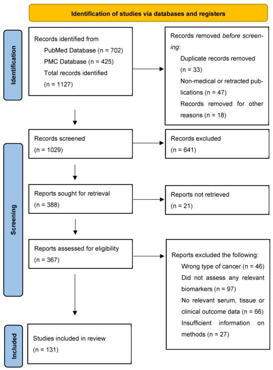
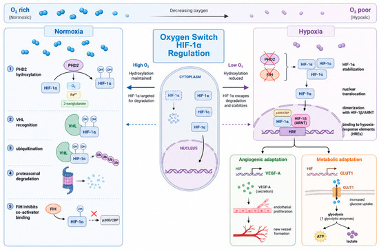
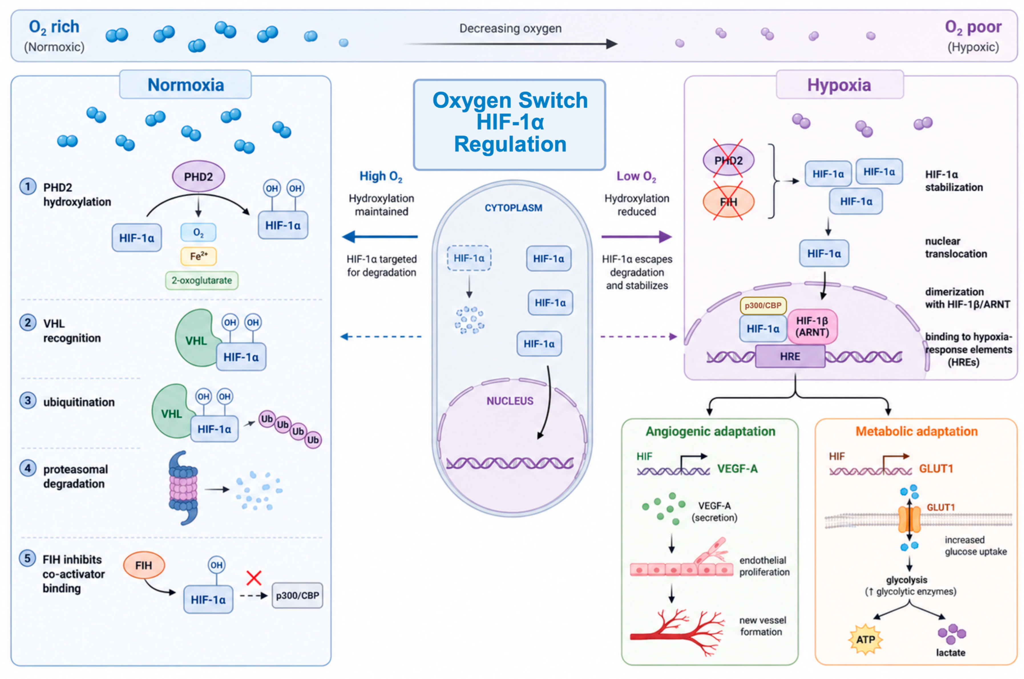
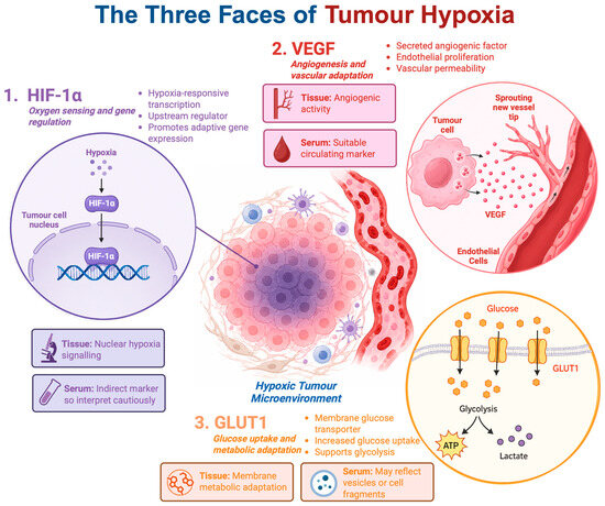
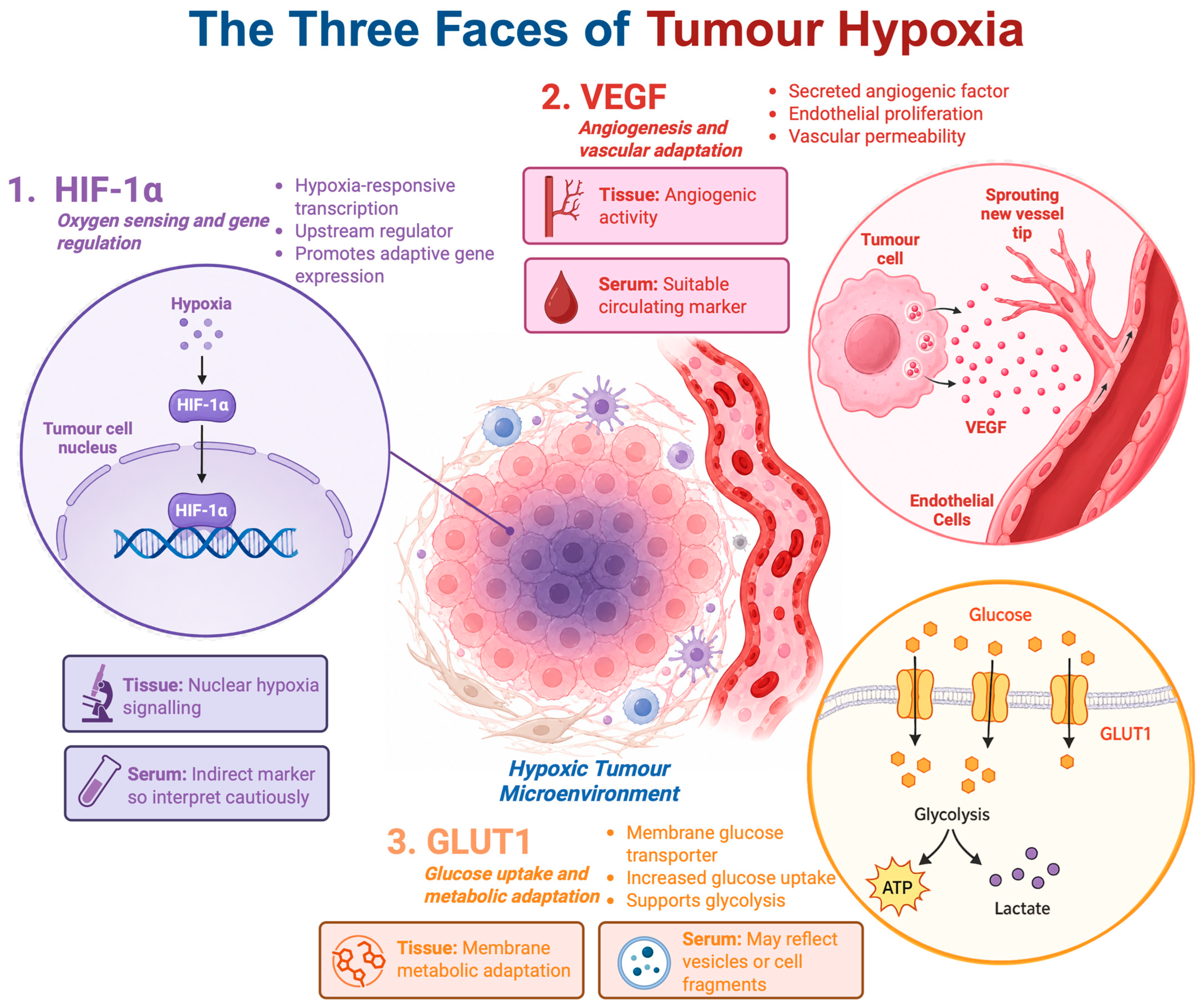
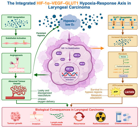
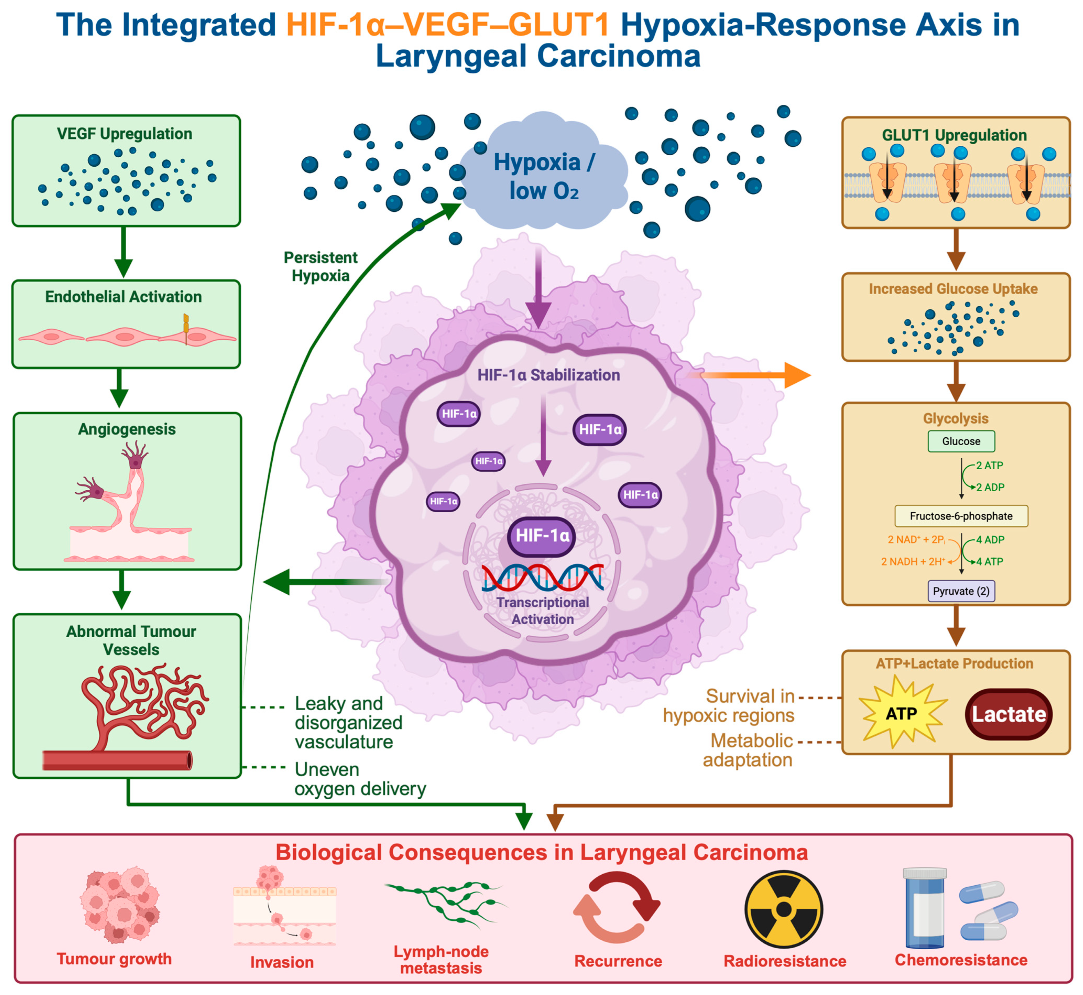

Disclaimer/Publisher’s Note: The statements, opinions and data contained in all publications are solely those of the individual author(s) and contributor(s) and not of MDPI and/or the editor(s). MDPI and/or the editor(s) disclaim responsibility for any injury to people or property resulting from any ideas, methods, instructions or products referred to in the content. |
© 2026 by the authors. Licensee MDPI, Basel, Switzerland. This article is an open access article distributed under the terms and conditions of the Creative Commons Attribution (CC BY) license.