Review

Triggering Pyroptosis in Cancer

by
Daniel E. Johnson
1,2 and
Zhibin Cui
3,*
1
Department of Otolaryngology—Head and Neck Surgery, University of California at San Francisco, San Francisco, CA 94143, USA
2
Helen Diller Family Comprehensive Cancer Center, University of California at San Francisco, San Francisco, CA 94143, USA
3
Department of Oral Biology, School of Dental Medicine, University at Buffalo, The State University of New York, Buffalo, NY 14214, USA
*
Author to whom correspondence should be addressed.
Biomolecules 2025, 15(3), 348; https://doi.org/10.3390/biom15030348
Submission received: 14 December 2024 / Revised: 19 February 2025 / Accepted: 25 February 2025 / Published: 28 February 2025

Abstract

:
Pyroptosis is an inflammatory programmed cell death recently identified as a crucial cellular process in various diseases, including cancers. Unlike other forms of cell death, canonical pyroptosis involves the specific cleavage of gasdermin by caspase-1, resulting in cell membrane damage and the release of the pro-inflammatory cytokines IL-1β and IL-18. Initially observed in innate immune cells responding to external pathogens or internal death signals, pyroptotic cell death has now been observed in numerous cell types. Recent studies have extensively explored different ways to trigger pyroptotic cell death in solid tumors, presenting a promising avenue for cancer treatment. This review outlines the mechanisms of both canonical and noncanonical pyroptosis pertinent to cancer and primarily focuses on various biomolecules that can induce pyroptosis in malignancies. This strategy aims not only to eliminate cancer cells but also to promote an improved tumor immune microenvironment. Furthermore, emerging research indicates that targeting pyroptotic pathways may improve the effectiveness of existing cancer treatments, making them more potent against resistant tumor types, offering new hope for overcoming treatment resistance in aggressive malignancies.

1. Introduction to Pyroptosis

Homeostasis of cell numbers in eukaryotes is maintained via a balance between cellular proliferation and cell death pathways. In cancer, this balance is lost, either as a result of aberrant proliferation or the loss of normal cell death processes, or both. Multiple forms of cell death have been described, with apoptosis being the most extensively characterized. Numerous studies have shown that apoptotic cell death is frequently abrogated in human tumors, resulting in tumor growth and resistance to anti-cancer drugs and biologics. More recently, additional forms of cell death have been identified, including autophagy, necroptosis, pyroptosis, ferroptosis, cuproptosis, and disulfidptosis. In this review, we summarize the current state of knowledge regarding the mechanisms of pyroptosis and the molecules and drugs that can be used to induce this cell death process.
The term pyroptosis was introduced by Cookson and Brennan in 2001 to describe a novel form of cell death distinct from apoptosis [1]. While the hallmark cell death features of nuclear condensation and DNA fragmentation are observed in both pyroptosis and apoptosis, the process of pyroptosis is uniquely dependent on caspase-1 protease, whereas apoptosis is typically dependent on the caspase-3 or caspase-7 proteases [1,2,3]. Caspase-1 is responsible for proteolytic production of the pro-inflammatory cytokines interleukin-1β (IL-1β) and interleukin-18 (IL-18) [4]. Hence, in contrast to apoptosis, which does not induce inflammation, cell death resulting from pyroptosis is pro-inflammatory. Moreover, pyroptosis is associated with rapid damage to the cell membrane, leading to the early release of pro-inflammatory cytokines [3]. This also contrasts with apoptotic cell death, where the loss of cell membrane integrity is a late event in the death process. Collectively, pyroptosis is distinguished from apoptosis by caspase-1 dependency, pro-inflammatory cytokine production, and early cell membrane rupture.
Pyroptosis was initially described as a cellular suicide process that macrophages undergo in response to interaction with Salmonella or other pathogens [2,5,6]. Later, the process was identified in other innate immune cells, including neutrophils and the barrier intestinal epithelial cells [7]. The induction of pyroptosis during an innate immune response is believed to function in fighting infections by promoting the release of pro-inflammatory cytokines (i.e., IL-1β and IL-18), resulting in the attraction of other immune cells to the site of infection and effective pathogen elimination. However, due to its inflammatory-associated properties, pyroptosis has also been found to be associated with several chronic inflammatory diseases, such as arthritis, pneumonia, hepatitis, colitis, and cardiovascular disease [8,9]. In addition, significant associations have been reported between pyroptosis and various cancers, highlighting the prospect of inducing this unique form of cell death as a novel therapeutic approach for cancer. In this review, we discuss the role of pyroptosis in cancer and explore promising biomolecules that might be utilized to induce pyroptosis in human malignancies.

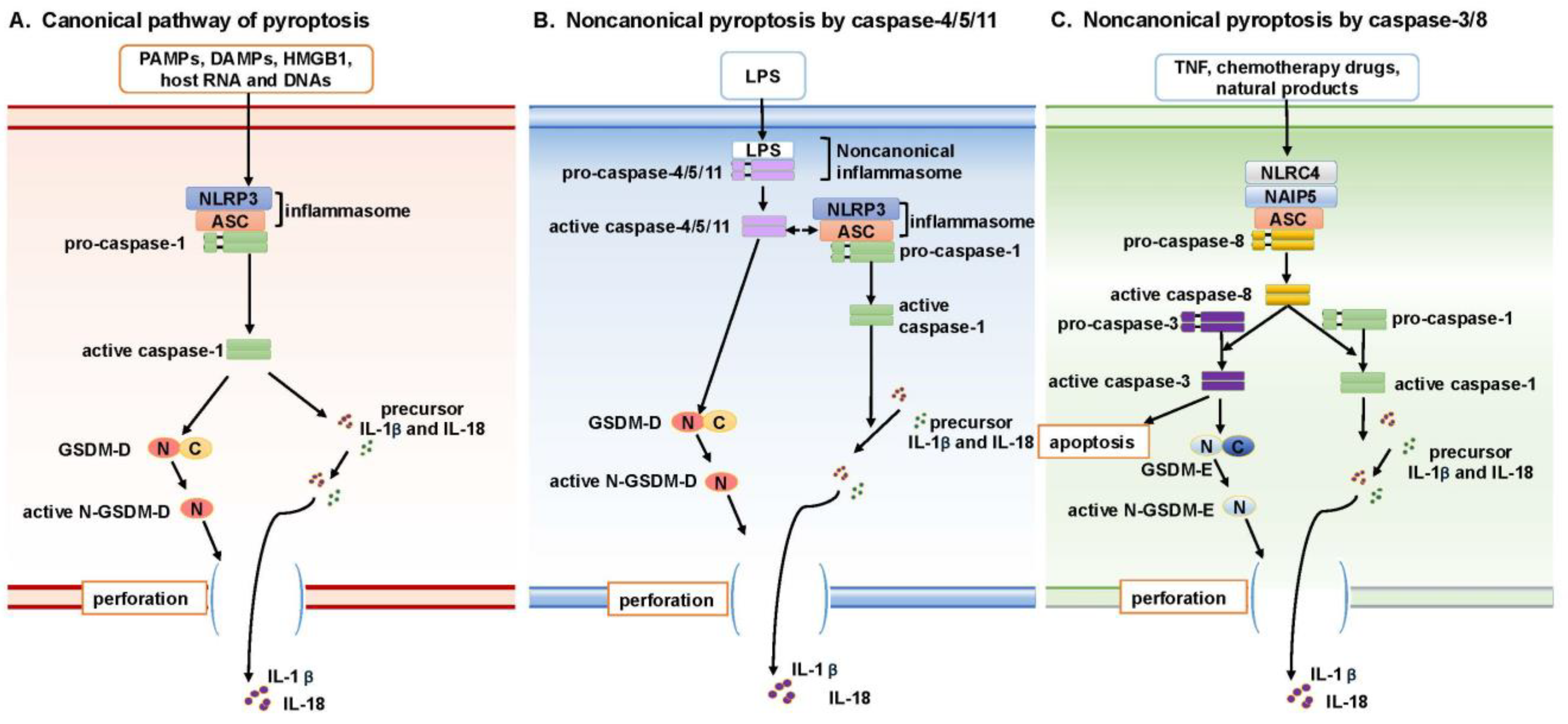
2. Pyroptosis Signaling Pathways

Pyroptosis is characterized by rapid cell membrane rupture, driven by the gasdermin (GSDM) protein family (particularly GSDM-D), along with secretion of the pro-inflammatory cytokines IL-1β and IL-18, which recruit other innate immune cells and enhance the cytolytic activities of NK and T-cells [10,11]. Initially, this process was thought to be mediated specifically by caspase-1, defining what is now known as the canonical pathway. However, in the past decade, additional mechanisms of pyroptosis activation have been identified, leading to the classification of these alternative routes as noncanonical pathways.

2.1. Canonical Pathway of Pyroptosis

The canonical pyroptosis pathway (Figure 1A) was originally identified in innate immune cells or antigen-presenting cells, such as macrophages and monocytes [12,13]. These cells play a defensive role by detecting “danger” signals—including pathogens, cell stress, cytokines, or disruptions to cellular homeostasis—which activate pyroptotic cell death and trigger inflammation [14].
During infection, microorganisms release pathogen-associated molecular patterns (PAMPs) like lipopolysaccharide (LPS), while stressed or damaged host cells release damage-associated molecular patterns (DAMPs), including histones, hyaluronic acid, high-mobility group protein B1 (HMGB1), and fragments of host cell RNA and DNA [15]. PAMPs and DAMPs are recognized by pattern recognition receptors (PRRs), leading to the activation of immune cells. Based on homologies in protein domains, several different PRRs have been defined, including Toll-like receptors (TLRs), nucleotide oligomerization domain (NOD)-like receptors (NLRs), retinoic acid-inducible gene-I (RIG-I)-like receptors (RLRs), C-type lectin receptors (CLRs), and absent in melanoma-2 (AIM2)-like receptors (ALRs). The most extensively studied PRR involved in the canonical pyroptosis pathway is NLRP3 (NOD-, LRR-, and pyrin domain-containing protein 3) [16,17]. Activated NLRP3 induces the assembly of the inflammasome complex by recruiting pro-caspase-1 and the adapter protein ASC (apoptosis-associated speck-like protein containing a caspase recruitment domain). The inflammasome complex recruits multiple pro-caspase-1 molecules, resulting in processing to generate active caspase-1. The activated caspase-1 then cleaves the substrate protein gasdermin D (GSDM-D), generating proteins encompassing the N-terminal domain (N-GSDM-D) and the C-terminal domain (C-GSDM-D). The N-GSDM-D protein is recruited to the cell membrane where it forms pores, resulting in the early rupture of the cell membrane, a defining feature of pyroptosis [18,19]. GSDM-D is an executor of pyroptosis and belongs to the gasdermin (GSDM) protein family, which includes six paralogues: GSDM-A to GSDM-E, and PJVK (DFNB59). In their inactive forms, GSDM proteins are primarily localized in the cytoplasm. Upon activation, the N-terminal domains translocate to the plasma membrane to execute their pore-forming functions. The expression of GSDMs is tissue- and cell-type-specific. For instance, GSDM-A is expressed in the skin, GSDM-A/B/C in the gastrointestinal tract, and GSDMB/C in the lungs and reproductive organs. GSDM-D and GSDM-E, however, are ubiquitously expressed in immune cells such as macrophages and dendritic cells, highlighting their essential role in innate immunity [20].
IL-1β and IL-18 are pro-inflammatory cytokines that belong to the interleukin-1 protein family. Both are synthesized as inactive precursor forms which are converted to active forms upon cleavage by caspase-1 [21]. During canonical pyroptosis signaling, cell membrane rupture mediated by GSDM-D facilitates the release of IL-1b and IL-18, which promotes the recruitment of innate immune cells like neutrophils and macrophages and modulates the activation of adaptive immune cells like CD4+ T-cells or even CD8+ T-cells [22,23,24].

2.2. Noncanonical Pyroptosis Mediated by Caspase-4/5/11

Following the identification and characterization of the canonical pyroptosis pathway, multiple studies revealed that pyroptosis can also be activated through alternative signaling mechanisms, collectively referred to as noncanonical pathways. One such noncanonical pathway is triggered directly in response to the PAMP molecule LPS and involves the formation of a ‘noncanonical inflammasome’, distinct from the inflammasome generated via the NLRP3/ASC/caspase-1 axis (Figure 1B). In this noncanonical pathway, LPS binds directly, and with high affinity, to caspase-11 in mouse macrophages, or to its homologs, caspase-4 and caspase-5, in human monocytes, without the need for NLRP3-mediated PAMP sensing [25,26].
The binding of LPS to caspase-11 (or caspase-4/5 in humans) induces the formation of a “noncanonical inflammasome”, which is sufficient to induce the oligomerization and activation of these caspases [25]. In mouse macrophages, activated caspase-11 directly cleaves GSDM-D, resulting in cell membrane perforation and pyroptotic cell death. Although caspase-11 does not directly cleave pro-IL-1β or pro-IL-18, evidence suggests it can activate a cascade involving the NLRP3/caspase-1 inflammasome, ultimately leading to IL-1β processing and release [26,27]. However, the precise mechanisms by which caspase-11 interacts with the canonical inflammasome remain unclear.

2.3. Noncanonical Pyroptosis Mediated by Caspase-3/8

In addition to pyroptosis induction by caspase-1 or caspases-4/5/11, the apoptotic mediators caspase-3 and caspase-8 also have been found to play roles in inducing a noncanonical pathway of pyroptosis (Figure 1C). The gasdermin (GSDM) protein family comprises six members (GSDM-A, -B, -C, -D, -E, and PJVK). Cleavage of these proteins generates N-terminal fragments (N-GSDM) that form pores in the cell membrane, leading to pyroptotic cell death [28]. Recent studies have shown that GSDM-E is a specific substrate of caspase-3. In cells expressing GSDM-E, tumor necrosis factor (TNF) can induce caspase-3-mediated cleavage of GSDM-E into active N-GSDM-E fragments, which perforate the cell membrane and induce pyroptosis [29].
A role for caspase-8 in noncanonical pyroptosis has come to light in studies of caspase-1 and caspase-11 double knockout mice. Infection of macrophages from these mice with Legionella pneumophila triggers the recruitment of caspase-8 to a noncanonical inflammasome complex, which consists of neuronal apoptosis inhibitory protein 5 (NAIP5), NLR family CARD-containing protein 4 (NLRC4), and ASC. Formation of this complex results in the activation of caspase-8 and subsequent cell membrane perforation via a GSDM-D-independent process [30]. Key questions regarding this pathway that remain unanswered are as follows: (1) whether caspase-8 activation is necessary to trigger caspase-3-mediated GSDM-E activation; and (2) whether the absence of caspase-1/11 and GSDM-D is essential for caspase-8-mediated membrane rupture. Additional findings reveal complex roles for caspase-8 in inflammasome activity. One study reported that expression of a catalytically inactive caspase-8 mutant (C362S) leads to ASC inflammasome complex formation and results in caspase-1 activation and IL-1β secretion. Embryonic expression of this caspase-8 mutant causes necroptosis-independent embryonic lethality due to pyroptosis [31]. Another study found that Yersinia infection, by inhibiting TGF-β-activated kinase 1 (TAK1) or IKK kinases, induces cell death mediated by both GSDM-D and GSDM-E. This pathway operates independently of caspase-1 and caspase-11 but is dependent on caspase-8 activity [32]. While the role of caspase-8 in this process is significant, it is unclear whether caspase-8 directly cleaves GSDM proteins.

3. Inducing Pyroptosis in Cancer

The role of pyroptosis in cancer development is not fully understood. Pyroptosis occurs in innate immune cells (primarily macrophages and neutrophils) in response to pathogen invasion or cellular stress, leading to the release of cytokines that trigger an inflammatory defense. Chronic inflammation, however, can promote cancer initiation [33]. During cancer progression, pyroptosis-induced immune cell infiltration may contribute to both pro-tumor and anti-tumor effects, with the balance of interactions between cancer cells and immune cells playing a critical role in cancer outcomes.
Pyroptosis has been observed not only in immune and barrier epithelial cells but also in various cancer cell types, including those representing liver cancer [34], breast cancer [35], esophageal cancer [36], lung cancer [37], colon cancer [38], and cervical cancer [39]. Despite the complex nature of pyroptosis within the tumor immune microenvironment, the induction of pyroptosis in cancer cells may present a promising opportunity for cancer therapy. The induction of pyroptosis in cancer cells not only leads to direct cell death but also releases cancer antigens, which enable antigen-presenting cells (APCs) to present these antigens to immune cells, including CD8+ T-cells. Additionally, the secretion of inflammatory cytokines can further support anti-tumor immunity to some extent.
Here, we describe biomolecules that have been shown to induce pyroptosis in cancers and the potential for incorporating these molecules in new therapeutic approaches.

3.1. Chemotherapy Drugs That Induce Pyroptosis

Chemotherapy drugs are primarily known to induce cell death via caspase-3-mediated apoptosis. However, recent studies have shown that certain chemotherapeutic agents can also induce pyroptosis. In particular, topotecan, etoposide, cisplatin, and CPT-11 have been observed to trigger pyroptosis in SH-SY5Y neuroblastoma cells and MeWo skin melanoma cells, both of which exhibit high levels of gasdermin E (GSDM-E) expression [29]. These chemotherapeutic agents promote pyroptosis by inducing the caspase-3-mediated cleavage of GSDM-E. Generally, GSDM-E expression is low in most cancer cells, leading to apoptosis as the primary cell death pathway. However, in cancer cells with elevated GSDM-E expression, the cell death mechanism is shifted towards pyroptosis following chemotherapy treatment. This switch highlights the potential of GSDM-E as a key factor in modulating the response of certain cancers to chemotherapy [29]. As summarized in Table 1, multiple chemotherapy drugs have now been shown to induce pyroptosis in a variety of cancer types via caspase-3-mediated cleavage of GSDM-E.

3.2. Metformin

Metformin, well known as an anti-diabetic medication, has shown potential as an anti-cancer agent by inducing pyroptosis in cancer cells. Wang et al. [45] demonstrated that metformin induces GSDM-D-mediated pyroptosis in esophageal squamous cell carcinoma (ESCC) following inhibition of the oncogenic scaffold protein PELP1, a biomarker of poor prognosis. However, the mechanisms linking PELP1- and GSDM-D-mediated pyroptosis were not fully elaborated in this study.
Further research has demonstrated that metformin can promote cancer cell pyroptosis through activation of the SIRT1/NF-κB signaling pathway. Metformin activates SIRT1, an NAD+-dependent deacetylase involved in regulating immunity and inflammatory responses. SIRT1 activation enhances the expression of NF-κB (p65), which then promotes the induction of BAX and the activation of caspase-3, leading to GSDM-E-mediated pyroptosis. This cascade of events underscores metformin’s capacity to harness the SIRT1/NF-κB signaling axis to drive pyroptosis and positions metformin as a novel therapeutic option to induce pyroptotic cell death in cancer cells, particularly those resistant to traditional apoptotic pathways [46].

3.3. Small Molecule Inhibitors That Induce Pyroptosis

3.3.1. DPP8/9 Inhibitors

Dipeptidyl peptidases (DPPs) are proteases that cleave the N-terminal dipeptide of their substrates. Inhibitors of DPP8 and DPP9 have attracted significant interest due to their ability to induce pyroptosis in immune cells [47]. DPP8/9 interacts with the inflammasome complex containing the PRR protein NLRP1 and CARD8 (caspase recruitment domain-containing 8 protein). When DPP8/9 binds to this inflammasome complex, CARD8 is retained within it. However, upon inhibition of DPP8/9, CARD8 is released from the complex, triggering caspase-1 activation and induction of pyroptosis [48]. The pyroptosis pathway induced by DPP8/9 inhibitors is primarily observed in monocytes and macrophages [47,49].
Several DPP8/9 inhibitors are currently available, including the nonselective DPP inhibitor Val-boroPro, the DPP8/9 selective inhibitor 1G244, as well as CQ31, and compounds 8j and L-allo-Ile-isoindoline [50,51,52,53]. Beyond immune cells, DPP8/9 inhibitors have been shown to induce pyroptosis in acute myeloid leukemia (AML) cell lines and primary AML samples [54]. While the effects of these inhibitors on solid tumors are not yet reported, DPP8 and DPP9 expressions have been detected in various normal tissues and a range of tumors, including brain tumors, gynecological malignancies, and liver cancer. Thus, DPP8/9 inhibitors represent a potential strategy for inducing pyroptotic cell death in solid cancers.

3.3.2. BRD4 Inhibitor

BRD4 is a member of the bromodomain and extra-terminal domain (BET) protein family, and it binds to acetylated lysine residues on histone tails, thereby altering chromatin structure. The BRD4 inhibitor JQ1, a thienotriazolodiazepine-based small molecule, competitively inhibits BET-histone binding, preventing BET proteins from interacting with chromatin. Recently, JQ1 has been shown to induce pyroptosis in renal cell carcinoma (RCC) via the activation of NF-κB, which subsequently activates the NLRP3 inflammasome, leading to caspase-1-dependent pyroptosis [55]. While the precise interaction between BRD4 and NLRP3 remains unclear, another study reported that BRD4 is an essential regulator of NLRC4 inflammasome activation in Salmonella-infected bone marrow-derived macrophages [56].

3.3.3. Other Inhibitors

Recent studies have demonstrated that inhibitors targeting EGFR, MEK, and ALK in lung cancer cells that express mutant forms of these proteins induce not only apoptosis but also pyroptosis. These inhibitors activate the intrinsic apoptotic pathway through the BIM-BAX-cytochrome c release signaling axis, which subsequently triggers caspase-3/GSDM-E-mediated pyroptosis. Notably, pyroptosis induced by these inhibitors occurs concurrently with caspase-3-mediated apoptosis, and there is a complex interplay among the proteins mediating both cell death pathways. However, the precise mechanisms underlying this crosstalk are incompletely understood [57].
In melanomas harboring the BRAF V600E/K mutation, the combined use of the BRAF inhibitor PLX4720 and the MEK inhibitor PD0325901 promotes GSDM-E cleavage and the release of HMGB1, along with an upregulation of IL-1β and IL-18, leading to pyroptosis induction [58]. This pyroptosis significantly enhances CD8+ T-cell infiltration into the tumor microenvironment, facilitating the immune-mediated inhibition of tumor growth. Interestingly, melanoma tumors that relapse after this combination therapy remain susceptible to etoposide- and doxorubicin-induced pyroptosis, highlighting the potential of pyroptosis induction as a therapeutic strategy not only for primary tumors but also for recurrent tumors resistant to targeted therapies [58].
Additional small molecule inhibitors that act to induce pyroptosis are summarized in Table 2.

3.4. Natural Products That Induce Pyroptosis

3.4.1. Curcumin

Curcumin is a natural compound derived from the rhizomes of turmeric (Curcuma longa), a hydrophobic polyphenol (Table 3). The United States Food and Drug Administration (FDA) has approved the use of curcumin as a safe flavoring and antioxidant agent in certain foods [68]. Over the past two decades, extensive research has shown that curcumin interferes with multiple cell signaling pathways, impacting cancer by activating apoptosis, inhibiting proliferation, reducing inflammation, and suppressing angiogenesis and metastasis [69,70,71]. In light of its promising impact in preclinical cancer models, curcumin-containing products have been investigated in numerous clinical trials for treating pancreatic, breast, prostate, and colorectal cancers [72,73,74].
Recent studies indicate that curcumin can induce pyroptosis in cancer cells. In HepG2 hepatocellular carcinoma cells, curcumin treatment activates the intrinsic apoptosis pathway, which in turn triggers caspase-3/GSDM-E-mediated pyroptotic cell death. This process results from elevation in the levels of reactive oxygen species (ROS) following curcumin treatment [75]. However, curcumin is far from a specific inducer of pyroptosis, as it also induces other forms of cell death, including apoptosis, necroptosis, and ferroptosis [86,87].

3.4.2. Iron

Iron plays a crucial role in both the formation and destruction of intracellular ROS, which serve as key mediators in multiple forms of cell death. Mitochondrial ROS, for example, drives intrinsic apoptosis, while ROS are also integral to necroptosis, ferroptosis, and, as recently identified, ROS-induced pyroptosis [88]. In melanoma cells, the combination of iron with the ROS-inducing agent carbonyl cyanide m-chlorophenyl hydrazone (CCCP) markedly amplifies ROS signaling, resulting in oxidation and oligomerization of the mitochondrial outer membrane protein TOM20. Oxidized TOM20 subsequently recruits BAX to the mitochondria, facilitating cytochrome c release into the cytosol and caspase-3 activation, which then leads to GSDM-E-mediated pyroptosis, highlighting a mechanistic pathway by which intracellular iron and enhanced ROS drive pyroptosis in melanoma [89].

3.4.3. Cucurbitacin B

Cucurbitacin B (CuB) is a bioactive compound derived from cucurbitaceae plants that has shown significant promise as an anti-tumor and anti-inflammatory agent. In non-small cell lung cancer (NSCLC), cucurbitacin B induces pyroptosis by binding directly to Toll-like receptor 4 (TLR4). This binding activates the NLRP3 inflammasome, resulting in the cleavage of GSDM-D and subsequent pyroptotic cell death. Cucurbitacin B further promotes pyroptosis by increasing the production of mitochondrial ROS, leading to the accumulation/oxidation of mitochondrial TOM20, as well as to the release of cytosolic calcium. In mouse models of NSCLC, cucurbitacin B effectively inhibits tumor growth and shows superior anti-tumor effects compared to standard treatments, underscoring its potential as a targeted therapy through the TLR4/NLRP3/GSDMD signaling axis [90].
Beyond its anti-cancer role, cucurbitacin B also exhibits anti-inflammatory properties, particularly in osteoarthritis. In osteoarthritis models, cucurbitacin B interacts with key signaling proteins, including NRF2 and NF-κB, to inhibit the secretion of pro-inflammatory cytokines such as IL-1β and IL-18, thereby reducing inflammation [91]. These effects indicate that the impact of cucurbitacin B treatment may differ according to tissue and disease type, acting as an anti-inflammatory agent in osteoarthritis and triggering pyroptosis in cancer. This adaptability makes cucurbitacin B a strong candidate for distinct therapeutic applications across multiple diseases settings.
Other natural products that induce pyroptosis are summarized in Table 3.

3.5. Pathogen or Pathogen-Derived Compounds That Induce Pyroptosis

Lipopolysaccharide

Lipopolysaccharide (LPS) is a bacterial endotoxin known to trigger pyroptosis in immune cells, particularly in monocytes and macrophages. In HT29 colon cancer cells, LPS exposure induces cleavage/activation of GSDM-D, leading to the secretion of IL-1β and IL-18 and, ultimately, pyroptotic cell death. Furthermore, LPS has been shown to increase the chemosensitivity of HT29 cells to oxaliplatin, and this combined treatment enhances GSDM-D-mediated pyroptosis. These findings suggest that combination of LPS with chemotherapy drugs may provide a strategy to promote pyroptosis and improve therapeutic outcomes in colon cancer [92].
For therapeutic purposes focused on the induction of pyroptosis, researchers have utilized bacterial outer membrane vesicles (OMVs) as natural carriers to deliver LPS into cancer cells. OMV-LPS has been shown to effectively induce GSDM-D-mediated pyroptosis in cancer cells, leading to a significant inhibition of tumor growth in vivo [93]. Additionally, OMV-LPS treatment promoted greater CD8+ T-cell infiltration into the tumor microenvironment, enhancing anti-tumor immunity. This approach represents a promising strategy for treating solid tumors by inducing cancer cell death and stimulating immune responses [93].
Other pathogen or pathogen-derived components that induce pyroptosis in cancer cells are summarized in Table 4.

3.6. Endogenous Metabolites That Induce Pyroptosis

Metabolites play pivotal roles in cancer development, influencing cellular processes like proliferation, survival, and immune evasion within the tumor microenvironment. Recently, several endogenous metabolites—including omega-3 docosahexaenoic acid, citric acid, α, β-unsaturated ketone, and α-ketoglutarate (α-KG)—were identified as inducers of pyroptosis (Table 5). These metabolites can engage inflammasome pathways or enhance ROS production, thereby activating pyroptotic signaling in cancer cells.
As an example, α-KG is a key metabolite in the tricarboxylic acid (TCA) cycle and is involved in lipid biosynthesis, oxidative stress response, protein modification, autophagy, and cell death. α-KG induces pyroptosis in cancer cells via caspase-8-mediated cleavage of GSDM-C, showing promise for limiting tumor growth and metastasis. Treatment with DM-α-KG, a cell-permeable α-KG derivative, elevates ROS levels, which oxidizes and triggers endocytosis of the plasma membrane receptor DR6. This process recruits pro-caspase-8 and GSDM-C into a “receptosome”, where active caspase-8 cleaves GSDM-C, initiating pyroptosis [103]. This mechanism relies on an acidic environment, where α-KG is converted to L-2-hydroxyglutarate (L-2HG) by malate dehydrogenase 1 (MDH1), boosting ROS production. The presence of lactic acid further intensifies this effect, sensitizing even pyroptosis-resistant cancer cells to α-KG-induced pyroptosis [103]. Together, these insights reveal the dual role of metabolites as drivers of tumor progression and as therapeutic agents. Further exploration into metabolite-driven pyroptosis could inspire innovative treatments that reshape the tumor immune microenvironment for more effective cancer therapy.

4. Challenges and Opportunities

The induction of pyroptosis shows great promise as a therapeutic strategy, especially for cancers resistant to conventional chemotherapies that induce apoptosis. Chemotherapy-induced cell death involves not only apoptosis but also necroptosis and pyroptosis, yet clear molecular mechanisms that differentiate these processes or highlight crosstalk are not well defined. Numerous chemotherapeutic agents trigger the intrinsic apoptotic pathway through caspase-3 activation. This, in turn, leads to the cleavage of multiple substrates, such as PARP-1, which promote apoptotic cell death, and GSDM-E, which initiates pyroptosis. Although apoptosis and pyroptosis may occur simultaneously during caspase-3 activation, pyroptosis is more immunogenic and promotes immune cell infiltration, particularly by CD8+ T-cells, thereby boosting anti-tumor responses. Nonetheless, further research is necessary to clarify how apoptosis might be switched toward pyroptosis to enhance therapeutic outcomes.
The role of pyroptosis in various stages of cancer development is still not fully understood. While chronic inflammation can facilitate cellular transformation, cytokine release due to pyroptosis has the potential to attract effector immune cells to combat cancer. A significant challenge is how to effectively induce pyroptosis in cancers with varied genetic backgrounds. For instance, many cancer cells demonstrate low GSDM-E expression, making pyroptosis induction more challenging. Overcoming this limitation requires a deeper understanding of how pyroptosis affects cancer progression across different genetic contexts and tumor types. To date, no clinical trials have been undertaken with the primary goal of targeting pyroptosis in cancer. Combined therapies that utilize agents capable of converting apoptosis into pyroptosis may enhance treatment efficacy, particularly in apoptosis-resistant cancers. Pyroptosis facilitates the conversion of immunologically “cold” tumors into more immunogenic phenotypes, thereby amplifying the advantages of immunotherapy. Merging pyroptosis-inducing strategies with immunotherapeutic methods might significantly advance cancer treatment. It is crucial to comprehend the mechanisms that regulate pyroptosis and its interplay with the immune microenvironment to advance this strategy. Since pyroptosis-induced inflammation may, under certain circumstances, contribute to tumor progression, combining anti-inflammatory drugs may serve as a complementary approach to enhance the efficacy of anti-pyroptosis strategies. This combinatorial approach could help mitigate the pro-tumorigenic effects of inflammation while simultaneously targeting pyroptosis, offering a more comprehensive therapeutic strategy for tumor suppression. By tackling these challenges, pyroptosis-based therapies may become vital for innovative cancer treatments, providing new hope for patients with resistant tumors.
Natural products, primarily derived from plants and microbes, have proven to be valuable sources of anti-cancer agents, including those capable of inducing pyroptosis. These compounds enhance ROS generation and activate pathways comprising signaling molecules like NLRP3, MEK/ERK1/2, and JNK, thereby promoting tumor cell death while fostering an immune-stimulatory tumor microenvironment. However, limitations such as low bioavailability and off-target effects have hindered the clinical application of natural products. Nanotechnology offers a potential solution by improving the solubility, stability, and specificity of natural products through nanosizing and targeted delivery systems. The incorporation of pyroptosis-inducing agents into nanoparticles enhances their efficacy and enables synergy with therapies like chemotherapy and immunotherapy. These advancements position natural product-based pyroptosis inducers as promising tools for precise and potent cancer immunotherapy.
Collectively, despite significant progress, challenges remain in translating pyroptosis-based therapies to clinical settings. Effective screening strategies are essential for identifying potent pyroptosis-inducing agents. Addressing tumor heterogeneity and the pro-inflammatory nature of pyroptosis is also critical to minimizing side effects and increasing treatment specificity. By integrating pyroptosis inducers with other therapeutic modalities, these combinatorial strategies may overcome treatment resistance, stimulate robust immune responses, and establish pyroptosis as a cornerstone in the fight against cancer.
Cancer cells exhibit unique vulnerabilities to cell death, and exploitation of these vulnerabilities has strong potential to lead to tumor-selective anti-cancer therapies. Pyroptosis represents a new area of cell death research that should be explored as a means of provoking cell death in cancer cells and tumors. At present, only a modest number of biomolecules have been identified to promote pyroptosis. More work is needed to identify pyroptosis-inducing agents, and combination of these agents with immune modulating therapies is warranted. Coincident with these investigations, it will be important to closely monitor whether selective induction of pyroptosis leads to adverse toxicities, and whether biomarkers that will help determine which patient populations are most likely to benefit from treatment with pyroptosis inducers can be identified. As research into these questions continues, we will learn the full utility of targeting pyroptosis to improve clinical outcomes in patients suffering from cancer.

Author Contributions

D.E.J.: Manuscript writing and editing, and figure and table designing; Z.C.: Manuscript writing and editing, and figure and table designing. All authors have read and agreed to the published version of the manuscript.

Funding

This work was supported by NIH grant R01 DE028289 (DEJ) and the New York State Foundation (ZC).

Institutional Review Board Statement

Not applicable.

Informed Consent Statement

Not applicable.

Data Availability Statement

Not applicable.

Acknowledgments

The grammar correction tool Grammarly Inc. was used to enhance the language by improving the quality of English and readability, all while being utilized under human oversight and control.

Conflicts of Interest

The authors declare no conflicts of interest.

References

  1. Cookson, B.T.; Brennan, M.A. Pro-inflammatory programmed cell death. Trends Microbiol. 2001, 9, 113–114. [Google Scholar] [CrossRef] [PubMed]
  2. Hersh, D.; Monack, D.M.; Smith, M.R.; Ghori, N.; Falkow, S.; Zychlinsky, A. The Salmonella invasin SipB induces macrophage apoptosis by binding to caspase-1. Proc. Natl. Acad. Sci. USA 1999, 96, 2396–2401. [Google Scholar] [CrossRef] [PubMed]
  3. Brennan, M.A.; Cookson, B.T. Salmonella induces macrophage death by caspase-1-dependent necrosis. Mol. Microbiol. 2000, 38, 31–40. [Google Scholar] [CrossRef] [PubMed]
  4. Arend, W.P.; Palmer, G.; Gabay, C. IL-1, IL-18, and IL-33 families of cytokines. Immunol. Rev. 2008, 223, 20–38. [Google Scholar] [CrossRef]
  5. Zychlinsky, A.; Prevost, M.C.; Sansonetti, P.J. Shigella flexneri induces apoptosis in infected macrophages. Nature 1992, 358, 167–169. [Google Scholar] [CrossRef]
  6. Man, S.M.; Hopkins, L.J.; Nugent, E.; Cox, S.; Gluck, I.M.; Tourlomousis, P.; Wright, J.A.; Cicuta, P.; Monie, T.P.; Bryant, C.E. Inflammasome activation causes dual recruitment of NLRC4 and NLRP3 to the same macromolecular complex. Proc. Natl. Acad. Sci. USA 2014, 111, 7403–7408. [Google Scholar] [CrossRef]
  7. Oh, C.; Spears, T.J.; Aachoui, Y. Inflammasome-mediated pyroptosis in defense against pathogenic bacteria. Immunol. Rev. 2024, 329, e13408. [Google Scholar] [CrossRef]
  8. Wu, Y.; Zhang, J.; Yu, S.; Li, Y.; Zhu, J.; Zhang, K.; Zhang, R. Cell pyroptosis in health and inflammatory diseases. Cell Death Discov. 2022, 8, 191. [Google Scholar] [CrossRef]
  9. Zhaolin, Z.; Guohua, L.; Shiyuan, W.; Zuo, W. Role of pyroptosis in cardiovascular disease. Cell Prolif. 2019, 52, e12563. [Google Scholar] [CrossRef]
  10. Dinarello, C.A. Immunological and inflammatory functions of the interleukin-1 family. Annu. Rev. Immunol. 2009, 27, 519–550. [Google Scholar] [CrossRef]
  11. Jorgensen, I.; Lopez, J.P.; Laufer, S.A.; Miao, E.A. IL-1beta, IL-18, and eicosanoids promote neutrophil recruitment to pore-induced intracellular traps following pyroptosis. Eur. J. Immunol. 2016, 46, 2761–2766. [Google Scholar] [CrossRef] [PubMed]
  12. Cordoba-Rodriguez, R.; Fang, H.; Lankford, C.S.; Frucht, D.M. Anthrax lethal toxin rapidly activates caspase-1/ICE and induces extracellular release of interleukin (IL)-1beta and IL-18. J. Biol. Chem. 2004, 279, 20563–20566. [Google Scholar] [CrossRef] [PubMed]
  13. Boyden, E.D.; Dietrich, W.F. Nalp1b controls mouse macrophage susceptibility to anthrax lethal toxin. Nat. Genet. 2006, 38, 240–244. [Google Scholar] [CrossRef] [PubMed]
  14. Man, S.M.; Kanneganti, T.D. Innate immune sensing of cell death in disease and therapeutics. Nat. Cell Biol. 2024, 26, 1420–1433. [Google Scholar] [CrossRef]
  15. Tang, D.; Kang, R.; Coyne, C.B.; Zeh, H.J.; Lotze, M.T. PAMPs and DAMPs: Signal 0s that spur autophagy and immunity. Immunol. Rev. 2012, 249, 158–175. [Google Scholar] [CrossRef]
  16. Wei, X.; Xie, F.; Zhou, X.; Wu, Y.; Yan, H.; Liu, T.; Huang, J.; Wang, F.; Zhou, F.; Zhang, L. Role of pyroptosis in inflammation and cancer. Cell Mol. Immunol. 2022, 19, 971–992. [Google Scholar] [CrossRef]
  17. Jin, C.; Flavell, R.A. Molecular mechanism of NLRP3 inflammasome activation. J. Clin. Immunol. 2010, 30, 628–631. [Google Scholar] [CrossRef]
  18. Ding, J.; Wang, K.; Liu, W.; She, Y.; Sun, Q.; Shi, J.; Sun, H.; Wang, D.C.; Shao, F. Pore-forming activity and structural autoinhibition of the gasdermin family. Nature 2016, 535, 111–116. [Google Scholar] [CrossRef]
  19. Liu, X.; Zhang, Z.; Ruan, J.; Pan, Y.; Magupalli, V.G.; Wu, H.; Lieberman, J. Inflammasome-activated gasdermin D causes pyroptosis by forming membrane pores. Nature 2016, 535, 153–158. [Google Scholar] [CrossRef]
  20. Chen, K.W.; Broz, P. Gasdermins as evolutionarily conserved executors of inflammation and cell death. Nat. Cell Biol. 2024, 26, 1394–1406. [Google Scholar] [CrossRef]
  21. Dinarello, C.A. Overview of the IL-1 family in innate inflammation and acquired immunity. Immunol. Rev. 2018, 281, 8–27. [Google Scholar] [CrossRef]
  22. Evavold, C.L.; Ruan, J.; Tan, Y.; Xia, S.; Wu, H.; Kagan, J.C. The Pore-Forming Protein Gasdermin D Regulates Interleukin-1 Secretion from Living Macrophages. Immunity 2018, 48, 35–44 e36. [Google Scholar] [CrossRef] [PubMed]
  23. Iwasaki, A.; Medzhitov, R. Control of adaptive immunity by the innate immune system. Nat. Immunol. 2015, 16, 343–353. [Google Scholar] [CrossRef] [PubMed]
  24. Van Den Eeckhout, B.; Tavernier, J.; Gerlo, S. Interleukin-1 as Innate Mediator of T Cell Immunity. Front. Immunol. 2020, 11, 621931. [Google Scholar] [CrossRef]
  25. Shi, J.; Zhao, Y.; Wang, Y.; Gao, W.; Ding, J.; Li, P.; Hu, L.; Shao, F. Inflammatory caspases are innate immune receptors for intracellular LPS. Nature 2014, 514, 187–192. [Google Scholar] [CrossRef]
  26. Kayagaki, N.; Warming, S.; Lamkanfi, M.; Vande Walle, L.; Louie, S.; Dong, J.; Newton, K.; Qu, Y.; Liu, J.; Heldens, S.; et al. Non-canonical inflammasome activation targets caspase-11. Nature 2011, 479, 117–121. [Google Scholar] [CrossRef]
  27. Kayagaki, N.; Stowe, I.B.; Lee, B.L.; O’Rourke, K.; Anderson, K.; Warming, S.; Cuellar, T.; Haley, B.; Roose-Girma, M.; Phung, Q.T.; et al. Caspase-11 cleaves gasdermin D for non-canonical inflammasome signalling. Nature 2015, 526, 666–671. [Google Scholar] [CrossRef]
  28. Zhu, C.; Xu, S.; Jiang, R.; Yu, Y.; Bian, J.; Zou, Z. The gasdermin family: Emerging therapeutic targets in diseases. Signal Transduct. Target. Ther. 2024, 9, 87. [Google Scholar] [CrossRef]
  29. Wang, Y.; Gao, W.; Shi, X.; Ding, J.; Liu, W.; He, H.; Wang, K.; Shao, F. Chemotherapy drugs induce pyroptosis through caspase-3 cleavage of a gasdermin. Nature 2017, 547, 99–103. [Google Scholar] [CrossRef]
  30. Mascarenhas, D.P.A.; Cerqueira, D.M.; Pereira, M.S.F.; Castanheira, F.V.S.; Fernandes, T.D.; Manin, G.Z.; Cunha, L.D.; Zamboni, D.S. Inhibition of caspase-1 or gasdermin-D enable caspase-8 activation in the Naip5/NLRC4/ASC inflammasome. PLoS Pathog. 2017, 13, e1006502. [Google Scholar] [CrossRef]
  31. Fritsch, M.; Gunther, S.D.; Schwarzer, R.; Albert, M.C.; Schorn, F.; Werthenbach, J.P.; Schiffmann, L.M.; Stair, N.; Stocks, H.; Seeger, J.M.; et al. Caspase-8 is the molecular switch for apoptosis, necroptosis and pyroptosis. Nature 2019, 575, 683–687. [Google Scholar] [CrossRef] [PubMed]
  32. Orning, P.; Weng, D.; Starheim, K.; Ratner, D.; Best, Z.; Lee, B.; Brooks, A.; Xia, S.; Wu, H.; Kelliher, M.A.; et al. Pathogen blockade of TAK1 triggers caspase-8-dependent cleavage of gasdermin D and cell death. Science 2018, 362, 1064–1069. [Google Scholar] [CrossRef] [PubMed]
  33. Pazhouhesh Far, N.; Hajiheidari Varnousafaderani, M.; Faghihkhorasani, F.; Etemad, S.; Abdulwahid, A.R.R.; Bakhtiarinia, N.; Mousaei, A.; Dortaj, E.; Karimi, S.; Ebrahimi, N.; et al. Breaking the barriers: Overcoming cancer resistance by targeting the NLRP3 inflammasome. Br. J. Pharmacol. 2024, 182, 3–25. [Google Scholar] [CrossRef]
  34. Wu, X.; Cao, J.; Wan, X.; Du, S. Programmed cell death in hepatocellular carcinoma: Mechanisms and therapeutic prospects. Cell Death Discov. 2024, 10, 356. [Google Scholar] [CrossRef]
  35. Yan, H.; Luo, B.; Wu, X.; Guan, F.; Yu, X.; Zhao, L.; Ke, X.; Wu, J.; Yuan, J. Cisplatin Induces Pyroptosis via Activation of MEG3/NLRP3/caspase-1/GSDMD Pathway in Triple-Negative Breast Cancer. Int. J. Biol. Sci. 2021, 17, 2606–2621. [Google Scholar] [CrossRef] [PubMed]
  36. Li, R.Y.; Zheng, Z.Y.; Li, Z.M.; Heng, J.H.; Zheng, Y.Q.; Deng, D.X.; Xu, X.E.; Liao, L.D.; Lin, W.; Xu, H.Y.; et al. Cisplatin-induced pyroptosis is mediated via the CAPN1/CAPN2-BAK/BAX-caspase-9-caspase-3-GSDME axis in esophageal cancer. Chem. Biol. Interact. 2022, 361, 109967. [Google Scholar] [CrossRef]
  37. Zhang, C.C.; Li, C.G.; Wang, Y.F.; Xu, L.H.; He, X.H.; Zeng, Q.Z.; Zeng, C.Y.; Mai, F.Y.; Hu, B.; Ouyang, D.Y. Chemotherapeutic paclitaxel and cisplatin differentially induce pyroptosis in A549 lung cancer cells via caspase-3/GSDME activation. Apoptosis 2019, 24, 312–325. [Google Scholar] [CrossRef]
  38. Yu, J.; Li, S.; Qi, J.; Chen, Z.; Wu, Y.; Guo, J.; Wang, K.; Sun, X.; Zheng, J. Cleavage of GSDME by caspase-3 determines lobaplatin-induced pyroptosis in colon cancer cells. Cell Death Dis. 2019, 10, 193. [Google Scholar] [CrossRef]
  39. Chen, J.; Ge, L.; Shi, X.; Liu, J.; Ruan, H.; Heng, D.; Ye, C. Lobaplatin Induces Pyroptosis in Cervical Cancer Cells via the Caspase-3/GSDME Pathway. Anticancer. Agents Med. Chem. 2022, 22, 2091–2097. [Google Scholar] [CrossRef]
  40. Peng, Z.; Wang, P.; Song, W.; Yao, Q.; Li, Y.; Liu, L.; Li, Y.; Zhou, S. GSDME enhances Cisplatin sensitivity to regress non-small cell lung carcinoma by mediating pyroptosis to trigger antitumor immunocyte infiltration. Signal Transduct. Target. Ther. 2020, 5, 159. [Google Scholar] [CrossRef]
  41. Wang, Y.; Yin, B.; Li, D.; Wang, G.; Han, X.; Sun, X. GSDME mediates caspase-3-dependent pyroptosis in gastric cancer. Biochem. Biophys. Res. Commun. 2018, 495, 1418–1425. [Google Scholar] [CrossRef] [PubMed]
  42. Chen, D.; Guo, S.; Tang, X.; Rong, Y.; Bo, H.; Shen, H.; Zhao, Z.; Qiao, A.; Shen, J.; Wang, J. Combination of ruthenium (II) polypyridyl complex Delta-Ru1 and Taxol enhances the anti-cancer effect on Taxol-resistant cancer cells through Caspase-1/GSDMD-mediated pyroptosis. J. Inorg. Biochem. 2022, 230, 111749. [Google Scholar] [CrossRef] [PubMed]
  43. Xiao, B.; Ying, C.; Chen, Y.; Huang, F.; Wang, B.; Fang, H.; Guo, W.; Liu, T.; Zhou, X.; Huang, B.; et al. Doxorubicin hydrochloride enhanced antitumour effect of CEA-regulated oncolytic virotherapy in live cancer cells and a mouse model. J. Cell Mol. Med. 2020, 24, 13431–13439. [Google Scholar] [CrossRef] [PubMed]
  44. Tang, Z.; Ji, L.; Han, M.; Xie, J.; Zhong, F.; Zhang, X.; Su, Q.; Yang, Z.; Liu, Z.; Gao, H.; et al. Pyroptosis is involved in the inhibitory effect of FL118 on growth and metastasis in colorectal cancer. Life Sci. 2020, 257, 118065. [Google Scholar] [CrossRef]
  45. Wang, L.; Li, K.; Lin, X.; Yao, Z.; Wang, S.; Xiong, X.; Ning, Z.; Wang, J.; Xu, X.; Jiang, Y.; et al. Metformin induces human esophageal carcinoma cell pyroptosis by targeting the miR-497/PELP1 axis. Cancer Lett. 2019, 450, 22–31. [Google Scholar] [CrossRef]
  46. Zheng, Z.; Bian, Y.; Zhang, Y.; Ren, G.; Li, G. Metformin activates AMPK/SIRT1/NF-kappaB pathway and induces mitochondrial dysfunction to drive caspase3/GSDME-mediated cancer cell pyroptosis. Cell Cycle 2020, 19, 1089–1104. [Google Scholar] [CrossRef]
  47. Okondo, M.C.; Johnson, D.C.; Sridharan, R.; Go, E.B.; Chui, A.J.; Wang, M.S.; Poplawski, S.E.; Wu, W.; Liu, Y.; Lai, J.H.; et al. DPP8 and DPP9 inhibition induces pro-caspase-1-dependent monocyte and macrophage pyroptosis. Nat. Chem. Biol. 2017, 13, 46–53. [Google Scholar] [CrossRef]
  48. Karakaya, T.; Slaufova, M.; Di Filippo, M.; Hennig, P.; Kundig, T.; Beer, H.D. CARD8: A Novel Inflammasome Sensor with Well-Known Anti-Inflammatory and Anti-Apoptotic Activity. Cells 2024, 13, 1032. [Google Scholar] [CrossRef]
  49. de Vasconcelos, N.M.; Vliegen, G.; Goncalves, A.; De Hert, E.; Martin-Perez, R.; Van Opdenbosch, N.; Jallapally, A.; Geiss-Friedlander, R.; Lambeir, A.M.; Augustyns, K.; et al. DPP8/DPP9 inhibition elicits canonical Nlrp1b inflammasome hallmarks in murine macrophages. Life Sci. Alliance 2019, 2, 313. [Google Scholar] [CrossRef]
  50. Coutts, S.J.; Kelly, T.A.; Snow, R.J.; Kennedy, C.A.; Barton, R.W.; Adams, J.; Krolikowski, D.A.; Freeman, D.M.; Campbell, S.J.; Ksiazek, J.F.; et al. Structure-activity relationships of boronic acid inhibitors of dipeptidyl peptidase IV. 1. Variation of the P2 position of Xaa-boroPro dipeptides. J. Med. Chem. 1996, 39, 2087–2094. [Google Scholar] [CrossRef]
  51. Lankas, G.R.; Leiting, B.; Roy, R.S.; Eiermann, G.J.; Beconi, M.G.; Biftu, T.; Chan, C.C.; Edmondson, S.; Feeney, W.P.; He, H.; et al. Dipeptidyl peptidase IV inhibition for the treatment of type 2 diabetes: Potential importance of selectivity over dipeptidyl peptidases 8 and 9. Diabetes 2005, 54, 2988–2994. [Google Scholar] [CrossRef] [PubMed]
  52. Van Goethem, S.; Van der Veken, P.; Dubois, V.; Soroka, A.; Lambeir, A.M.; Chen, X.; Haemers, A.; Scharpe, S.; De Meester, I.; Augustyns, K. Inhibitors of dipeptidyl peptidase 8 and dipeptidyl peptidase 9. Part 2: Isoindoline containing inhibitors. Bioorg. Med. Chem. Lett. 2008, 18, 4159–4162. [Google Scholar] [CrossRef] [PubMed]
  53. Rao, S.D.; Chen, Q.; Wang, Q.; Orth-He, E.L.; Saoi, M.; Griswold, A.R.; Bhattacharjee, A.; Ball, D.P.; Huang, H.C.; Chui, A.J.; et al. M24B aminopeptidase inhibitors selectively activate the CARD8 inflammasome. Nat. Chem. Biol. 2022, 18, 565–574. [Google Scholar] [CrossRef]
  54. Johnson, D.C.; Taabazuing, C.Y.; Okondo, M.C.; Chui, A.J.; Rao, S.D.; Brown, F.C.; Reed, C.; Peguero, E.; de Stanchina, E.; Kentsis, A.; et al. DPP8/DPP9 inhibitor-induced pyroptosis for treatment of acute myeloid leukemia. Nat. Med. 2018, 24, 1151–1156. [Google Scholar] [CrossRef]
  55. Tan, Y.F.; Wang, M.; Chen, Z.Y.; Wang, L.; Liu, X.H. Inhibition of BRD4 prevents proliferation and epithelial-mesenchymal transition in renal cell carcinoma via NLRP3 inflammasome-induced pyroptosis. Cell Death Dis. 2020, 11, 239. [Google Scholar] [CrossRef]
  56. Dong, X.; Hu, X.; Bao, Y.; Li, G.; Yang, X.D.; Slauch, J.M.; Chen, L.F. Brd4 regulates NLRC4 inflammasome activation by facilitating IRF8-mediated transcription of Naips. J. Cell Biol. 2021, 220, e202005148. [Google Scholar] [CrossRef]
  57. Lu, H.; Zhang, S.; Wu, J.; Chen, M.; Cai, M.C.; Fu, Y.; Li, W.; Wang, J.; Zhao, X.; Yu, Z.; et al. Molecular Targeted Therapies Elicit Concurrent Apoptotic and GSDME-Dependent Pyroptotic Tumor Cell Death. Clin. Cancer Res. 2018, 24, 6066–6077. [Google Scholar] [CrossRef]
  58. Erkes, D.A.; Cai, W.; Sanchez, I.M.; Purwin, T.J.; Rogers, C.; Field, C.O.; Berger, A.C.; Hartsough, E.J.; Rodeck, U.; Alnemri, E.S.; et al. Mutant BRAF and MEK Inhibitors Regulate the Tumor Immune Microenvironment via Pyroptosis. Cancer Discov. 2020, 10, 254–269. [Google Scholar] [CrossRef]
  59. Wu, M.; Wang, Y.; Yang, D.; Gong, Y.; Rao, F.; Liu, R.; Danna, Y.; Li, J.; Fan, J.; Chen, J.; et al. A PLK1 kinase inhibitor enhances the chemosensitivity of cisplatin by inducing pyroptosis in oesophageal squamous cell carcinoma. EBioMedicine 2019, 41, 244–255. [Google Scholar] [CrossRef]
  60. Huo, J.; Shen, Y.; Zhang, Y.; Shen, L. BI 2536 induces gasdermin E-dependent pyroptosis in ovarian cancer. Front. Oncol. 2022, 12, 963928. [Google Scholar] [CrossRef]
  61. Qiao, L.; Wu, X.; Zhang, J.; Liu, L.; Sui, X.; Zhang, R.; Liu, W.; Shen, F.; Sun, Y.; Xi, X. alpha-NETA induces pyroptosis of epithelial ovarian cancer cells through the GSDMD/caspase-4 pathway. FASEB J. 2019, 33, 12760–12767. [Google Scholar] [CrossRef] [PubMed]
  62. Deng, B.B.; Jiao, B.P.; Liu, Y.J.; Li, Y.R.; Wang, G.J. BIX-01294 enhanced chemotherapy effect in gastric cancer by inducing GSDME-mediated pyroptosis. Cell Biol. Int. 2020, 44, 1890–1899. [Google Scholar] [CrossRef] [PubMed]
  63. Long, K.; Gu, L.; Li, L.; Zhang, Z.; Li, E.; Zhang, Y.; He, L.; Pan, F.; Guo, Z.; Hu, Z. Small-molecule inhibition of APE1 induces apoptosis, pyroptosis, and necroptosis in non-small cell lung cancer. Cell Death Dis. 2021, 12, 503. [Google Scholar] [CrossRef] [PubMed]
  64. Huang, J.; Fan, P.; Liu, M.; Weng, C.; Fan, G.; Zhang, T.; Duan, X.; Wu, Y.; Tang, L.; Yang, G.; et al. Famotidine promotes inflammation by triggering cell pyroptosis in gastric cancer cells. BMC Pharmacol. Toxicol. 2021, 22, 62. [Google Scholar] [CrossRef]
  65. Kobayashi, T.; Mitsuhashi, A.; Hongying, P.; Shioya, M.; Kojima, K.; Nishikimi, K.; Yahiro, K.; Shozu, M. Bexarotene-induced cell death in ovarian cancer cells through Caspase-4-gasdermin E mediated pyroptosis. Sci. Rep. 2022, 12, 11123. [Google Scholar] [CrossRef]
  66. Zhao, W.; Zhang, L.; Zhang, Y.; Jiang, Z.; Lu, H.; Xie, Y.; Han, W.; Zhao, W.; He, J.; Shi, Z.; et al. The CDK inhibitor AT7519 inhibits human glioblastoma cell growth by inducing apoptosis, pyroptosis and cell cycle arrest. Cell Death Dis. 2023, 14, 11. [Google Scholar] [CrossRef]
  67. Huntington, K.E.; Louie, A.D.; Srinivasan, P.R.; Schorl, C.; Lu, S.; Silverberg, D.; Newhouse, D.; Wu, Z.; Zhou, L.; Borden, B.A.; et al. GSK-3 inhibitor elraglusib enhances tumor-infiltrating immune cell activation in tumor biopsies and synergizes with anti-PD-L1 in a murine model of colorectal cancer. bioRxiv 2023. [Google Scholar] [CrossRef]
  68. Prasad, S.; Tyagi, A.K.; Aggarwal, B.B. Recent developments in delivery, bioavailability, absorption and metabolism of curcumin: The golden pigment from golden spice. Cancer Res. Treat. 2014, 46, 2–18. [Google Scholar] [CrossRef]
  69. Alexandrow, M.G.; Song, L.J.; Altiok, S.; Gray, J.; Haura, E.B.; Kumar, N.B. Curcumin: A novel Stat3 pathway inhibitor for chemoprevention of lung cancer. Eur. J. Cancer Prev. 2012, 21, 407–412. [Google Scholar] [CrossRef]
  70. Panahi, Y.; Darvishi, B.; Ghanei, M.; Jowzi, N.; Beiraghdar, F.; Varnamkhasti, B.S. Molecular mechanisms of curcumins suppressing effects on tumorigenesis, angiogenesis and metastasis, focusing on NF-kappaB pathway. Cytokine Growth Factor. Rev. 2016, 28, 21–29. [Google Scholar] [CrossRef]
  71. Cho, J.W.; Lee, K.S.; Kim, C.W. Curcumin attenuates the expression of IL-1beta, IL-6, and TNF-alpha as well as cyclin E in TNF-alpha-treated HaCaT cells; NF-kappaB and MAPKs as potential upstream targets. Int. J. Mol. Med. 2007, 19, 469–474. [Google Scholar] [PubMed]
  72. Dhillon, N.; Aggarwal, B.B.; Newman, R.A.; Wolff, R.A.; Kunnumakkara, A.B.; Abbruzzese, J.L.; Ng, C.S.; Badmaev, V.; Kurzrock, R. Phase II trial of curcumin in patients with advanced pancreatic cancer. Clin. Cancer Res. 2008, 14, 4491–4499. [Google Scholar] [CrossRef] [PubMed]
  73. Kanai, M.; Yoshimura, K.; Asada, M.; Imaizumi, A.; Suzuki, C.; Matsumoto, S.; Nishimura, T.; Mori, Y.; Masui, T.; Kawaguchi, Y.; et al. A phase I/II study of gemcitabine-based chemotherapy plus curcumin for patients with gemcitabine-resistant pancreatic cancer. Cancer Chemother. Pharmacol. 2011, 68, 157–164. [Google Scholar] [CrossRef] [PubMed]
  74. Passildas-Jahanmohan, J.; Eymard, J.C.; Pouget, M.; Kwiatkowski, F.; Van Praagh, I.; Savareux, L.; Atger, M.; Durando, X.; Abrial, C.; Richard, D.; et al. Multicenter randomized phase II study comparing docetaxel plus curcumin versus docetaxel plus placebo in first-line treatment of metastatic castration-resistant prostate cancer. Cancer Med. 2021, 10, 2332–2340. [Google Scholar] [CrossRef]
  75. Liang, W.F.; Gong, Y.X.; Li, H.F.; Sun, F.L.; Li, W.L.; Chen, D.Q.; Xie, D.P.; Ren, C.X.; Guo, X.Y.; Wang, Z.Y.; et al. Curcumin Activates ROS Signaling to Promote Pyroptosis in Hepatocellular Carcinoma HepG2 Cells. In Vivo 2021, 35, 249–257. [Google Scholar] [CrossRef]
  76. Yue, E.; Tuguzbaeva, G.; Chen, X.; Qin, Y.; Li, A.; Sun, X.; Dong, C.; Liu, Y.; Yu, Y.; Zahra, S.M.; et al. Anthocyanin is involved in the activation of pyroptosis in oral squamous cell carcinoma. Phytomedicine 2019, 56, 286–294. [Google Scholar] [CrossRef]
  77. Zhang, X.; Zhang, P.; An, L.; Sun, N.; Peng, L.; Tang, W.; Ma, D.; Chen, J. Miltirone induces cell death in hepatocellular carcinoma cell through GSDME-dependent pyroptosis. Acta Pharm. Sin. B 2020, 10, 1397–1413. [Google Scholar] [CrossRef]
  78. An, H.; Heo, J.S.; Kim, P.; Lian, Z.; Lee, S.; Park, J.; Hong, E.; Pang, K.; Park, Y.; Ooshima, A.; et al. Tetraarsenic hexoxide enhances generation of mitochondrial ROS to promote pyroptosis by inducing the activation of caspase-3/GSDME in triple-negative breast cancer cells. Cell Death Dis. 2021, 12, 159. [Google Scholar] [CrossRef]
  79. Cai, J.; Yi, M.; Tan, Y.; Li, X.; Li, G.; Zeng, Z.; Xiong, W.; Xiang, B. Natural product triptolide induces GSDME-mediated pyroptosis in head and neck cancer through suppressing mitochondrial hexokinase-IotaIota. J. Exp. Clin. Cancer Res. 2021, 40, 190. [Google Scholar] [CrossRef]
  80. Tan, Y.; Xiang, J.; Huang, Z.; Wang, L.; Huang, Y. Trichosanthin inhibits cell growth and metastasis by promoting pyroptosis in non-small cell lung cancer. J. Thorac. Dis. 2022, 14, 1193–1202. [Google Scholar] [CrossRef]
  81. Li, T.; Shi, L.; Liu, W.; Hu, X.; Hui, Y.; Di, M.; Xue, S.; Zheng, Y.; Yao, M.; Li, C.; et al. Aloe-Emodin Induces Mitochondrial Dysfunction and Pyroptosis by Activation of the Caspase-9/3/Gasdermin E Axis in HeLa Cells. Front. Pharmacol. 2022, 13, 854526. [Google Scholar] [CrossRef] [PubMed]
  82. Han, J.; Cheng, C.; Zhang, J.; Fang, J.; Yao, W.; Zhu, Y.; Xiu, Z.; Jin, N.; Lu, H.; Li, X.; et al. Myricetin activates the Caspase-3/GSDME pathway via ER stress induction of pyroptosis in lung cancer cells. Front. Pharmacol. 2022, 13, 959938. [Google Scholar] [CrossRef] [PubMed]
  83. Chen, Y.; Ma, S.; Pi, D.; Wu, Y.; Zuo, Q.; Li, C.; Ouyang, M. Luteolin induces pyroptosis in HT-29 cells by activating the Caspase1/Gasdermin D signalling pathway. Front. Pharmacol. 2022, 13, 952587. [Google Scholar] [CrossRef] [PubMed]
  84. Dai, X.; Sun, F.; Deng, K.; Lin, G.; Yin, W.; Chen, H.; Yang, D.; Liu, K.; Zhang, Y.; Huang, L. Mallotucin D, a Clerodane Diterpenoid from Croton crassifolius, Suppresses HepG2 Cell Growth via Inducing Autophagic Cell Death and Pyroptosis. Int. J. Mol. Sci. 2022, 23, 14217. [Google Scholar] [CrossRef]
  85. Ju, X.; Zhang, H.; Wang, J.; Sun, Z.; Guo, L.; Wang, Q. Shikonin triggers GSDME-mediated pyroptosis in tumours by regulating autophagy via the ROS-MAPK14/p38alpha axis. Phytomedicine 2023, 109, 154596. [Google Scholar] [CrossRef]
  86. Lee, Y.J.; Park, K.S.; Lee, S.H. Curcumin Targets Both Apoptosis and Necroptosis in Acidity-Tolerant Prostate Carcinoma Cells. Biomed. Res. Int. 2021, 2021, 8859181. [Google Scholar] [CrossRef]
  87. Li, R.; Zhang, J.; Zhou, Y.; Gao, Q.; Wang, R.; Fu, Y.; Zheng, L.; Yu, H. Transcriptome Investigation and In Vitro Verification of Curcumin-Induced HO-1 as a Feature of Ferroptosis in Breast Cancer Cells. Oxid. Med. Cell Longev. 2020, 2020, 3469840. [Google Scholar] [CrossRef]
  88. Dixon, S.J.; Stockwell, B.R. The role of iron and reactive oxygen species in cell death. Nat. Chem. Biol. 2014, 10, 9–17. [Google Scholar] [CrossRef]
  89. Zhou, B.; Zhang, J.Y.; Liu, X.S.; Chen, H.Z.; Ai, Y.L.; Cheng, K.; Sun, R.Y.; Zhou, D.; Han, J.; Wu, Q. Tom20 senses iron-activated ROS signaling to promote melanoma cell pyroptosis. Cell Res. 2018, 28, 1171–1185. [Google Scholar] [CrossRef]
  90. Yuan, R.; Zhao, W.; Wang, Q.Q.; He, J.; Han, S.; Gao, H.; Feng, Y.; Yang, S. Cucurbitacin B inhibits non-small cell lung cancer in vivo and in vitro by triggering TLR4/NLRP3/GSDMD-dependent pyroptosis. Pharmacol. Res. 2021, 170, 105748. [Google Scholar] [CrossRef]
  91. Lou, C.; Fang, Y.; Mei, Y.; Hu, W.; Sun, L.; Jin, C.; Chen, H.; Zheng, W. Cucurbitacin B attenuates osteoarthritis development by inhibiting NLRP3 inflammasome activation and pyroptosis through activating Nrf2/HO-1 pathway. Phytother. Res. 2024, 38, 3352–3369. [Google Scholar] [CrossRef] [PubMed]
  92. Wu, L.S.; Liu, Y.; Wang, X.W.; Xu, B.; Lin, Y.L.; Song, Y.; Dong, Y.; Liu, J.L.; Wang, X.J.; Liu, S.; et al. LPS Enhances the Chemosensitivity of Oxaliplatin in HT29 Cells via GSDMD-Mediated Pyroptosis. Cancer Manag. Res. 2020, 12, 10397–10409. [Google Scholar] [CrossRef] [PubMed]
  93. Chen, L.; Ma, X.; Liu, W.; Hu, Q.; Yang, H. Targeting Pyroptosis through Lipopolysaccharide-Triggered Noncanonical Pathway for Safe and Efficient Cancer Immunotherapy. Nano Lett. 2023, 23, 8725–8733. [Google Scholar] [CrossRef] [PubMed]
  94. Sannino, F.; Sansone, C.; Galasso, C.; Kildgaard, S.; Tedesco, P.; Fani, R.; Marino, G.; de Pascale, D.; Ianora, A.; Parrilli, E.; et al. Pseudoalteromonas haloplanktis TAC125 produces 4-hydroxybenzoic acid that induces pyroptosis in human A459 lung adenocarcinoma cells. Sci. Rep. 2018, 8, 1190. [Google Scholar] [CrossRef]
  95. Zhu, X.; Wu, T.; Chi, Y.; Ge, Y.; Wu, B.; Zhou, M.; Zhu, F.; Ji, M.; Cui, L. Pyroptosis induced by enterovirus A71 infection in cultured human neuroblastoma cells. Virology 2018, 521, 69–76. [Google Scholar] [CrossRef]
  96. Zhang, X.; Li, C.; Chen, D.; He, X.; Zhao, Y.; Bao, L.; Wang, Q.; Zhou, J.; Xie, Y.H. pylori CagA activates the NLRP3 inflammasome to promote gastric cancer cell migration and invasion. Inflamm. Res. 2022, 71, 141–155. [Google Scholar] [CrossRef]
  97. Liu, Z.; Li, Y.; Zhu, Y.; Li, N.; Li, W.; Shang, C.; Song, G.; Li, S.; Cong, J.; Li, T.; et al. Apoptin induces pyroptosis of colorectal cancer cells via the GSDME-dependent pathway. Int. J. Biol. Sci. 2022, 18, 717–730. [Google Scholar] [CrossRef]
  98. Wang, G.; Wei, W.; Jiang, Z.; Jiang, J.; Han, J.; Zhang, H.; Hu, J.; Zhang, P.; Li, X.; Chen, T.; et al. Talaromyces marneffei activates the AIM2-caspase-1/-4-GSDMD axis to induce pyroptosis in hepatocytes. Virulence 2022, 13, 963–979. [Google Scholar] [CrossRef]
  99. Zhang, Y.; Xu, T.; Tian, H.; Wu, J.; Yu, X.; Zeng, L.; Liu, F.; Liu, Q.; Huang, X. Coxsackievirus Group B3 Has Oncolytic Activity against Colon Cancer through Gasdermin E-Mediated Pyroptosis. Cancers 2022, 14, 6206. [Google Scholar] [CrossRef]
  100. Pizato, N.; Luzete, B.C.; Kiffer, L.; Correa, L.H.; de Oliveira Santos, I.; Assumpcao, J.A.F.; Ito, M.K.; Magalhaes, K.G. Omega-3 docosahexaenoic acid induces pyroptosis cell death in triple-negative breast cancer cells. Sci. Rep. 2018, 8, 1952. [Google Scholar] [CrossRef]
  101. Wang, X.; Yin, Y.; Qian, W.; Peng, C.; Shen, S.; Wang, T.; Zhao, S. Citric acid of ovarian cancer metabolite induces pyroptosis via the caspase-4/TXNIP-NLRP3-GSDMD pathway in ovarian cancer. FASEB J. 2022, 36, e22362. [Google Scholar] [CrossRef] [PubMed]
  102. Zhu, M.; Wang, J.; Xie, J.; Chen, L.; Wei, X.; Jiang, X.; Bao, M.; Qiu, Y.; Chen, Q.; Li, W.; et al. Design, synthesis, and evaluation of chalcone analogues incorporate alpha,beta-Unsaturated ketone functionality as anti-lung cancer agents via evoking ROS to induce pyroptosis. Eur. J. Med. Chem. 2018, 157, 1395–1405. [Google Scholar] [CrossRef] [PubMed]
  103. Zhang, J.Y.; Zhou, B.; Sun, R.Y.; Ai, Y.L.; Cheng, K.; Li, F.N.; Wang, B.R.; Liu, F.J.; Jiang, Z.H.; Wang, W.J.; et al. The metabolite alpha-KG induces GSDMC-dependent pyroptosis through death receptor 6-activated caspase-8. Cell Res. 2021, 31, 980–997. [Google Scholar] [CrossRef] [PubMed]
Figure 1. Canonical and noncanonical pyroptosis signaling pathways. (A) The canonical pyroptosis pathway in innate immune cells, like macrophages, is initiated by PAMPs and DAMPs that activate pattern recognition receptors (PRRs), primarily the NLRP3 receptor. When NLRP3 is engaged, it forms the inflammasome complex with ASC, which subsequently recruits and activates caspase-1. The active form of caspase-1 cleaves GSDM-D, leading to the formation of active N-terminal fragments (N-GSDM-D) that create pores in the cell membrane, culminating in pyroptotic cell death. Additionally, caspase-1 cleaves and activates the pro-inflammatory cytokines IL-1β and IL-18, which are released upon cell rupture and serve to attract and activate both innate and adaptive immune cells, thereby promoting inflammation. (B) Noncanonical pyroptosis pathways operate independently of the NLRP3/ASC/caspase-1 axis and involve the direct detection of LPS by caspase-11 in mice or by caspase-4/5 in humans. This engagement leads to the formation of a “noncanonical inflammasome”, activating these caspases and resulting in GSDM-D cleavage and pyroptotic cell death. While caspase-11 does not directly process IL-1β or IL-18, it can activate the canonical NLRP3/caspase-1 inflammasome to enhance cytokine release. (C) Apoptotic mediators such as caspase-3 and caspase-8 can also provoke pyroptosis via noncanonical pathways triggered by TNF, chemotherapy agents, or natural products. Caspase-3 cleaves GSDM-E into N-terminal fragments, and caspase-8 can induce pyroptosis in macrophages that lack caspase-1 and -11 through a GSDM-D-independent mechanism that involves the NAIP5/NLRC4/ASC inflammasome.
Biomolecules 15 00348 g001
Table 1. Chemotherapy drugs that induce pyroptosis.
ReagentsCategoryMechanismCellsReference
TopotecanTopoisomerase 1 inhibitorcaspase-3/GSDM-ESH-SY5Y, MeWo[29]
EtoposideTopoisomerase II inhibitorcaspase-3/GSDM-ESH-SY5Y, MeWo[29]
CisplatinAlkylating agentcaspase-3/GSDM-ESH-SY5Y, MeWo, A549[37,40]
5-FuAnti-metabolitescaspase-3/GSDM-ESGC-7901 MKN-45[41]
PaclitaxelMicrotubules interferencecaspase-3/GSDM-EA549[41]
Ru (II) polypyridyl + Paclitaxelcaspase-1/GSDM-DHeLa[42]
LobaplatinPlatinumcaspase-3/GSDM-EHT-29, HCT116[38]
DoxorubicinAnthracyclinecaspase-3/GSDM-EHepG2, Hep 3B[43]
FL118Camptothecin_Colon cancer cells[44]
Table 2. Small-molecule inhibitors that induce pyroptosis.
NameTargetMechanismsCancer TypesReference
BI2536PLK1caspase-3/GSDM-EEsophageal[59,60]
Alpha-NETACholine acetylcholine transferasecaspase-4/GSDM-DOvarian[61]
BIX-01294 (BIX)G9acaspase-3/GSDM-EGastric[62]
NO. 0449-0145APE1caspase-4/GSDM-DLung[63]
FamotidineHistamine H2-receptorGSDM-Egastric[64]
BexaroteneRXRcaspase-4/GSDM-Eovarian[65]
AT7519CDKcaspase-3/GSDM-EGlioblastoma[66]
ElraglusibGSK-3GSDM-BColon[67]
Table 3. Natural products that induce pyroptosis.
NameCategoryMechanismsCancer TypesReference
CurcuminPolyphenolcaspase-3/GSDM-ELiver[75]
AnthocyaninsFlavonoidcaspase-1/GSDM-DOral[76]
MiltironeAbietane-type diterpenoidscaspase-3/GSDM-ELiver[77]
Tetraarsenic hexoxideArsenic oxidecaspase-3/GSDM-EBreast[78]
TriptolideDiterpenoid epoxidecaspase-3/GSDM-EOral[79]
TrichosanthinRibosome-inactivating proteincaspase-1/GSDM-DLung[80]
Aloe-emodinAnthraquinonecaspase-3/GSDM-ECervical[81]
MyricetinFlavonoidcaspase-3/GSDM-ELung[82]
LuteolinFlavonoidcaspase-1/GSDM-DColon[83]
Mallotucin DFlavonoid glycosidecaspase-3/GSDM-ELiver[84]
ShikoninNaphthoquinonecaspase-3/GSDM-EGastric[85]
Table 4. Pathogen or pathogen-derived compound that induce pyroptosis.
NameCategoryMechanismsCancer TypesReference
4-hydroxybenzoic acidBenzoic acid derivativescaspase-1/GSDM-D Lung[94]
EV-A71Enteroviruscaspase-1/GSDM-D Neuroblastoma[95]
CagAProtein from Helicobacter pyloricaspase-1/GSDM-D Gastric[96]
ApoptinProtein from chicken anemia viruscaspase-3/GSDM-E Colon[97]
Talaromyces marneffeiFunguscaspase-1/GSDM-D Liver[98]
Coxsackievirus group B3 Enteroviruscaspase-3/GSDM-E Colon[99]
Table 5. Endogenous metabolites that induce pyroptosis.
NameCategoryMechanismsCancer TypesReference
Omega-3 docosahexaenoic acidPolyunsaturated fatty acidcaspase-1/GSDM-DBreast[100]
Citric acidTricarboxylic acidcaspase-4/GSDM-DOvarian[101]
α, β-Unsaturated ketoneUnsaturated carbonyl compoundcaspase-3/GSDM-ELung cancer[102]
Disclaimer/Publisher’s Note: The statements, opinions and data contained in all publications are solely those of the individual author(s) and contributor(s) and not of MDPI and/or the editor(s). MDPI and/or the editor(s) disclaim responsibility for any injury to people or property resulting from any ideas, methods, instructions or products referred to in the content.

