Patient-Friendly Real-Time Optical Tomographic Imaging System (LOTIS) for Lupus Arthritis

 , , , , , , , and
Abstract
1. Introduction
2. Materials and Methods
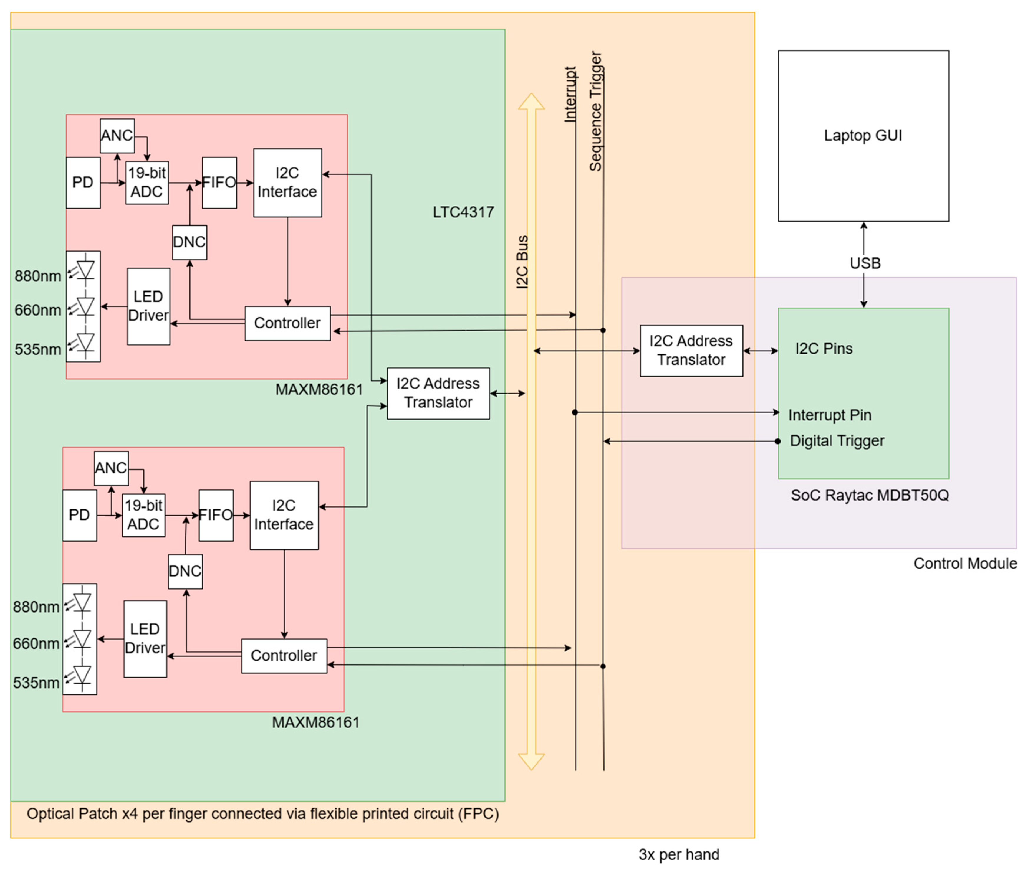
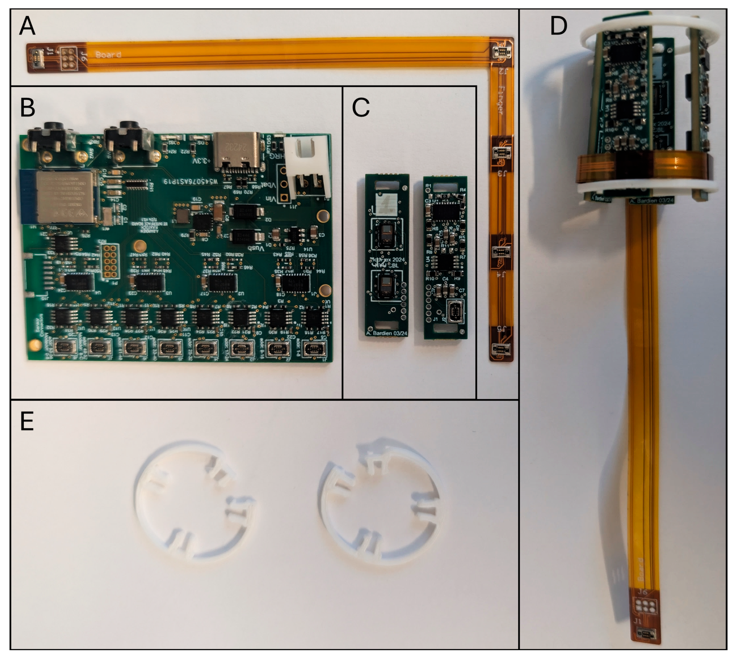
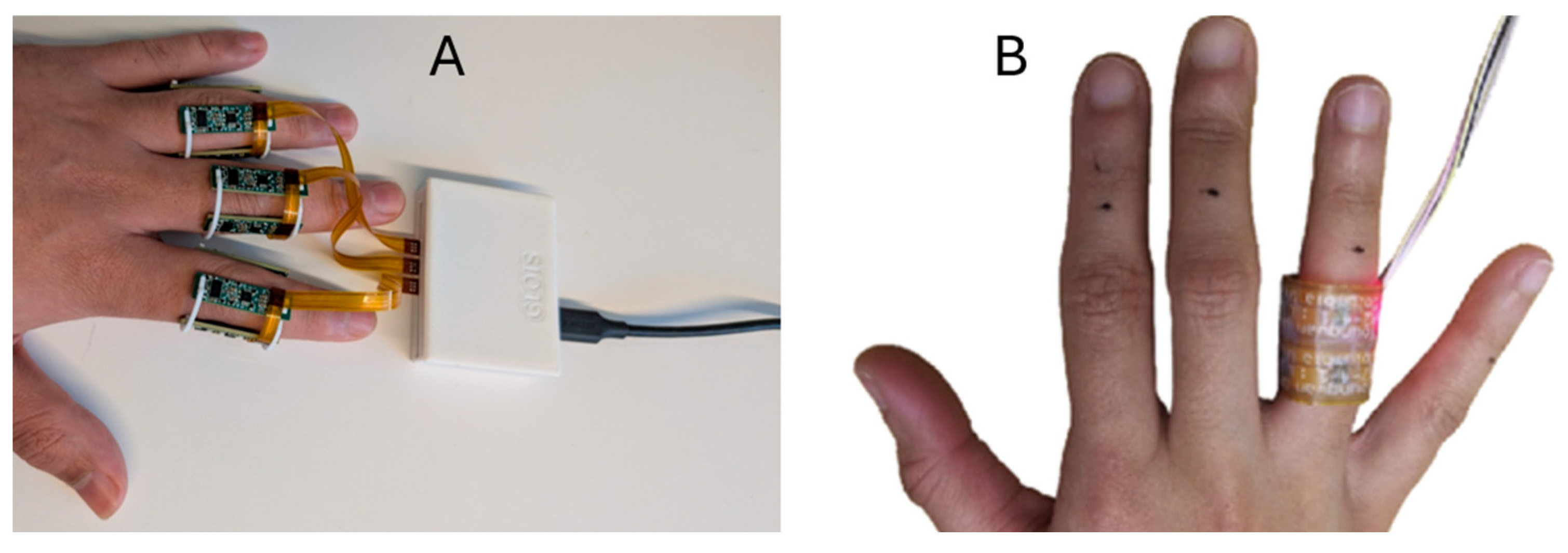
2.1. Instrumentation
2.2. Data Acquisition and Firmware
2.3. Real-Time Reconstruction Algorithm
2.3.1. Background
2.3.2. Methods
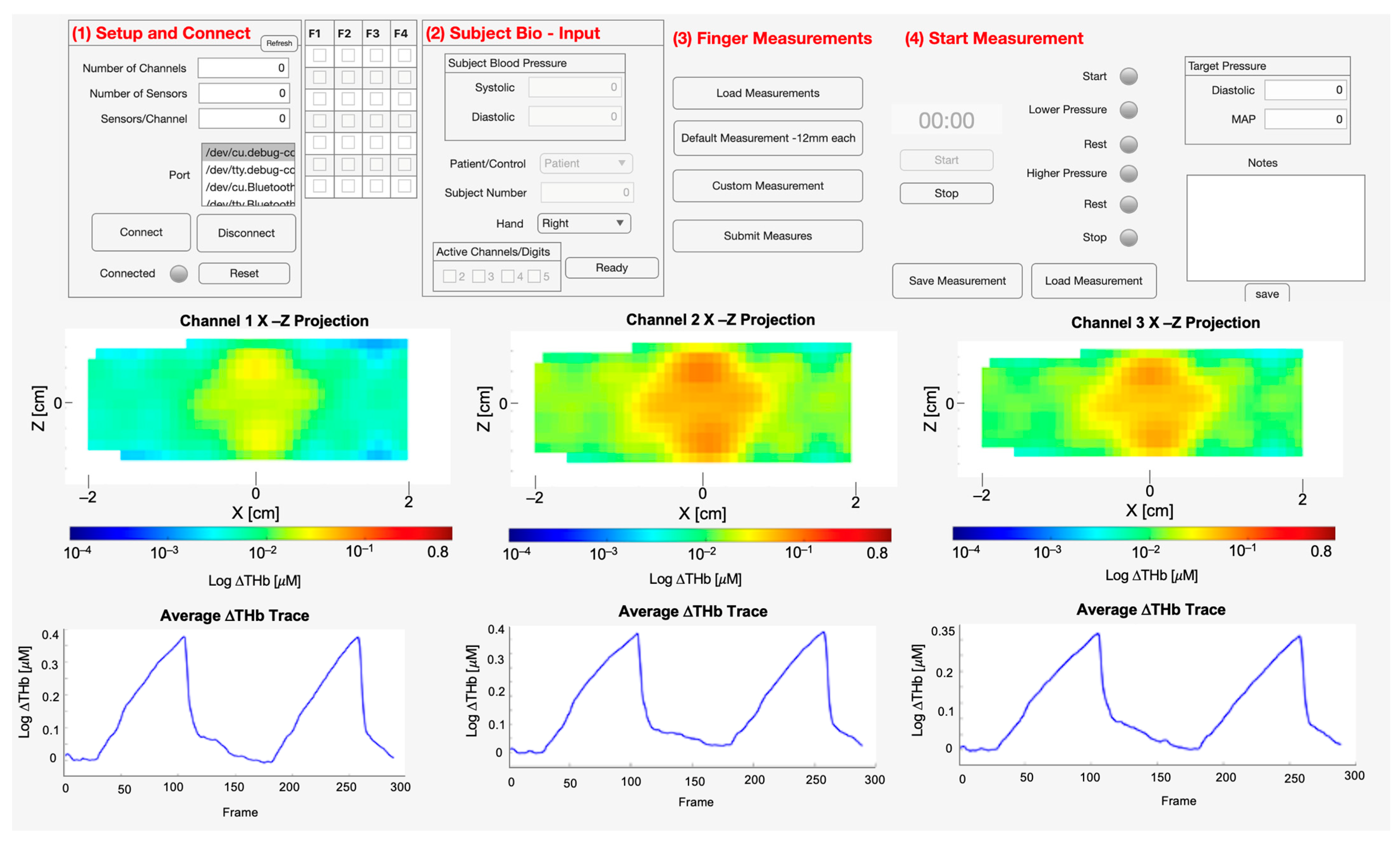
2.4. GUI
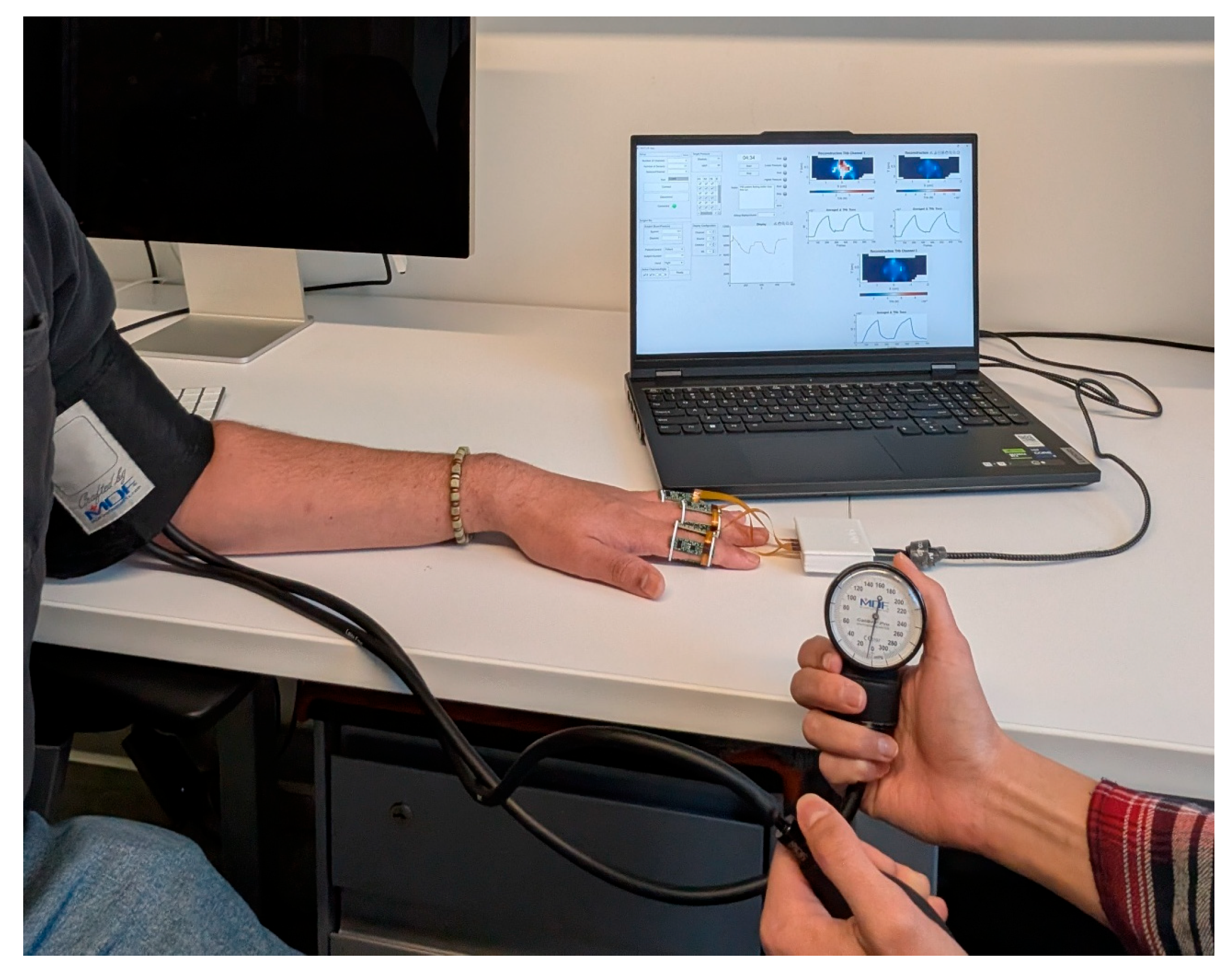
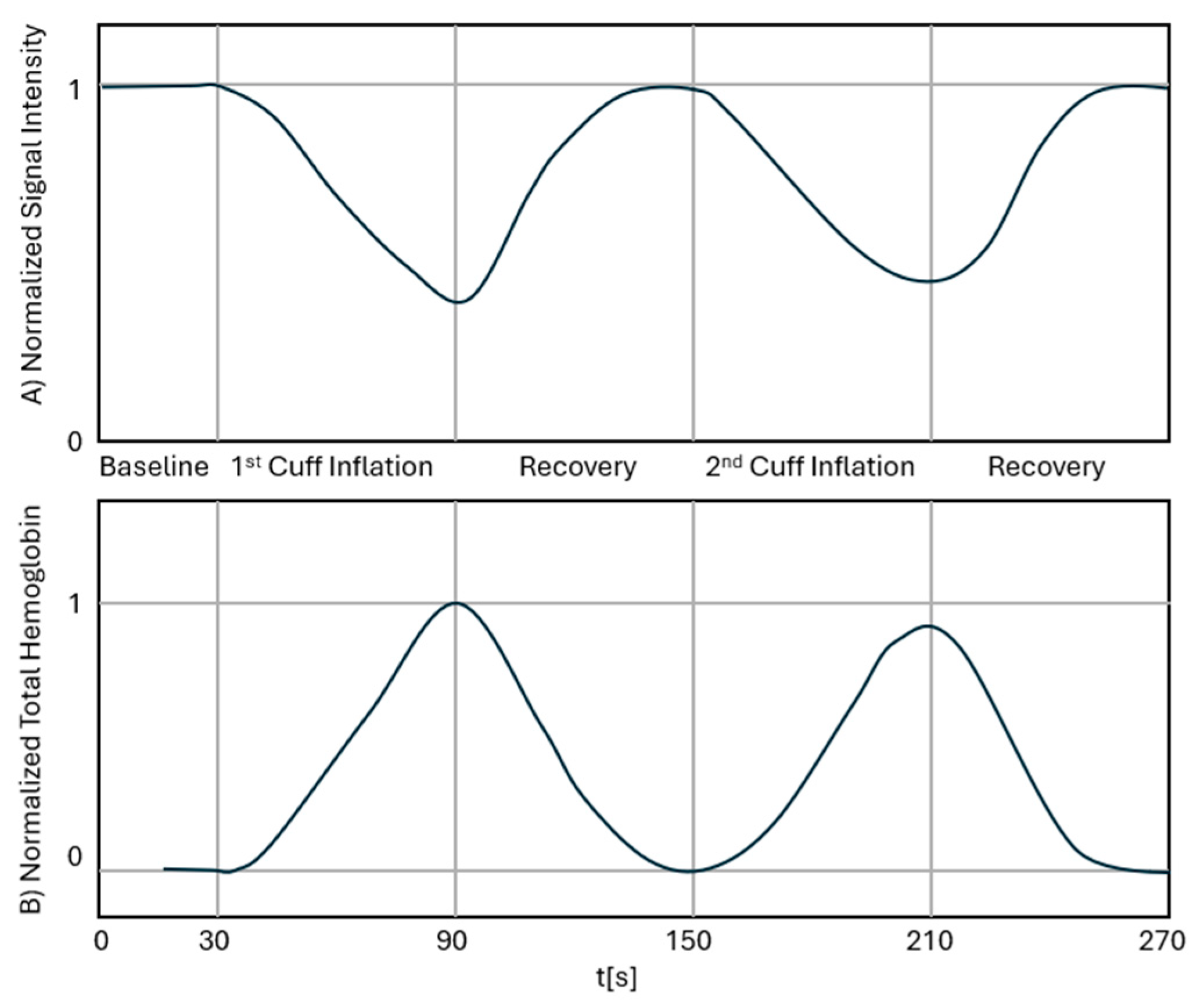
2.5. Measurement Protocol
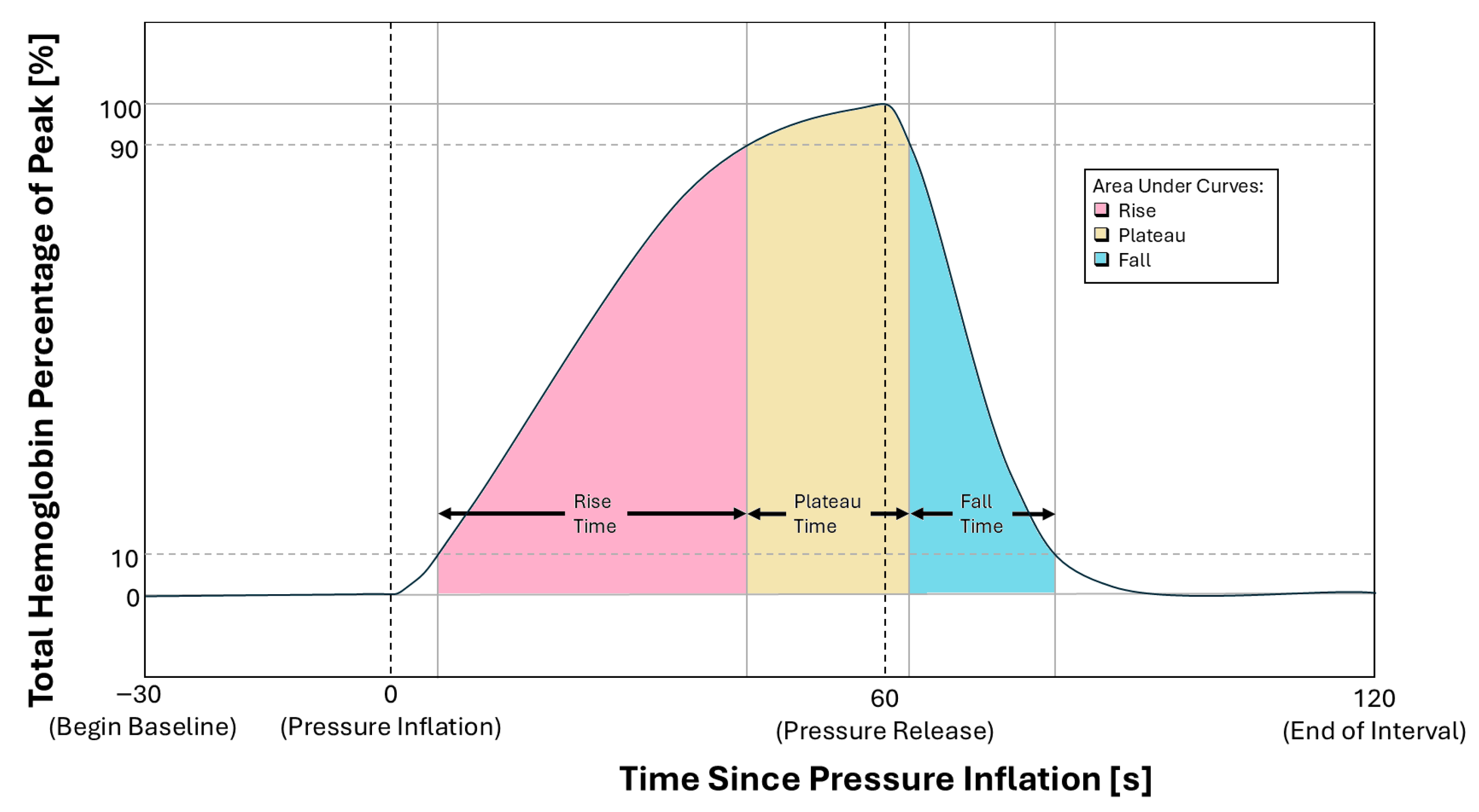
2.6. Parameters of Interest
3. Results and Discussion
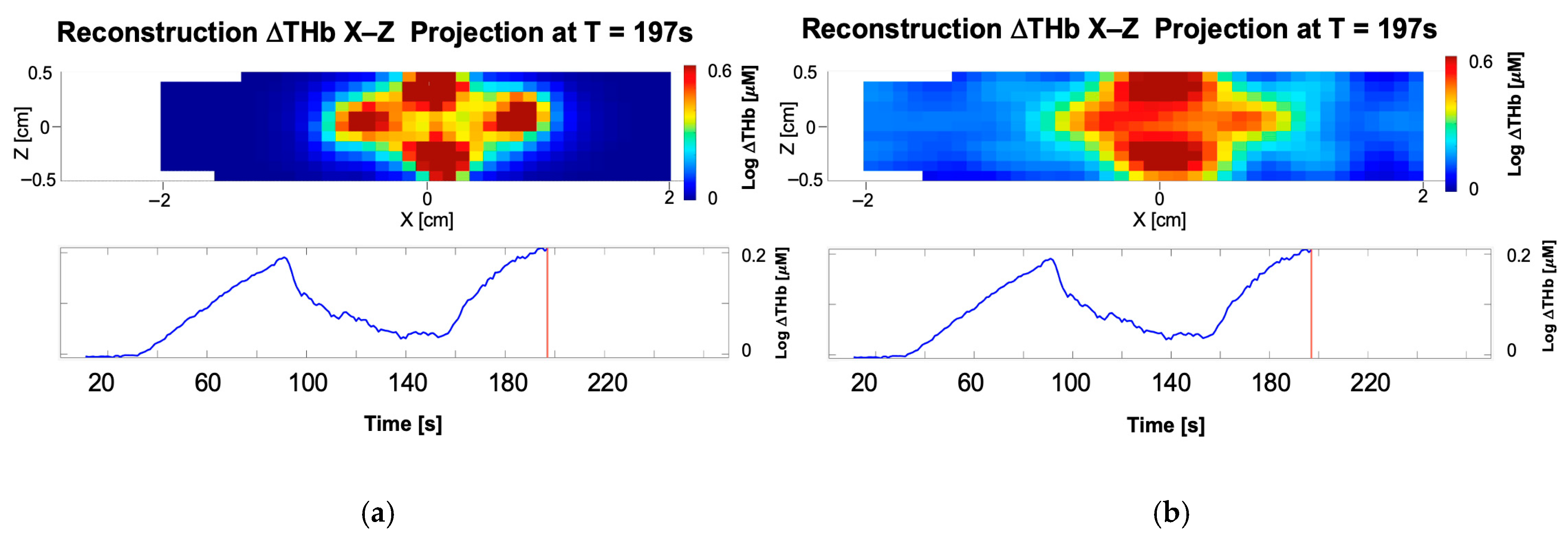
3.1. Comparison of LOTIS to FOIS
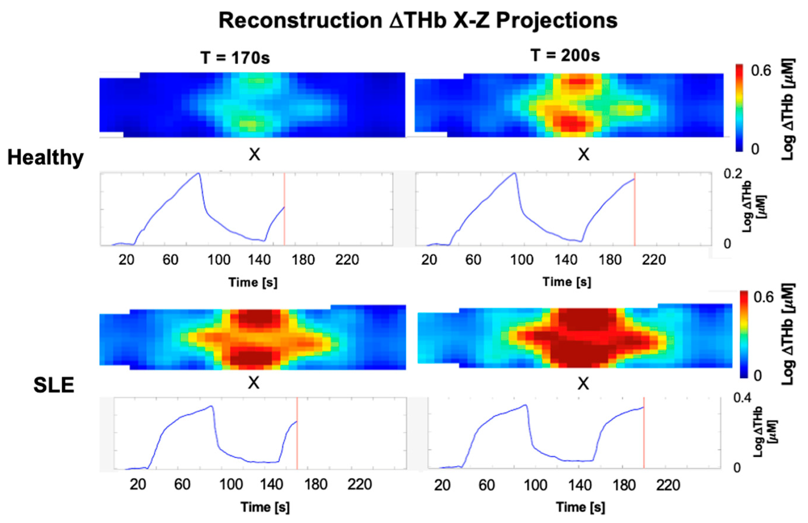
3.2. Initial Human Measurements Demonstrating System Capability
4. Conclusions
5. Patents
Supplementary Materials
Author Contributions
Funding
Institutional Review Board Statement
Informed Consent Statement
Data Availability Statement
Acknowledgments
Conflicts of Interest
Abbreviations
| ADC | Analog-to-digital converter |
| ANC | Ambient Noise Cancellation |
| AUCF | Area Under the Curve (Fall) |
| AUCP | Area Under the Curve (Plateau) |
| AUCR | Area Under the Curve (Rise) |
| BILAG | British Isles Lupus Assessment Group |
| CDC | Centers for Disease Control and Prevention |
| CW | Continuous wave |
| DCT | Discrete Cosine Transform |
| DNC | Digital Noise Cancellation |
| DOT | Diffuse Optical Tomography |
| EMI | Electromagnetic interference |
| FIFO | First In First Out Register |
| FOIS | Flexible Optical Imaging System |
| FPC | Flexible printed circuit |
| FT | Fall Time |
| GUI | Graphical user interface |
| Hb | Deoxygenated hemoglobin |
| HbO2 | Oxygenated hemoglobin |
| LED | Light-emitting diode |
| LOTIS | Lupus Optical Tomography Imaging System |
| MAP | Mean Arterial Pressure |
| MRI | Magnetic resonance imaging |
| MSK-US | Musculoskeletal ultrasound |
| NIR | Near-infrared |
| PD | Photodiode |
| PIP | Proximal interphalangeal (joint) |
| PT | Plateau Time |
| RA | Rheumatoid arthritis |
| RT | Rise Time |
| SD | Source–detector |
| SENSOR | Sensitivity-Equation-Based Non-Iterative Sparse Optical Reconstruction |
| SLE | Systemic Lupus Erythematosus |
| SLEDAI | Systemic lupus erythematosus disease activity index |
| SoC | System on a chip |
| SNR | Signal-to-noise ratio |
| THb | Total hemoglobin |
References
- Izmirly, P.M.; Parton, H.; Wang, L.; McCune, W.J.; Lim, S.S.; Drenkard, C.; Ferucci, E.D.; Dall’Era, M.; Gordon, C.; Helmick, C.G.; et al. Prevalence of Systemic Lupus Erythematosus in the United States: Estimates From a Meta-Analysis of the Centers for Disease Control and Prevention National Lupus Registries. Arthritis Rheumatol. 2021, 73, 991–996. [Google Scholar] [CrossRef] [PubMed]
- Grossman, J.M. Lupus arthritis. Best Pract. Res. Clin. Rheumatol. 2009, 23, 495–506. [Google Scholar] [CrossRef]
- Alarcón, G.S.; Friedman, A.W.; Straaton, K.V.; Moulds, J.M.; Lisse, J.; Bastian, H.M.; McGwin, G.; Bartolucci, A.A.; Roseman, J.M.; Reveille, J.D. Systemic lupus erythematosus in three ethnic groups: III A comparison of characteristics early in the natural history of the LUMINA cohort. Lupus 1999, 8, 197–209. [Google Scholar] [CrossRef]
- Zhu, T.Y.; Tam, L.S.; Lee, V.W.Y.; Lee, K.K.; Li, E.K. Relationship Between Flare and Health-related Quality of Life in Patients with Systemic Lupus Erythematosus. J. Rheumatol. 2010, 37, 568–573. [Google Scholar] [CrossRef]
- Frade-Sosa, B.; Sarmiento-Monroy, J.C.; Salman-Monte, T.C.; Corzo, P.; Gómez-Puerta, J.A. Diagnosis and treatment of articular manifestations of systemic lupus erythematosus. Rev. Colomb. Reumatol. 2021, 28, 90–100. [Google Scholar] [CrossRef]
- Carter, L.M.; Gordon, C.; Yee, C.S.; Bruce, I.; Isenberg, D.; Skeoch, S.; Vital, E.M. Easy-BILAG: A new tool for simplified recording of SLE disease activity using BILAG-2004 index. Rheumatology 2022, 61, 4006. [Google Scholar] [CrossRef]
- Gladman, D.D.; Ibañez, D.; Urowitz, M.B. Systemic lupus erythematosus disease activity index 2000. J. Rheumatol. 2002, 29, 288–291. [Google Scholar] [PubMed]
- Isenberg, D.A.; Rahman, A.; Allen, E.; Farewell, V.; Akil, M.; Bruce, I.N.; D’Cruz, D.; Griffiths, B.; Khamashta, M.; Maddison, P.; et al. BILAG 2004. Development and initial validation of an updated version of the British Isles Lupus Assessment Group’s disease activity index for patients with systemic lupus erythematosus. Rheumatology 2005, 44, 902–906. [Google Scholar] [CrossRef]
- Inês, L.S.; Fredi, M.; Jesus, D.; Shumilova, A.; Franceschini, F.; Vital, E.M. What is the best instrument to measure disease activity in SLE?—SLE-DAS vs Easy BILAG. Autoimmun. Rev. 2024, 23, 103428. [Google Scholar] [CrossRef]
- Arora, S.; Isenberg, D.A.; Castrejon, I. Measures of Adult Systemic Lupus Erythematosus: Disease Activity and Damage. Arthritis Care Res. 2020, 72, 27–46. [Google Scholar] [CrossRef]
- Sudoł-Szopińska, I.; Żelnio, E.; Olesińska, M.; Gietka, P.; Ornowska, S.; Power, D.J.; Taljanovic, M.S. Update on Current Imaging of Systemic Lupus Erythematous in Adults and Juveniles. J. Clin. Med. 2022, 11, 5212. [Google Scholar] [CrossRef]
- Rowbotham, E.L.; Grainger, A.J. Rheumatoid arthritis: Ultrasound versus MRI. Am. J. Roentgenol. 2011, 197, 541–546. [Google Scholar] [CrossRef]
- Chiara, T.; Dario, D.A.; Niccolò, P.; Andrea, D.S.; Davide, C.; Stefano, B.; Marta, M. MRI pattern of arthritis in systemic lupus erythematosus: A comparative study with rheumatoid arthritis and healthy subjects. Skelet. Radiol. 2015, 44, 261–266. [Google Scholar] [CrossRef]
- Di Matteo, A.; Corzo, P.; Filippucci, E. The role of ultrasound and magnetic resonance imaging in the assessment of musculoskeletal involvement in systemic lupus erythematosus: A review of recent advances and insights. Best Pract. Res. Clin. Rheumatol. 2025, 39, 102042. [Google Scholar] [CrossRef]
- Golovko, D.; Meier, R.; Rummeny, E.; Daldrup-Link, H. Optical imaging of rheumatoid arthritis. Int. J. Clin. Rheumatol. 2011, 6, 67. [Google Scholar] [CrossRef]
- Jacques, S.L. Optical properties of biological tissues: A review. Phys. Med. Biol. 2013, 58, R37–R61. [Google Scholar] [CrossRef]
- Welch, A.J.; van Gemert, M.J.C. Optical-Thermal Response of Laser-Irradiated Tissue; Plenum: New York, NY, USA, 1995. [Google Scholar]
- Hielscher, A.H.; Bluestone, A.Y.; Abdoulaev, G.S.; Klose, A.D.; Lasker, J.; Stewart, M.; Netz, U.; Beuthan, J. Near-infrared diffuse optical tomography. Dis. Markers 2002, 18, 313–337. [Google Scholar] [CrossRef] [PubMed]
- Hielscher, A.H.; Hyun Keol, K.; Montejo, L.D.; Blaschke, S.; Netz, U.J.; Zwaka, P.A.; Illing, G.; Muller, G.A.; Beuthan, J. Frequency-Domain Optical Tomographic Imaging of Arthritic Finger Joints. IEEE Trans. Med. Imaging 2011, 30, 1725–1736. [Google Scholar] [CrossRef] [PubMed]
- Kim, Y.; Marone, A.; Tang, W.; Gartshteyn, Y.; Kim, H.K.; Askanase, A.D.; Kymissis, I.; Hielscher, A.H. Flexible optical imaging band system for the assessment of arthritis in patients with systemic lupus erythematosus. Biomed. Opt. Express 2021, 12, 1651–1665. [Google Scholar] [CrossRef] [PubMed]
- Bluestone, A.; Abdoulaev, G.; Schmitz, C.; Barbour, R.; Hielscher, A. Three-dimensional optical tomography of hemodynamics in the human head. Opt. Express 2001, 9, 272–286. [Google Scholar] [CrossRef] [PubMed]
- Maheshwari, N.; Marone, A.; Altoé, M.; Kim, S.H.K.; Bajakian, D.R.; Hielscher, A.H. Postintervention monitoring of peripheral arterial disease wound healing using dynamic vascular optical spectroscopy. J. Biomed. Opt. 2022, 27, 125002. [Google Scholar] [CrossRef]
- Kim, H.K.; Zhao, Y.; Raghuram, A.; Veeraraghavan, A.; Robinson, J.; Hielscher, A.H. Ultrafast and Ultrahigh-Resolution Diffuse Optical Tomography for Brain Imaging with Sensitivity Equation based Noniterative Sparse Optical Reconstruction (SENSOR). J. Quant. Spectrosc. Radiat. Transf. 2021, 276, 107939. [Google Scholar] [CrossRef]
- Arridge, S.R. Optical tomography in medical imaging. Inverse Probl. 1999, 15, R41–R93. [Google Scholar] [CrossRef]
- Xu, R.; Povoski, S. Near-infrared imaging in cancer: A new frontier for personalized medicine. Expert Rev. Med. Devices 2007, 4, 83–94. [Google Scholar] [CrossRef]
- Kim, H.K.; Flexman, M.L.; Yamashiro, D.J.; Kandel, J.J.; Hielscher, A.H. Hand-held, breast-dedicated, diffuse optical tomography system. Biomed. Opt. Express 2010, 1, 812–827. [Google Scholar] [CrossRef]
- Corlu, A.; Choe, R.; Durduran, T.; Lee, K.; Schweiger, M.; Arridge, S.R.; Hillman, E.M.C.; Yodh, A.G. Unstructured grid, three-dimensional diffuse optical tomography of fingers in vivo. Appl. Opt. 2005, 44, 2082–2092. [Google Scholar] [CrossRef] [PubMed]
- Corlu, A.; Durduran, T.; Choe, R.; Schweiger, M.; Hillman, E.M.C.; Arridge, S.R.; Yodh, A.G. Three-dimensional in vivo fluorescence diffuse optical tomography of breast cancer in humans. Opt. Express 2003, 28, 2339–2341. [Google Scholar] [CrossRef] [PubMed]
- Wang, F.; Kim, S.H.; Zhao, Y.; Raghuram, A.; Veeraraghavan, A.; Robinson, J.; Hielscher, A.H. High-Speed Time-Domain Diffuse Optical Tomography With a Sensitivity Equation-Based Neural Network. IEEE Trans. Comput. Imaging 2023, 9, 459–474. [Google Scholar] [CrossRef]
- Karadima, O.; Rahman, M.; Sotiriou, I.; Ghavami, N.; Lu, P.; Ahsan, S.; Kosmas, P. Experimental Validation of Microwave Tomography with the DBIM-TwIST Algorithm for Brain Stroke Detection and Classification. Sensors 2020, 20, 840. [Google Scholar] [CrossRef] [PubMed]
- Nocedal, J.; Wright, S.J. Numerical Optimization; Springer: New York, NY, USA, 2006. [Google Scholar]
- Gilmore, C.; Mojabi, P.; Zakaria, A.; Ostadrahimi, M.; Kaye, C.; Noghanian, S.; Shafai, L.; Pistorius, S.; Lovetri, J. A Wideband Microwave Tomography System With a Novel Frequency Selection Procedure. IEEE Trans. Biomed. Eng. 2010, 57, 894–904. [Google Scholar] [CrossRef]




| Feature | LOTIS | FOIS |
|---|---|---|
| Light Source | 2 LEDs (660 nm, 880 nm) | 3 LEDs (530 nm, 655 nm, 940 nm) |
| Simultaneous Channels | Configurable (Used in a tri-channel configuration) | Single channel only |
| Frame Rate | 1.25 Hz (in tri-channel mode; higher if fewer channels are used) | 0.75 Hz |
| Signal Conditioning | On-module ambient light & digital noise cancellation | No on-board filtering |
| A/D Conversion | On-module | On a separate control board |
| Construction | Modular: Rigid sensor PCBs with flexible interconnects | Integrated: Single flexible PCB assembly |
| Fixation Method | 3D-Printed flexible rings | Micropore™ surgical tape |
| SNR (range, mean) | Range: 18–46 dB; Mean: 23 dB | Range: 7–28 dB; Mean: 17 dB |
| Inverse Model Computation | <0.2 s (2× speed up via DCT) | Conventional Elementwise |
| Reconstruction Time | <0.5 s per frame | 5 s per frame |
| Parameter | Description | Healthy Joints (Representative Range) | SLE Joints (Representative Range) |
|---|---|---|---|
| Rise Time (RT) | Time to 90% peak signal | 44–48 s | 40–46 s |
| Plateau Duration (PT) | Duration of peak occlusion | 8–13 s | 9–16 s |
| Fall Time (FT) | Time to return to baseline | 33–44 s | 19–40 s |
| Rise AUC (AUCR) | Blood volume accumulation | 1250–1600 | 1100–1400 |
| Plateau AUC (AUCP) | Blood volume sustained | 470–760 | 550–880 |
| Fall AUC (AUCF) | Blood volume outflow | 2600–3800 | 1400–3300 |
Disclaimer/Publisher’s Note: The statements, opinions and data contained in all publications are solely those of the individual author(s) and contributor(s) and not of MDPI and/or the editor(s). MDPI and/or the editor(s) disclaim responsibility for any injury to people or property resulting from any ideas, methods, instructions or products referred to in the content. |
© 2026 by the authors. Licensee MDPI, Basel, Switzerland. This article is an open access article distributed under the terms and conditions of the Creative Commons Attribution (CC BY) license.
Share and Cite
Bardien, M.A.; Pinar, L.; Marone, A.; Nordmann-Gomes, A.; Khalili, L.; Suh, S.; Kim, S.H.; Askanase, A.D.; Hielscher, A.H. Patient-Friendly Real-Time Optical Tomographic Imaging System (LOTIS) for Lupus Arthritis. Biosensors 2026, 16, 184. https://doi.org/10.3390/bios16040184
Bardien MA, Pinar L, Marone A, Nordmann-Gomes A, Khalili L, Suh S, Kim SH, Askanase AD, Hielscher AH. Patient-Friendly Real-Time Optical Tomographic Imaging System (LOTIS) for Lupus Arthritis. Biosensors. 2026; 16(4):184. https://doi.org/10.3390/bios16040184
Chicago/Turabian StyleBardien, Moegammad A., Lara Pinar, Alessandro Marone, Alberto Nordmann-Gomes, Leila Khalili, Stephen Suh, Stephen H. Kim, Anca D. Askanase, and Andreas H. Hielscher. 2026. "Patient-Friendly Real-Time Optical Tomographic Imaging System (LOTIS) for Lupus Arthritis" Biosensors 16, no. 4: 184. https://doi.org/10.3390/bios16040184
APA StyleBardien, M. A., Pinar, L., Marone, A., Nordmann-Gomes, A., Khalili, L., Suh, S., Kim, S. H., Askanase, A. D., & Hielscher, A. H. (2026). Patient-Friendly Real-Time Optical Tomographic Imaging System (LOTIS) for Lupus Arthritis. Biosensors, 16(4), 184. https://doi.org/10.3390/bios16040184