Abstract
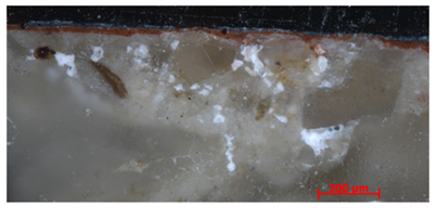
This paper reports the investigation of six microsamples collected from the vault of the San Panfilo Church in Tornimparte (AQ). The aim was to detect the composition of the pigments and protective/varnishes, and to investigate the executive technique, the conservation state, and the evidence of the restoration works carried out in the past. Six microsamples were analyzed by optical microscopy, scanning electron microscopy coupled with energy-dispersive spectroscopy (EDS), X-ray fluorescence (XRF), and infrared and Raman spectroscopy. The investigations were carried out within the framework of the Tornimparte project “Archeometric investigation of the pictorial cycle of Saturnino Gatti in Tornimparte (AQ, Italy)” sponsored in 2021 by the Italian Association of Archeometry (AIAr).
1. Introduction
This paper contributes to the Special Issue “Results of the II National Research project of AIAr: Archaeometric study of the frescoes by Saturnino Gatti and workshop at the church of San Panfilo in Villagrande di Tornimparte (AQ, Italy)”, in which the scientific results of II National Research Project conducted by members of the Italian Association of Archaeometry (AIAr (Associazione Italiana di Archeometria. http://www.associazioneaiar.com/wp/home-2/, accessed on 20 February 2023)) are discussed and collected. For in-depth details on the aims of the project, see the introduction of the Special Issue [1]. This work is a first attempt to understand the historical past of these frescoes on the basis of a direct observation of the current state of conservation and a study of the (fragmentary) documentation preserved in the archive [2,3]. The frame in which the execution of the pictorial cycle can be placed begins with a note of an advanced payment given to the artist in 1489; it was 1491 when Saturnino started to work on the frescoes which were completed in 1494 [2]. Art historians consider this cycle of frescoes the cornerstone of Saturnino Gatti’s activity as a painter; it is also considered his masterpiece for the close and evident similarities that the frescoes demonstrate with the models of his famous master, the Florentine sculptor and painter Andrea del Verrocchio. New information from the recent analysis campaign is added [4,5,6]. The pre-restoration analysis is an invaluable contribution, setting objective data on the quality and nature of the original and restoration materials and degradation processes, leading us on a parallel path that confirms and enriches the information reported on the documents in our possession. In the long life of an artwork, the conservation events are difficult to reconstruct [7,8], on one hand, because the news is often fragmentary and inaccurate, and, on the other, because it is not possible to see beyond the mere aesthetic aspect through only visual inspection. In addition to the normal degradation to which the constituent materials are subjected, we have to deal with clumsy and invasive interventions and inadequate maintenance work. In this regard, there is not much to add to the description made by Giuseppina Vecchioli where she describes how the frescoes of the vault appear “grossly repainted”, and how the figures are excessively reintegrated [9]. This is how the most important pictorial cycle of Saturnino Gatti appears today. Its history is not only made up of localized and limited interventions responding to the scarce economic resources of the moment, since other factors have determined uneven intervention, always characterized by different criteria. It is then necessary, as spectators, to take a step back and try to take a view of the most important events that have marked the history of these frescoes. Through the examination of some documents, we can verify how there have been episodes causing degradation in paintings over the years. Some of these events are listed in a letter signed by Mons. Bernardino Santucci dated 12 February 1964, and addressed to the Superintendent of Medieval and Modern Art and the Minister of Education: “the earthquake of 15 January 1915, devastating war of 1940–1944, very hard winter of 1962–1963, and permanent moisture penetrating from the roofs and the floor under which are burials of the dead” (12 February 1964). The first documented information placed in the records of this Superintendency concerning this pictorial cycle dates back to 1950 and after the note signed by the Superintendent which describes: “Some bombs that exploded nearby damaged the roof and the windows in various points”, attaching an appraisal of the amount of 40,000 ITL “to prevent the next winter season from damaging the paintings”. Another note of 3 August 1950 for the financing “will follow… on the chap. 257 relating to war damages”. The total amount of 3,400,000 ITL concerned three of the most damaged sacred buildings in the area, including S. Panfilo di Tornimparte (Letter from Soprintendenza ai Monumenti e Gallerie degli Abruzzi e Molise, addressed to Ministero della Pubblica Istruzione e Direzione Generale delle Belle Arti, 3 August 1950). To the catastrophic events are added others dictated by the lack of maintenance and neglect (such as the fire of October 1958), or little availability of funds, all leading us to the same result, i.e., “the frescoes of Gatti are deteriorating visibly”. A letter written by the then parish priest Bernardino Santucci recalls the damage suffered by the building: “After the restorations of 1929, nothing more remarkable has been done for the conservation and decoration of the Holy Temple and its sacred works of art of which it is rich”. It continues: “Because of the bad weather of the mountain and of the war, everything is in a state of decay and ruin and urgent action is required as the pictorial cycle continues to deteriorate” (Letter from Sac. Bernardino Santucci, addressed to Direttore Generale, 15 February 1951). At the previous request, and, after some exchanges between the superintendency and the Ministry of Public Education, in August, the Superintendent requested and communicated that it would be sufficient to carry out the work on the frescoes, that another estimate was received, that an allocation was approved, and that the announcement of the forthcoming start of the work was given (Letter from Soprintendente Dott. Arch. Umberto Chierici, addressed to Reverendo Parroco di S. Panfilo, 13 September 1951). In 1951, the allocation of the necessary funds for the restoration of the frescoes by Saturnino Gatti in Tornimparte was approved. For the approval of these funds, an expense report is found in the documents that describe in detail the intervention proposed on 30 August 1951: “cleaning of the painted surface and removal of vast in paintings altered by time, fixing of the crumbling and crumbling color and consolidation of swollen plaster areas with injections of lime caseate, filling of cracks or gaps in backgrounds with local glazes”.
The Wall Paintings of the Apse of San Panfilo
These paintings were restored for the first time in 1929 by Domenico Brizi. Another intervention to mention is that by Enrico Vivio, following the fire of 1958–1959. A new communication dated 5 November 1958 resumes the need for a restoration of the pictorial cycle, this time due to the fire caused by electric damage in the night between 5 and 6 October in the parish sacristy, with consequent damage to the frescoes in the apse [5]. On 12 November 1958, the Superintendent announced that a technician had been sent to carry out the necessary investigations, confirming in a subsequent note dated 12 November 1956 the start of “cleaning and restoration” work and confirmed by a letter of thanks dated 14 April 1959, written by the parish priest of San Panfilo Mons. Bernardino Santucci (Letter from Mons. Bernardino Santucci, addressed to Soprintendente all’arte Medievale e Moderna L’Aquila, 10 April 1959). About 15 years after this last restoration, a new overall intervention of the paintings was entrusted to the restorers Antonio Liberti and Umberto Marini, and, in a letter addressed to B. Santucci, the architect Mario Moretti specifies that the intervention would begin, but only once the roof was restored “… and, nevertheless, not before the spring of 1971” (29 December 1970). “The frescoes of the vault, due to the infiltration of moisture from the roof, have large oxidations and blooms of carbonates and nitrates and the presence of mildew, which have seriously altered the blues. In some areas the color has fallen; in others, the color is curled and unsafe” (arch. Mario Moretti, 17 April 1972). The architect Moretti describes a very bad state of conservation of the paintings of the vault. Among other things, he mentions the serious alteration “of the blues”, covering a large area; hence, at the time, a trace of the original coloring of the sky was perhaps still visible. It is worth mentioning that, with the cleaning carried out on that occasion, according to Liberati, the original colors were recovered as much as possible, freeing the frescoes of the superfetations. According to the judgment of Ferdinando Bologna, the cleaning was excessive, compromising the legibility of important details. Unfortunately, details of the appraisal presented are sparse and do not offer useful information to trace the cleaning system used or if a pre-consolidation work of the pictorial film was performed before cleaning the surface, deducing that the frequent infiltrations had generated a serious problem of decohesion of the pictorial layer. At first glance, the pictorial work still manages to convey the beauty of the composition and the accurate stroke of its figures. Conducting a more accurate visual inspection, the numerous “interferences” against the pictorial film creep into the eyes of the viewer; the celestial vault appears covered by a heavy and compact dark-blue application and many interventions on some figures, distorted by interpretative integrations. On the walls, there are gaps, abrasions of the pictorial film and humidity problems. The investigations conducted, using different methods, analyzed each situation and found answers that are not handed down in the documents. In the apse, the one that first catches the eye is the background of a very compact blue. The sky is heavily repainted, leaving a set of figures “cut out and outlined” by heavy coloring. On the other hand, the figures floating in the sky show both signs of degradation, such as abrasions and small gaps, and repaints that overload the features of hands and faces. The succession of winter seasons, frost and thaw, and repeated episodes of infiltration from the roof have caused the degradation of the supporting plaster and serious decohesion of the pictorial film as described by the architect Mario Moretti in his report. However, it is to be assumed that these episodes have been a constant in the long life of these paintings, as demonstrated by the same repainting already mentioned. Therefore, the aim of this work is to identify the pigments, binders, and protective/varnishes, to study the stratigraphy, and to investigate the executive technique and the conservation state of the paintings at the Vault. Attention was paid to evaluate the original pigments or eventually the ones used during a restoration work performed in the past. Six microsamples collected in the Vault were analyzed with the main aim of understanding the stratigraphy and providing a post quem term for the realization of the extensive integrations and additions, which altered the colors and original reading of the fresco. The adopted methodology is based on a combined use of several techniques, widely used for elemental and molecular investigations of pigments in the field of Cultural Heritage: observation of cross-sections under optical microscopy and scanning electron microscopy coupled with EDS detection, X-ray fluorescence (XRF), and Raman spectroscopy, along with an investigation of each layer thought μ-infrared spectroscopy [10,11,12].
2. Materials and Methods
2.1. Materials
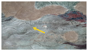
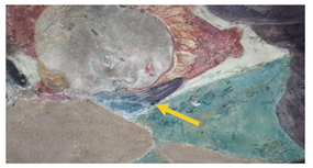
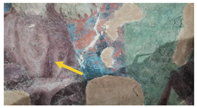
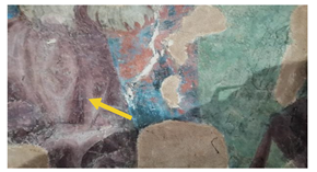
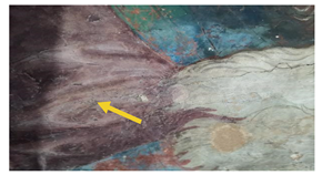
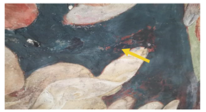
The six microsamples were selected on the basis of the results given by the multispectral investigation, reported in [4,5,6]. All microsamples were collected in the upper part of the vault, as reported in Figure 1.
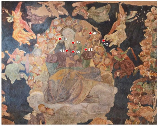
Figure 1.
Area of the vault where the microsamples have been collected. The number corresponds to the ID microsamples.
The ID samples and a photo of each microsample, together with the aim of the specific investigation, are reported in Table 1.

Table 1.
ID sample, area of the sampling, photo of the microsamples, and their macroscopic description.
2.2. Methods
The cross-sections were prepared by embedding the collected microsamples into epoxy resin (bicomponent Prochima E30) and curing until solidification at room temperature. The resin was then cut and polished to get the stratigraphy across the center of the objects.
Optical microscopy observation was performed using a LEICA EZ24W optical microscope in reflection mode. Sample 39B cross-section was observed in reflected light by means of a ZEISS Axio Scope A1 microscope, equipped with a video camera (5 MP resolution), and image analysis software AxioVision (V1).
SEM investigation coupled with energy-dispersive X-ray spectroscopy (EDS) analysis was performed using a Phenom Pro X, Phenom-World (The Netherlands) with an optical magnification range of 20×–135×, electron magnification range of 80,000×–130,000×, maximal digital zoom of 12×, and acceleration voltage of 15 kV, as well as an EDS detector, with a nominal resolution of 10 nm or less. The microscope was equipped with a temperature-controlled (25 °C) sample holder. The samples were positioned on an aluminum stub using adhesive carbon tape. Morphological and semiquantitative microchemical analyses of sample 39B were performed using a SEM-EDS electronic microscope (ZEISS EVO MA 15) with a W-filament equipped with an analysis system in energy dispersion EDS/SDD, Oxford Ultimax 40 (40 mm2 with resolution 127 eV @ 5.9 keV) and the Aztec 5.0 SP1 software. Measurements were performed on a carbon-metallized cross-section of the sample under the following operating conditions: accelerating potential of 15 kV, 500 pA beam current, working distance between 9 and 8.5 mm, 20 s live time as an acquisition rate useful for archiving at least 600,000 cts, on co-standard and process time 4 for point analyses, and 500 μs pixel dwell time for recording 1024 × 768 pixel resolution maps. The software for the microanalysis was Aztec 5.0 SP1 software, which uses the XPP matrix correction scheme developed by Pouchou and Pichoir in 1991 [13].
XRF spectra were acquired using a Tracer III SD Bruker AXS portable spectrometer. The irradiation by a Rhodium Target X-Ray tube operating at 40 kV and 11 µA and the detection of fluorescence X-rays by a 10 mm2 silicon drift X-Flash detector allowed the detection of elements with atomic number Z > 11. A window of 3–4 mm in diameter determined the sampled area. Each spectrum was acquired for 30 s. The S1PXRF® software (https://s1pxrf.software.informer.com/, accessed on 14 April 2023) was used for data acquisition and spectral assignments. The fluorescence signal area was estimated once the deconvolution of the whole spectrum was performed using the software ARTAX 7. Ar, Ni, Pd, and Rh signals, due to the atmosphere and instrumental components, were also present in all spectra.
Raman spectra were acquired through a LabRAM HR-800 Jobin-Yvon spectrometer equipped with a laser at 633 nm, and a Bravo Bruker spectrometer equipped with two lasers at 785 and 853 nm.
The μ-IR spectra were acquired through a micro FT-IR Lumos Bruker spectrophotometer equipped with a Platinum ATR unit using a germanium crystal operating in the spectral range between 4000 and 600 cm−1, with a spectral resolution of 2 cm−1 and 120 scans. The size of the investigated area was determined by the size of the crystal tip (~50 microns). In all spectra, a baseline correction of scattering was made. Data analysis was performed using the OPUS 7.5® software.
3. Results and Discussion
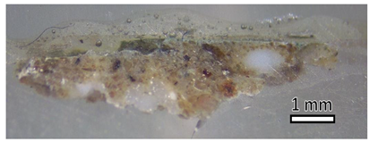
The ID samples, the macro-photos of the cross sections observed under the optical microscope, and the description of the observed layers are reported in Table 2.

Table 2.
ID sample, macro-photos of the cross-section observed under the optical microscope, and description of the observed layers.
For all samples, the plaster made of calcium carbonate (CaCO3), consisting of aggregates of variable grain size and different colors, and the presence of a protective copolymer of vinyl-acrylate acetate/Mowilith DM912 [14], ascribed to a previous restoration, were identified. In most of the microsamples, the presence of weddelite or whewellite, attributable to the growth of microorganisms responsible for biological degradation [15], was identified.
The obtained results for each microsample are reported separately below.
3.1. SG_37B Microsample
This microsample is constituted by two layers:
- –
- Plaster made with a light-colored binder (calcium carbonate, CaCO3), consisting of aggregates of variable grain size and color;
- –
- Continuous and homogeneous dark-gray layer.
The XRF spectra show the peaks of calcium (Ca), iron (Fe), and lead (Pb), suggesting that the gray layer is made up of a mixture of several parts of ochres (iron oxides) and a black pigment (such as carbon black), probably the so-called caput mortuum pigment, also known as morellone (a mixture of red ochre and organic black). The presence of lead (Pb) indicates the use of white lead pigment (Figure 2). The FT-IR spectrum shows absorption bands at 1783, 1428, 870, and 716 cm−1, attributed to the characteristic vibrations of the carbonate ion (CO32−) [16], used as a binder, and the absorption bands at 2973, 2926, 1731, 1433, 1370, 1227, 1120, 1020, 945, 865, 795, 632, and 604 cm−1 ascribable to a synthetic resin known as Mowilith DM 912 [17].
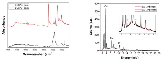
Figure 2.
(left) FT-IR and (right) XRF spectra acquired on the front and back of the SG_37B micro sample before the preparation of the cross-section.
3.2. SG_38 Microsample
This microsample is constituted by three layers:
- –
- Plaster made with a light-colored binder (calcium carbonate), consisting of aggregates of variable grain size and color;
- –
- Continuous gray layer;
- –
- Green layer.
The XRF spectra (Figure 3) show the peaks of calcium (Ca) and iron (Fe), suggesting that the gray layer was obtained by mixing a white pigment with a black pigment probably obtained from natural earth (organic black, a mixture of clay and calcium carbonate, iron, and manganese). For the green pigment, the presence of iron (Fe) and silicon (Si) in the XRF spectra suggests the use of green earth (ferrous and ferric silicates of potassium, manganese, and aluminum plus oxides of Fe, Mg, Al, and K). As with the previous microsample, the FT-IR analysis showed the presence of calcium carbonate and a synthetic resin known as Mowilith DM 912. The bands of whewellite (1623, 1320, and 780 cm−1) were also present [15].
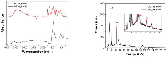
Figure 3.
(left) FT-IR and (right) XRF spectra acquired on the front and back of the SG_38 microsample before the preparation of the cross-section.
3.3. SG_39A Microsample
This microsample is constituted by three layers:
- –
- Plaster made with a light-colored binder (calcium carbonate);
- –
- First layer of white drafting;
- –
- Second layer of red drafting (red ochre/hematite);
- –
- Third layer of white drafting (calcite);
- –
- Red layer applied a secco, retouching;
- –
- Blue layer (Prussian blue) applied a secco, retouching.
The EDS analysis, acquired along the dashed line reported in the SEM micrograph (Figure 4), showed the elements present in the different layers. The results mainly indicated the presence of calcium (Ca), lead (Pb), and iron (Fe). From the distribution of the elements along the cross-section of the sample, from the plaster toward the surface pictorial layers, a high Ca content can be observed, which is attributable to the calcite present in the plaster and in the first white layer, followed by Fe in correspondence of the second red layer, which suggests the use of red ochre. The use of minium or other lead-based pigments such as litharge or massicot cannot be excluded [18]. However, the presence of lead (Pb) may also indicate the use of lead white pigment. An increase in Ca was observed again, in correspondence with the third white layer, followed by the Fe presence for the second red layer and the last blue layer, which suggests the use of red ochre and Prussian blue (Fe4[Fe(CN)6]3), respectively.
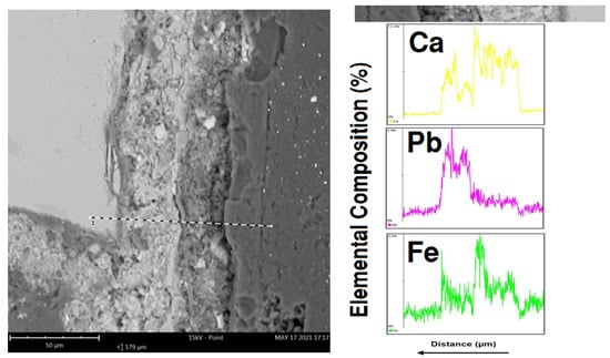
Figure 4.
(left) SEM micrograph and (right) the corresponding EDS profile of SG_39A cross-section. The EDS investigation was performed along the dashed line to evaluate the distribution of Ca, Pb, and Fe from the back to front of the sample.
As for the previous microsamples, the FT-IR analysis showed the presence of calcium carbonate, a synthetic resin known as Mowilith DM 912, and whewellite and weddellite. In addition, the IR bands at about 2100 cm−1, attributable to the stretching of the triple bond –C≡N. and two Raman bands at 520 and 2148 cm−1, attributable to the ferric hexacyanoferrate, Fe4[Fe(CN)6]3, are ascribable to the Prussian blue pigment. The Raman band at 1008 cm−1 is typical of gypsum (CaSO4)∙2H2O. The presence of Prussian blue is ascribable to a retouching, applied a secco (Figure 5).
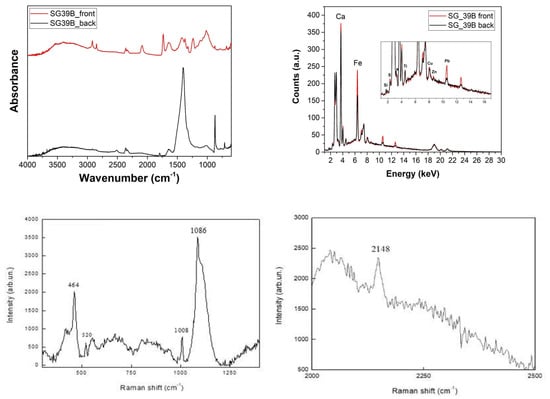
Figure 5.
(top left) FT-IR and (top right) XRF spectra acquired on the front and back of the SG_39A microsample. (bottom left, right) Raman spectra acquired on SG_39A before the preparation of the cross-section.
3.4. SG_39B Microsample
This microsample is constituted by three layers:
- –
- Plaster containing rounded and squared grains;
- –
- First layer of red coating, probably the so-called morellone or caput murtuum (a mixture of red ochre and organic black) [19];
- –
- Blue layer (approximately 20 µm) with white grains inside.
The EDS analysis was acquired on the three layers and, when required, in correspondence with specific colored grains, in order to characterize every single pigment (Figure 6). The acquired spectrum on the observed underlayer (plaster) showed large amounts of calcium (Ca), in addition to silicon (Si), aluminum (Al), sodium (Na), magnesium (Mg), potassium (K), and iron (Fe). Concerning the above red layer (SEM-EDS not reported), the only detected chromophore element was Fe, in relation to the use of red ochre, which supports the hypothesis made for morellone. The analysis of the blue layer showed the presence of a high percentage of lead (Pb), in white grains, due to the use of lead white pigment, as well as Si, Al, Na, and sulfur (S), in blue grains, all distinctive elements for ultramarine blue.
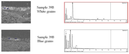
Figure 6.
(left) SEM micrographs and (right) the corresponding EDS spectra of the SG_39B microsample cross-section.
3.5. SG_41A Microsample
This microsample is constituted by three layers:
- –
- Plaster made of a light-colored binder (calcium carbonate), consisting of aggregates of variable grain size and color;
- –
- White layer;
- –
- Continuous, compact, and homogeneous purplish layer.
The EDS analysis, acquired along the dashed line in the SEM micrograph, showed the trends of the characteristic elements Ca, Fe, and a slightly higher amount of manganese (Mn), Figure 7. From the distribution of the elements along the cross-section of the sample, from the plaster toward the superficial pictorial layers, a high Ca content could be observed, attributable to the calcite present in the plaster and in the first white layer, followed by an intense peak of Fe in correspondence with the purplish layer, which, together with the presence of small quantities of Mn, suggests the use of umber (iron and manganese oxides). This hypothesis was confirmed by the XRF spectra because the same elements were identified. As for the previous microsamples, the IR analysis showed the presence of calcium carbonate, Mowilith DM 912, and whewellite and weddellite. The FT-IR bands at 2900, 2839, 1620, 1121, 1033, 940, and 876 cm−1 confirmed the presence of the pigment caput mortuum mixed with gypsum [20].
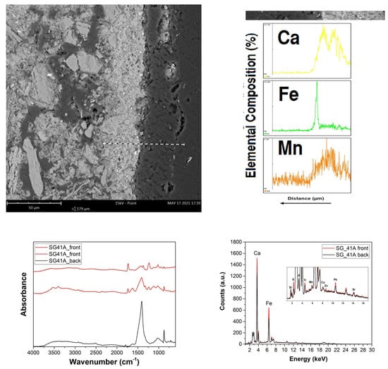
Figure 7.
(top left,right) SEM micrograph and the corresponding EDS profile of the SG_41 microsample cross-section. The EDS investigation was carried out on the dashed line to evaluate the distribution of Ca, Fe, and Mn from back to front of the sample. (bottom left) FT-IR and (bottom right) XRF acquired on the front and back of the SG_41 microsample before preparation of the cross-section.
3.6. SG_42 Microsample
The microsample is constituted by four layers:
- –
- Plaster made of a light-colored binder (calcium carbonate), consisting of aggregates of variable grain size and color;
- –
- First continuous and homogeneous white layer (zinc white);
- –
- Second continuous and homogeneous purplish-red layer (caput mortuum);
- –
- Continuous and inhomogeneous blue layer. The presence of grains of variable size and of different colors, particularly green, can be observed.
The EDS analysis, acquired along the dashed line reported in the SEM micrograph (Figure 8), showed the trends of the distribution of the characteristic elements Ca, zinc (Zn), Fe, and copper (Cu) along the cross-section of the sample from the plaster toward the surface paint layers. A high Ca content, attributable to the calcite of the plaster, was observed, followed by an increase in the Zn and Fe content, indicating the presence of zinc oxides in the white layer and of iron oxides in the red purplish layer, respectively, along with a subsequent increase in the Cu content in correspondence with the blue layer, attributable to the presence of a copper-based pigment such as azurite. Since the azurite is a water-soluble substance and reacts with the lime present in the fresh plaster, it was not applied a fresco, but a secco. On the other hand, the presence of green grains was evident under the optical microscope (Figure 9), which could be an indication of malachite, and an indication of the well-known reaction of conversion of azurite in malachite [21]. However, the results did not allow excluding the presence of other green–blue Cu-based pigments [22]. The barium (Ba) signals in the XRF spectra could indicate the presence of impurities in the copper-based pigment.
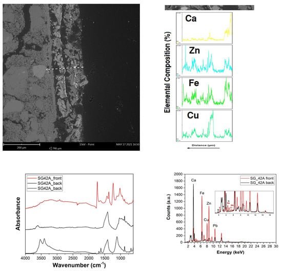
Figure 8.
(top left,right) SEM micrograph and the corresponding EDS profile of the SG_42 microsample cross-section. The EDS investigation was performed on the dashed line. (bottom left) FT-IR and (bottom right) XRF spectra acquired on the front and back of the SG_42 microsample before preparation of the cross-section.
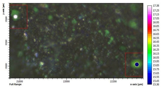
Figure 9.
Image of the surface observed under the optical microscope.
The presence of zinc oxide in the first white layer and of iron oxide added with manganese oxides in the second purplish-red layer was confirmed by the XRF spectra, suggesting the presence of the pigment caput mortuum and umber pigment.
As for the previously described microsamples, the FT-IR analysis showed the presence of calcium carbonate, Mowilith DM 912, and the pigment caput mortuum or morellone mixed with gypsum.
4. Conclusions
The investigation here reported gave an overview of the pigments, the conservation state, the executive technique, and the trace of past retouching. For all samples, the plaster made of calcium carbonate (CaCO3), consisting of aggregates of variable grain size and different colors, and the presence of a copolymer of vinyl-acrylate acetate/Mowilith DM912 [14], probably used in previous restoration work as protective, were identified. The presence of weddellite and whewellite is ascribable to some microbiological attack, and the presence of gypsum is an indication of the not good conservation state.
The painter used the traditional fresco technique with the white primer of calcium carbonate before applying the pigments.
In the vault, the following original pigments have been identified:
- – Black pigment probably obtained from natural earth (black earth, a mixture of clay and calcium carbonate, iron and manganese)
- – Dark gray: caput mortuum or morellone (mixture of organic black and ochre)
- – Red pigment: red ochre/hematite
- – White pigment: lead white
- – Red–violet primer (caput mortuum or morellone)
- – Green pigment: green earth
- – Blue pigment: ultramarine blue
- – The pigments, used during previous restoration work, are:
- – White pigment: zinc white
- – Red pigment (red ochre), applied a secco, retouching;
- – Blue pigment (Prussian blue), applied a secco, retouching.
The blue pigment azurite applied a secco, partially converted into the green malachite, could be both original and applied by retouching.
The same pigments have been identified on other areas of the painting in the framework of the same campaign [23].
Through the recent campaign of investigations, representing the most complete study ever on the cycle of Saturnino Gatti in San Panfilo, we added some important details to the written information. From the beginning, the aim of carrying out scientific investigations was to find answers and acquire new information on the basis of objective data to elaborate the next restoration project. However, it cannot be excluded that future intervention on the paintings may still reveal some surprises, and that new details can be highlighted, as the restoration has always represented a privileged moment to increase knowledge of the work. Since the painting had some dry finishings (original), which unfortunately have been lost, the future restoration will also help in their identification and in their differentiation from the repaintings.
Author Contributions
M.L.S., conceptualization and methodology; M.F.F.M. historical research; V.C.C., M.L.S., A.P.S., E.P. and F.B., data curation and writing; V.C.C., μ-IR and OM; F.A., XRF and SEM investigations. A.P.S., E.P. and F.B., OM and SEM investigations; D.G., Raman investigation; R.C.P. and M.L.S., writing—review and editing. All authors have read and agreed to the published version of the manuscript.
Funding
This work was performed in the framework of the Project Torninparte “Archeometric investigation of the pictorial cycle of Saturnino Gatti in Tornimparte (AQ, Italy)” sponsored in 2021 by the Italian Association of Archeometry AIAR (www.associazioneaiar.com). F.A. thanks the MIUR for the Project PON Ricerca e Innovazione 2014–2020—Avviso DD 407/2018 “AIM Attrazione e Mobilità Internazionale” (AIM1808223).
Institutional Review Board Statement
Not applicable.
Informed Consent Statement
Not applicable.
Data Availability Statement
The datasets generated during and/or analyzed during the current study are available from the corresponding author on reasonable request. Samples are stored at the STEBICEF Department, University of Palermo (Italy).
Acknowledgments
The cross-sections were prepared by LabSTONE snc (http://www.labstone.it, accessed on 16 June 2023).
Conflicts of Interest
The authors declare no conflict of interest.
References
- Galli, A.; Alberghina, M.F.; Re, A.; Magrini, D.; Grifa, C.; Ponterio, R.C.; La Russa, M.F. Special Issue: Results of the II National Research project of AIAr: Archaeometric study of the frescoes by Saturnino Gatti and workshop at the church of San Panfilo in Tornimparte (AQ, Italy). Appl. Sci. 2023; under review. [Google Scholar]
- Ricci, S. Tornimparte, a Mimesis of Florence in Abruzzo. New insights into Saturnino Gatti’s art. Appl. Sci. 2023; under review. [Google Scholar]
- Arbace, L.; Di Paolo, G. I Volti Dell’anima, Saturnino Gatti: Vita e Opere Di Un Artista Del Rinascimento; Paolo De Siena Editore: Pescara, Italy, 2012; ISBN 8896341116. [Google Scholar]
- Bonizzoni, L.; Caglio, S.; Galli, A.; Lanteri, L.; Pelosi, C. Materials and technique: The first look at Saturnino Gatti. Appl. Sci. 2023, 13, 6842. [Google Scholar] [CrossRef]
- Bonizzoni, L.; Caglio, S.; Galli, A.; Germinario, C.; Izzo, F.; Magrini, D. Identifying original and restoration materials through spectroscopic analyses on Saturnino Gatti mural paintings: How far a non-invasive approach can go. Appl. Sci. 2023, 13, 6638. [Google Scholar] [CrossRef]
- Andreotti, A.; Izzo, F.C.; Bonaduce, I. Archaeometric study of the mural paintings by Saturnino Gatti and workshop in the Church of San Panfilo-Tornimparte (AQ). The study of organic materials. Appl. Sci. 2023; under review. [Google Scholar]
- Defeyt, C.; Marechal, D.; Vandepitte, F.; Strivay, D. Rethinking Jacques-Louis David’s Marat assassiné through material evidences. Herit. Sci. 2023, 11, 21. [Google Scholar] [CrossRef]
- Dal Fovo, A.; Mattana, S.; Ramat, A.; Riitano, P.; Cicchi, R.; Fontana, R. Insights into the stratigraphy and palette of a painting by Pietro Lorenzetti through non-invasive methods. J. Cult. Herit. 2023, 61, 91–991. [Google Scholar] [CrossRef]
- Mannetti, T.R.; Chelli, N.; Vecchioli, G. Saturnino Gatti Nella Chiesa Di San Panfilo a Tornimparte; L’Aquila Publishing: L’Aquila, Italy, 1992. [Google Scholar]
- Saladino, M.L.; Ridolfi, S.; Carocci, I.; Chillura Martino, D.; Lombardo, R.; Spinella, A.; Traina, G.; Caponetti, E. A multi-analytical non-invasive and micro-invasive approach to canvas oil paintings. General considerations from a specific case. Microchem. J. 2017, 133, 607–613. [Google Scholar] [CrossRef]
- Mollica Nardo, V.; Renda, V.; Parrotta, F.; Bonanno, S.; Anastasio, G.; Caponetti, E.; Saladino, M.L.; Vasi, C.S.; Ponterio, R.C. Non invasive investigation of the pigments of wall painting in S. Maria delle Palate di Tusa (Messina, Italy). Heritage 2019, 2, 2398–2407. [Google Scholar] [CrossRef]
- Armetta, F.; Cardenas, J.; Caponetti, E.; Alduina, R.; Presentato, A.; Vecchioni, L.; di Stefano, P.; Spinella, A.; Saladino, M.L. Materials, conservation state and environmental monitoring of paintings in the Santa Margherita cliff cave. Environ. Sci. Pollut. Res. J. 2021. [Google Scholar] [CrossRef]
- Pouchou, J.L.; Pichoir, F. Quantitative Analysis of Homogeneous or Stratified Microvolumes Applying the Model “PAP”. In Electron Probe Quantitation; Springer: Boston, MA, USA, 1991; pp. 31–75. [Google Scholar]
- Horton-James, D.; Walston, S.; Zounis, S. Evaluation of the Stability, Appearance and Performance of Resins for the Adhesion of Flaking Paint on Ethnographic Objects. Stud. Conserv. 1991, 36, 203–221. [Google Scholar] [CrossRef]
- Frost, R.L. Raman spectroscopy of natural oxalates. Anal. Chim. Acta 2004, 517, 207214. [Google Scholar] [CrossRef]
- Henderson, E.J.; Helwig, K.; Read, S.; Rosendahl, S.M. Infrared chemical mapping of degradation products in cross-sections from paintings and painted objects. Herit. Sci. 2019, 7, 1–15. [Google Scholar] [CrossRef]
- Wei, S.; Pintus, V.; Schreiner, M. Photochemical degradation study of polyvinyl acetate paints used in artworks by Py–GC/MS. J. Anal. Appl. Pyrolysis 2012, 97, 158–163. [Google Scholar] [CrossRef] [PubMed]
- Guglielmi, V.; Andreoli, M.; Comite, V.; Baroni, A.; Fermo, P. The combined use of SEM-EDX, Raman, ATR-FTIR and visible reflectance techniques for the characterisation of Roman wall painting pigments from Monte d’Oro area (Rome): An insight into red, yellow and pink shades. Environ. Sci. Pollut. Res. 2022, 29, 29419–29437. [Google Scholar] [CrossRef] [PubMed]
- Giannini, C. Dizionario del Restauro. Tecniche, Diagnostica, Conservazione; Nardini Editore: Firenze, Italy, 2010; pp. 113–114. [Google Scholar]
- De Oliveira, L.F.; Edwards, H.G.; Frost, R.L.; Kloprogge, J.T.; Middleton, P.S. Caput mortuum: Spectroscopic and structural studies of an ancient pigment. Analyst 2002, 127, 536–541. [Google Scholar] [CrossRef] [PubMed]
- Saladino, M.L.; Ridolfi, S.; Carocci, I.; Chirco, G.; Caramanna, S.; Caponetti, E. A multi-disciplinary investigation of the “Tavolette fuori posto” of the main hall wooden ceiling of the “Steri” (Palermo, Italy). Microchem. J. 2016, 126, 132–137. [Google Scholar] [CrossRef]
- Zaffino, C.; Guglielmi, V.; Faraone, S.; Vinaccia, A.; Bruni, S. Exploiting external reflection FTIR spectroscopy for the in-situ identification of pigments and binders in illuminated manuscripts. Brochantite and posnjakite as a case study. Spectrochim. Acta Part A Mol. Biomol. Spectrosc. 2015, 136, 1076–1085. [Google Scholar] [CrossRef] [PubMed]
- Briani, F.; Caridi, F.; Ferella, F.; Gueli A., M.; Marchegiani, F.; Nisi, S.; Paladini, G.; Pecchioni, E.; Politi, G.; Santo, A.P.; et al. Multi-Technique Characterization of Painting Drawings of the Pictorial Cycle at the San Panfilo Church in Tornimparte (AQ). Appl. Sci. 2023, 13, 6492. [Google Scholar] [CrossRef]
Disclaimer/Publisher’s Note: The statements, opinions and data contained in all publications are solely those of the individual author(s) and contributor(s) and not of MDPI and/or the editor(s). MDPI and/or the editor(s) disclaim responsibility for any injury to people or property resulting from any ideas, methods, instructions or products referred to in the content. |
© 2023 by the authors. Licensee MDPI, Basel, Switzerland. This article is an open access article distributed under the terms and conditions of the Creative Commons Attribution (CC BY) license (https://creativecommons.org/licenses/by/4.0/).
