Abstract
Microplastics introduced into freshwater environments create novel surfaces that select for specific microbial colonizers and exclude others. In urban rivers, these biofilms can act as reservoirs of antimicrobial resistance and contain potential enzymes for polymer degradation. We studied microbial communities associated with microplastics in the Setun River and examined how their composition changes during laboratory enrichment on plastic substrates. Native river specimen and cultures enriched on low-density polyethylene (LDPE) and polycaprolactone (PCL) were analyzed using mWGS and full-length 16S rRNA nanopore sequencing. Enrichment led to a pronounced shift toward nearly monoculture of Bacillota, more specifically Bacillus cereus, while native plastisphere communities were dominated by Pseudomonadota. Microscopy revealed clear degradation of PCL but not LDPE, and functional screening of native metagenomes uncovered a diverse resistome, including oqxAB efflux operons, mcr-3-like phosphoethanolamine transferases, various -lactamases, and class 1 integron genes, demonstrating that the Setun River plastisphere already contained clinically relevant AMR determinants. These findings suggest that certain bacteria such as Bacillus cereus can thrive and dominate on plastic surfaces in urban rivers, while many other taxa cannot persist there, highlighting that microplastics strongly reshape plastisphere communities and emphasize the role of river-borne microplastics as potential vectors of antibiotic resistance.
1. Introduction
Recently, the problem of contamination of freshwater systems with microplastics has become particularly relevant [1]. The content level of microplastics is often used as an indirect indicator of the ecological state of water bodies; however, there are currently no established regulations defining permissible concentrations of microplastics in aquatic environments. Numerous studies have investigated plastic pollution in rivers and marine systems [2,3,4,5], determining the concentration and composition of polymer particles, analyzing their spatial distribution in relation to potential pollution sources, and comparing observations across diverse geographic regions. At the same time, the impact of microplastics on aquatic ecosystems is diverse and potentially hazardous.
One major concern is the ability of microplastics to adsorb and transport various chemicals, including toxic pollutants, as well as biological entities such as bacteria and other microorganisms. Such interactions can alter the physicochemical properties of polymer particles and modify their ecological behavior, influencing their interactions with aquatic biota [6,7]. Microplastics also serve as substrates for the development of distinct microbial biofilms, giving rise to the so-called “plastisphere” [8]. With increasing microplastic loads in freshwater systems worldwide, there is growing interest in understanding how biofilm formation on plastic surfaces shapes microbial community composition and alters ecosystem processes [9,10,11].
Recent findings indicate that microplastic-associated biofilms can harbor microorganisms with clinically relevant traits, including antibiotic resistance genes (ARGs), mobile genetic elements, and opportunistic pathogens capable of persisting in diverse aquatic environments [12,13,14]. These plastisphere communities often differ substantially from surrounding planktonic microbiota and may facilitate horizontal gene transfer due to their high cell density and surface-attached growth. Such observations raise concerns that microplastics could act as vectors for the environmental dissemination of antimicrobial resistance, particularly in urban rivers exposed to stormwater runoff and wastewater inputs.
Despite increasing scientific attention, current knowledge of the microbial diversity, ecological roles, and functional potential of plastisphere communities remains fragmented. Understanding the taxonomic and functional attributes of microorganisms inhabiting microplastic surfaces is essential for assessing their potential environmental impact. Of particular interest are microorganisms capable of degrading polymeric materials or modulating the fate and toxicity of microplastics in natural environments.
In this study, we provide the first integrated assessment of microplastic pollution in the Setun River, a major urban tributary of the Moskva River. Our goals were to (i) identify polymer composition of microplastics, (ii) characterize the native riverine microbiota and compare it with plastic-enriched communities, and (iii) identify antimicrobial resistance determinants and potential plastic-degrading genes present in the river metagenomes. Together, these analyses offer new insight into the ecological and public-health relevance of the Setun River plastisphere.
2. Materials and Methods
2.1. Sample Collection
The Setun River is one of the largest rivers in Moscow, flowing predominantly through urbanized landscapes. Its chemical and biological characteristics are shaped by multiple sources of anthropogenic pollution, including urban runoff, stormwater drainage carrying road-derived contaminants, and potential wastewater discharges from surrounding infrastructure.
Surface water samples were collected to assess microplastic concentrations using a trawl net designed specifically for capturing microplastic particles [15,16]. Such sampling approaches commonly rely on plankton nets [17,18] or MANTA nets adapted for surface microplastic collection. During sampling, a net approximately 2 m in length [18] was deployed from a vessel for a designated period, with its inlet partially submerged to allow efficient collection of particles from the uppermost water layer. A spinner mechanism integrated into the system measured the volume of filtered water. After each trawling run, all retained material was transferred into containers for laboratory processing.
In this study, we used the LEI-MANTA300 net set (EkoInstrument LLC, San Jose, CA, USA), equipped with 300 µm mesh filtration bags (Figure 1). The net inlet (30 cm × 15 cm) was fitted with a current meter to estimate the filtered water volume. The LEI-MANTA300 was towed behind the vessel at a speed of approximately 5 km/h, with towing duration adjusted according to the expected abundance of microplastics.
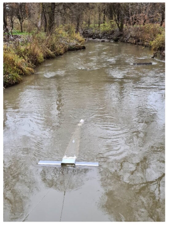
Figure 1.
Sampling of microplastics using the LEI-MANTA300 net.
Following each sampling event, the net was thoroughly flushed to recover the entire filtered material into a detachable beaker. The contents were sieved through stainless-steel meshes of 5 mm (top) and 0.3 mm (bottom). Larger debris retained on the 5 mm sieve was discarded only after rinsing off microplastic particles. The fraction between 0.3 and 5 mm, consisting of microplastics and organic residues, was transferred to sterile containers and stored under refrigeration for subsequent microbiological analyses.
Due to the high organic content, samples underwent chemical digestion prior to microplastic identification. Each sample was transferred into a 2 L glass beaker placed on a magnetic stirrer with heating. A 30% NaOH solution was added and heated to 75–80 °C, after which 30% H2O2 was gradually introduced under continuous stirring until the sample became fully discolored, indicating the removal of organic matter. Digestion time ranged from 1 h to several hours depending on sample composition.
Because synthetic polymers remain unaffected by this treatment, the digested samples were sieved at 100 µm. A saturated saline solution was then applied for density separation using a funnel to isolate microplastics from mineral particles. The retained particles were examined under a stereomicroscope at up to 80× magnification. Microplastics were visually identified and categorized into three types: fragments, fibers, and films. This workflow enabled quantitative assessment of microplastic abundance and export from the river.
In addition, three water samples were collected from the Setun River for mWGS analysis and designated as C1, C2, and C3 (Figure 2). Field sampling was conducted on 9 November 2022 to assess microplastic concentrations and microbial communities during autumn baseflow conditions. Water temperature ranged from 7 °C to 9 °C across stations, and the sampling period was chosen to represent hydrologically stable conditions prior to winter ice formation.

Figure 2.
Sampling locations of specimen C1–C3.
2.2. Composition of Plastics
Individual plastic fragments were examined by differential scanning calorimetry (DSC), which allows recording characteristic phase transitions in polymer materials. The measurement was performed on a DSC device (DSC 402 F1 Phoenix, Netzsch, Selb, Germany). The suspended plastic fragment was placed in an aluminum crucible and heated in the temperature range from 25 to 200 °C, at a rate of 10 S/min in an argon current of 50 mL/min. An empty aluminum crucible was used as a comparison sample. The peaks on the curves represent melting peaks, the kinks correspond to the glass transition, and the temperature of both can be compared with the literature data.
2.3. Culture Media
Bacterial cultures were maintained in two media: Luria–Bertani (LB) broth consisting of tryptone (10 g/L), yeast extract (5 g/L), and NaCl (10 g/L); Minimal M9 medium containing Na2HPO4 (6.8 g/L), KH2PO4 (3 g/L), NaCl (0.5 g/L), NH4Cl (1 g/L), glucose (0.1 g/L), MgSO4·7H2O (2.465 mg/L), thiamine-HCl (3.37 mg/L), CaCl2·2H2O (0.147 mg/L), adjusted to pH 7.0.
2.4. Experimental Design
There were two rounds of experiments. In the first experiment, cells from the initial river samples (C1–C3) were first cultivated overnight for 11 h in LB medium (37 °C, 220 rpm). An aliquot of each culture approximately 300–400 µL was transferred into 30 mL of M9 medium to a final OD600 of 0.1. Sterilized and pre-weighed plastic fragments of two polymer types, low-density polyethylene (LDPE) and polycaprolactone (PCL), were added to each culture. Cultivation was performed for 90–100 h (37 °C, 220 rpm). After growth, cells were transferred according to the following procedure (hereafter referred to as “passaging”): (i) aliquots were inoculated into fresh LB (10 mL) and incubated for 5–6 h to accumulate biomass; (ii) residual M9 medium was removed from the plastic, which was then replenished with fresh M9; (iii) biomass from LB cultures (OD600 ≈ 5–7) was reinoculated into the plastic-containing M9 to a final OD600 of 0.1. This cycle was repeated up to five times. Two cell subcultures were extracted from each sample: cells from liquid suspension were denoted as “Liq” subculture, while cells which remain attached to the plastic surface were denoted as “Pl” subculture. Surface-associated cells were detached using M9 salts supplemented with 0.05% SDS (1 h, 37 °C, 200 rpm). Both Liq and Pl subcultures were then cultivated in LB (37 °C, 200 rpm, 10–11 h). Plastic fragments were subsequently washed with ethanol, dried overnight, and re-weighed.
In the second experiment, passaging in LB was omitted. Instead, cultures were maintained long-term with plastic in M9 medium. Aliquots of LB-grown biomass (initial OD600 = 0.1) were inoculated into 30 mL of M9 containing plastics samples, which were used at the first step. Additionally, in one setup (of C2 origin), the biosurfactant surfactin was added at a final concentration of 30 mg/mL to test its effect on plastic biodegradation. Cultures were incubated for up to seven weeks with monitoring of OD600. Plastic fragments were washed, dried, and weighed at the end of the experiment to assess mass loss. Cells from the resulting suspensions were collected and used for microbial community analysis by full-length 16S rRNA gene sequencing using Oxford Nanopore technology.
2.5. Microscopy of Plastic Surfaces
Plastic fragments were imaged before and after microbial cultivation using a standard light microscope microscope at magnifications of 10× and 20×. Both LDPE and PCL fragments were photographed. For each experimental sample (C1–C3), two to three replicate images were taken per plastic type. Images of the “before” state correspond to plastics prior to inoculation, while the “after” state corresponds to the end of Round I of cultivation. No additional imaging was performed after Round II. Representative images were archived for subsequent analysis of surface alterations.
2.6. Sequencing Experiments
Sequencing experiments were performed to characterize microbial communities associated with plastic fragments and liquid cultures, as well as the native Setun river microbial composition. Genomic DNA extraction was performed using Monarch DNA purification kit (NE BioLabs, Ipswich, MA, USA), DNA quality was then evaluated by A260/A280 and A260/A230 absorbance ratios using a Nanodrop spectrophotometer (Thermo Scientific, Waltham, MA, USA). DNA library for the nanopore sequencing was prepared according to Native barcoding genomic DNA protocol (with EXP-NBD104, EXP-NBD 114, and SQK-LSK109), provided by Oxford nanopore (Oxford, UK), with the exception that at the barcoding ligation step samples were left overnight. Nanopore sequencing was performed using “MinION” with R9.4.1. flow cell (Oxford nanopore). Full-length 16S rRNA gene sequencing (samples VT_1 to VT_16) was performed using Oxford Nanopore sequencing chemistry R10.4.1.
At the end of the first cultivation round, two distinct fractions were recovered from each experimental condition: Liq (liquid fraction)—cells collected from the suspended culture medium after the fifth passage. Pl (plastic-attached fraction)—cells detached from the surface of plastic fragments by washing. Both fractions were sequenced in order to compare planktonic and plastic-associated communities. The complete set of sequenced samples is summarized in Table 1.

Table 1.
Sequenced samples from liquid (Liq) and plastic-attached (Pl) fractions. “S” indicates surfactin supplementation; entries without “S” correspond to no surfactin.
Planned comparisons were structured as follows:
- Liq vs. Pl within the same sample and polymer type. For each inoculum (C1–C3) and each polymer (LDPE or PCL), paired Liq and Pl samples were compared. These comparisons reveal community differences between free-floating and plastic-attached cells. In total, eight such Liq–Pl pairs were analyzed.
- Between-inoculum comparisons within the same fraction. To evaluate the effect of inoculum origin, Liq samples from C1, C2, and C3 were compared with each other, and the same was performed for Pl samples. These comparisons highlight community-level differences attributable to distinct river water sources.
- Effect of surfactin. For inoculum C2, additional cultures were prepared with (+) and without (−) surfactin. Although secondary to the main design, these comparisons provide insight into the effect of surfactin on microbial colonization of plastics.
2.7. Sequencing Data Processing
Dorado [19] (Oxford Nanopore Technologies, 0.9.1+c8c2c9f, model dna_r9.4.1_e8_sup@v3.6 for basecalling and dna_r9.4.1_e8_sup@v3.3_5mCG_5hmCG@v0 for modified basecalling), was used for basecalling, demultiplexing and adapter trimming. FastQC [20] (v0.12.1) was used to assess the quality of reads. Chopper [21] (v0.8.0) was used to filter out low quality reads with a threshold of Q15, and to filter by read length in 16S gene sequencing (1300 < read length < 1600 bp). Raw and processed fastq files were first assessed for sequencing quality and basic statistics using seqkit v2.10.1 [22] (Table A1).
2.8. Metagenome Assembly and Polishing
Long-read metagenome assemblies for samples C1–C3 were generated using Flye [23] (v2.9.5-b1801) with the –meta option to enable metagenome-aware repeat resolution. Dorado basecalled reads (.fastq files) were used as input. The resulting contigs were then polished with Medaka [24] (v2.0.1, model r941_prom_sup_g507) under default parameters to refine errors. The final polished contigs were used for all downstream taxonomic and functional analyses.
2.9. Taxonomic Classification
Taxonomic profiling of native river and plastic-associated microbial communities was performed using a combination of read classification and abundance estimation tools. For taxonomic classification, long-read datasets were analyzed with MetaMaps v0.1 [25], which performs mapping of nanopore reads to a reference database followed by probabilistic assignment of reads to taxonomic nodes. The default MetaMaps database (“miniSeq+H”, 8 G compressed) includes 12,000 microbial genomes and the human reference genome. This approach provided species-level profiles of community composition and was used to compute relative abundances for samples C1–C3.
Contigs polished with Medaka were taxonomically classified using Kraken2 v2.1.6 [26] with the Standard-16 database (Kraken2 Standard database capped at 16 GB, release 14 July 2025). The database includes complete bacterial, archaeal, viral, and human reference genomes from RefSeq. Classification reports were generated for each sample (C1–C3) and used to estimate the percentage of fragments covered by the clade rooted at each taxonomic level.
In parallel, reads from bacterial 16S rRNA gene sequencing (samples VT_1 to VT_16) were classified with EMU [27], which applies expectation–maximization for abundance estimation from nanopore reads. EMU uses a curated full-length 16S rRNA reference database, a combination of rrnDB v5.6 and NCBI 16S RefSeq from 17 September 2020. Taxonomy is also from NCBI on the same date. The resulting database contains 49,301 sequences from 17,555 unique bacterial and archaeal species.
Alpha and beta diversity metrics were computed using the scikit-bio v0.7.0 [28] Python package. Alpha diversity indices included Shannon entropy, Simpson index, and observed richness. Beta diversity was estimated using Bray–Curtis dissimilarities. Distance matrices were used to compare community similarity across native and enrichment samples.
2.10. Screening for AMR and Plastic-Degrading Enzymes
Protein sequences were extracted from polished contigs using Prodigal v2.6.3 [29]. Native communities C1–C3 were checked for antimicrobial resistance genes using AMRFinderPlus v4.0.23 [30].
To find previously reported plastic-degrading proteins in native communities C1–C3 we used PlasticDB [31]—a database of microorganisms and proteins linked to plastic biodegradation. PlasticDB contains records for 1701 protein sequences which are known to degrade plastics, records also contain information on taxonomic label and type of plastic. Protein sequences from Prodigal v2.6.3 were aligned to PlasticDB database using DIAMOND v2.1.13 [32].
3. Results
3.1. Microplastic Abundance and Composition in the Setun River
The quantitative analysis showed an average concentration of microplastic particles of approximately 4.6 pieces per cubic meter of river water. The sample mainly contained irregularly shaped fragments formed as a result of the breakdown of larger macroplastic pollutants. The polymer composition of the fragments was determined using differential scanning calorimetry (DSC) based on their phase transition temperatures. Most particles were identified as polyethylene (both low-density polyethylene, LDPE, with a melting point around 110 °C, and high-density polyethylene, HDPE, with around 130 °C) and polypropylene fragments ( around 160 °C). The measurement results for all selected elements are shown in Table 2.
Table 2.
Thermophysical characteristics of polymer samples. Here, denotes the melting temperature as determined by differential scanning calorimetry (DSC).
3.2. Growth of River Microbiota on Plastics: Plastic Mass Loss and Microscopic Observations of Plastic Surfaces
Bacterial communities from Setun River samples (C1–C3) were cultivated in M9 medium in the presence of low-density polyethylene (LDPE) or polycaprolactone (PCL) granules. Pre-weighed plastic fragments (Table 3) were incubated with microbial inocula for 90–100 h at 37 °C with shaking.
Table 3.
Initial mass of LDPE and PCL fragments used for cultivation experiments.
After the first round of cultivation, moderate growth was observed (OD600 = 0.21–0.45 for LDPE; 0.33–0.41 for PCL; Table 4). Serial passaging was performed up to five times. Cell density increased during the second and third passages, reaching OD600 = 0.34–0.46 (LDPE) and 0.40–0.44 (PCL). A slight decline was noted in later passages (IV–V).
Table 4.
OD600 of cultures grown with LDPE and PCL during serial passaging.
After the fifth passage, cultures were divided into suspended (Liq) and plastic-attached (Pl) subcultures. Both fractions were regrown in LB medium at 37 °C. High biomass yields were obtained (OD600 up to 8.4), with similar performance for Liq and Pl subcultures (Table 5).
Table 5.
Final OD600 of suspended (Liq) and plastic-attached (Pl) fractions in LB medium.
Dry weight measurements of plastic fragments indicated minimal mass changes (Table 6), suggesting limited polymer degradation during the first round. Nevertheless, cultures derived from sample C2 showed the most pronounced decrease in plastic mass, particularly for PCL, where weight loss reached approximately 1.1% of the initial mass. This suggests that the C2 community exhibited slightly higher potential for plastic degradation compared to C1 and C3.
Table 6.
Final mass of LDPE and PCL fragments and changes () after cell growth.
In the second round of experiment (Table 7), bacteria were cultivated long-term (7 weeks) in M9 medium without LB passaging. Both Liq and Pl fractions were monitored for growth. To test the effect of biosurfactants, surfactin was added to sample C2 (+sf) at 30 mg/mL.
Table 7.
Initial plastic mass at the beginning of the second experiment (Liq—suspension; Pl—plastic with attached cells).
After two weeks, moderate growth was observed across samples (OD600 = 0.20–0.69). By the end of 7 weeks, cell density decreased to OD600 = 0.08–0.34 (Table 8). The mass changes of plastics during this experiment are shown in Table 9.
Table 8.
OD600 values of cultures grown with LDPE and PCL during the second round of the experiment (Liq—suspension; Pl—plastic with attached cells).
Table 9.
Plastic mass changes () for LDPE and PCL after the second round of cultivation (Liq—suspension; Pl—plastic with attached cells).
Gravimetric analysis revealed slight reductions in plastic mass after 7 weeks (Table 9). PCL fragments showed higher apparent weight loss ( up to 17.3 mg) compared with LDPE ( generally below 1 mg). The addition of surfactin in C2 appeared to enhance PCL degradation.
Microscopic examination revealed clear differences between plastic fragments before and after microbial growth (Figure A1). Prior to inoculation, both LDPE and PCL surfaces showed irregular textures with visible pits and grooves. After cultivation with river microbiota, the surface of PCL became noticeably smoother, likely due to partial surface restructuring during biodegradation, whereas LDPE surfaces showed no visible changes and remained morphologically similar to their initial state. The microscopic data corroborate the gravimetric analysis, supporting the interpretation that PCL underwent microbial colonization and partial degradation.
3.3. Taxonomic Classification, Abundance and Diversity
Metagenomic profiling (Table 10) of the three Setun River samples (C1–C3) using MetaMaps revealed microbial communities dominated by Pseudomonadota, with Bacillota detected at lower abundances. At the phylum level, Pseudomonadota comprised 91.1–97.8% of the communities, while Bacillota accounted for 1.9–8.7%.
Table 10.
Relative abundances of bacterial taxa in native river samples at different taxonomic ranks obtained with MetaMaps.
At the genus level, the most abundant groups were Aeromonas (40.8–63.3%) and Escherichia (20.6–38.9%). Other taxa consistently detected included Klebsiella (6.3–11.8%), Enterobacter (1.6–8.1%), and Citrobacter (1.1–3.2%). Members of the genus Bacillus (phylum Bacillota) were present at lower abundances, ranging from 1.6% in C2 to 8.3% in C1.
Species-level classification showed that the dominant taxa belonged to the families Enterobacteriaceae and Aeromonadaceae, including Aeromonas veronii, Escherichia coli, Aeromonas hydrophila, and Klebsiella pneumoniae. In C1, the community was relatively balanced across several taxa, whereas C2 was strongly enriched in Aeromonas veronii (53.6%). Sample C3 was dominated by E. coli (38.9%) and A. veronii (32.7%).
Overall, native river samples contained diverse communities dominated by Pseudomonadota, with substantial contributions from Aeromonas and Enterobacteriaceae. Bacillota were consistently present at lower relative abundances, primarily represented by Bacillus. This composition contrasts with plastic-associated enrichment cultures, where Bacillota (notably Bacillus cereus) became dominant.
Kraken2 classification produced genus-level profiles that mirrored the trends observed with MetaMaps. As shown in Table 11, more than half of the classified fragments in all three samples were assigned to the genus Aeromonas (51.5–57.2%). Additional coverage was observed for Enterobacteriaceae, including Klebsiella (up to 12.3% in C2), Citrobacter (1.7–3.4%), and Enterobacter (2.5% in C1). Acinetobacter contributed 0.9–2.7% of classified fragments across the samples, while Bacillus was more prominent in C3 (6.2%). These fragment coverage estimates confirm the predominance of Pseudomonadota phylum in the Setun River communities and support the consistency of results across independent taxonomic classification methods.
Table 11.
Percentage of fragments covered by the clade rooted at each taxon in Setun River samples C1–C3 according to Kraken2 classification. Values are directly taken from the Kraken2 reports (rounded to one decimal place).
In the plastic-enrichment cultures, community structure shifted sharply away from the Pseudomonadota-dominated river microbiomes toward near-monocultures of Bacillota across all starting communities (C1–C3), inoculum states (Liq vs. Pl), and polymers (LDPE vs. PCL). In C1 sets, Bacillota comprised 98.9–99.8% with only 0.2–1.1% Pseudomonadota, mirroring >98.8% relative abundance of the genus Bacillus and species-level dominance of B. cereus (0.820–0.852), accompanied by smaller contributions of B. thuringiensis and trace Ralstonia pickettii (Table 12). C2 without surfactin showed the same pattern (Bacillota 99.4–99.9%; Bacillus 99.4–99.9%), with B. cereus (0.693–0.820) prevailing and variable B. thuringiensis (0.097–0.306); R. pickettii was rare (≤0.006) (Table 13). Adding surfactin partially relaxed this dominance: in PCL variants, Pseudomonadota increased to 7.7% driven by Ralstonia (0.020–0.076), and low-abundance Paenibacillus and Brevibacillus emerged; a trace of Escherichia appeared in Liq_C2_LDPE_S (Table 14). C3 enrichments remained almost entirely Bacillus-dominated (Bacillota 99.2–99.9%) with only minor Ralstonia in PCL (0.002–0.007) (Table 15). Together, these results indicate a reproducible convergence from Pseudomonadota-rich river communities to low-diversity Bacillota/Bacillus consortia under M9 + plastic incubation.
Table 12.
Relative abundances of bacterial taxa in community C1 (Liq and Pl, LDPE and PCL) obtained with EMU.
Table 13.
Relative abundances of bacterial taxa in community C2 (without surfactin) obtained with EMU.
Table 14.
Relative abundances of bacterial taxa in community C2 (with surfactin) obtained with EMU.
Table 15.
Relative abundances of bacterial taxa in community C3 (Liq and Pl, LDPE and PCL) obtained with EMU.
Alpha-diversity metrics corroborated this selection (Table 16). Native samples displayed higher diversity (Shannon 1.35–1.90; Simpson 0.63–0.81; richness 7–11). In contrast, most enrichments had markedly reduced diversity (typically Shannon < 1.0, Simpson ≤ 0.49, richness 1–6). The lowest values occurred in C3 on plastics (e.g., Liq_C3_LDPE: Shannon 0.01, richness 2; PL_C3_LDPE: Shannon 0.00, richness 1), consistent with near-pure Bacillus cultures. Surfactin modestly increased diversity: Liq_C2_PCL_S reached Shannon 0.95 versus 0.62 without surfactin, and PL_C2_LDPE_S showed the highest richness (7) compared to 2 in PL_C2_LDPE, suggesting partial retention/expansion of minor taxa in the presence of the biosurfactant. Polymer effects were weaker than medium/inoculum effects; PCL was more often associated with a small Ralstonia increase under surfactin, but did not overturn the overall Bacillus dominance.
Table 16.
Alpha diversity indices (Shannon, Simpson, Richness) for native river samples and cultivation experiment samples, with separate blocks for surfactin-treated samples.
Beta diversity analysis using Bray–Curtis distances revealed clear differentiation among the native river communities (Table 17). The lowest dissimilarity was observed between C2 and C3 (0.26), suggesting greater similarity in their species composition, while C1 was more distinct from both C2 (0.34) and C3 (0.27). These values indicate that although all three communities share a core of dominant taxa, sample C1 harbors a comparatively different assemblage, consistent with its more balanced distribution of taxa compared to the stronger enrichment of Aeromonas veronii in C2 and Escherichia coli in C3.
Table 17.
Bray–Curtis beta diversity distances between native river samples at the species level (metamaps).
3.4. Functional Analysis
In a representative, non-redundant set of high-confidence AMR hits (Table 18), we recovered ARGs spanning tetracycline, macrolide, sulfonamide, aminoglycoside, trimethoprim, phenicol/quinolone (efflux), fosfomycin and streptothricin classes. Sequence similarity to curated references was uniformly high: 10/13 representatives showed ≥99% amino-acid identity, and 8/13 were exact (100% identity, 100% coverage). Coverage was complete for 11/13 entries (two exceptions: catA, 93.6%; satA, 98.9%). Notably, we observed the complete oqxAB multidrug efflux operon (both oqxA and oqxB25 at 100% identity/coverage), the tetracycline MFS efflux tet(E) (99.8%/100%), macrolide mph(A) (100%/100%), and multiple modifying enzymes for aminoglycosides (aadA5 and aac(3)-IId, both 100%/100%). Antifolate resistance was represented by sul1 (100%/100%) together with dfrA12 (100%/100%) and dfrA17 (99.4%/100%). We also detected two fosfomycin transferases—fosA5 (100%/100%) and a fosA/FosA2-family variant (95.7%/100%)—and phenicol catA, which, despite lower identity (65.2%), remained within the expected family-level range. Collectively, these representatives indicate a broad resistome with multiple high-confidence, near-clonal matches to known ARGs and intact multidrug efflux capability.
Table 18.
Representative high-confidence antibiotic resistance genes detected by AMRFinderPlus (protein mode). One representative per gene.
The -lactamase repertoire spanned all Ambler classes, including serine enzymes (A, C, D) and metallo--lactamases (B1, B2), with 11 non-redundant representatives (Table 19). Five entries were allele-level exact matches (100% identity/100% coverage): TEM-1, SHV-11 (class A), ACT-95 (class C), and OXA-1163/OXA-912 (class D). The remaining calls were high-identity matches, all full-length (100% coverage) except Bla1 (99.39% coverage), and included FOX-5 and MOX-13 (class C; 98.17–99.22% identity), an OXA-12 family member (class D; 99.24%), BcII (class B1; 95.70%), and two CphA-family hits (class B2; 98.03–99.21%). Together, these data indicate the presence of broad-spectrum class A enzymes, multiple AmpC-like class C cephalosporinases, OXA-type class D enzymes, and both B1 and B2 metallo--lactamases—all with near-complete sequence concordance to curated references.
Table 19.
Detected -lactamases grouped by Ambler class with quality metrics.
The metagenome of samples C1–C3 harbours several high-priority AMR determinants (Table 20). We detected colistin resistance signatures comprising a full-length mcr-3–like phosphoethanolamine transferase (HMM call; 80.0% identity, 100% coverage; contig_209_131) and a second high-identity but truncated mcr-3–like sequence (99.6% identity, 51.4% coverage; contig_289_21), consistent with either a fragmented ORF or contig boundary. Carbapenem resistance potential was supported by two metallo--lactamase lineages: BcII (B1) and CphA (B2), each recovered at full length (100% coverage) with high amino-acid identity (95.7% and 98.0–99.2%, respectively). We also observed an intact oqxAB multidrug efflux operon (oqxA/oqxB25, both 100% identity/coverage) on adjacent ORFs of the same contig, alongside the tetracycline MFS efflux determinant tet(E) (99.8% identity, 100% coverage). Together, these markers indicate the presence of last-line (polymyxin) and carbapenem resistance mechanisms and broad MDR efflux capacity, underscoring the clinical relevance of the resistome.
Table 20.
Clinically critical resistance markers observed in the metagenome.
The assembly contains two contigs with gene arrangements diagnostic of class 1 integron cassettes [33] (Table 21). On contig_1109, we observed the canonical 3′ conserved segment marker qacEΔ1 adjacent to sul1, flanked by cassette-type ARGs aadA5 and dfrA17 (identity/coverage: 100/100 for sul1, qacEΔ1, aadA5; 99.36/100 for dfrA17). contig_1108 showed a second instance, aadA2–qacEΔ1–sul1, with similarly high concordance (99.61/98.48, 100/100, and 100/100, respectively). Although qacEΔ1 is annotated as a biocide/“stress” determinant, its co-occurrence with sul1 and aadA/dfrA is a well-known signature of the class 1 integron 3′ conserved region [34], indicating integron-associated, potentially mobile MDR modules in this community.
Table 21.
Putative class 1 integron-associated cassettes inferred from gene co-occurrence on contigs.
A homology screen for plastic-degrading enzymes using PlasticDB yielded a single, high-confidence hit consistent with a polyhydroxyalkanoate (PHA)/polyhydroxybutyrate (PHB) depolymerase Table 22. On contig_502, we detected a protein homolog annotated as PHB/PHA depolymerase (Bacillus thuringiensis) with 96.3% amino-acid identity across a 300-aa ungapped alignment (E ; bit score ). The alignment spans the query from residue 1 to 300 and maps to residues 6–305 of the reference, indicating a contiguous match across the core region without indels. Given the very high sequence identity and the functional annotation of the best-hit subject, we infer that the assembly might encode a putative PHB/PHA depolymerase, implying potential for bioplastic (PHA/PHB) depolymerization in this community. Though this should not be extrapolated to activity against recalcitrant petrochemical polymers (e.g., PE/PP/PET).
Table 22.
Homolog of a plastic-degrading enzyme detected by protein homology search.
4. Discussion
The analysis revealed that polyethylene and polypropylene were the predominant polymers across all samples. Both materials were present as fragments of varying morphology and color, suggesting multiple sources and indicating that household plastic waste is likely the major contributor of microplastics in the river. Some fragments exhibited no detectable thermal transitions within the studied temperature range. This may indicate that their transition temperatures lie outside the experimental window—for instance, rubbers with glass transition temperatures below room temperature, or more thermally stable polymers with transitions above the analyzed range. The absence of visible transitions may also point to the presence of thermosetting polymers, such as certain polyurethanes, phenol–formaldehyde, or epoxy resins, which do not soften or melt before thermal decomposition. It is also important to note that all samples were collected from the surface water layer, which naturally enriches the dataset with polymers of density equal to or lower than that of water—primarily polyethylene, polypropylene, and polystyrene—whereas denser materials, such as polyethylene terephthalate, are less likely to be encountered under these sampling conditions.
This study also set out to examine whether riverine microbiota from the Setun can colonise and degrade representative polymers (LDPE and PCL), and how the community structure shifts between the native water column, plastic-attached biofilms, and the corresponding liquid phases. Several consistent patterns emerge.
First, native Setun communities were Pseudomonadota-dominated, with Aeromonas and Enterobacteriaceae as major constituents, a composition typical of temperate freshwater systems influenced by urban inputs [35]. It should be noted that enrichment cultures in this study were incubated at 37 °C, a temperature substantially higher than the in situ river conditions (7–9 °C). This laboratory setting accelerates microbial growth but also might selectively favor mesophilic taxa.
After enrichment on plastics in minimal medium, however, virtually all cultures converged toward low-diversity Bacillota communities dominated by the Bacillus cereus group across inocula, fractions (Liq vs. Pl), and polymers. This shift was accompanied by a marked loss of alpha diversity and near-monoculture states. Such strong selection is consistent with the ability of spore-forming Gram-positives (such as Bacillus cereus) to withstand nutrient stress and repeated handling [36], the ready deployment of secreted hydrolases/esterases [37], and rapid biofilm development on hydrophobic surfaces in minimal medium, as reported for other plastic-enrichment experiments [38].
The dominance of Bacillota observed in the enrichment cultures can be attributed, at least in part, to the incubation temperature of 37 °C, which differs markedly from the ambient river temperature (7–9 °C). Such conditions might preferentially stimulate the growth of mesophilic microorganisms, including many Bacillota representatives, and therefore influence the resulting community composition independently of the original environmental structure. Although specific summer temperature records for the Setun River are not available, surface waters of rivers in the Moscow region typically reach approximately 18–22 °C during the warm season. Because the Setun River is expected to follow a similar seasonal pattern, repeating the enrichment experiment under these warmer, seasonally relevant thermal conditions could provide a more ecologically representative assessment of microbial dynamics and plastic-degrading potential.
Second, the materials behaved as expected from polymer chemistry. PCL, an aliphatic polyester with hydrolysable ester bonds, exhibited visible surface erosion and the largest gravimetric losses (up to 17 mg over seven weeks: ), whereas LDPE—an inert C–C backbone polyolefin—showed negligible and often sub-mg changes over comparable time frames. Microscopy corroborated this contrast: after cultivation, PCL surfaces became smoother, consistent with partial surface restructuring during biodegradation, whereas LDPE surfaces showed no visible changes and remained morphologically identical to their initial state. The pattern aligns with a broad literature showing that PCL is readily degraded by bacterial and fungal lipases/cutinases [39,40], while LDPE generally requires oxidative/photochemical pretreatment before measurable biotic mass loss occurs [41,42].
Third, adding the lipopeptide biosurfactant surfactin modestly diversified communities and reproducibly increased the relative abundance of Pseudomonadota (notably Ralstonia) in some PCL setups, with a corresponding tendency toward larger PCL mass loss compared with surfactant-free controls. Surfactants can increase the apparent bioavailability of hydrophobic substrates by lowering surface tension and improving wetting [43], and they can restructure biofilms—effects that plausibly underlie the trends observed here [44]. While our design was not powered for formal inference about surfactants, the directionality suggests that biosurfactant dosing could serve as a useful experimental lever for future work.
Our findings are consonant with the emerging view that plastics create distinctive microbial niches (the “plastisphere”) whose composition is shaped by polymer type, environmental context, and time [45,46]. Field and mesocosm studies consistently report rapid biofilm formation on plastics and divergence from surrounding planktonic communities, often with selection for taxa equipped for biofilm life and surface-associated metabolism [38]. The pronounced convergence toward Bacillus observed here under laboratory enrichment indicates that medium composition and transfer regime can strongly override the river’s native signal—an important caveat when extrapolating enrichment results back to natural settings.
In this study, we deliberately cross-validated taxonomic composition and relative abundances with three orthogonal pipelines that differ in input molecules, algorithms, and reference space: (1) long-read shotgun read mapping with MetaMaps (miniSeq+H database) to assign individual nanopore reads probabilistically and derive species-level community profiles for C1–C3, (2) contig-level k-mer classification of polished assemblies using Kraken2 (Standard-16 GB RefSeq build) to summarize the percentage of fragments covered by each clade (a contig-coverage-style abundance), and (3) full-length 16S rRNA amplicon profiling with EMU (expectation–maximization on a curated rrnDB+NCBI 16S reference) to estimate species-level proportions for VT1–VT16. Together these methods consistently recovered Pseudomonadota-rich native river microbiota and Bacillus-dominated enrichment cultures, but they are not numerically interchangeable: MetaMaps yields read-based relative abundances, Kraken2 on contigs reflects assembly-weighted fragment coverage (thereby down-weighting low-abundance/assembly-refractory taxa), and EMU infers proportions from marker-gene reads though does not account for rRNA copy-number and primer biases. In practice, we therefore interpret concordant directional trends across methods (e.g., Pseudomonadota → Bacillota shift; surfactin-linked Ralstonia increases on PCL) as robust signals, while treating absolute percentages with caution, especially for clades that are hard to resolve by 16S alone or that may be under-assembled. Going forward, we can make the three methods agree better by using the same taxonomy in all databases, correcting 16S results for rRNA gene copy number, adding internal standards to calibrate abundances, and double-checking key taxa with genome-resolved bins (MAGs). This keeps the strengths of read mapping, contig classification, and 16S profiling without forcing everything into one tool.
Our assemblies of samples C1–C3 encode a broad spectrum of antimicrobial resistance determinants spanning efflux, drug-modifying enzymes, antifolate, sulfonamide, tetracycline, fosfomycin, phenicol, and multiple -lactamase classes (A, C, D, and metallo--lactamases B1/B2). Notably, we detected an intact oqxAB multidrug efflux operon, high-identity tetracycline MFS efflux tet(E), macrolide mph(A), sul1 with trimethoprim dfrA12/dfrA17, fosA variants, and a phenicol catA, with the majority of representatives showing ≥99–100% amino-acid identity and full coverage against curated references. In addition, we recovered -lactamases across all Ambler classes, including TEM-1/SHV-11 (A), ACT/FOX/MOX (C), OXA variants (D), and metallo--lactamases BcII (B1) and CphA family (B2), plus two mcr-3-like phosphoethanolamine transferases (one full-length call and one truncated high-identity fragment). Two contigs also exhibited the canonical class 1 integron 3′-conserved region signature (sul1–qacEΔ1 with aadA/dfr cassettes), consistent with potentially mobile MDR modules. Taken together, these data place last-line polymyxin (mcr-like) and carbapenem resistance mechanisms alongside broad efflux and cassette-borne ARGs in the same metagenome, underscoring the public-health relevance of the Setun plastisphere resistome.
These genomic signals align with the emerging consensus that microplastic biofilms can enrich ARGs and mobile genetic elements and act as vectors across aquatic habitats (the “plastisphere” as AMR reservoir) [12,13,14]. Reviews and field studies report thicker biofilms on plastics, elevated ARG burdens (e.g., intI1/sul1), and conditions that favor horizontal gene transfer relative to natural substrates. The oqxAB operon we observed is a well-characterized plasmid-mediated PMQR determinant [47] conferring reduced susceptibility to quinolones, chloramphenicol/phenicols, nitrofurantoin, and more, and it has spread widely among Enterobacterales; its presence is therefore a plausible contributor to multidrug phenotypes.
The mcr-3-like hits are notable because aquatic bacteria and environmental waters have been repeatedly implicated in the emergence and circulation of mcr genes and their mobilization on plasmids across Enterobacteriaceae [48,49]. The co-occurrence of sul1 with qacEΔ1 in integron contexts further suggests co-selection by biocides and disinfectants, a mechanism demonstrated in environmental matrices and wastewater where quaternary ammonium compounds enrich class 1 integrons [50,51].
Mechanistically, the metallo--lactamases we recovered map onto known environmental and clinical lineages. CphA (subclass B2) is a carbapenemase native to Aeromonas spp. and has been associated with clinical, occasionally under-detected, carbapenem resistance; BcII is the canonical Bacillus cereus group MBL, prevalent as an intrinsic determinant across the clade [52,53]. Given the strong Bacillus cereus-group dominance in our enrichment cultures, the presence of BcII-type MBLs is also congruent with community structure.
Aligning proteins from C1-C3 to PlasticDB yielded one high-confidence enzyme: a PHB/PHA depolymerase homolog most similar to Bacillus thuringiensis PhaZ. This is consistent with the well-documented capacity of Bacillus spp. to encode PHB/PHA depolymerases and de-polymerize intracellular or extracellular PHAs [54,55]. Because PHAs are aliphatic polyesters designed to be biodegradable, their enzymatic depolymerization is widespread in soils and waters; by contrast, abiotic pretreatment is usually required before microbes can attack recalcitrant polyolefins (e.g., LDPE). Thus, the enzyme-level signal we found should not be over-interpreted as evidence for polyethylene catabolism. Instead, it aligns with the material behavior we observed experimentally: negligible loss of LDPE and clear surface erosion with mass loss for the aliphatic polyester PCL, which is known to be susceptible to lipases and cutinases.
5. Conclusions
This study shows that riverine microbiota from the Setun (Moscow) readily colonise plastics, but laboratory enrichment in minimal medium drives a strong and repeatable shift from Pseudomonadota-dominated native communities toward low-diversity Bacillus cereus-group consortia across inocula, plastic types, and fractions. Consistent with polymer chemistry, we observed clear surface erosion and mass loss for the biodegradable aliphatic polyester PCL, whereas LDPE showed negligible change; addition of the lipopeptide biosurfactant surfactin modestly diversified communities and, in some PCL setups, coincided with greater mass loss. A high-confidence PHB/PHA depolymerase homolog indicates capacity for bioplastic depolymerisation within the enriched plastisphere, but provides no direct evidence for polyethylene catabolism.
Functional profiling of native communities recovered a clinically relevant resistome: a complete oqxAB multidrug efflux operon, tet(E), mph(A), sul1 with dfrA12/dfrA17, fosA variants, multiple aminoglycoside-modifying enzymes, and -lactamases spanning Ambler classes, including BcII (B1) and CphA (B2). Notably, we detected two mcr-3–like phosphoethanolamine transferases and a class 1 integron 3′-conserved-region signature (sul1–qacEΔ1 with aadA/dfr cassettes), underscoring the potential of river-borne plastics to intersect recognised AMR dissemination routes.
Looking ahead, we will move beyond 16S profiling and perform whole-genome metagenomic shotgun sequencing (mWGS) of the microplastic-enriched community.
Author Contributions
Conceptualization, M.Z.; methodology, V.T., A.E. (Andrey Eremin) and M.Z.; sample collection, A.L., A.S. (Alexey Sazonov) and A.E. (Anna Efimova); formal analysis, A.E. (Andrey Eremin) and A.S. (Alexander Sergeev); investigation, A.L., V.R., V.T., T.P., A.B. and A.S. (Alexey Sazonov); data curation, A.E. (Andrey Eremin) and A.S. (Alexander Sergeev); writing—original draft preparation, A.E. (Andrey Eremin); writing—review and editing, M.Z. and A.E. (Andrey Eremin); sequencing, V.R. and T.P.; visualization, A.E. (Andrey Eremin); plastic calorimetric analysis, A.B.; microscopic and macroscopic analysis, V.T., A.E. (Anna Efimova) and A.S. (Alexander Sergeev); supervision, M.Z.; project administration, M.Z.; funding acquisition, A.E. (Anna Efimova), M.Z. and M.G.K. All authors have read and agreed to the published version of the manuscript.
Funding
This research was funded by the project of the Interdisciplinary Scientific and Educational School of Moscow State University “Molecular technologies of living systems and synthetic biology” (23-Sh04-45); analysis of polymeres supported by the project “Modern Problems of Chemistry and Physical Chemistry of Macromolecular Compounds” (State Assignment No. AAAA-A21-121011990022-4).
Data Availability Statement
The sequencing reads generated in this study have been submitted to the NCBI BioProject database https://www.ncbi.nlm.nih.gov/bioproject/?term=PRJNA1333826 (accessed on 16 December 2025) under accession number PRJNA1333826.
Acknowledgments
The authors are deeply grateful to the students of the Lomonosov Moscow State University Gymnasium who were actively involved in the sampling process and quantitative analysis of microplastics: Luka Rogov, Daria Sokolova, Ekaterina Zheliabina, Anastasia Kurbatova, Sofia Andronova, Ayganat Alieva, Maxim Artamonov, and Vera Nifantieva.
Conflicts of Interest
The authors declare no conflicts of interest.
Abbreviations
The following abbreviations are used in this manuscript:
| AMR | Antimicrobial Resistance |
| PMQR | plasmid-mediated quinolone resistance |
| ARG | Antimicrobial Resistance Gene |
| PHA | Polyhydroxyalkanoate |
| PHB | Polyhydroxybutyrate |
| PCL | Polycaprolactone |
| LDPE | Low-Density Polyethylene |
| rRNA | Ribosomal Ribonucleic Acid |
| MDR | Multidrug Resistance |
| MAG | Metagenome assembeled genome |
| RND | Resistance–Nodulation–Division (efflux system) |
| ORF | Open Reading Frame |
| HMM | Hidden Markov Model |
Appendix A

Figure A1.
Microscopic images of LDPE (c,d) and PCL (a,b) fragments before (a,c) and after (b,d) microbial cultivation at 20× zoom.
Table A1.
Sequencing statistics for raw and processed FASTQ files.
Table A1.
Sequencing statistics for raw and processed FASTQ files.
| File | Num_SEQS | N50 | Min_Len | Max_Len | Avg_Len | Q20 (%) | Q30 (%) | AvgQual | GC (%) |
|---|---|---|---|---|---|---|---|---|---|
| C1.fastq | 493,192 | 11,880 | 5 | 98,414 | 6952.10 | 65.36 | 31.98 | 13.73 | 55.24 |
| C1_processed.fastq | 181,035 | 11,786 | 89 | 53,387 | 7218.50 | 75.49 | 41.48 | 16.08 | 54.37 |
| C2.fastq | 138,313 | 14,774 | 5 | 100,047 | 6661.60 | 64.91 | 32.11 | 13.65 | 56.11 |
| C2_processed.fastq | 47,916 | 14,518 | 102 | 62,480 | 6901.50 | 75.24 | 41.79 | 16.02 | 55.61 |
| C3.fastq | 428,216 | 13,694 | 5 | 83,029 | 6653.00 | 65.67 | 32.60 | 13.77 | 54.65 |
| C3_processed.fastq | 161,308 | 13,578 | 123 | 62,270 | 6910.60 | 75.63 | 42.04 | 16.09 | 53.96 |
| VT_1.fastq | 101,605 | 1476 | 5 | 187,657 | 1437.20 | 78.38 | 68.02 | 14.63 | 53.45 |
| VT_1_filtered.fastq | 67,184 | 1475 | 1300 | 1600 | 1486.10 | 88.40 | 78.63 | 18.80 | 53.54 |
| VT_2.fastq | 102,374 | 1477 | 5 | 155,410 | 1475.40 | 76.58 | 66.09 | 14.14 | 53.56 |
| VT_2_filtered.fastq | 65,439 | 1475 | 1300 | 1600 | 1486.40 | 88.26 | 78.39 | 18.73 | 53.53 |
| VT_3.fastq | 67,799 | 1479 | 5 | 88,309 | 1513.10 | 76.51 | 65.90 | 14.17 | 53.47 |
| VT_3_filtered.fastq | 40,571 | 1475 | 1300 | 1600 | 1485.20 | 88.34 | 78.53 | 18.74 | 53.59 |
| VT_4.fastq | 108,150 | 1478 | 5 | 444,867 | 1504.00 | 75.93 | 65.49 | 13.91 | 53.54 |
| VT_4_filtered.fastq | 69,263 | 1475 | 1300 | 1600 | 1489.50 | 88.22 | 78.38 | 18.69 | 53.59 |
| VT_5.fastq | 85,154 | 1479 | 5 | 144,555 | 1545.80 | 74.51 | 63.81 | 13.78 | 53.51 |
| VT_5_filtered.fastq | 51,105 | 1475 | 1300 | 1600 | 1485.00 | 88.20 | 78.27 | 18.70 | 53.57 |
| VT_6.fastq | 45,537 | 1497 | 5 | 118,349 | 1533.70 | 74.87 | 64.23 | 13.76 | 53.45 |
| VT_6_filtered.fastq | 26,279 | 1476 | 1300 | 1600 | 1489.10 | 88.19 | 78.34 | 18.66 | 53.54 |
| VT_7.fastq | 51,953 | 1481 | 6 | 49,572 | 1523.50 | 74.17 | 63.49 | 13.66 | 53.44 |
| VT_7_filtered.fastq | 29,696 | 1475 | 1300 | 1600 | 1485.90 | 87.92 | 77.92 | 18.59 | 53.55 |
| VT_8.fastq | 69,985 | 1482 | 5 | 476,092 | 1509.20 | 75.23 | 64.74 | 13.80 | 53.53 |
| VT_8_filtered.fastq | 42,468 | 1475 | 1300 | 1600 | 1488.60 | 88.21 | 78.35 | 18.67 | 53.60 |
| VT_9.fastq | 80,702 | 1479 | 5 | 173,764 | 1510.30 | 75.52 | 64.92 | 13.92 | 53.58 |
| VT_9_filtered.fastq | 48,491 | 1475 | 1300 | 1600 | 1487.80 | 88.30 | 78.45 | 18.73 | 53.57 |
| VT_10.fastq | 85,935 | 1478 | 5 | 236,639 | 1522.60 | 73.33 | 62.68 | 13.55 | 53.39 |
| VT_10_filtered.fastq | 49,347 | 1475 | 1300 | 1600 | 1483.10 | 88.18 | 78.22 | 18.70 | 53.46 |
| VT_11.fastq | 62,074 | 1515 | 6 | 256,856 | 1580.00 | 73.96 | 63.36 | 13.58 | 53.47 |
| VT_11_filtered.fastq | 35,165 | 1476 | 1300 | 1600 | 1490.50 | 88.28 | 78.48 | 18.71 | 53.50 |
| VT_12.fastq | 96,361 | 1478 | 5 | 227,820 | 1502.30 | 75.40 | 64.85 | 13.92 | 53.56 |
| VT_12_filtered.fastq | 59,150 | 1475 | 1300 | 1600 | 1487.80 | 88.21 | 78.35 | 18.69 | 53.63 |
| VT_13.fastq | 84,870 | 1478 | 5 | 110,861 | 1491.90 | 76.51 | 66.00 | 14.13 | 53.44 |
| VT_13_filtered.fastq | 53,505 | 1475 | 1300 | 1600 | 1488.40 | 88.22 | 78.37 | 18.69 | 53.55 |
| VT_14.fastq | 94,972 | 1480 | 5 | 53,872 | 1484.90 | 78.86 | 68.32 | 14.88 | 53.50 |
| VT_14_filtered.fastq | 64,476 | 1476 | 1300 | 1600 | 1494.70 | 88.24 | 78.38 | 18.68 | 53.55 |
| VT_15.fastq | 51,324 | 1513 | 5 | 144,409 | 1568.00 | 74.30 | 63.48 | 13.56 | 53.53 |
| VT_15_filtered.fastq | 28,248 | 1475 | 1300 | 1600 | 1488.40 | 88.12 | 78.19 | 18.66 | 53.60 |
| VT_16.fastq | 72,592 | 1482 | 6 | 49,906 | 1534.60 | 75.34 | 64.55 | 14.05 | 53.58 |
| VT_16_filtered.fastq | 42,947 | 1475 | 1300 | 1600 | 1487.70 | 88.14 | 78.22 | 18.66 | 53.65 |
References
- Science Advice for Policy by European Academies (SAPEA). A Scientific Perspective on Microplastics in Nature and Society; SAPEA: Berlin, Germany, 2019. [Google Scholar] [CrossRef]
- Horton, A.A. Plastic pollution: When do we know enough? J. Hazard. Mater. 2022, 422, 126885. [Google Scholar] [CrossRef]
- Lisina, A.A.; Platonov, M.M.; Lomakov, O.I.; Sazonov, A.A.; Shishova, T.V.; Berkovich, A.K.; Frolova, N.L. Microplastic Abundance in Volga River: Results of a Pilot Study in Summer 2020. Geogr. Environ. Sustain. 2021, 14, 82–93. [Google Scholar] [CrossRef]
- Frank, Y.A.; Vorobiev, E.D.; Vorobiev, D.S.; Trifonov, A.A.; Antsiferov, D.V.; Soliman Hunter, T.; Wilson, S.P.; Strezov, V. Preliminary Screening for Microplastic Concentrations in the Surface Water of the Ob and Tom Rivers in Siberia, Russia. Sustainability 2020, 13, 80. [Google Scholar] [CrossRef]
- Yakushev, E.; Gebruk, A.; Osadchiev, A.; Pakhomova, S.; Lusher, A.; Berezina, A.; van Bavel, B.; Vorozheikina, E.; Chernykh, D.; Kolbasova, G.; et al. Microplastics distribution in the Eurasian Arctic is affected by Atlantic waters and Siberian rivers. Commun. Earth Environ. 2021, 2, 23. [Google Scholar] [CrossRef]
- Fred-Ahmadu, O.H.; Bhagwat, G.; Oluyoye, I.; Benson, N.U.; Ayejuyo, O.O.; Palanisami, T. Interaction of chemical contaminants with microplastics: Principles and perspectives. Sci. Total Environ. 2020, 706, 135978. [Google Scholar] [CrossRef]
- Cao, J.; Yang, Q.; Jiang, J.; Dalu, T.; Kadushkin, A.; Singh, J.; Fakhrullin, R.; Wang, F.; Cai, X.; Li, R. Coronas of micro/nano plastics: A key determinant in their risk assessments. Part. Fibre Toxicol. 2022, 19, 55. [Google Scholar] [CrossRef]
- Yang, Y.; Liu, W.; Zhang, Z.; Grossart, H.P.; Gadd, G.M. Microplastics provide new microbial niches in aquatic environments. Appl. Microbiol. Biotechnol. 2020, 104, 6501–6511. [Google Scholar] [CrossRef]
- Mughini-Gras, L.; van der Plaats, R.Q.; van der Wielen, P.W.; Bauerlein, P.S.; de Roda Husman, A.M. Riverine microplastic and microbial community compositions: A field study in the Netherlands. Water Res. 2021, 192, 116852. [Google Scholar] [CrossRef]
- Miao, L.; Gao, Y.; Adyel, T.M.; Huo, Z.; Liu, Z.; Wu, J.; Hou, J. Effects of biofilm colonization on the sinking of microplastics in three freshwater environments. J. Hazard. Mater. 2021, 413, 125370. [Google Scholar] [CrossRef]
- Galafassi, S.; Sabatino, R.; Sathicq, M.B.; Eckert, E.M.; Fontaneto, D.; Dalla Fontana, G.; Mossotti, R.; Corno, G.; Volta, P.; Di Cesare, A. Contribution of microplastic particles to the spread of resistances and pathogenic bacteria in treated wastewaters. Water Res. 2021, 201, 117368. [Google Scholar] [CrossRef] [PubMed]
- Tuvo, B.; Scarpaci, M.; Bracaloni, S.; Esposito, E.; Costa, A.L.; Ioppolo, M.; Casini, B. Microplastics and Antibiotic Resistance: The Magnitude of the Problem and the Emerging Role of Hospital Wastewater. Int. J. Environ. Res. Public Health 2023, 20, 5868. [Google Scholar] [CrossRef]
- Wu, J.; Liu, D.F.; Wu, J.; He, R.L.; Cheng, Z.H.; Li, W.W. Underestimated Risks of Microplastics on the Environmental Spread of Antibiotic Resistance Genes. ACS ES T Water 2023, 3, 1976–1979. [Google Scholar] [CrossRef]
- Zadjelovic, V.; Wright, R.J.; Borsetto, C.; Quartey, J.; Cairns, T.N.; Langille, M.G.I.; Wellington, E.M.H.; Christie-Oleza, J.A. Microbial hitchhikers harbouring antimicrobial-resistance genes in the riverine plastisphere. Microbiome 2023, 11, 225. [Google Scholar] [CrossRef]
- Liedermann, M.; Gmeiner, P.; Pessenlehner, S.; Haimann, M.; Hohenblum, P.; Habersack, H. A Methodology for Measuring Microplastic Transport in Large or Medium Rivers. Water 2018, 10, 414. [Google Scholar] [CrossRef]
- Bruge, A.; Dhamelincourt, M.; Lanceleur, L.; Monperrus, M.; Gasperi, J.; Tassin, B. A first estimation of uncertainties related to microplastic sampling in rivers. Sci. Total Environ. 2020, 718, 137319. [Google Scholar] [CrossRef]
- McCormick, A.; Hoellein, T.J.; Mason, S.A.; Schluep, J.; Kelly, J.J. Microplastic is an Abundant and Distinct Microbial Habitat in an Urban River. Environ. Sci. Technol. 2014, 48, 11863–11871. [Google Scholar] [CrossRef]
- Campanale, C.; Stock, F.; Massarelli, C.; Kochleus, C.; Bagnuolo, G.; Reifferscheid, G.; Uricchio, V.F. Microplastics and their possible sources: The example of Ofanto river in southeast Italy. Environ. Pollut. 2020, 258, 113284. [Google Scholar] [CrossRef] [PubMed]
- github.com. GitHub—Nanoporetech/Dorado: Oxford Nanopore’s Basecaller. Available online: https://github.com/nanoporetech/dorado (accessed on 14 July 2025).
- Babraham Bioinformatics. FastQC: A Quality Control Tool for High Throughput Sequence Data. Available online: https://www.bioinformatics.babraham.ac.uk/projects/fastqc/ (accessed on 3 June 2024).
- De Coster, W.; Rademakers, R. NanoPack2: Population-scale evaluation of long-read sequencing data. Bioinformatics 2023, 39, btad311. [Google Scholar] [CrossRef] [PubMed]
- Shen, W.; Le, S.; Li, Y.; Hu, F. SeqKit: A Cross-Platform and Ultrafast Toolkit for FASTA/Q File Manipulation. PLoS ONE 2016, 11, e0163962. [Google Scholar] [CrossRef] [PubMed]
- Kolmogorov, M.; Yuan, J.; Lin, Y.; Pevzner, P.A. Assembly of long, error-prone reads using repeat graphs. Nat. Biotechnol. 2019, 37, 540–546. [Google Scholar] [CrossRef]
- Technologies, O.N. Medaka: A Tool for Polishing Nanopore Assemblies. Available online: https://github.com/nanoporetech/medaka (accessed on 18 January 2025).
- Dilthey, A.T.; Jain, C.; Koren, S.; Phillippy, A.M. Strain-level metagenomic assignment and compositional estimation for long reads with MetaMaps. Nat. Commun. 2019, 10, 3066. [Google Scholar] [CrossRef]
- Wood, D.E.; Lu, J.; Langmead, B. Improved metagenomic analysis with Kraken 2. Genome Biol. 2019, 20, 257. [Google Scholar] [CrossRef] [PubMed]
- Curry, K.D.; Wang, Q.; Nute, M.G.; Tyshaieva, A.; Reeves, E.; Soriano, S.; Wu, Q.; Graeber, E.; Finzer, P.; Mendling, W.; et al. Emu: Species-level microbial community profiling of full-length 16S rRNA Oxford Nanopore sequencing data. Nat. Methods 2022, 19, 845–853. [Google Scholar] [CrossRef]
- Rideout, J.R.; Bolyen, E.; McDonald, D.; Baeza, Y.V.; Alastuey, J.C.; Pitman, A.; Morton, J.; Zhu, Q.; Navas, J.; Gorlick, K.; et al. Scikit-Bio/Scikit-Bio: Scikit-Bio 0.7.0. 2025. Available online: https://zenodo.org/records/15988672 (accessed on 16 December 2025).
- Hyatt, D.; Chen, G.L.; LoCascio, P.F.; Land, M.L.; Larimer, F.W.; Hauser, L.J. Prodigal: Prokaryotic gene recognition and translation initiation site identification. BMC Bioinform. 2010, 11, 119. [Google Scholar] [CrossRef] [PubMed]
- Feldgarden, M.; Brover, V.; Gonzalez-Escalona, N.; Frye, J.G.; Haendiges, J.; Haft, D.H.; Hoffmann, M.; Pettengill, J.B.; Prasad, A.B.; Tillman, G.E.; et al. AMRFinderPlus and the Reference Gene Catalog facilitate examination of the genomic links among antimicrobial resistance, stress response, and virulence. Sci. Rep. 2021, 11, 12728. [Google Scholar] [CrossRef] [PubMed]
- Gambarini, V.; Pantos, O.; Kingsbury, J.M.; Weaver, L.; Handley, K.M.; Lear, G. PlasticDB: A database of microorganisms and proteins linked to plastic biodegradation. Database 2022, 2022, baac008. [Google Scholar] [CrossRef] [PubMed]
- Buchfink, B.; Reuter, K.; Drost, H.G. Sensitive protein alignments at tree-of-life scale using DIAMOND. Nat. Methods 2021, 18, 366–368. [Google Scholar] [CrossRef]
- Sabbagh, P.; Rajabnia, M.; Maali, A.; Ferdosi-Shahandashti, E. Integron and its role in antimicrobial resistance: A literature review on some bacterial pathogens. Iran. J. Basic Med. Sci. 2021, 24, 136. [Google Scholar] [CrossRef]
- Rosser, S.J.; Young, H.K. Identification and characterization of class 1 integrons in bacteria from an aquatic environment. J. Antimicrob. Chemother. 1999, 44, 11–18. [Google Scholar] [CrossRef]
- Numberger, D.; Zoccarato, L.; Woodhouse, J.; Ganzert, L.; Sauer, S.; Márquez, J.R.G.; Domisch, S.; Grossart, H.P.; Greenwood, A.D. Urbanization promotes specific bacteria in freshwater microbiomes including potential pathogens. Sci. Total Environ. 2022, 845, 157321. [Google Scholar] [CrossRef]
- Guha, M.; Singh, A.; Butzin, N.C. Gram-positive bacteria are primed for surviving lethal doses of antibiotics and chemical stress. bioRxiv 2024. [Google Scholar] [CrossRef] [PubMed]
- Gao, W.; Xu, M.; Zhao, W.; Yang, X.; Xin, F.; Dong, W.; Jia, H.; Wu, X. Microbial Degradation of (Micro)plastics: Mechanisms, Enhancements, and Future Directions. Fermentation 2024, 10, 441. [Google Scholar] [CrossRef]
- Howard, S.A.; Carr, C.M.; Sbahtu, H.I.; Onwukwe, U.; López, M.J.; Dobson, A.D.W.; McCarthy, R.R. Enrichment of native plastic-associated biofilm communities to enhance polyester degrading activity. Environ. Microbiol. 2023, 25, 2698–2718. [Google Scholar] [CrossRef]
- Ravi, J.; Ponnuraj, K.; Ragunathan, P. Enzymatic biodegradation of Poly(ε-Caprolactone) (PCL) by a thermostable cutinase from a mesophilic bacteria Mycobacterium marinum. Sci. Total Environ. 2025, 972, 179066. [Google Scholar] [CrossRef]
- Khan, I.; Nagarjuna, R.; Dutta, J.R.; Ganesan, R. Enzyme-Embedded Degradation of Poly(ε-caprolactone) using Lipase-Derived from Probiotic Lactobacillus plantarum. ACS Omega 2019, 4, 2844–2852. [Google Scholar] [CrossRef]
- Buron-Moles, G.; Vandenbossche, V.; Gorret, N.; Santonja-Blasco, L.; González-Aranda, T.; Cameleyre, X.; Guillouet, S. Biodegradation of pre-treated low-density polyethylene (LDPE) by Yarrowia lipolytica determined by oxidation and molecular weight reduction. Polym. Degrad. Stab. 2025, 236, 111292. [Google Scholar] [CrossRef]
- Cai, Z.; Li, M.; Zhu, Z.; Wang, X.; Huang, Y.; Li, T.; Gong, H.; Yan, M. Biological Degradation of Plastics and Microplastics: A Recent Perspective on Associated Mechanisms and Influencing Factors. Microorganisms 2023, 11, 1661. [Google Scholar] [CrossRef]
- Pardhi, D.S.; Panchal, R.R.; Raval, V.H.; Joshi, R.G.; Poczai, P.; Almalki, W.H.; Rajput, K.N. Microbial surfactants: A journey from fundamentals to recent advances. Front. Microbiol. 2022, 13, 982603. [Google Scholar] [CrossRef]
- Gill, S.P.; Hunter, W.R.; Coulson, L.E.; Banat, I.M.; Schelker, J. Synthetic and biological surfactant effects on freshwater biofilm community composition and metabolic activity. Appl. Microbiol. Biotechnol. 2022, 106, 6847–6859. [Google Scholar] [CrossRef] [PubMed]
- Bocci, V.; Galafassi, S.; Levantesi, C.; Crognale, S.; Amalfitano, S.; Congestri, R.; Matturro, B.; Rossetti, S.; Di Pippo, F. Freshwater plastisphere: A review on biodiversity, risks, and biodegradation potential with implications for the aquatic ecosystem health. Front. Microbiol. 2024, 15, 1395401. [Google Scholar] [CrossRef]
- Gambardella, C.; Basili, M.; Castelli, F.; Miroglio, R.; Manini, E.; Quero, G.M.; Almeda, R.; Regoli, F.; Faimali, M.; Garaventa, F. Early biofilm colonization on traditional and biodegradable plastics in the Baltic Sea using a mesocosm approach. Mar. Environ. Res. 2025, 212, 107592. [Google Scholar] [CrossRef]
- Li, J.; Zhang, H.; Ning, J.; Sajid, A.; Cheng, G.; Yuan, Z.; Hao, H. The nature and epidemiology of OqxAB, a multidrug efflux pump. Antimicrob. Resist. Infect. Control 2019, 8, 44. [Google Scholar] [CrossRef]
- Kalová, A.; Gelbíčová, T.; Overballe-Petersen, S.; Litrup, E.; Karpíšková, R. Characterisation of Colistin -Resistant Enterobacterales and Acinetobacter Strains Carrying mcr Genes from Asian Aquaculture Products. Antibiotics 2021, 10, 838. [Google Scholar] [CrossRef] [PubMed]
- Yin, W.; Li, H.; Shen, Y.; Liu, Z.; Wang, S.; Shen, Z.; Zhang, R.; Walsh, T.R.; Shen, J.; Wang, Y. Novel Plasmid-Mediated Colistin Resistance Gene mcr-3 in Escherichia coli. mBio 2017, 8, e00543-17. [Google Scholar] [CrossRef]
- Wan, M.; Chou, C. Class 1 Integrons and the Antiseptic Resistance Gene (qacEΔ1) in Municipal and Swine Slaughterhouse Wastewater Treatment Plants and Wastewater—Associated Methicillin-Resistant Staphylococcus aureus. Int. J. Environ. Res. Public Health 2015, 12, 6249–6260. [Google Scholar] [CrossRef]
- Tang, K.H.D.; Li, R. Aged Microplastics and Antibiotic Resistance Genes: A Review of Aging Effects on Their Interactions. Antibiotics 2024, 13, 941. [Google Scholar] [CrossRef] [PubMed]
- Mills, E.; Sullivan, E.; Kovac, J. Comparative Analysis of Bacillus cereus Group Isolates’ Resistance Using Disk Diffusion and Broth Microdilution and the Correlation between Antimicrobial Resistance Phenotypes and Genotypes. Appl. Environ. Microbiol. 2022, 88, e02302-21. [Google Scholar] [CrossRef]
- Hilt, E.E.; Fitzwater, S.P.; Ward, K.; de St. Maurice, A.; Chandrasekaran, S.; Garner, O.B.; Yang, S. Carbapenem Resistant Aeromonas hydrophila Carrying blacphA7 Isolated From Two Solid Organ Transplant Patients. Front. Cell. Infect. Microbiol. 2020, 10, 563482. [Google Scholar] [CrossRef]
- Paloyan, A.; Tadevosyan, M.; Ghevondyan, D.; Khoyetsyan, L.; Karapetyan, M.; Margaryan, A.; Antranikian, G.; Panosyan, H. Biodegradation of polyhydroxyalkanoates: Current state and future prospects. Front. Microbiol. 2025, 16, 1542468. [Google Scholar] [CrossRef] [PubMed]
- Tseng, C.L.; Chen, H.J.; Shaw, G.C. Identification and Characterization of the Bacillus thuringiensi s phaZ Gene, Encoding New Intracellular Poly-3-Hydroxybutyrate Depolymerase. J. Bacteriol. 2006, 188, 7592–7599. [Google Scholar] [CrossRef]
Disclaimer/Publisher’s Note: The statements, opinions and data contained in all publications are solely those of the individual author(s) and contributor(s) and not of MDPI and/or the editor(s). MDPI and/or the editor(s) disclaim responsibility for any injury to people or property resulting from any ideas, methods, instructions or products referred to in the content. |
© 2026 by the authors. Licensee MDPI, Basel, Switzerland. This article is an open access article distributed under the terms and conditions of the Creative Commons Attribution (CC BY) license.