Long-Term Follow-Up in Patients with Large-Vessel Vasculitis Applying Extracranial and Transcranial Duplex Sonography

 , , ,
, , ,  , and
, and
Abstract
1. Introduction
2. Materials and Methods
2.1. Data Source, Patient Selection, and Cohort Development
2.2. Demographic, Clinical, and Sonographic Characteristics
2.3. Sonographic Outcome Definitions
2.4. Statistical Analysis
3. Results
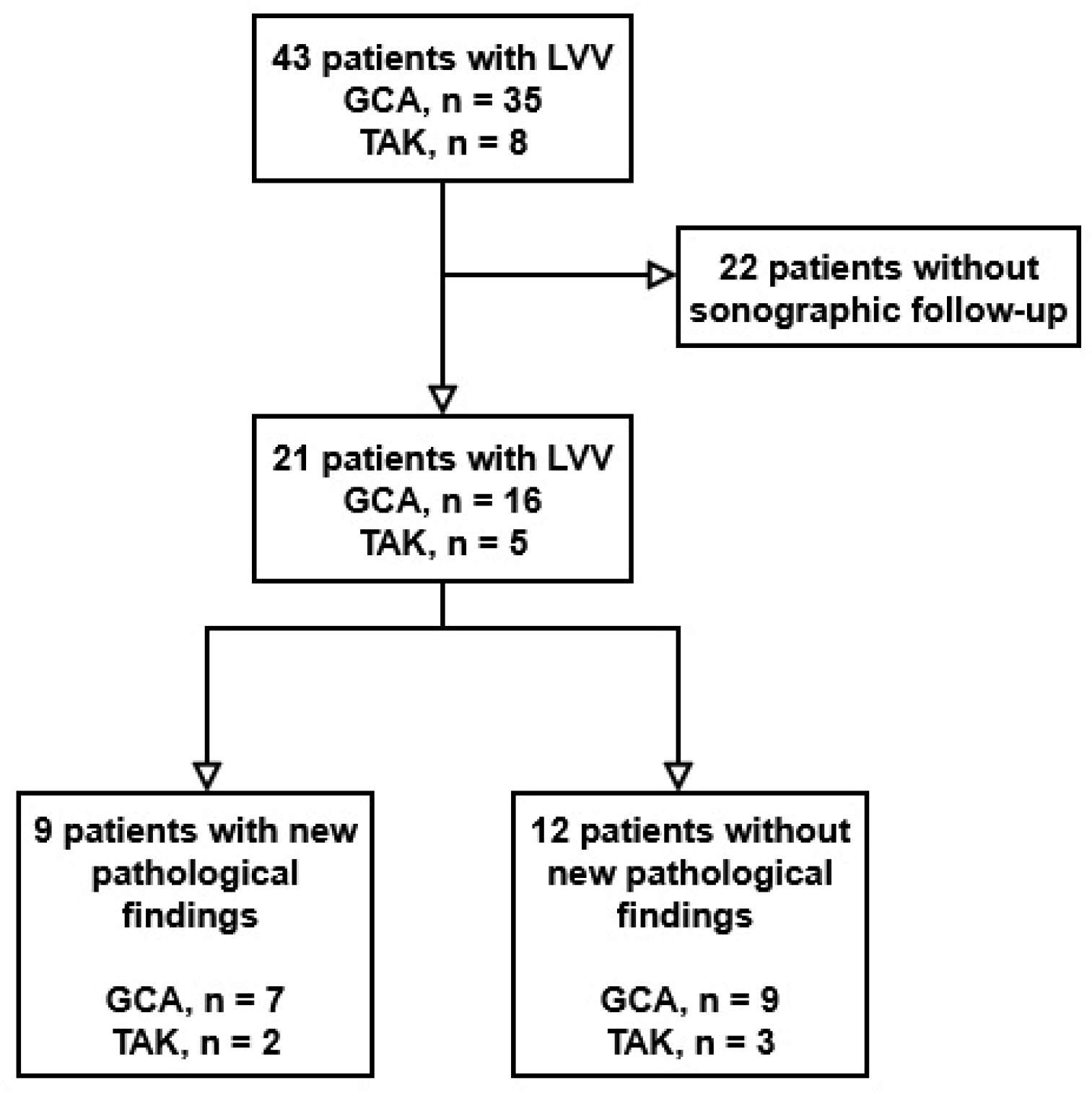
3.1. Patient Cohort
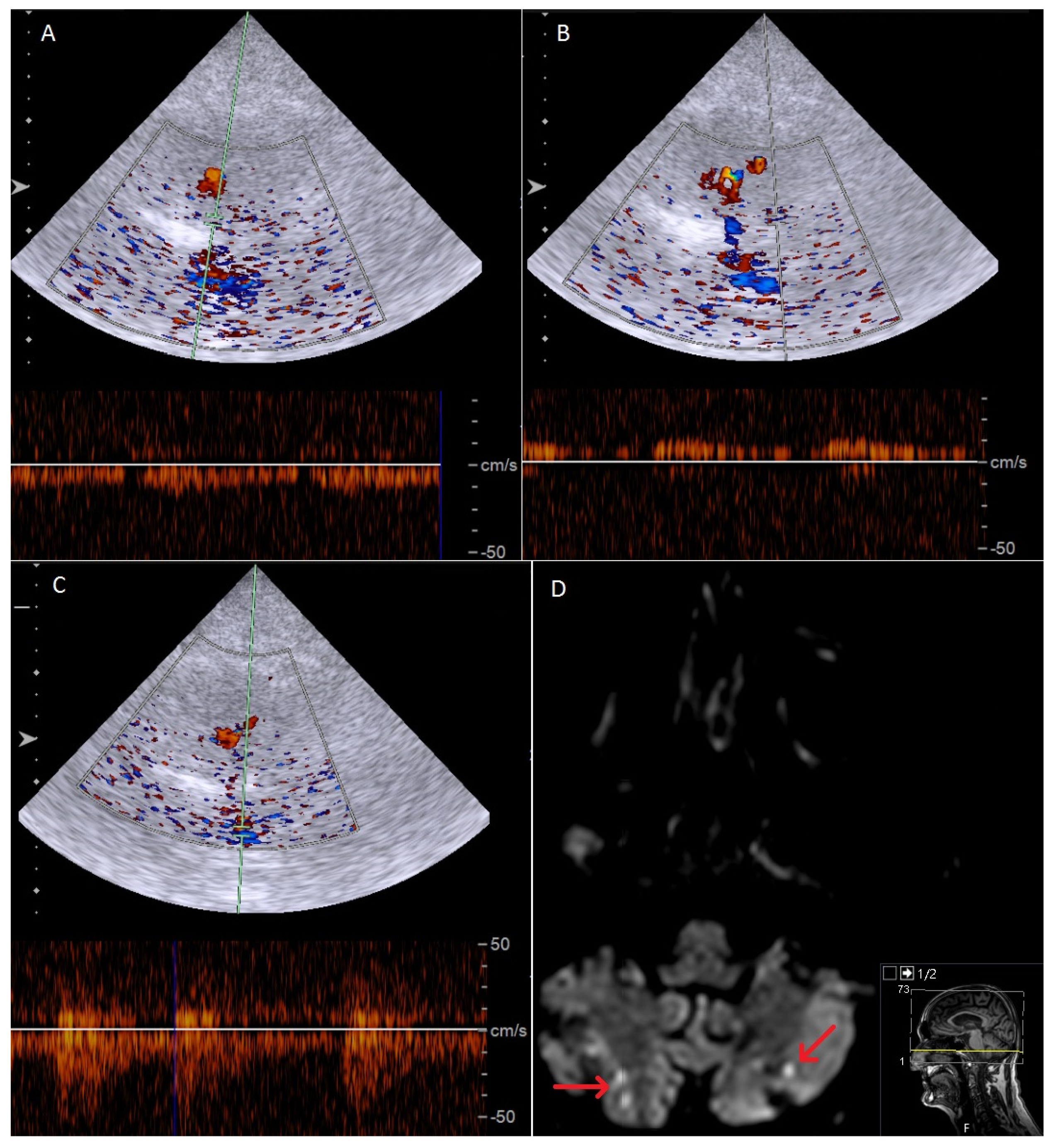
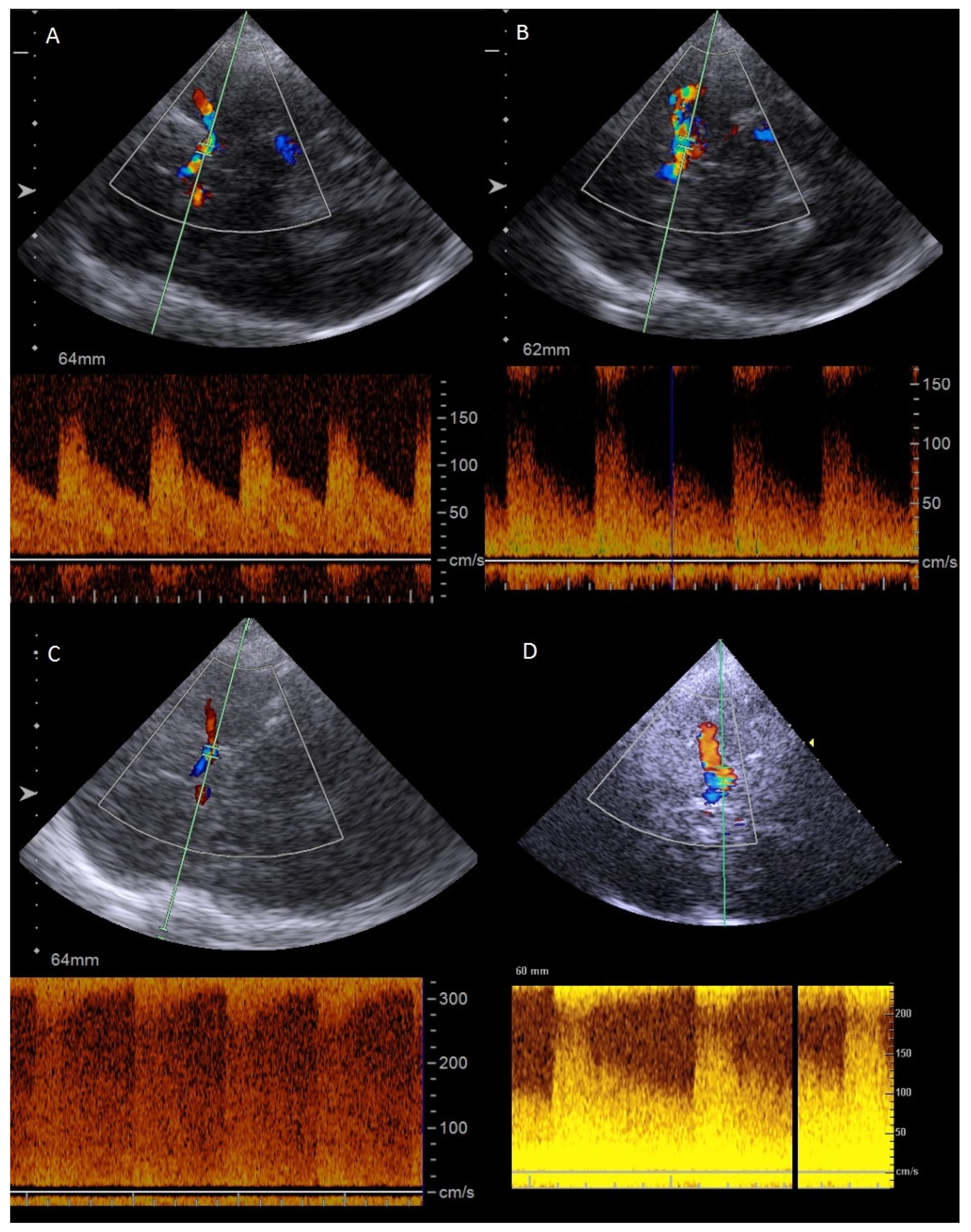
3.2. Patients with New-Onset or Progressive Inflammatory Artery Disease in GCA
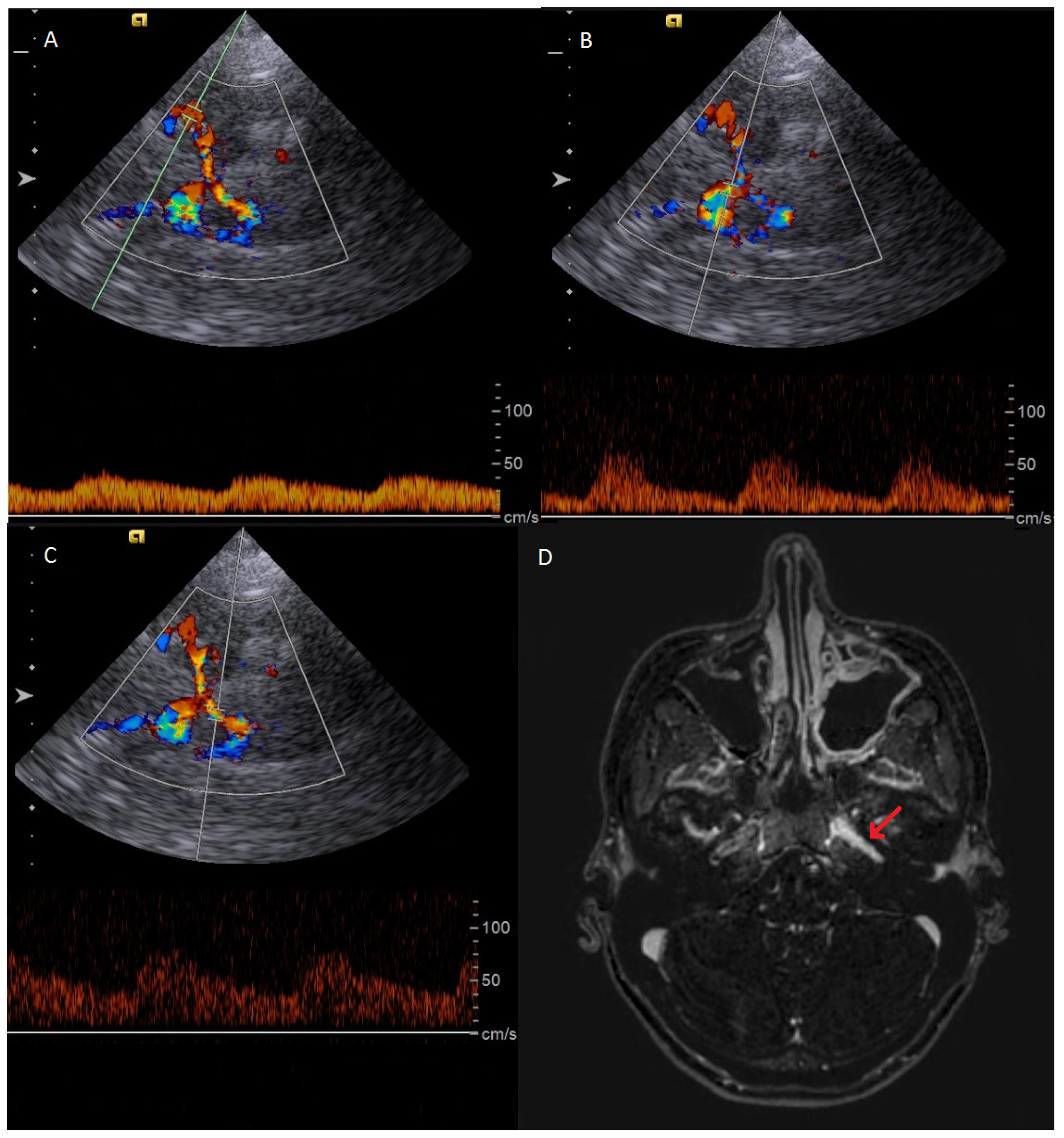
3.3. Patients with New-Onset or Progressive Inflammatory Artery Disease in TAK
4. Discussion
5. Conclusions
Supplementary Materials
Author Contributions
Funding
Institutional Review Board Statement
Informed Consent Statement
Data Availability Statement
Acknowledgments
Conflicts of Interest
Abbreviations
| ACA | anterior cerebral artery |
| BA | basilar artery |
| CCA | common carotid artery |
| GCA | giant cell arteritis |
| ICA | internal carotid artery |
| IMT | intima-media thickness |
| LVV | large-vessel vasculitis |
| MCA | middle cerebral artery |
| MRI | magnetic resonance imaging |
| OGUS | giant cell arteritis ultrasonography score |
| PCA | posterior cerebral artery |
| PSV | peak systolic velocity |
| SA | subclavian artery |
| TA | temporal artery |
| TAK | Takayasu arteritis |
| TCD | transcranial duplex sonography |
| VA | vertebral artery |
References
- Schäfer, V.S.; Dejaco, C.; Karakostas, P.; Behning, C.; Brossart, P.; Burg, L.C. Follow-up ultrasound examination in patients with newly diagnosed giant cell arteritis. Rheumatology 2025, 64, 732–739. [Google Scholar] [CrossRef] [Scilit] [PubMed]
- Schmidt, W.A.; Schäfer, V.S. Diagnosing vasculitis with ultrasound: Findings and pitfalls. Ther. Adv. Musculoskelet. Dis. 2024, 16, 1759720x241251742. [Google Scholar] [CrossRef] [Scilit] [PubMed]
- Kirby, C.; Flood, R.; Mullan, R.; Murphy, G.; Kane, D. Evolution of ultrasound in giant cell arteritis. Front. Med. 2022, 9, 981659. [Google Scholar] [CrossRef] [Scilit] [PubMed]
- Luqmani, R.; Lee, E.; Singh, S.; Gillett, M.; Schmidt, W.A.; Bradburn, M.; Dasgupta, B.; Diamantopoulos, A.P.; Forrester-Barker, W.; Hamilton, W.; et al. The Role of Ultrasound Compared to Biopsy of Temporal Arteries in the Diagnosis and Treatment of Giant Cell Arteritis (TABUL): A diagnostic accuracy and cost-effectiveness study. Health Technol. Assess. 2016, 20, 1–238. [Google Scholar] [CrossRef] [Scilit]
- Dejaco, C.; Ramiro, S.; Duftner, C.; Besson, F.L.; Bley, T.A.; Blockmans, D.; Brouwer, E.; Cimmino, M.A.; Clark, E.; Dasgupta, B.; et al. EULAR recommendations for the use of imaging in large vessel vasculitis in clinical practice. Ann. Rheum. Dis. 2018, 77, 636–643. [Google Scholar] [CrossRef] [Scilit]
- Martins-Martinho, J.; Sopa, I.; Ponte, C. The value of ultrasound for monitoring disease activity in giant cell arteritis. Vessel. Plus 2024, 8, 34. [Google Scholar] [CrossRef] [Scilit]
- Dejaco, C.; Ponte, C.; Monti, S.; Rozza, D.; Scirè, C.A.; Terslev, L.; Bruyn, G.A.W.; Boumans, D.; Hartung, W.; Hočevar, A.; et al. The provisional OMERACT ultrasonography score for giant cell arteritis. Ann. Rheum. Dis. 2023, 82, 556–564. [Google Scholar] [CrossRef] [Scilit]
- Monti, S.; Ponte, C.; Schäfer, V.S.; Rozza, D.; Scirè, C.; Franchi, G.; Milanesi, A.; Khmelinskii, N.; Petzinna, S.M.; Carrara, G.; et al. The giant cell arteritis (GCA) ultrasound score (OGUS) at diagnosis and after initial treatment predicts future relapses in GCA patients: Results of a multicentre prospective study. Ann. Rheum. Dis. 2025, 84, 823–832. [Google Scholar] [CrossRef] [Scilit]
- Kirby, C.; Molloy, D.; Cowley, S.; Flood, R.; Gheta, D.; Mullan, R.; Murphy, G.; Kane, D. A prospective cohort study evaluating the role of quantifying sonographic inflammatory changes in GCA. Rheumatol. Adv. Pract. 2025, 9, rkaf085. [Google Scholar] [CrossRef] [Scilit]
- Hansen, M.; Hansen, I.T.; Keller, K.K.; Therkildsen, P.; Hauge, E.M.; Nielsen, B.D. Serial assessment of ultrasound sensitivity and scores in patients with giant cell arteritis before and 3 and 10 days after treatment. Rheumatology 2025, 64, 3895–3899. [Google Scholar] [CrossRef] [Scilit]
- Haaversen, A.C.B.; Brekke, L.K.; Kermani, T.A.; Molberg, Ø.; Diamantopoulos, A.P. Vascular ultrasound as a follow-up tool in patients with giant cell arteritis: A prospective observational cohort study. Front. Med. 2024, 11, 1436707. [Google Scholar] [CrossRef] [Scilit]
- Petzinna, S.M.; Burg, L.C.; Terheyden, J.H.; Bauer, C.J.; Karakostas, P.; Behning, C.; Holz, F.G.; Finger, R.P.; Schäfer, V.S. Transorbital ultrasonography reveals persistent decrease in retinal artery flow and optic nerve diameter in treated giant cell arteritis. Rheumatology 2025, 65, keaf456. [Google Scholar] [CrossRef] [Scilit] [PubMed]
- Petzinna, S.M.; Terheyden, J.H.; Burg, L.C.; Bauer, C.J.; Karakostas, P.; Behning, C.; Holz, F.G.; Finger, R.P.; Schäfer, V.S. Imaging of ophthalmic manifestations: Optical coherence tomography angiography and transorbital ultrasound in giant cell arteritis. Rheumatol. Int. 2025, 45, 48. [Google Scholar] [CrossRef] [Scilit] [PubMed]
- Sun, Y.; Yip, P.-K.; Jeng, J.-S.; Hwang, B.-S.; Lin, W.-H. Ultrasonographic Study and Long-term Follow-up of Takayasu’s Arteritis. Stroke 1996, 27, 2178–2182. [Google Scholar] [CrossRef] [Scilit] [PubMed]
- Dong, Y.; Wang, Y.; Wang, Y.; Tian, X.; Li, J.; Yang, Y.; Ge, Z.; Zhang, L.; Zou, M.; Wang, H.; et al. Ultrasonography and contrast-enhanced ultrasound for activity assessment in 115 patients with carotid involvement of Takayasu arteritis. Mod. Rheumatol. 2023, 33, 1007–1015. [Google Scholar] [CrossRef] [Scilit]
- Herlin, B.; Baud, J.M.; Chadenat, M.L.; Pico, F. Contrast-enhanced ultrasonography in Takayasu arteritis: Watching and monitoring the arterial inflammation. BMJ Case Rep. 2015, 2015, 211094. [Google Scholar] [CrossRef] [Scilit]
- Keleşoğlu Dinçer, A.B.; Kılıç, L.; Erden, A.; Kalyoncu, U.; Hazirolan, T.; Kiraz, S.; KaradaĞ, Ö. Imaging modalities used in diagnosis and follow-up of patients with Takayasu’s arteritis. Turk. J. Med. Sci. 2021, 51, 224–230. [Google Scholar] [CrossRef] [Scilit]
- Svensson, C.; Eriksson, P.; Zachrisson, H. Vascular ultrasound for monitoring of inflammatory activity in Takayasu arteritis. Clin. Physiol. Funct. Imaging 2020, 40, 37–45. [Google Scholar] [CrossRef] [Scilit]
- Yıldırım, F.; Tastekin, F.; Bakkal, S.; Kutu, M.E.; Ocak, T.; San, S.; Avcu, A.; Kocaayan, H.; Işık, S.; Uludogan, B.C.; et al. Prognostic significance and rationality of regular non-invasive imaging follow-up in Takayasu arteritis: A large multicenter cohort study. Semin. Arthritis Rheum. 2025, 74, 152813. [Google Scholar] [CrossRef] [Scilit]
- Johnson, A.; Emery, D.; Clifford, A. Intracranial Involvement in Takayasu’s Arteritis. Diagnostics 2021, 11, 1997. [Google Scholar] [CrossRef] [Scilit]
- Bond, K.M.; Nasr, D.; Lehman, V.; Lanzino, G.; Cloft, H.J.; Brinjikji, W. Intracranial and Extracranial Neurovascular Manifestations of Takayasu Arteritis. AJNR Am. J. Neuroradiol. 2017, 38, 766–772. [Google Scholar] [CrossRef] [Scilit] [PubMed]
- Samaan, M.; Abramyan, A.; Sundararajan, S.; Nourollah-Zadeh, E.; Sun, H.; Nanda, A.; Roychowdhury, S.; Gupta, G. Cerebrovascular implications of takayasu arteritis: A review. Neuroradiology 2025, 67, 125–136. [Google Scholar] [CrossRef] [Scilit] [PubMed]
- Huo, J.; Wang, B.; Yu, L.; Gao, D.; Cheng, R.; Wang, J.; Zhou, X.; Tian, T.; Gao, L. Clinical characteristics and outcomes in patients with Takayasu arteritis coexisting with myocardial ischemia and neurological symptoms: A multicenter, long-term, follow-up study. Front. Cardiovasc. Med. 2022, 9, 948124. [Google Scholar] [CrossRef] [Scilit] [PubMed]
- Guggenberger, K.V.; Vogt, M.L.; Song, J.W.; Fröhlich, M.; Schmalzing, M.; Venhoff, N.; Werner, R.A.; Hillenkamp, J.; Pham, M.; Meckel, S.; et al. High-resolution magnetic resonance imaging visualizes intracranial large artery involvement in giant cell arteritis. Rheumatology 2024, 64, 842–848. [Google Scholar] [CrossRef] [Scilit]
- Patel, S.; Okaj, I.; Scott, J.; Atwal, S.; Stark, C.; Tahir, R.; Khalidi, N.; Junek, M. Intracranial GCA: A comprehensive systematic review. Rheumatology 2025, 64, 4517–4524. [Google Scholar] [CrossRef] [Scilit]
- Grayson, P.C.; Ponte, C.; Suppiah, R.; Robson, J.C.; Gribbons, K.B.; Judge, A.; Craven, A.; Khalid, S.; Hutchings, A.; Danda, D.; et al. 2022 American College of Rheumatology/EULAR Classification Criteria for Takayasu Arteritis. Arthritis Rheumatol. 2022, 74, 1872–1880. [Google Scholar] [CrossRef] [Scilit]
- Ponte, C.; Grayson, P.C.; Robson, J.C.; Suppiah, R.; Gribbons, K.B.; Judge, A.; Craven, A.; Khalid, S.; Hutchings, A.; Watts, R.A.; et al. 2022 American College of Rheumatology/EULAR Classification Criteria for Giant Cell Arteritis. Arthritis Rheumatol. 2022, 74, 1881–1889. [Google Scholar] [CrossRef] [Scilit]
- Ponte, C.; Serafim, A.S.; Monti, S.; Fernandes, E.; Lee, E.; Singh, S.; Piper, J.; Hutchings, A.; McNally, E.; Diamantopoulos, A.P.; et al. Early variation of ultrasound halo sign with treatment and relation with clinical features in patients with giant cell arteritis. Rheumatology 2020, 59, 3717–3726. [Google Scholar] [CrossRef] [Scilit]
- Seitz, L.; Christ, L.; Lötscher, F.; Scholz, G.; Sarbu, A.C.; Bütikofer, L.; Kollert, F.; Schmidt, W.A.; Reichenbach, S.; Villiger, P.M. Quantitative ultrasound to monitor the vascular response to tocilizumab in giant cell arteritis. Rheumatology 2021, 60, 5052–5059. [Google Scholar] [CrossRef] [Scilit]
- Ford, J.A.; DiIorio, M.A.; Huang, W.; Sobiesczcyk, P.; Docken, W.P.; Tedeschi, S.K. Follow-up vascular ultrasounds in patients with giant cell arteritis. Clin. Exp. Rheumatol. 2020, 38, 107–111. [Google Scholar]
- Nielsen, B.D.; Therkildsen, P.; Keller, K.K.; Gormsen, L.C.; Hansen, I.T.; Hauge, E.M. Ultrasonography in the assessment of disease activity in cranial and large-vessel giant cell arteritis: A prospective follow-up study. Rheumatology 2023, 62, 3084–3094. [Google Scholar] [CrossRef] [Scilit] [PubMed]
- Aschwanden, M.; Schegk, E.; Imfeld, S.; Staub, D.; Rottenburger, C.; Berger, C.T.; Daikeler, T. Vessel wall plasticity in large vessel giant cell arteritis: An ultrasound follow-up study. Rheumatology 2018, 58, 792–797. [Google Scholar] [CrossRef] [Scilit] [PubMed]
- Zachrisson, H.; Svensson, C.; Dremetsika, A.; Eriksson, P. An extended high-frequency ultrasound protocol for detection of vessel wall inflammation. Clin. Physiol. Funct. Imaging 2018, 38, 586–594. [Google Scholar] [CrossRef] [Scilit] [PubMed]
- Schmidt, W.A. Role of ultrasound in the understanding and management of vasculitis. Ther. Adv. Musculoskelet. Dis. 2014, 6, 39–47. [Google Scholar] [CrossRef] [Scilit]
- Siemonsen, S.; Brekenfeld, C.; Holst, B.; Kaufmann-Buehler, A.K.; Fiehler, J.; Bley, T.A. 3T MRI reveals extra- and intracranial involvement in giant cell arteritis. AJNR Am. J. Neuroradiol. 2015, 36, 91–97. [Google Scholar] [CrossRef] [Scilit]
- Samson, M.; Jacquin, A.; Audia, S.; Daubail, B.; Devilliers, H.; Petrella, T.; Martin, L.; Durier, J.; Besancenot, J.F.; Lorcerie, B.; et al. Stroke associated with giant cell arteritis: A population-based study. J. Neurol. Neurosurg. Psychiatry 2015, 86, 216–221. [Google Scholar] [CrossRef] [Scilit]
- Larivière, D.; Sacre, K.; Klein, I.; Hyafil, F.; Choudat, L.; Chauveheid, M.P.; Papo, T. Extra- and intracranial cerebral vasculitis in giant cell arteritis: An observational study. Medicine 2014, 93, e265. [Google Scholar] [CrossRef] [Scilit]
- Pariente, A.; Guédon, A.; Alamowitch, S.; Thietart, S.; Carrat, F.; Delorme, S.; Capron, J.; Cacciatore, C.; Soussan, M.; Dellal, A.; et al. Ischemic stroke in giant-cell arteritis: French retrospective study. J. Autoimmun. 2019, 99, 48–51. [Google Scholar] [CrossRef] [Scilit]
- Penet, T.; Lambert, M.; Baillet, C.; Outteryck, O.; Hénon, H.; Morell-Dubois, S.; Hachulla, E.; Launay, D.; Pokeerbux, M.R. Giant cell arteritis-related cerebrovascular ischemic events: A French retrospective study of 271 patients, systematic review of the literature and meta-analysis. Arthritis Res. Ther. 2023, 25, 116. [Google Scholar] [CrossRef] [Scilit]



| Patient Characteristics | |||
|---|---|---|---|
| Patients | GCA | TAK | |
| Number of patients included | 16 | 5 | |
| Age (years), mean (±SD) | 72.8 ± 7.4 | 37.6 ± 8.4 | |
| Gender | |||
| Female n (%) | 9 (56.3%) | 3 (60%) | |
| Male n (%) | 7 (43.8%) | 2 (40%) | |
| Follow-up period (months), median (range) | 28 (2–106) | 75 (33–255) | |
| Number of sonographic assessments during follow-up, median (range) | 4.5 (2–33) | 14 (4–60) | |
| Baseline sonography | Halo sign TA, n (%) | 13 (81.3%) | - |
| Involvement of VA, n (%) | 8 (50.0%) | 2 (40%) | |
| Involvement of SA, n (%) | - | 5 (100%) | |
| Involvement of CCA, n (%) | - | 2 (40%) | |
| Involvement of extracranial ICA, n (%) | - | 2 (40%) | |
| Involvement of anterior circulation IA, n (%) | 3 (18.8%) | 2 (40%) | |
| New or progressive artery involvement detected in follow-up sonography | Total, n (%) | 7 (43.8%) | 2 (40%) |
| Symptomatic progress, n (%) | 4 (25.0%) | 1 (20%) | |
| Progress or new-onset involvement of VA, n (%) | 3 (18.8%) | - | |
| Progress or new-onset involvement of anterior circulation IA, n (%) | 3 (18.8%) | 1 (20%) | |
| Progress or new-onset involvement of CCA, n (%) | - | 1 (20%) | |
| Change of medical treatment following progress, n (%) | 4 (25.0%) | - | |
| Interventional treatment following progress, n (%) | 2 (12.5%) | 1 (20%) | |
| Clinical and Sonographic Findings in Patients with Progressive Inflammatory Artery Involvement in LVV and Their Therapeutic Impact | |||||||||||
|---|---|---|---|---|---|---|---|---|---|---|---|
| Clinical Information | Progressive Inflammatory Artery Disease During Sonographic Follow-Up | Medical and Interventional Treatment Following Progress | |||||||||
| Age | Sex | Follow-up Period (Months) | Number of Sonographic Examinations | Baseline Pathology | Time from First Diagnosis (Months) | Symptomatic Progress | Sonographic Findings | Baseline | Change | Interventional Treatment | |
| GCA | 68 | m | 105 | 20 | Halo TA Extracranial VA stenosis | 36 | - | Progressive halo sign TA | Prednisolone + MTX | - | - |
| GCA | 67 | f | 55 | 19 | Occlusion intracranial right VA | 3 | Recurrent left-hemispheric strokes | High-grade stenosis of both ICAs (petrous segment) Stenosis MCA left | Prednisolone | MPS pulse (1000 mg for 5 days), MTX | Stent angioplasty of the left intracranial ICA |
| GCA | 64 | m | 30 | 33 | Halo TA Intracranial right VA stenosis Intracranial, bilateral ICA stenosis | 1 | Recurrent bihemispheric strokes | Progressive stenosis ICA and MCA left Progressive stenosis VA right extracranial and both intracranial VA | Prednisolone | MTX, TOC, CYC | Balloon dilatation of both ICAs |
| GCA | 83 | m | 60 | 3 | Halo TA | 41 | - | Progressive halo TA | Prednisolone + TOC | - | - |
| GCA | 80 | f | 4 | 4 | Halo TA Extracranial VA stenosis | 1 | - | Progressive extracranial, right-side VA stenosis | Prednisolone + MTX | - | - |
| GCA | 72 | m | 23 | 6 | Halo TA Intracranial VA stenosis | 4 | Recurrent vertebro-basilar strokes | Intracranial, bilateral VA occlusion Intracranial, bilateral ICA stenosis | Prednisolone + TOC | MPS pulse (1000 mg for 5 days), CYC, MTX | - |
| GCA | 80 | m | 12 | 2 | Halo TA | 5 | Headache | Progressive halo TA | Prednisolone + TOC | Prednisolone dose increase | - |
| TAK | 26 | f | 75 | 14 | VA, SA, CCA, ICA (extracranial) | 57 | - | Intracranial left-side ICA stenosis | Prednisolone + Certolizumab | - | - |
| TAK | 33 | f | 254 | 60 | SA, ACC | 95 | Right-hemispheric stroke | Progressive right-side CCA stenosis | Prednisolone + MTX | - | Stent angioplasty right CCA |
Disclaimer/Publisher’s Note: The statements, opinions and data contained in all publications are solely those of the individual author(s) and contributor(s) and not of MDPI and/or the editor(s). MDPI and/or the editor(s) disclaim responsibility for any injury to people or property resulting from any ideas, methods, instructions or products referred to in the content. |
© 2026 by the authors. Licensee MDPI, Basel, Switzerland. This article is an open access article distributed under the terms and conditions of the Creative Commons Attribution (CC BY) license.
Share and Cite
Härtl, J.; Lambrecht, S.; Hess, F.; Berthele, A.; Wunderlich, S.; Baki, E. Long-Term Follow-Up in Patients with Large-Vessel Vasculitis Applying Extracranial and Transcranial Duplex Sonography. Diagnostics 2026, 16, 455. https://doi.org/10.3390/diagnostics16030455
Härtl J, Lambrecht S, Hess F, Berthele A, Wunderlich S, Baki E. Long-Term Follow-Up in Patients with Large-Vessel Vasculitis Applying Extracranial and Transcranial Duplex Sonography. Diagnostics. 2026; 16(3):455. https://doi.org/10.3390/diagnostics16030455
Chicago/Turabian StyleHärtl, Johanna, Sebastian Lambrecht, Felix Hess, Achim Berthele, Silke Wunderlich, and Enayatullah Baki. 2026. "Long-Term Follow-Up in Patients with Large-Vessel Vasculitis Applying Extracranial and Transcranial Duplex Sonography" Diagnostics 16, no. 3: 455. https://doi.org/10.3390/diagnostics16030455
APA StyleHärtl, J., Lambrecht, S., Hess, F., Berthele, A., Wunderlich, S., & Baki, E. (2026). Long-Term Follow-Up in Patients with Large-Vessel Vasculitis Applying Extracranial and Transcranial Duplex Sonography. Diagnostics, 16(3), 455. https://doi.org/10.3390/diagnostics16030455