Abstract
Environmental DNA (eDNA) has become a promising tool for phytoplankton surveys. However, the accuracy of eDNA-based detection is related to primer selection across diverse environments, and optimal primer pairs selection on phytoplankton community in human impacted ecosystems is still lacking. The aim of this study is to evaluate how primer selection shapes phytoplankton community profiles by eDNA biomonitoring diverse anthropogenically disturbed aquatic systems (rivers, reservoirs, and seas). Four primer pairs targeting the 18S rRNA (V9-1 and V9-2), chloroplast rbcL, and ITS regions, were explored and our results revealed that primer choice critically governed the accuracy of phytoplankton profiling. Significant variations in annotated phytoplankton eDNA sequences in different groups of primer pairs were observed, where the primers 18SV9-1 and rbcL demonstrated superior specificity, amplifying >90% of phytoplankton OTUs. 18S-targeted primers detected the highest species richness, while the ITS primer showed the lowest. Alpha diversity was highest and most consistent for 18S primers. Beta diversity ordination (nMDS/Bray–Curtis) further highlighted primer-dependent community structuring in which 18S primers effectively clustered reservoir and marine samples separately, whereas primer rbcL discriminated habitat-specific signatures across three ecosystems. The primer ITS failed to distinguish among different habitats. Overall, our data demonstrated the critical role of primer optimization in eDNA-based phytoplankton studies, and could provide methodological guidelines for the design of effective monitoring protocols in rapidly urbanizing aquatic systems.
1. Introduction
Phytoplankton, as the primary producers in ecosystems such as oceans, rivers, and reservoirs, form the foundation of aquatic food webs by driving energy transfer from microbial loops to higher trophic levels, and play pivotal roles in maintaining the ecological equilibrium through oxygen production and carbon sequestration via the biological pump. Their community composition and structural distribution directly or indirectly respond to and influence ecosystem characteristics [1,2]. Due to their rapid response to changes in aquatic environmental factors, such as water temperature, nutrient concentration, and pH, phytoplankton are often used as indicator organisms in aquatic ecological studies [1,3,4]. These attributes make phytoplankton community dynamics critical for the adaptive management of water resources, particularly in urbanizing regions where compounded pressures caused by industrialization and climate change threaten aquatic biodiversity [5]. Therefore, investigating phytoplankton communities holds significant scientific and practical value for deciphering ecosystem resilience and guiding sustainable governance strategies.
Current methodologies for phytoplankton community monitoring primarily include microscopic morphological identification, remote sensing, and metagenomics. However, traditional morphological approaches are often time-consuming, labor-intensive, and costly, requiring taxonomic expertise. Additionally, they have limited capacity to detect cryptic species (e.g., morphologically indistinct taxa) or rare taxa in environmental samples, and are susceptible to subjective bias [6]. Meanwhile, conventional satellite-based phytoplankton monitoring faces inherent limitations. Firstly, satellite imagery suffers from relatively coarse spatial and temporal resolution. Secondly, correcting spectral biases induced by atmospheric and ionospheric interference requires complex procedures that rely heavily on empirical equations and parameter estimations. Consequently, satellite data with high spatial, temporal, and spectral resolution, that are essential for localized applications, are often prohibitively expensive [7]. Although metagenomic approaches offer species-level resolution insights, they are accompanied by notable drawbacks, including high costs and prolonged processing times. Environmental DNA (eDNA) metabarcoding, by integrating DNA-based identification with high-throughput sequencing, has emerged as a powerful tool for characterizing biodiversity across the Tree of Life and resolving intraspecific genetic variation critical to population-level analyses [8,9,10,11,12,13]. Its high sensitivity and time efficiency, and lower subjectivity, have driven widespread adoption in monitoring phytoplankton community dynamics across diverse aquatic ecosystems. For instance, Zeng et al. integrated eDNA metabarcoding with machine learning to map spatial variations in phytoplankton community composition along China’s Dongjiang River, demonstrating the capacity of this approach to resolve fine-scale ecological patterns [8]. Similarly, Hou et al. applied this approach in the Minghu National Wetland Park (China) to compare planktonic community diversity and assembly mechanisms between natural and artificial lakes, thereby highlighting its utility in disentangling environmental drivers of community structure [14]. Further exemplifying its global applicability, Gaonkar et al. employed eDNA metabarcoding to decode phytoplankton genetic diversity patterns in the Gulf of Mexico, underscoring its versatility in both coastal and open-water systems [15].
The performance of phytoplankton biomonitoring by eDNA metabarcoding critically depends on the selection of primer pairs [16,17,18]. Effective primers must possess specificity to avoid cross-hybridization with non-target sequences or the formation of primer-dimers, thereby preventing amplification failures; adaptability to monitoring goals as universal primers necessitate broad taxonomic coverage while maintaining the resolution to differentiate closely related species, whereas species-specific primers demand rigorous validation of amplification efficiency to minimize false negatives (e.g., missed targets) and false positives (e.g., off-target binding); and technical robustness to ensure reliable performance across heterogeneous environmental samples containing inhibitors or trace DNA [19]. Current eDNA-based phytoplankton monitoring predominantly utilizes primers targeting 12 distinct genetic regions, including V3 18S, V4 18S, V4-V5 18S, V7 18S, V7-V8 18S, V8-V9 18S, V9 18S, V9-ITS1, ITS2, rbcL, 23S, and V4 16S [20]. Nuclear regions (e.g., V3, V4, V7, V9, and V9-ITS1) were employed to analyze entire eukaryotic communities and microalgal assemblages, with specific applications in targeting individual algal groups such as dinoflagellates and diatoms [21]. In Antarctic studies, the ITS2 region has been selected for investigating green algae s.l. (Viridiplantae) [22]. Plastid regions are also widely adopted—for instance, rbcL is extensively used for diatom metabarcoding, and primers have been specifically designed and validated for identifying Eustigmatophyceae [23]. Additionally, the universal plastid barcode 23S has been applied to study cyanobacterial and eukaryotic algal diversity, while the 16S rRNA gene serves as a universal marker for both prokaryotes and eukaryotic algae [20]. However, previous studies have often applied primers in a context-specific manner without benchmarking their efficacy, biases, and practical applicability against one another in complex, real-world settings. This gap hinders the development of standardized, reliable eDNA-based monitoring, particularly in rapidly urbanizing regions where accurate biodiversity assessment is critical. Consequently, how primer pair selection influences phytoplankton biomonitoring across diverse environmental samples remains underexplored, particularly in the context of complex aquatic systems subjected to high-intensity anthropogenic disturbances, such as seas, rivers, and reservoirs in rapidly urbanizing regions like Shenzhen, China. The applicability and biases of primer pairs for eDNA-based phytoplankton detection in these human-altered ecosystems require systematic evaluation to optimize taxonomic resolution and ecological interpretability.
To elucidate the applicability of eDNA technology in high-disturbance aquatic systems and refine methodologies for urban aquatic biomonitoring, we selected four pairs of primers widely used by scholars in the ribosomal 18S rRNA, chloroplast, and internal transcribed spacer gene regions (Table S2), and established a total of 44 sets of PCR tests (Table S3). Surface water eDNA samples were collected from different habitats in Shenzhen, China (Dapeng River, Pingshan River, Longkou Reservoir, Shiyan Reservoir, and Shenzhen Bay), and the impact of primers on the biological monitoring of eukaryotic phytoplankton was analyzed. The objectives of this study mainly include the following three aspects: (1) to evaluate the specificity of different primers for eukaryotic phytoplankton by calculating the ratio of eDNA OTUs of eukaryotic and non-eukaryotic phytoplankton; (2) to compare the taxonomic specificity and richness revealed by different primers through analyzing the composition of eukaryotic phytoplankton taxa at different taxonomic levels; and (3) to reveal the impact of different primers on the structural differences in eukaryotic phytoplankton communities by calculating the Bray dissimilarity matrix.
2. Materials and Methods
2.1. Study Area and Sampling
Surface water samples were collected from 11 sites across the Dapeng River (DP), Pingshan River (PS), Longkou Reservoir (LK), Shiyan Reservoir (SY), and Shenzhen Bay (SZ) in Shenzhen, China, during November and December 2023 (Table 1). At each site, 1 L of surface water was collected using sterile bottles (Thermo Fisher Scientific, Waltham, MA, USA) and immediately transferred to a portable cooler with ice packs (0–4 °C) for transport. Filtration was completed within 24 h of collection. The water was vacuum-filtered through 0.45 μm hydrophilic nylon membranes (Merck Millipore, Burlington, MA, USA) to capture environmental DNA (eDNA). During sampling, sterile water was used as a negative control and processed in parallel through all subsequent stages: filtration, DNA extraction, and PCR amplification. Filtered eDNA samples were stored in 5.0 mL centrifuge tubes, flash-frozen in liquid nitrogen, and preserved at −80 °C until DNA extraction.

Table 1.
Longitude and latitude of sampling points.
2.2. DNA Extraction, PCR Amplification, and Sequencing
Total DNA was extracted from the membranes using the DNeasy PowerWater Kit (QIAGEN, Hilden, Germany). DNA concentration was quantified with a Qubit Flex Fluorometer (Thermo Fisher Scientific, Waltham, MA, USA) and the Equalbit dsDNA HS Assay Kit (Vazyme Biotech Co., Ltd., Nanjing, China). Purified DNA extracts were subsequently used for PCR amplification, which was performed using primer pairs targeting specific genetic regions (Table 2). Each primer was fused with unique barcode for sample indexing. Thermal cycling conditions for different primer pairs are detailed in Table S1. Based on Qubit-quantified DNA concentrations, equimolar volumes of PCR products from each sample were pooled and purified by gel extraction. Sequencing libraries were constructed following the ALFA-SEQ Amplicon Library Prep Kit protocol (Fangzhou Biosafety Science & Technology (Guangzhou) Co., Ltd, Guangzhou, China). Library quality was assessed using a Qubit Flex Fluorometer (Thermo Fisher Scientific, Waltham, MA, USA) and the QSEP400 High-throughput Nucleic Acid and Protein Analysis System. Following library preparation, paired-end sequencing was performed on the Illumina NovaSeq 6000 platform (Illumina, Inc., San Diego, CA, USA). To monitor sequencing accuracy and potential contamination, both negative controls (using sterile water) and positive controls (provided in the sequencing kit) were incorporated during library preparation and sequencing. Raw sequencing data generated from base calling were converted into FASTQ-formatted files containing sequence reads and corresponding Phred quality scores.

Table 2.
Summary of oligonucleotides used in this study, including primer name, target region, amplicon size, original references, and primer sequences.
2.3. Bioinformatic Analysis
Raw sequencing reads were processed through Fastp (v0.12.4) for adaptor removal and quality filtering. Low-quality reads (1) shorter than 100 bp, (2) containing >5 ambiguous bases (N), or (3) with an average Phred score < 20, were discarded. Read quality was evaluated pre and post filtering via FastQC (v0.11.9). Paired-end reads were assembled (USEARCH v11.0.667), trimmed of primers (VSEARCH, exact match), and filtered to retain sequences within expected amplicon lengths with ≤1% sequencing errors. Dereplication (VSEARCH) retained representative sequences (abundance ≥ 4), followed by chimera removal (uchime3_denovo, USEARCH) and OTU clustering at 98% similarity (UPARSE). Taxonomic assignment was performed using BLAST (v2.2.31) against eukaryotic phytoplankton databases (BOLD, SILVA, NCBI, R-Syst: Diatom, Figshare, and the ITS2 Database), and sequences with alignment length < 90% or similarity < 80% were further discarded. Taxonomic ranks (from species to class) were assigned at 98%, 95%, 90%, 85%, and 80% similarity thresholds, respectively. The algae species annotations were then corrected according to AlageBase (https://www.algaebase.org, accessed on 28 October 2025). Final OTU tables excluded sequences with relative abundance < 0.01% to minimize spurious detections.
2.4. Statistical Analysis
To assess the sequence specificity of eukaryotic phytoplankton across PCR assay groups, we analyzed the relative abundance of eDNA sequences taxonomically classified as phytoplankton versus non-phytoplankton organisms. The taxonomic resolution of each primer pair was evaluated by quantifying assigned sequences at multiple hierarchical levels (species, genus, and family). Phytoplankton diversity was characterized through alpha diversity metrics including species richness, the Shannon index, the Gini–Simpson index, and Pielou’s evenness index, with statistical significance determined by Student’s t-test. Beta diversity patterns were visualized using non-metric multidimensional scaling (nMDS) based on Bray–Curtis dissimilarity matrices to examine community structure variations among groups. All statistical analyses were conducted in R (v4.1.3) with the vegan package, while data visualization was implemented using ggplot2.
3. Results
3.1. Analysis of Eukaryotic Phytoplankton eDNA Sequence Specificity
Based on the sequencing results, the primers 18SV9-1, 18SV9-2, and rbcL successfully amplified their target products in river, reservoir, and sea samples. In contrast, the ITS primer produced amplifications only in two river samples (PS2 and PS3), indicating its limited applicability in riverine environments (Table S2).
Furthermore, primers 18SV9-1 and 18SV9-2 generated more OTUs than primers rbcL and ITS. Specifically, primer 18SV9-1 yielded a total of raw reads ranging from 112,488 to 543,854, clean reads from 103,898 to 317,745, and OTUs from 2216 to 3343. Meanwhile, primer 18SV9-2 detected raw reads ranging from 76,837 to 721,155, clean reads from 72,115 to 423,557, and OTUs from 3005 to 5734. For primer rbcL, raw reads ranged from 217,548 to 498,128, clean reads from 173,765 to 405,809, and OTUs from 1448 to 1754. Lastly, primer ITS identified raw reads ranging from 351,281 to 1,178,256, clean reads from 254,390 to 935,456, and OTUs from 1001 to 2003 (Table S3).
Our analysis showed that, within all groups, the percentage of eukaryotic phytoplankton OTUs amplified by primers 18SV9-1 and rbcL significantly exceeded 90%, with rbcL demonstrating the highest yield of phytoplankton OTUs. Following closely behind was 18SV9-2, with eukaryotic phytoplankton OTUs accounting for about 80% of the total OTUs. Conversely, the eukaryotic group, associated with the primer ITS, showed the lowest proportion, with a successful amplification rate of less than 25% (Figure S1). The results may be related to database limitations, PCR amplification efficiency, or the actual community composition of sampling points.
3.2. Species Composition and Taxonomic Richness
The 18S rRNA primer pairs exhibited the broadest taxonomic coverage at all phylogenetic levels (species to phylum), surpassing primers rbcL and ITS (Table S4). Upon analyzing the same environmental specimens, different primer pairs revealed strikingly disparate community composition profiles of eukaryotic phytoplankton (Figure S2; Table S4). Notably, 18S primers (18SV9-1/18SV9-2) were able to detect a considerable abundance of Chlorophyta in diverse habitats, whereas primer rbcL consistently demonstrated a tendency to underestimate the presence of this phylum, highlighting a substantial disparity in the performance of these two primer pairs. As for primer rbcL, evident habitat-specific amplification patterns were observed. Compared to the other primer pairs, the amplification results of primer rbcL revealed that Crypista emerged as the dominant taxon in marine and riverine samples, while reservoir ecosystems exhibited heightened abundances of Euglenophyta and Haptophyta. Regarding primer ITS, it preferentially amplified Heterokontophyta, Chlorophyta, and Dinoflagellata.
To further elaborate on primer performance, we analyzed river water samples and observed pronounced discrepancies in eukaryotic phytoplankton communities across different primer pairs (Figure 1a and Figure S2a). The two 18S primers generated similar detection results with a more uniform species richness at both the phylum and order levels, and consistently outperformed other primers in capturing phylum diversity (Table S4). Conversely, the primer rbcL predominantly identified Heterokontophyta (16.2–76.2% relative abundance) and Crypista (11.3–69.4%). In particular, it showed a higher relative abundance of Cryptista compared to other primers (Figure S2a) and exhibited a strong preference for detecting phytoplankton within the order Cryptomonadales (Figure 1a). Meanwhile, the primer ITS detected the fewest species (Table S4) and only successfully amplified specific bands, i.e., Heterokontophyta (73.4–94.3%) and Chlorophyta (5.7–26.4%) in the PS2 and PS3 samples (Figure S2a).


Figure 1.
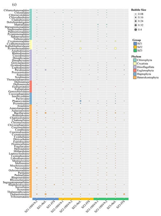
Monitoring the taxonomic diversity of phytoplankton using eDNA technology: (a) Rivers, (b), reservoirs, and (c) the sea. The colors refer to the phyla of different phytoplankton and the size of the bubbles reflects the relative sequence readings of each group.
Reservoir samples followed a similar trend, such that 18S primers detected the highest species richness, whereas the primer ITS exhibited the poorest taxonomic resolution (Figure 1b and Figure S2b, and Table S4). Specifically, the 18S primers primarily identified Cryptista (17.9–54.4%), Heterokontophyta (23.4–57.4%), Dinoflagellata (12.5–16.8%), and Chlorophyta at the phylum level, while the primer rbcL preferred to detect Cryptista (24.8–58.6%), Heterokontophyta (31.5–55.2%), Haptophyta, and Euglenophyta. The primer ITS, in contrast, was largely restricted to detecting Heterokontophyta (37.8–96.2%), Chlorophyta (3.8–31.9%), and Dinoflagellata (Figure S2b).
For samples collected from the marine ecosystem, the 18S primers also detected the greatest species diversity, followed by the primer rbcL, with the primer ITS again showing the narrowest taxonomic scope (Figure S2c and Figure 1c, and Table S4). Furthermore, the relative abundance of detected species was notably influenced by the choice of primer. Specifically, the primer rbcL demonstrated a preference for detecting Cryptista, with a pronounced tendency towards Pyrenomonadales and Cryptomonadales at the order level (Figure 1c). Conversely, the primer ITS remained predominantly inclined towards the identification of diatoms (Heterokontophta), underscoring the distinct taxonomic biases associated with each primer pair (Figure 1c).
3.3. The Alpha Diversity
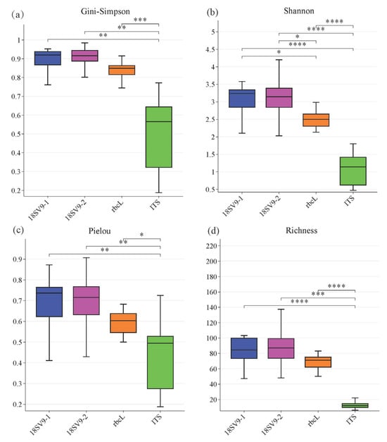
Based on genus-level relative abundance profiles derived from sequencing data, α-diversity indices (Gini–Simpson, Shannon, Pielou, and Richness) were systematically compared across the four primer pairs (Figure 2). The primer ITS consistently yielded the lowest diversity values across all metrics, highlighting its inherent constraints in phytoplankton studies. This limited performance, which was closely related to the low taxonomic resolution and substantial underestimation of species, renders the primer ITS suboptimal for robust α-diversity characterization in phytoplankton communities.

Figure 2.
Alpha diversity results of different primers. Indexes: (a) Gini–Simpson, (b) Shannon, (c) Pielou, and (d) Richness. *: p-value < 0.05; **: p-value < 0.01; ***: p-value < 0.001, and ****: p-value < 0.0001.
In contrast, the primers 18SV9-1 and 18SV9-2 exhibited superior detection capacity, generating the highest diversity values with strong inter-primer consistency. Meanwhile, the primer rbcL displayed moderate efficacy. Notably, statistically significant differences between the primers 18S and rbcL were only observed in the Shannon index. This indicated intrinsic disparities in detection efficiency between the primers 18S and rbcL for rare taxonomic groups, as the Shannon index demonstrates higher sensitivity to rare taxa. In contrast, the Gini–Simpson index primarily reflects dominant species, and the Pielou index characterizes community evenness. The lack of statistically significant differences in these indices suggested compositional similarity in the dominant communities detected by the primers 18S and rbcL. The Richness index, representing the total count of identified species, revealed that the three primer pairs achieved comparable numbers of genus-level taxonomic units.
3.4. Community Structure
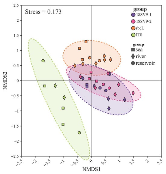
The NMDS ordination plots were plotted based on the Bray matrix. The results show that the application of different primers led to the identification of eukaryotic phytoplankton communities with different structures (Figure 3). In particular, the structural differences in eukaryotic phytoplankton communities detected by the primer ITS were significantly different from those detected by the other primer pairs. The sample points of the primers 18SV9-1 and 18SV9-2 were closer in the ordination plots, indicating that the monitored eukaryotic phytoplankton community structures were more similar to each other.

Figure 3.
The Non-Metric Multidimensional Scale (NMDS) plot shows the structural differences in phytoplankton communities among the different primer pairs.
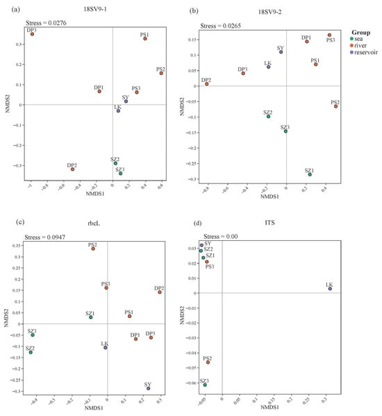
To further evaluate the discriminatory capabilities of each primer pair, NMDS ordination plots specifically tailored for each primer pair were constructed (Figure 4). Notably, the primers 18SV9-1 and 18SV9-2 demonstrated a pronounced ability to coherently cluster samples from reservoir and marine ecosystems separately, whereas river samples exhibited a more dispersed distribution pattern. Meanwhile, the primer rbcL showed remarkable potential in differentiating sample sites from the three distinct habitats—reservoirs, rivers, and seas—with each displaying an obvious spatial dispersion pattern. Conversely, the primer ITS failed to demonstrate such clear habitat-specific segregation. These findings underscore the variable influence that different primer pairs exert on the eukaryotic phytoplankton communities residing in diverse aquatic environments.

Figure 4.
The Non-Metric Multidimensional Scale (NMDS) plots showing structural dissimilarities of phytoplankton communities across the groups of primer pairs in river samples: (a) 18SV9-1, (b) 18SV9-2, (c) rbcL, and (d) ITS.
4. Discussion
In this study, we used four pairs of primers, 18SV9-1, 18SV9-1, rbcL, and ITS, to monitor the environmental DNA of eukaryotic phytoplankton in different habitats. The results showed that, compared to the effects of habitat variations, primer selection exerted a stronger influence on eukaryotic phytoplankton biomonitoring in river, reservoir, and sea samples.
Our data indicate that, compared to the primer ITS, the primers 18SV9-1, 18SV9-2, and rbcL exhibited higher percentages of eukaryotic planktonic OTUs and more specific classification resolution. This outcome may be attributable to several factors for the primers 18SV9-1 and 18SV9-2. Firstly, the annealing temperature of the primers 18SV9-1 and 18SV9-2 is relatively low, which reduces the preferential amplification, resulting in a more balanced monitoring of eukaryotic phytoplankton species [26]. Secondly, the 18S region, which encompasses nine hypervariable regions (V1 to V9, with V6 being more conserved in eukaryotes and thus an exception), is widely recognized as an effective target for eukaryotic identification [27]. These hypervariable regions serve as “short barcodes” for species discrimination. Thirdly, research has shown that the V9 region is the most suitable for biodiversity assessment and, compared to other segments of 18S, V9 can detect a wider range of higher taxonomic groups [28].
Among the primers targeting the 18SV9 region, the primers 18SV9-1 and 18SV9-2 share the same reverse primer, 1510R, but differ by only nine nucleotides in their forward primers. However, this subtle difference leads to significant differences in their detection efficacies. Specifically, primer 18SV9-1 can detect a higher proportion of eukaryotic phytoplankton, demonstrating superior performance, likely due to its forward primer’s higher degree of matching and specificity with the target sequences. In contrast, the forward primer 1389F of 18SV9-2 may contribute to the amplification of non-eukaryotic phytoplankton sequences due to the highly conserved nature at the three-domain level, resulting in a higher proportion of non-eukaryotic phytoplankton tags being recovered by the primer 18SV9-2 [24]. Simultaneously, this characteristic enables primer 18SV9-2 to detect unique OTUs that remain undetected when using the primer 18SV9-1. This indicates that they all have important value in identifying OTUs of eukaryotic phytoplankton, but with different emphasis and sensitivity.
Furthermore, our results demonstrate that the 18S primers exhibited superior amplification efficacy for Chlorophyta phytoplankton compared to the primers rbcL and ITS (Figure 1). These findings aligned with Zhou’s [29] comparative assessment of barcoding markers, wherein 18S outperformed rbcL, tufA, ITS, and 16S in resolving taxonomically critical genera such as Chlorella and Scenedesmus, confirming its utility for species-level discrimination within the Chlorophyta phylum. Our observation is also consistent with the conclusions reported by Tragin Margot [30], reinforcing the reliability of 18S primers for characterizing green algal communities. Nevertheless, Hall et al. [31] claimed that ITS-based methods are superior for green algae identification, and that, of the markers tested, ITS2 (in chlorophytes) is the most promising candidate for use for DNA barcodes. This methodological divergence may be attributed to environmental or biogeographic differences among the sampled communities. Given our empirical validation in lentic environments where Chlorophyta dominates, such as lakes and reservoirs, we recommend prioritizing the use of 18S primers for phytoplankton diversity assessments due to their optimal detection efficiency in characterizing these communities.
The superiority of the primer rbcL in generating higher eukaryotic phytoplankton OTU percentages and enhanced taxonomic resolution stems from three key advantages: straightforward recovery of the gene sequence, the large amounts of easily accessible data, and good discriminative power [32]. Apothéloz-Perret-Gentil et al. compared two markers (a fragment of the rbcL gene and the V4 region of the 18S rRNA gene) for the inference of the molecular diatom index [33]. It was revealed that, generally, the rbcL marker was more taxonomically resolutive and enabled more precise discrimination between diatoms and other species, showing a slightly better correlation with the morphological reference. The generated species lists based on rbcL were more exhaustive than the ones generated by the 18S marker. In the authors’ opinion, rbcL so far represents the ideal candidate for the implementation of metabarcoding methods for routine river monitoring, primarily due to the dominance of diatoms in river ecosystems. Similarly, in our experiment, primer rbcL detected a richer number of diatom species in river, reservoir, and marine samples compared to other primers (Figure 1). These findings also aligned well with Wang Binliang’s proposition that rbcL serves as a robust genetic marker for diatom species delineation [34].
Hence, for samples dominated by diatoms, such as river ecosystems, we advocate employing the primer rbcL for monitoring eukaryotic phytoplankton. Based on our previous investigation of eukaryotic phytoplankton communities in reservoirs, which are primarily composed of Chlorophyta, Heterokontophyta, and Cryptista [3], we propose incorporating either the primer 18SV9-1 or 18SV9-2, in conjunction with the primer rbcL, to devise a comprehensive and robust monitoring approach that encapsulates the entire diversity of these organisms.
The ITS region is universally acknowledged as the DNA barcode for fungi and a robust locus for delineating or identifying species from various algal groups, encompassing Chlorophyta, Dinophyceae, Chrysophyceae, Xanthophyceae, and Eustigmatophyceae [20,25]. Consequently, employing this region for metabarcoding holds promising prospects, with a high likelihood of achieving species-level nucleotide sequence identification. An intensive analysis of intra- and inter-species genetic distances within the ITS region, utilizing 81 dinoflagellate species spanning 14 genera, revealed that “the sequence of the prevalent ITS region allele possesses the capability to function as a distinctive, species-specific ‘DNA barcode,’ facilitating rapid dinoflagellate identification” [35]. This concept has garnered support from additional dinoflagellate research endeavors [20].
Our findings indicated that the percentage and classification of eukaryotic phytoplankton OTUs amplified by the primer ITS were relatively modest, and in most river samples, the amplification reactions did not result in any identifiable specific bands (Table S1). Moreover, ITS failed to detect the more diverse Dinoflagellata species, which may be attributed not only to sample-specific factors but also to the inherent limitations of the primer ITS in phytoplankton monitoring. Among the four primer pairs evaluated, the primer ITS exhibited a longer amplicon length and higher annealing temperature, both of which likely compromised their amplification efficiency for phytoplankton targets. Furthermore, fungi are the main species in the public database of ITS sequences, and current methodological constraints exacerbate the issue that, compared to the well-curated ITS2 databases, the ITS1 region suffers from insufficient reference sequences and a lack of specialized bioinformatic pipelines, directly impairing taxonomic annotation accuracy for phytoplankton communities [25,36]. Therefore, the primer ITS should not be a suitable candidate for eukaryotic phytoplankton eDNA monitoring in river and reservoir environments. Nevertheless, given that the dominant species in Shenzhen Bay mainly comprise Heterokontophyta and Dinoflagellata, the combined utilization of the primer rbcL and the primer ITS presents an effective approach for eukaryotic phytoplankton monitoring [37]. This combination leverages the strengths of both primers, and therefore could potentially enhance the accuracy and efficiency of species identification in this particular ecosystem.
The adoption of eDNA metabarcoding for routine monitoring hinges on its cost-effectiveness and practical applicability. Our results demonstrate that primers 18SV9-1, 18SV9-2, and rbcL represent the most efficient options in this regard. By delivering specific and ecologically informative data through a standardized protocol, they provide the highest informational return on laboratory investment. Their reliability minimizes the risk of project delays and the need for costly repeat analyses—a major hidden expense in molecular ecology. Conversely, ITS primers offer negligible ecological insights for equivalent resources, making them inefficient for phytoplankton monitoring. In terms of implementation, we recommend, for targeted studies, using a single robust primer like 18S or rbcL, which offers a straightforward and efficient solution; for comprehensive surveys, the combined use of 18S and rbcL is optimal. Although this doubles the sequencing costs, it efficiently captures complementary community profiles and avoids the fundamental inefficiency of using less informative primers.
Overall, we investigated how different primers affect the effectiveness of eDNA monitoring of eukaryotic phytoplankton across various aquatic habitats during specific periods, which is crucial for establishing reliable and comprehensive datasets. To fully understand these dynamic systems, future work must incorporate multi-season sampling. This longitudinal design is essential to tracking temporal succession, verifying primer reliability across seasons, and elucidating how environmental changes influence eDNA detection efficacy. Nevertheless, similarly to Min and Xingyue’s viewpoint [19], we also believe that blindly copying methods from others’ research can lead to inaccurate data, and before investigating eDNA in eukaryotic phytoplankton, especially in underexplored biodiversity areas, primer pairs must be carefully selected. We were not looking for the ‘best’ primer pair, as there is not one that fits all solutions perfectly in the entire ecosystem [19]. Instead, multi-primer strategies are more recommended to improve the efficiency and accuracy of species detection in unknown or underexplored ecosystems.
5. Conclusions
The performance of four primer pairs for characterizing eukaryotic phytoplankton communities via environmental DNA in human-impacted aquatic ecosystems were systematically evaluated in the present study. The results revealed that primer choice critically governed phytoplankton profiling accuracy, and striking primer-dependent disparities were observed. Generally, primers 18SV9-1 and rbcL exhibited exceptional specificity, targeting over 90% of phytoplankton OTUs, while 18S-targeted assays (V9-1/V9-2) achieved superior species richness detection compared to the primer ITS. Alpha diversity patterns confirmed the reliability of 18S primers in resolving community complexity. Meanwhile, beta diversity ordination presented distinct habitat discrimination capacities of different primers, with 18S primers delineating reservoir and marine communities, while primer rbcL resolved fine-scale spatial signatures across rivers, reservoirs, and marine areas. Notably, primer ITS showed negligible ecological resolution. By quantifying primer-specific biases in taxonomic coverage and habitat differentiation efficacy, it can be concluded that the primer rbcL is most suitable for eukaryotic phytoplankton eDNA monitoring from river habitat, a combination of the primers 18S and rbcL is recommended for reservoir samples, and combined utilization of the primers ITS and rbcL is optimal for marine samples. Nevertheless, the concurrent utilization of multiple primers is strongly advocated. Our findings established an empirical framework for optimizing primer selection in eDNA-based phytoplankton monitoring. This advances methodological standardization for assessing ecosystem resilience and informing management strategies in rapidly urbanizing aquatic systems.
Supplementary Materials
The following supporting information can be downloaded at: https://www.mdpi.com/article/10.3390/w17213173/s1, Figure S1: The proportion of eDNA sequences annotated (Success, blue box) and unassigned (Failure, grey box) into phytoplankton communities for four primer pairs.; Figure S2. The proportion of phytoplankton eDNA sequences at the phylum level: (a) rivers, (b) reservoirs, and (c) the sea; Table S1. PCR amplification program for primers, including the set time and temperature of the denaturation, annealing, and extension processes; Table S2. Results of successful PCR assays between primer pairs, namely, the agarose gel electrophoresis have specific bands and correct amplification size; Table S3. Raw Reads, Clean Reads, and OTU Statistics in Phytoplankton eDNA Annotation; Table S4. The number and proportion of eDNA sequences successfully annotating different taxonomic classifications of phytoplankton communities.
Author Contributions
Conceptualization: Y.L. and Q.L. Developing methods: Q.L. and Y.L. Conducting the research: Q.L., S.W., J.C. and J.F. Data analysis: Q.L. and Y.L. Preparation of figures and tables: Q.L., J.W. and C.C., Writing: Q.L. and Y.L. All authors have read and agreed to the published version of the manuscript.
Funding
This research was funded by the Shenzhen Science and Technology Program, grant number JCYJ20230807152300002, KCXFZ20240903093659003, the National Natural Science Foundation of China 32000268.
Data Availability Statement
Data will be made available on request from the corresponding author.
Conflicts of Interest
The authors declare no conflicts of interest.
References
- Henson, S.A.; Cael, B.B.; Allen, S.R.; Dutkiewicz, S. Future phytoplankton diversity in a changing climate. Nat. Commun. 2021, 12, 5372. [Google Scholar] [CrossRef] [PubMed]
- Domis, L.N.D.; Van de Waal, D.B.; Helmsing, N.R.; Van Donk, E.; Mooij, W.M. Community stoichiometry in a changing world: Combined effects of warming and eutrophication on phytoplankton dynamics. Ecology 2014, 95, 1485–1495. [Google Scholar] [CrossRef] [PubMed]
- Liang, Q.T.; Jin, X.L.; Feng, J.; Wu, S.H.; Wu, J.J.; Liu, Y.; Xie, Z.X.; Li, Z.; Chen, C.X. Spatial and Temporal Characteristics of Phytoplankton Communities in Drinking Water Source Reservoirs in Shenzhen, China. Plants 2023, 12, 3933. [Google Scholar] [CrossRef] [PubMed]
- Huang, M.J.; Xu, F.; Xia, J.; Yang, X.; Zhang, F.B.; Liu, S.Y.; Zhang, T. Evaluation of the current status and risks of aquatic ecology in the Jialing River Basin based on the characteristics and succession trends of phytoplankton communities. Ecol. Indic. 2025, 170, 113121. [Google Scholar] [CrossRef]
- Li, X.X.; Chen, K.; Wang, C.; Zhuo, T.Y.; Li, H.T.; Wu, Y.; Lei, X.H.; Li, M.; Chen, B.; Chai, B.B. Deciphering environmental factors influencing phytoplankton community structure in a polluted urban river. J. Environ. Sci. 2025, 148, 375–386. [Google Scholar] [CrossRef]
- Zhang, L.J.; Yang, J.H.; Zhang, Y.; Shi, J.Z.; Yu, H.X.; Zhang, X.W. eDNA biomonitoring revealed the ecological effects of water diversion projects between Yangtze River and Tai Lake. Water Res. 2022, 210, 117994. [Google Scholar] [CrossRef]
- Wu, D.; Li, R.P.; Zhang, F.Y.; Liu, J. A review on drone-based harmful algae blooms monitoring. Environ. Monit. Assess. 2019, 191, 211. [Google Scholar] [CrossRef]
- Zeng, L.P.; Wen, J.; Huang, B.J.; Yang, Y.; Huang, Z.W.; Zeng, F.T.; Fang, H.Y.; Du, H.W. Environmental DNA metabarcoding reveals the effect of environmental selection on phytoplankton community structure along a subtropical river. Environ. Res. 2024, 243, 117708. [Google Scholar] [CrossRef]
- Gibson, T.I.; Baillie, C.; Collins, R.A.; Wangensteen, O.S.; Corrigan, L.; Ellison, A.; Heddell-Cowie, M.; Westoby, H.; Byatt, B.; Lawson-Handley, L.; et al. Environmental DNA reveals ecologically relevant spatial and temporal variation in fish assemblages between estuaries and seasons. Ecol. Indic. 2024, 165, 112215. [Google Scholar] [CrossRef]
- Zhang, Z.S.; Bao, Y.C.; Fang, X.Y.; Ruan, Y.L.; Rong, Y.; Yang, G. A circumpolar study of surface zooplankton biodiversity of the Southern Ocean based on eDNA metabarcoding. Environ. Res. 2024, 255, 119183. [Google Scholar] [CrossRef]
- Hu, H.; Wei, X.Y.; Liu, L.; Wang, Y.B.; Bu, L.K.; Jia, H.J.; Pei, D.S. Biogeographic patterns of meio- and micro-eukaryotic communities in dam-induced river-reservoir systems. Appl. Microbiol. Biotechnol. 2024, 108, 130. [Google Scholar] [CrossRef]
- Buxton, A.; Diana, A.; Matechou, E.; Griffin, J.; Griffiths, R.A. Reliability of environmental DNA surveys to detect pond occupancy by newts at a national scale. Sci. Rep. 2022, 12, 1295. [Google Scholar] [CrossRef]
- Specchia, V.; Zangaro, F.; Tzafesta, E.; Saccomanno, B.; Vadrucci, M.R.; Pinna, M. Environmental DNA detects biodiversity and ecological features of phytoplankton communities in Mediterranean transitional waters. Sci. Rep. 2023, 13, 15192. [Google Scholar] [CrossRef]
- Hou, T.Y.; Lu, S.C.; Shao, J.; Dong, X.H.; Yang, Z.C.; Yang, Y.W.; Yao, D.D.; Lin, Y.H. Assessment of planktonic community diversity and stability in lakes and reservoirs based on eDNA metabarcoding--A case study of Minghu National Wetland Park, China. Environ. Res. 2025, 271, 121025. [Google Scholar] [CrossRef] [PubMed]
- Gaonkar, C.C.; Campbell, L. Metabarcoding reveals high genetic diversity of harmful algae in the coastal waters of Texas, Gulf of Mexico. Harmful Algae 2023, 121, 102368. [Google Scholar] [CrossRef] [PubMed]
- Orberg, S.B.; Krause-Jensen, D.; Geraldi, N.R.; Ortega, A.; Díaz-Rúa, R.; Duarte, C.M. Fingerprinting Arctic and North Atlantic Macroalgae with eDNA—Application and perspectives. Environ. DNA 2022, 4, 385–401. [Google Scholar] [CrossRef]
- Zhang, G.L.; Guo, Z.L.; Ke, Y.; Li, H.Y.; Xiao, X.L.; Lin, D.; Lin, L.J.; Wang, Y.H.; Liu, J.C.; Lu, H.L.; et al. Comparative analysis of size-fractional eukaryotic microbes in subtropical riverine systems inferred from 18S rRNA gene V4 and V9 regions. Sci. Total Environ. 2024, 953, 175972. [Google Scholar] [CrossRef]
- Zimmermann, H.H.; Haròardóttir, S.; Ribeiro, S. Assessing the performance of short 18S rDNA markers for environmental DNA metabarcoding of marine protists. Environ. DNA 2024, 6, e580. [Google Scholar] [CrossRef]
- Min, X.Y.; Li, F.L.; Zhang, X.F.; Guo, F.; Zhang, F.; Zhang, Y. Choice of primer pairs and PCR polymerase affect the detection of fish eDNA. Environ. Sci. Eur. 2023, 35, 103. [Google Scholar] [CrossRef]
- Kezlya, E.; Tseplik, N.; Kulikovskiy, M. Genetic Markers for Metabarcoding of Freshwater Microalgae: Review. Biology 2023, 12, 1038. [Google Scholar] [CrossRef]
- Nolte, V.; Pandey, R.V.; Jost, S.; Medinger, R.; Ottenwälder, B.; Boenigk, J.; Schlötterer, C. Contrasting seasonal niche separation between rare and abundant taxa conceals the extent of protist diversity. Mol. Ecol. 2010, 19, 2908–2915. [Google Scholar] [CrossRef] [PubMed]
- Câmara, P.; Menezes, G.C.A.; Pinto, O.H.B.; Silva, M.C.; Convey, P.; Rosa, L.H. Using metabarcoding to assess Viridiplantae sequence diversity present in Antarctic glacial ice. An. Da Acad. Bras. De Cienc. 2022, 94, e20201736. [Google Scholar] [CrossRef]
- Fawley, M.W.; Fawley, K.P.; Cahoon, A.B. Finding needles in a haystack-Extensive diversity in the eustigmatophyceae revealed by community metabarcode analysis targeting the rbcL gene using lineage-directed primers. J. Phycol. 2021, 57, 1636–1647. [Google Scholar] [CrossRef] [PubMed]
- Amaral-Zettler, L.A.; McCliment, E.A.; Ducklow, H.W.; Huse, S.M. A Method for Studying Protistan Diversity Using Massively Parallel Sequencing of V9 Hypervariable Regions of Small-Subunit Ribosomal RNA Genes. PLoS ONE 2009, 4, e6372. [Google Scholar] [CrossRef]
- Wang, X.C.; Liu, C.; Huang, L.; Bengtsson-Palme, J.; Chen, H.M.; Zhang, J.H.; Cai, D.Y.; Li, J.Q. ITS1: A DNA barcode better than ITS2 in eukaryotes? Mol. Ecol. Resour. 2015, 15, 573–586. [Google Scholar] [CrossRef]
- Sipos, R.; Székely, A.J.; Palatinszky, M.; Révész, S.; Márialigeti, K.; Nikolausz, M. Effect of primer mismatch, annealing temperature and PCR cycle number on 16S rRNA gene-targetting bacterial community analysis. Fems Microbiol. Ecol. 2007, 60, 341–350. [Google Scholar] [CrossRef]
- Tanabe, A.S.; Nagai, S.; Hida, K.; Yasuike, M.; Fujiwara, A.; Nakamura, Y.; Takano, Y.; Katakura, S. Comparative study of the validity of three regions of the 18S-rRNA gene for massively parallel sequencing-based monitoring of the planktonic eukaryote community. Mol. Ecol. Resour. 2016, 16, 402–414. [Google Scholar] [CrossRef]
- Stoeck, T.; Bass, D.; Nebel, M.; Christen, R.; Jones, M.D.M.; Breiner, H.W.; Richards, T.A. Multiple marker parallel tag environmental DNA sequencing reveals a highly complex eukaryotic community in marine anoxic water. Mol. Ecol. 2010, 19, 21–31. [Google Scholar] [CrossRef]
- Zou, S.M.; Fei, C.; Yang, W.N.; Huang, Z.; He, M.L.; Wang, C.H. High-efficiency 18S microalgae barcoding by coalescent, distance and character-based approaches: A test in Chlorella and Scenedesmus. J. Oceanol. Limnol. 2018, 36, 1771–1777. [Google Scholar] [CrossRef]
- Tragin, M.; Zingone, A.; Vaulot, D. Comparison of coastal phytoplankton composition estimated from the V4 and V9 regions of the 18S rRNA gene with a focus on photosynthetic groups and especially Chlorophyta. Environ. Microbiol. 2018, 20, 506–520. [Google Scholar] [CrossRef]
- Hall, J.D.; Fucíková, K.; Lo, C.; Lewis, L.A.; Karol, K.G. An assessment of proposed DNA barcodes in freshwater green algae. Cryptogam. Algol. 2010, 31, 529–555. [Google Scholar]
- Li, X.W.; Yang, Y.; Henry, R.J.; Rossetto, M.; Wang, Y.T.; Chen, S.L. Plant DNA barcoding: From gene to genome. Biol. Rev. 2015, 90, 157–166. [Google Scholar] [CrossRef]
- Apothéloz-Perret-Gentil, L.; Bouchez, A.; Cordier, T.; Cordonier, A.; Guéguen, J.; Rimet, F.; Vasselon, V.; Pawlowski, J. Monitoring the ecological status of rivers with diatom eDNA metabarcoding: A comparison of taxonomic markers and analytical approaches for the inference of a molecular diatom index. Mol. Ecol. 2021, 30, 2959–2968. [Google Scholar] [CrossRef] [PubMed]
- Wang, B.L.; Li, R.R.; Lan, X.; Kong, D.N.; Liu, X.D.; Xie, S.L. Benthic diatom eDNA metabarcoding for ecological assessment of an urban river: A comparison with morphological method. Ecol. Indic. 2024, 166, 112302. [Google Scholar] [CrossRef]
- Litaker, R.W.; Vandersea, M.W.; Kibler, S.R.; Reece, K.S.; Stokes, N.A.; Lutzoni, F.M.; Yonish, B.A.; West, M.A.; Black, M.N.D.; Tester, P.A. Recognizing dinoflagellate species using ITS rDNA sequences. J. Phycol. 2007, 43, 344–355. [Google Scholar] [CrossRef]
- Tzafesta, E.; Saccomanno, B.; Zangaro, F.; Vadrucci, M.R.; Specchia, V.; Pinna, M. DNA Barcode Gap Analysis for Multiple Marker Genes for Phytoplankton Species Biodiversity in Mediterranean Aquatic Ecosystems. Biology 2022, 11, 1277. [Google Scholar] [CrossRef]
- Chen, S.; Chen, H.g.; Tian, F.; Li, Y.t.; Zhang, L.b.; Zhang, Z.; Wang, X.F.; Cai, W.G. Community structure of phytoplankton and its relationship to environmental factors in Shenzhen Bay. Ecol. Sci. 2021, 40, 9–16. [Google Scholar]
Disclaimer/Publisher’s Note: The statements, opinions and data contained in all publications are solely those of the individual author(s) and contributor(s) and not of MDPI and/or the editor(s). MDPI and/or the editor(s) disclaim responsibility for any injury to people or property resulting from any ideas, methods, instructions or products referred to in the content. |
© 2025 by the authors. Licensee MDPI, Basel, Switzerland. This article is an open access article distributed under the terms and conditions of the Creative Commons Attribution (CC BY) license (https://creativecommons.org/licenses/by/4.0/).