Abstract
The nuclear envelope (NE) consists of the inner and outer nuclear membranes (INM and ONM), and the nuclear pore complex (NPC), which penetrates the double membrane. ONM continues with the endoplasmic reticulum (ER). INM and NPC can interact with chromatin to regulate the genetic activities of the chromosome. Studies in the fission yeast Schizosaccharomyces pombe have contributed to understanding the molecular mechanisms underlying heterochromatin formation by the RNAi-mediated and histone deacetylase machineries. Recent studies have demonstrated that NE proteins modulate heterochromatin formation and functions through interactions with heterochromatic regions, including the pericentromeric and the sub-telomeric regions. In this review, we first introduce the molecular mechanisms underlying the heterochromatin formation and functions in fission yeast, and then summarize the NE proteins that play a role in anchoring heterochromatic regions and in modulating heterochromatin formation and functions, highlighting roles for a conserved INM protein, Lem2.
1. Introduction
The nuclear envelope (NE) is a membrane structure that surrounds chromosomes and plays a role in providing an appropriate physicochemical environment for chromosomes to modulate their genetic activities. The NE is composed of three components: The double-membrane, the nuclear pore complex (NPC), and the nuclear lamina. The double membrane is composed of an inner nuclear membrane (INM) and an outer nuclear membrane (ONM). ONM is continuous with the endoplasmic reticulum (ER), and thus contains integral membrane proteins common to those of the ER membrane. In contrast, the INM contains various NE-specific integral membrane proteins. The majority of the proteins interact with the nuclear lamina, chromatin, and other NE proteins, and thus can be involved in various chromosomal processes such as heterochromatin formation. The NPC exists as a structure piercing through the INM and ONM. The NPC is a large protein complex with 8-fold rotational symmetry and acts as a gateway for nucleocytoplasmic transport. The nuclear lamina exists beneath the INM, and is known to play an important role in the structure and function of chromosomes by interacting with the chromosome region named lamina-associated domain (LAD) [1]. However, the nuclear lamina exists only in metazoans, including humans, but not in unicellular organisms such as yeasts, and plants [2,3].
A number of NE proteins have been identified in mammalian cells. One of the classical NE proteins is lamin B receptor (LBR), which was first identified as an INM protein from turkey erythrocyte cells [4]. LBR is known to play a role in attaching chromatin to NE and to form heterochromatin underneath the NE in mammalian cells [5,6,7]. LBR exists in various species of metazoans, but does not exist in those outside of metazoan. Another classic NE protein is the LEM domain protein, which was named as an acronym for three proteins: Lamina-associated polypeptide 2 (Lap2), emerin, and MAN1 because the domains in the N-terminal region (approximately 40 amino acid residues) of these proteins are similar [8,9,10]. LEM domain proteins include Lem2, several splicing variants of Lap2, and others, in addition to the three proteins (Lap2, emerin and MAN1) described above [8,9,10]. LEM domain proteins have been reported to be involved in various cellular processes, including retroviral infection, cell cycle control, NE assembly, chromatin organization, and heterochromatin formation [11,12]. Among the LEM domain proteins, Lem2 is highly conserved among various species, from Tetrahymena (belonging to SAR) to yeasts and humans (belonging to Opisthokonta) [13], suggesting that it may have conserved functions on the chromosome. In addition to these NE proteins, over 600 NE proteins have been identified in mammalian cells [14], and their cellular functions remain largely unknown.
The NPC is composed of multiple sets of approximately 30 different proteins, called nucleoporins (Nups), which are generally conserved across species. Nups are classified into three groups based on their structural and functional features: Phenylalanine-glycine (FG) repeat Nups, transmembrane Nups, and scaffold Nups. FG repeat Nups are involved in nucleocytoplasmic transport through pores. Transmembrane Nups have transmembrane domains and attach the NPCs to the NE. Scaffold Nups form two ring structures: Inner ring and outer ring, which serve as the NPC structural core [15,16,17,18] and associate with the membrane through interactions with the transmembrane Nups [19,20,21]. Although these NPC structures and most Nups are generally conserved among eukaryotes [22,23,24,25,26,27,28,29], some differences have been found among the species [23,30]. Recently, it has been reported that some of the Nups play a critical role in attaching heterochromatin to the NE [31,32].
Fission yeast Schizosaccharomyces pombe is a model organism that has advantages in studies of nuclear membrane proteins as it has a relatively small number of nuclear membrane proteins, and genetic analysis makes it easier to investigate their functions. Recent studies in S. pombe demonstrated that some NE and NPC proteins play a role in modulating heterochromatin formation [31,32,33,34,35]. In this review, we highlight the proteins that modulate heterochromatin formation and functions in S. pombe. Because these proteins are highly conserved among a wide range of eukaryotes, findings from studies of S. pombe will provide general insights into heterochromatin formation in eukaryotes.
2. Nuclear Membrane Proteins and Heterochromatin Formation in Fission Yeast
2.1. Organization of Chromosome in the Nucleus
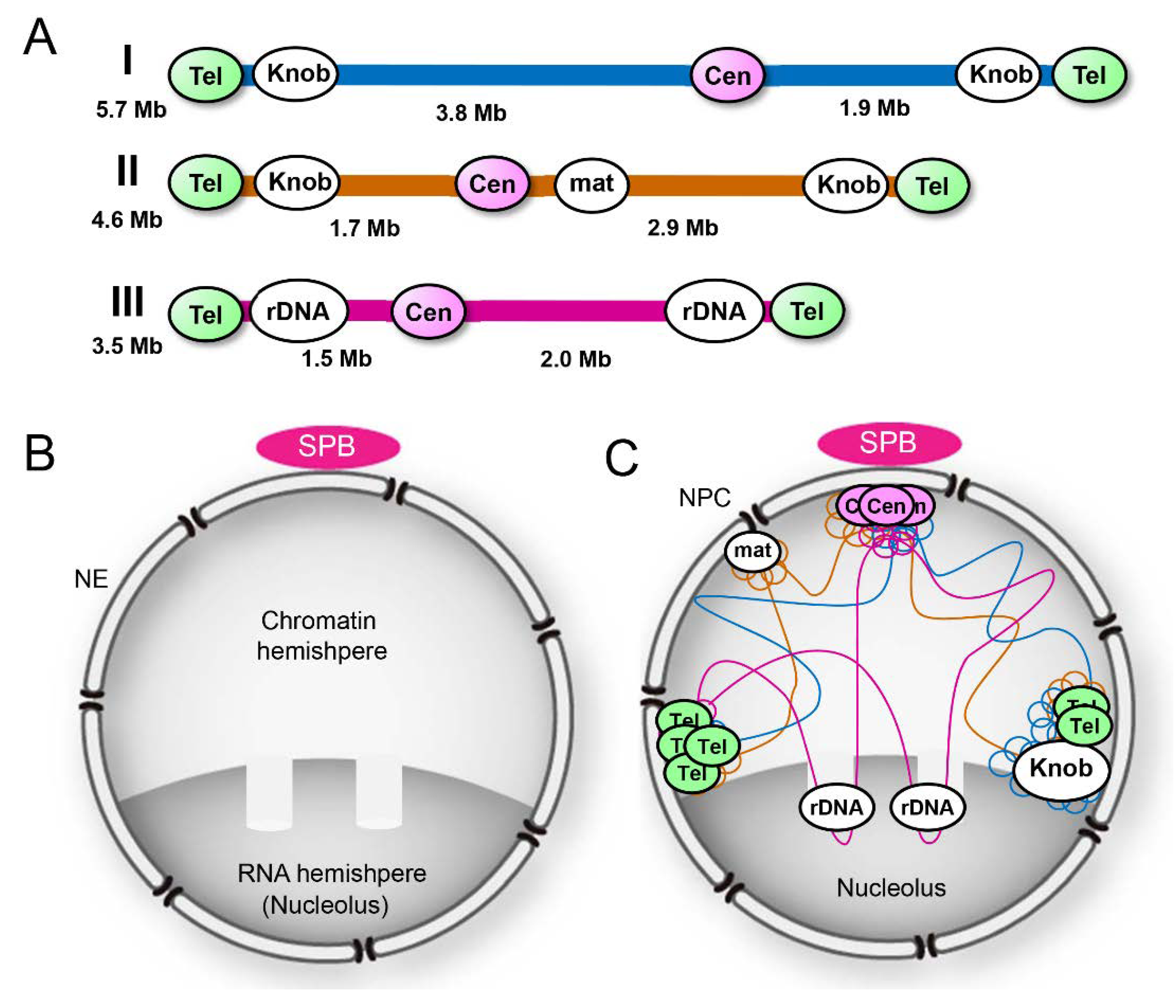
S. pombe cells grow with a haploid genome consisting of three chromosomes: I, II, and III [36]. These chromosomes have a centromere at their middle and telomere repeat sequences at both ends; rDNA repeats, which code for ribosomal RNA, are present at both ends of chromosome III flanked by telomere repeat sequences (Figure 1A). In mitotic interphase, these chromosomes are packed in the hemispherical half of the nucleus; the other half of the nucleus is rich in RNA, corresponding to the nucleolus in higher eukaryotes; two protrusions of chromatin are embedded in the RNA-rich hemisphere (Figure 1B). Centromeres are associated with the spindle pole body (SPB; a centrosome-equivalent structure in fungi) located on the cytoplasmic side of NE, and telomeres are on the NE near the nucleolus located at the opposite side of the SPB [37,38,39,40,41,42,43] (Figure 1C).
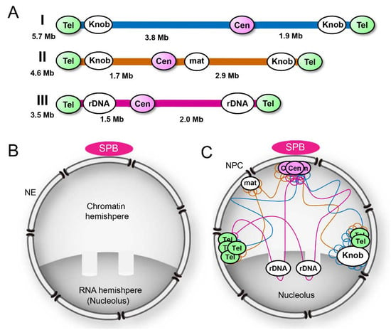
Figure 1.
The overall structure of the Schizosaccharomyces pombe nucleus. (A) Genomic organization of S. pombe chromosomes. Cen, centromere; Tel, telomere; mat, mat locus; and rDNA, repeat sequences coding ribosomal RNA. (B) S. pombe nucleus: A chromatin hemisphere and an RNA hemisphere with two protrusions of chromatin. (C) Spatial organization of chromosomes in the nucleus.
2.2. Heterochromatin as Transcriptionally Silent Regions
In metazoan cells, a typical electron-dense heterochromatin is observed beneath the NE or around the nucleoli [44]. Such an electron-dense heterochromatin is hardly seen in the nucleus of S. pombe. In this organism, heterochromatic regions have been identified as transcriptionally silent chromatin regions: The pericentromeric region, the sub-telomeric region, and the silent mating type locus (hereafter referred to as mat locus) [45,46,47]. These heterochromatic regions in S. pombe share histone modification marks (post-translational modifications such as methylation and acetylation) similar to those in other eukaryotes, but do not completely match with the cytological definition of heterochromatin as condensed chromatin domains [48]. These regions are located proximally to the NE (Figure 1C), and the NE proteins anchoring them to the NE are described in Section 4.
2.3. “Knob” Regions
A distinct region of chromatin was recognized as a “knob,” which shows a highly condensed chromatin region near the sub-telomeric region in S. pombe [48] (Figure 1A,C; also see Figure 4). The “knob” region does not share typical histone modification marks of heterochromatin, but matches the cytological definition of heterochromatin and shows unique properties different from other constitutive heterochromatin regions [39]. In this review, we define the knob region as heterochromatin and introduce its properties in a later section (see Section 3.4).
3. Mechanisms for Heterochromatin Formation in Fission Yeast
In S. pombe, mat locus was first recognized as the most striking region of gene silencing [49] and was predicted as an inheritable element for silencing [50], which is now recognized as an epigenetic regulation. A group of proteins required for the inheritance of mating types was identified as Swi1‒Swi9 [49]. Among them, Swi6 was identified as a key player in the epigenetic regulation of mating types [51] and was later identified as a homolog of the heterochromatin protein 1 (HP1) in Drosophila and mammals [52]. These pioneering studies have contributed to the unveiling of the molecular mechanisms underlying heterochromatin formation [45,46,47,53,54]. In this section, we summarize the molecular machineries involved in heterochromatin formation.
3.1. RNAi-Mediated Silencing Machinery
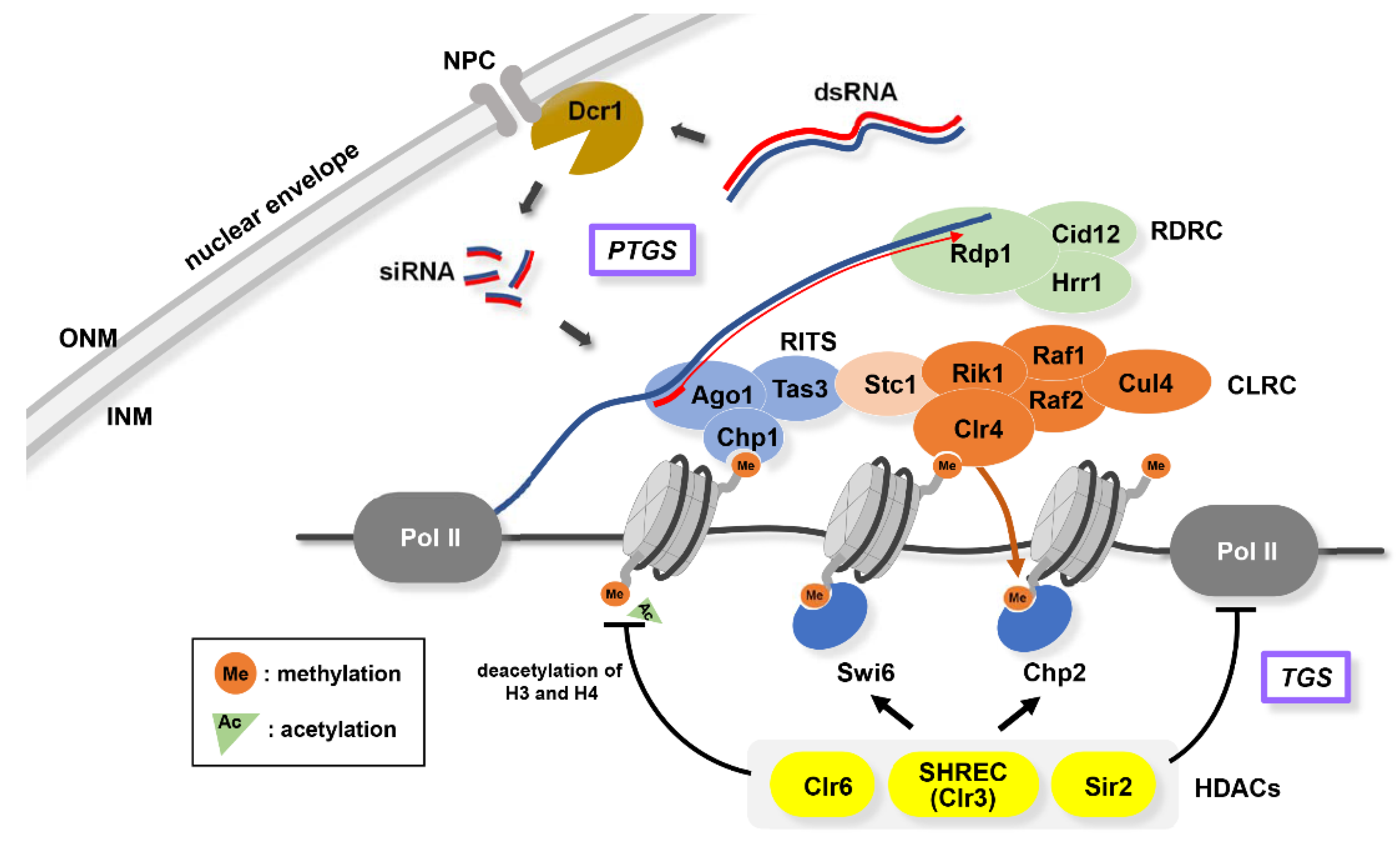
In S. pombe, constitutive heterochromatin is nucleated, propagated, and maintained at limited genomic loci such as the pericentromere, the sub-telomere, and the mat locus, all of which contain a repetitive DNA sequence. To accomplish heterochromatin formation in these regions, RNA processing and chromatin modification play a pivotal role in a mutually dependent manner (Figure 2). RNA interference (RNAi) is a post-transcriptional gene silencing (PTGS) regulation that has been initially considered as a system for degrading protein-coding transcripts [55,56]. However, genetic analyses in S. pombe led to the intriguing finding that the core components of RNAi machinery are essential for pericentromeric heterochromatin formation [57]. With this discovery, numerous studies have revealed the function of RNAi and its interplay with chromatin proteins in the context of heterochromatin formation [47,58].
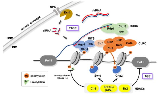
Figure 2.
Principles of heterochromatin formation. RNAi-mediated PTGS (post-transcriptional gene silencing) and HDAC-mediated TGS (transcriptional gene silencing). PTGS involves Dicer (Dcr1) located at the NPC; RITS (RNA-induced initiation of transcriptional silencing) complex consist of Ago1, Chp1, and Tas3; RDRC (RNA-dependent RNA polymerase complex); CLRC (Clr4 methyltransferase complex) consist of Clr4, Rik1, Cul4, Raf1, and Raf2. TGS involves HDACs (Clr6, SHREC, and Sir2) and HP1 (Swi6 and Chp2).
PTGS involves long non-coding RNAs (lncRNAs) transcribed by RNA polymerase II (Pol II) from repetitive sequences located in the heterochromatic regions: dg/dh, dh-like, and cenH for pericentromere, sub-telomere, and mat locus, respectively [57,59,60,61]. lncRNAs are cleaved by Dicer (Dcr1), generating small interfering RNA (siRNA). siRNAs are incorporated into an RNA-induced initiation of transcriptional silencing (RITS) complex, which consists of Ago1, Chp1, and Tas3 [62]. Ago1 belongs to the Argonaute family of proteins that can directly interact with siRNAs. RITS complex recruits an RNA-directed RNA polymerase complex (RDRC) that facilitates siRNA generation with Dicer to increase siRNA-bound RITS complex [63,64]. Thus, RITS complex functions as a guide for nucleation of heterochromatin assembly. RITS complex is also important in recruiting a Clr4 methyltransferase complex (CLRC) consisting of Clr4, Rik1, Cul4, Raf1, and Raf2, through an interaction with Stc1 [64,65]. Clr4 is the sole methyltransferase for the 9th lysine residue of histone H3 (H3K9) in S. pombe and shares functional similarity with mammalian Suv39h [66,67]. Note that H3K9 dimethylation (H3K9me2), instead of trimethylation (H3K9me3), is a major heterochromatic mark in S. pombe. H3K9me2 assembles chromodomain-containing proteins, such as Chp1, Chp2, and Swi6 (homologs of human HP1) [68]. Once histones are marked by H3K9me2, a positive feedback loop reinforces heterochromatin assembly: siRNA-bound RITS complex stably binds to H3K9me2 via the chromodomain of Chp1, and consequently accelerates CLRC and Swi6 recruitment [62]. Conversely, it has been shown that siRNA generation and localization of RNAi components to heterochromatin is dependent on chromatin factors, including Clr4 and HP1 homologs [69]. Therefore, RNAi and chromatin factors function in an interdependent manner with regards to heterochromatin formation, thereby raising a “Chicken and Egg” problem of which one works first [70]. The finding that Ago1 can bind with heterochromatin- and Dicer-independent species of dg siRNAs provides a clue to solving this conundrum, suggesting that the primal amplification of siRNAs by RNAi machinery acts as a seed to create heterochromatin, even in the absence of heterochromatin. Dcr1 is localized to NE through interaction with NPC components [34] (Figure 2; see Section 6.1).
In addition to the RNAi machinery, RNAi-independent mechanisms involving nuclear RNA processing and degradation factors such as the TRAMP (Trf4-Air1/Air2-Mtr4 polyadenylation) complex also contribute to establishing heterochromatin. TRAMP containing Cid14, a member of the Trf4 family of poly(A) polymerases, targets RNAs into degradation machineries that include the exosome. Both RNAi-dependent and RNAi-independent mechanisms work in parallel to target CLRC to the repetitive DNA sequences located in the constitutive heterochromatin domains [71,72,73,74,75,76,77].
3.2. HDAC-Mediated Silencing Machinery
Transcriptional gene silencing (TGS) pathways also contribute to heterochromatin formation in addition to the above RNAi-mediated PTGS machinery (Figure 2). Chp2 and Swi6 bound to H3K9me2 can target SHREC, a class II histone deacetylase (HDAC) complex containing Clr3 and Mit1 (Snf2 family ATPase) [78]. Swi6 can also recruit another protein complex containing class I HDAC Clr6 to the heterochromatin region [79]. Hypoacetylation of the lysine residues of histone H3 and H4 via HDAC, which is localized at the heterochromatin region, suppresses chromatin remodeling, restricting the access of Pol II to suppress transcription. Recent studies have reported that Swi6/HP1 protein contributes to the liquid-liquid phase separation associated with the reshaping of the nucleosome core, which accordingly quarantines heterochromatin to restrict Pol II accessibility [80].
In addition to these general TGS pathways, it is also known in mat locus that several DNA binding proteins, such as CENP-B homologs (Abp1 and Cbh1) and ATF/CREB family proteins (Atf1 and Pcr1), recruit HDACs to their binding sites within the locus [81,82,83].
Ubiquitination of histone H3 also affects heterochromatin formation through the modulation of H3K9 methylation. Cul4, Rik1, and Raf1 of S. pombe CLRC are presumed to have a ubiquitin E3 ligase activity owing to their structural similarity with CUL4-DDB1-DDB2 of other eukaryotes [84,85]. Indeed, a recent report demonstrates that CLRC ubiquitylates lysine 14 of histone H3 (H3K14ub) and H3K14ub promotes H3K9 methylation by Clr4 [86].
3.3. Boundary Elements Between Heterochromatin and Euchromatin
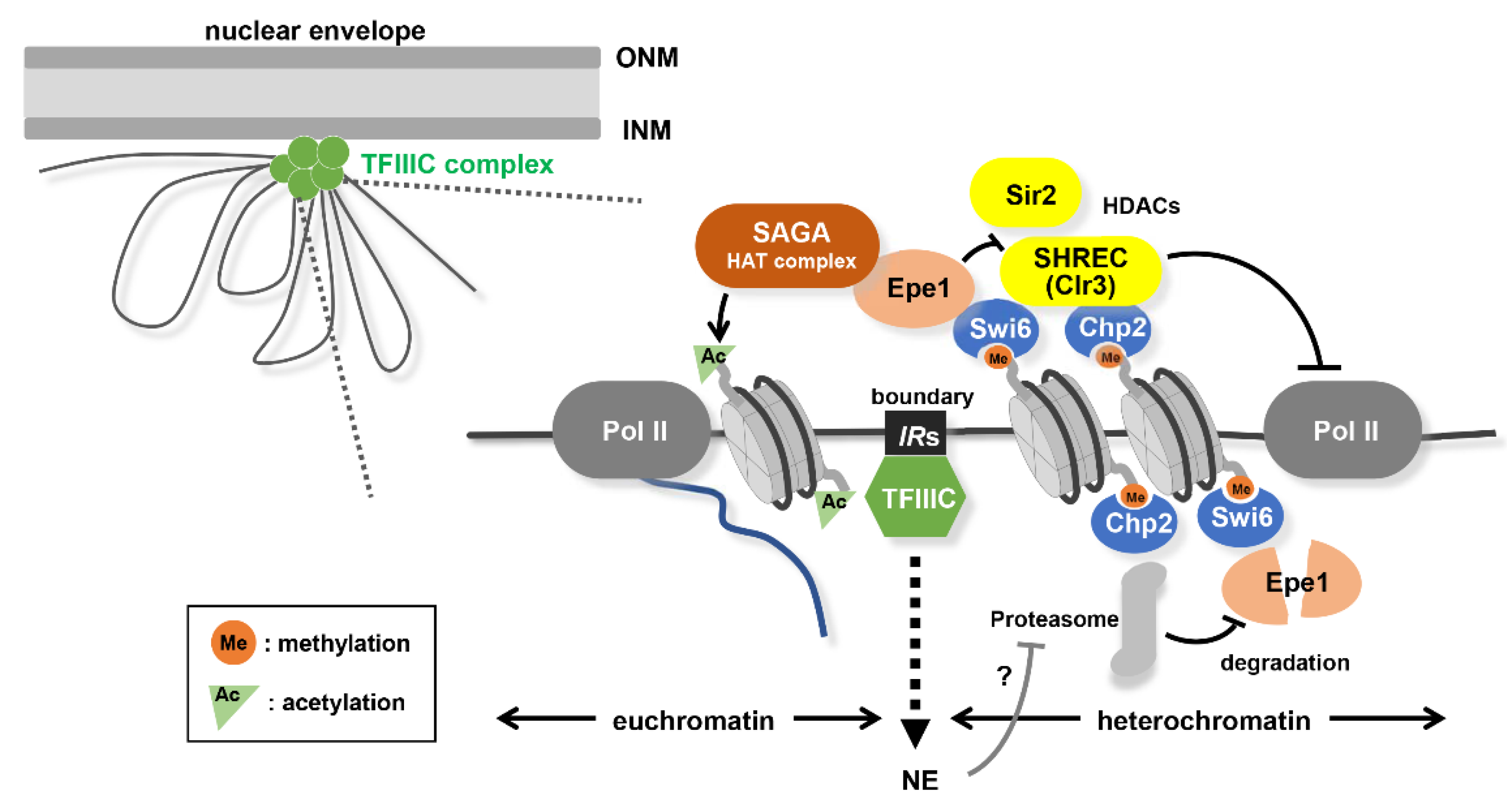
The propagation of the heterochromatin is strongly restricted within its locus to avoid the leakage of the silencing effect on the neighboring genes. Inverted repeat sequences called IRs (IRC or IR-L/R at the pericentromere or mat locus, respectively) work as boundaries in demarcating the heterochromatin and euchromatin regions [59,87,88] (Figure 3). The function of this boundary is partly conferred by the action of TFIIIC, an RNA polymerase III (Pol III) transcription initiation factor, but in a Pol III transcription-independent manner [89]. DNA regions including IRs in which TFIIIC binds irrelevant to Pol III tend to form a cluster and accumulate around the nuclear periphery [89]. This peripheral nuclear tethering is supposed to be necessary in its function as a boundary.
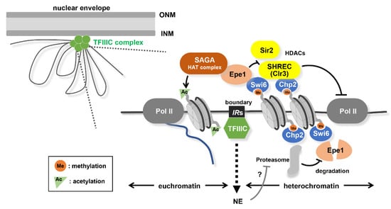
Figure 3.
Formation of heterochromatin/euchromatin boundary. Boundary elements (IRs) clustered at the nuclear periphery through TFIIIC binding. Epe1 (anti-silencing factor) is specifically accumulated at the boundary (IRs) owing to the proteasome-mediated selective elimination of Epe1 from heterochromatin. A heterochromatin/euchromatin boundary is formed owing to a balance between the opposing activity of histone acetyltransferase (SAGA recruited by Epe1) and histone deacetylase (SHREC or Sir2 recruited by Swi6 and Chp2).
The border between the heterochromatin and euchromatin is also controlled by the anti-silencing factor, Epe1 [90,91]. Epe1 has a JmjC-domain, which is the catalytic domain of histone demethylase; however, demethylase activity in vitro has not been detected in S. pombe Epe1 [91,92,93]. Epe1 is recruited to the heterochromatic regions in a Swi6-dependent manner [94] but enriched only around the boundary elements owing to the degradation by the proteasome inside the heterochromatin region [95] (Figure 3). It has been proposed that Epe1 might modulate H3K9 methylation indirectly, thus, compelling the localization of HDAC Clr3 bound to Swi6 [96,97,98]. Epe1 also recruits SAGA, a histone acetyltransferase complex, to counteract HDAC Sir2 [99]. These combined activities prevent the spreading of heterochromatin beyond the boundary (Figure 3). TFIIIC and Epe1 have been suggested to act redundantly for effective boundary formation [100].
3.4. DAPI-Dense “Knob” Region
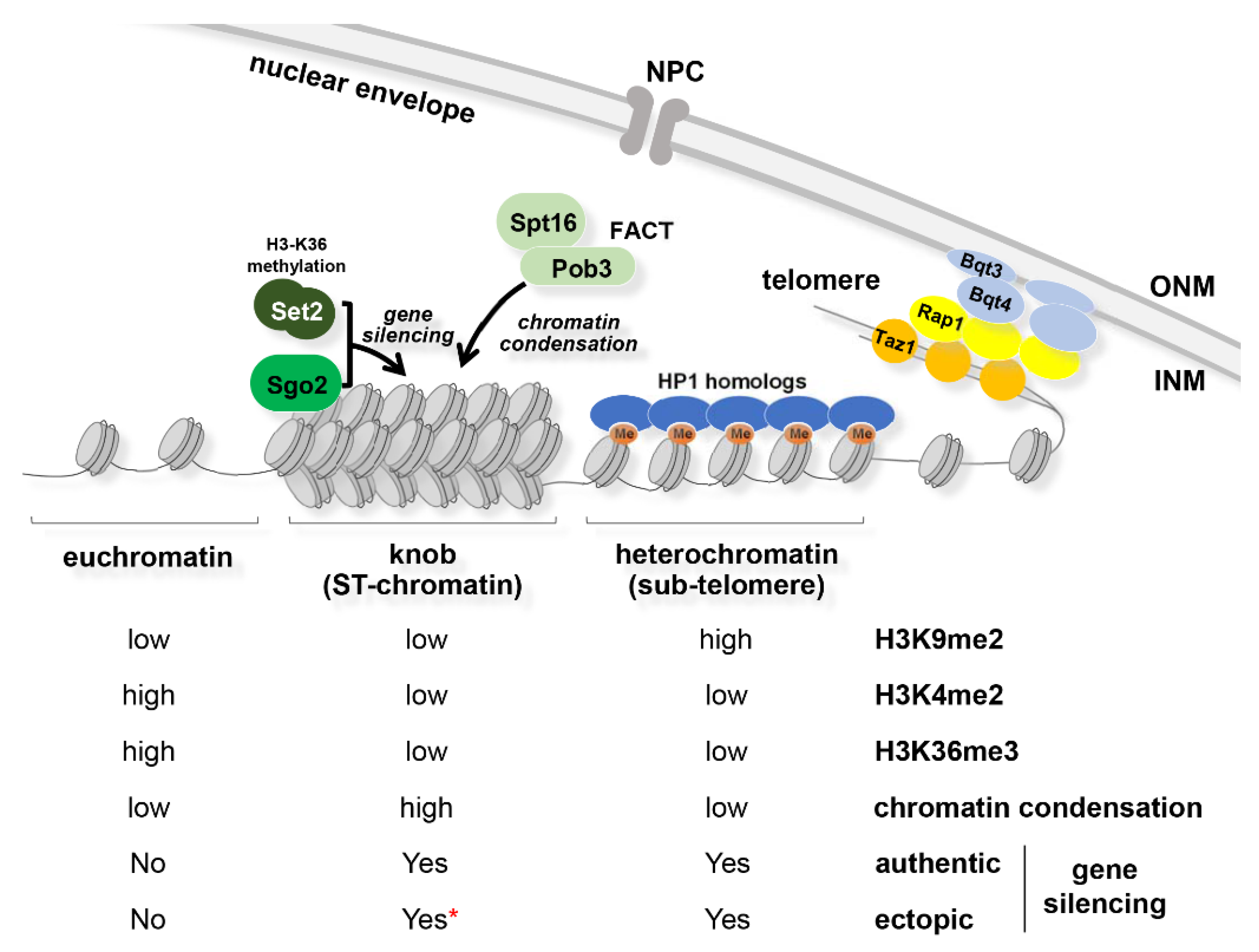
A “knob” is recognized as a distinct region of highly condensed chromatin that is densely stained with a DNA-specific fluorescent dye, 4′,6-diamidino-2-phenylindole (DAPI). This highly condensed chromatin is formed near the telomere, spanning approximately 50 kb next to the sub-telomeric heterochromatin regions of chromosomes I and II [48], which corresponds to a special chromatin region called “ST-chromatin” [101]. This knob region (ST-chromatin) shows characteristic histone modification patterns: Low levels of methylation at H3K4, H3K9, and H3K36 and acetylation at H3K14, H4K5, H4K12, and H4K16 [48,101,102] (Figure 4). Despite the low levels of H3K36 methylation, interestingly, knob formation requires Set2, a sole methyltransferase of H3K36 in S. pombe. It has also been shown that the kinetochore protein Sgo2, FACT (facilitates chromatin transcription), which is an essential histone chaperon consisting of Spt16 and Pob3, and the mono ubiquitinated histone H2B (H2Bub) are required for knob formation [102,103]. H2Bub facilitates histone H2A/H2B dimer deposition by FACT. In addition, FACT maintains the nucleosome density in the sub-telomeric region [103], which may be the key to knob formation.
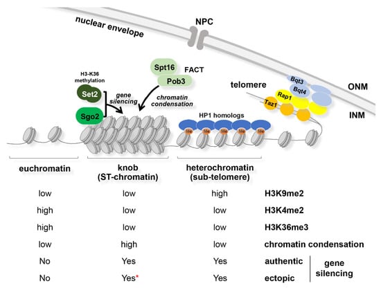
Figure 4.
Chromatin states in telomeric regions. A highly condensed “knob” at ST-chromatin region formed by a cooperative action of Sgo2 and Set2, and a FACT-dependent nucleosome assembly pathway. (Lower columns) Levels of histone modifications (H3K9me2, H3K4me2, and H3K36me3). Levels of chromatin condensation. Gene silencing: “authentic”, expression of endogenous genes upon nitrogen starvation; “ectopic”, expression of the ura4+ reporter gene ectopically inserted; * silencing in the knob region depends on the position of ura4+ insertion.
Genes inside the knob region are transcribed at a very low level in vegetative cells, but they can be upregulated upon nitrogen starvation, accompanied with a loss of knob formation [48,104]. Curiously, a reporter gene inserted within the knob region of the right arm of chromosome II, but not in the right arm of chromosome I, was slightly silenced, depending on Sgo2 and Set2 [48,102]. These results suggest that the knob region has the potential to locally suppress gene expression, depending on the adequate localization pattern of Sgo2 and histone modifications. Deletion of Set2 or Sgo2 not only increases the expression of the genes located in the knob but also alters the replication timing of knob chromatin from late to early S-phase [102], suggesting that knob condensation appears to be important in modulating transcription and DNA replication.
Intriguingly, neocentromere, which is a newly generated centromere that escaped the catastrophe of an authentic centromere deletion, is frequently formed in this knob region [105]. Neocentromere formation requires RNAi-machinery-mediated heterochromatin formation in sub-telomeric region [105]. Therefore, knob is a unique chromatin structure that has the potential to provide a flexible platform in response to various cellular reactions.
4. Proteins Attaching Heterochromatic Regions to the NE
INM proteins play a key role in attaching heterochromatin loci and facilitating/maintaining heterochromatin formation [106]. Heterochromatic regions in S. pombe are located proximally to NE, as shown in Figure 1C. In this section, we describe S. pombe NE proteins that attach heterochromatic regions to the NE.
4.1. Proteins Attaching Heterochromatic Regions to the NE in Mitosis
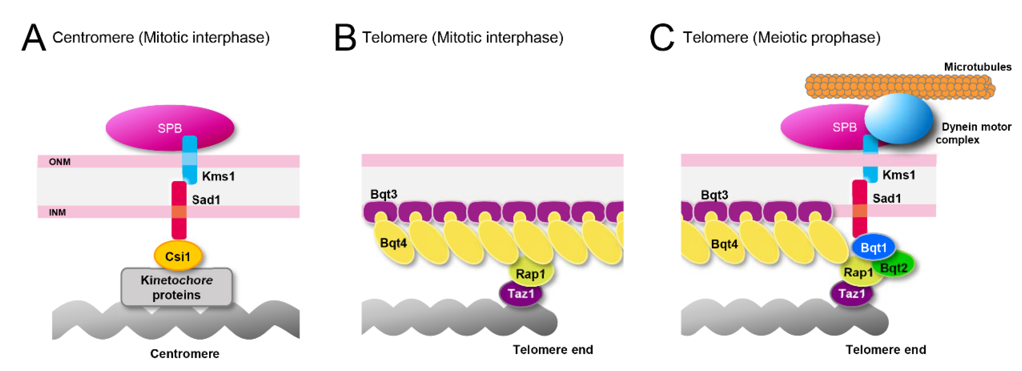
Centromeres are attached to the SPB through the interaction with Sad1/Unc84 (SUN)-domain protein Sad1 [107]. Sad1 interacts with the outer kinetochore through the interaction with Csi1 [108,109]. Sad1 also interacts with Klarsicht/ANC-1/Syne homology (KASH) domain-containing proteins Kms1/2 and forms a complex termed as “linker of nucleoskeleton and cytoskeleton (LINC) complex,” which is localized exclusively at the SPB during mitotic interphase [110,111,112,113]. Centromeres detach from the SPB in Lem2 and Csi1 double deletion mutants [33,114]. Thus, centromeres are attached to the SPB through interactions among Lem2, Csi1, and the LINC complex across the NE (Figure 5A).
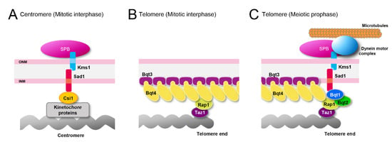
Figure 5.
NE proteins attaching heterochromatin regions to the NE. (A) Centromeres attached to the SPB in mitotic interphase. (B) Telomeres attached to the NE in mitotic interphase. (C) Telomeres attached to the SPB in meiotic prophase.
Telomeres are connected to the NE through the interaction of the INM protein, Bqt4, with the telomere protein, Rap1 [115] (Figure 5B). Mutation or loss of these proteins causes dissociation of telomeres from the NE [109,115]. Telomeres also detach from the NE in the absence of Lem2 [33,116], and the double deletion of Lem2 and Dsh1 exacerbates the phenotype of telomere detachment, suggesting that Lem2 and Dsh1 play a role in attaching telomeres to the NE [33].
The chromatin region containing the mat locus is localized at the nuclear periphery through the cluster formation of boundary elements on the inverted repeat sequence IR-L/R [89]. A recent report demonstrated that the NE localization of mat locus is mediated through an interaction with nucleoporin Amo1 [31] (see Section 6.2).
Knob is also located near the NE, but in a Bqt4-independent manner [117]. A conserved subfamily of SNF2 chromatin remodeler protein Fft3 is involved in the localization of knob at the NE because loss of Fft3 results in the detachment of the sub-telomeric region from the NE [117]. Fft3, which binds to a solo long-terminal repeat (LTR) located at the border region between knob and euchromatin, interacts with a LEM/HEH-domain inner nuclear membrane protein Man1 [117], suggesting that an interaction between Fft3 and Man1 is involved in attaching knob to the NE.
4.2. Proteins Attaching Heterochromatic Regions to the NE in Meiosis
As described in Section 2.1 (Figure 1), centromeres form a cluster with the SPB, while telomeres are located on the opposite side of the nucleus in mitotic interphase (the configuration called a Rabl orientation). Upon entering meiosis, centromeres and telomeres switch their positions in the nucleus: Telomeres become associated with the SPB, while centromeres detach from the SPB (called a bouquet configuration). During the process of centromere–telomere switching, meiosis-specific telomere proteins, Bqt1 and Bqt2, interact with the telomere protein Rap1. Bqt1 and Bqt2 also interact with the NE protein complex, Sad1, and Kms1 (LINC complex), to attach telomeres to the NE [118]. Kms1 interacts with cytoplasmic dynein motor complex on microtubules, which tethers telomeres toward the SPB across the NE [119,120] (Figure 5C). This phenomenon provides a striking example, in which interactions between chromatin and NE play an important role in the organization of the chromosomes within the nucleus.
5. NE Proteins Modulating Heterochromatin Formation
5.1. Lem2 Functions in Heterochromatin Formation
5.1.1. Lem2 is a Conserved Protein
Lem2 is one of the most broadly conserved INM proteins, sharing two transmembrane (TM) domains and a MAN1/Src1 C-terminal (MSC) domain at the C-terminal region [3,11,121] (Figure 6A). By in silico homology search using MSC domain as a query followed by experimental localization analysis, members of the Lem2 protein family were found in different eukaryotic supergroups: Opisthokonta (yeasts to humans) [11], Amoebozoa (Src1 in Dictyostelium discoideum) [122,123], SAR (Lem2 and MicLem2 in Tetrahymena thermophila) [13], and Archaeplastida (plants) [13]. Lem2 in metazoans has the LEM domain and Lem2 in fungus has the LEM-like HEH (helix-extended-helix) domain at the N-terminal region; no obvious LEM or HEH domains were found in other supergroups.

Figure 6.
Lem2 for heterochromatin formation. (A) Molecular domains of Lem2. TM, transmembrane domain. (B) Lem2 on pericentromeric heterochromatin. White circles represent chromatin at the central core of the centromere and blue circles represent chromatin at the pericentromeric heterochromatin. Lem2 is localized at the central core and promote heterochromatin formation at the pericentromeric regions. Elo2 and Lnp1 compensate Lem2 functions. VLCFA, very-long-chain fatty acid. (C) Lem2 on sub-telomeric heterochromatin. Lem2 is localized to the NE through interaction with Bqt4, and promotes heterochromatin formation at the sub-telomeric regions attached to the NE through an interaction between Rap1 and Bqt4.
Since overexpression or deletion of Lem2 in S. pombe alters chromosome functions and NE morphology and integrity, Lem2 plays an important role in both chromosome and NE functions [33,35,107]. Recent reports show that Lem2 interacts with chromatin and regulates its functions, especially in heterochromatin formation [33,35,124]. This function seems to be unique in Lem2 because the deletion of Man1 or Ima1 does not show any effect on heterochromatin formation [33].
5.1.2. Lem2 on Centromeric Heterochromatin
One of the most prominent phenotypes of the Lem2 deletion mutant is severe chromosome instability in a nutrient-dependent manner [35]. Deletion of Lem2, but not Man1 and Ima1, results in a high rate of minichromosome loss; the phenotype appears only in rich medium culture conditions. In contrast, the deletion of Csi1, which is a crucial protein that attaches centromeres to the SPB, displays a high rate of minichromosome loss in both rich and minimum medium culture conditions. This high loss rate is probably caused by the deficiency of pericentromeric heterochromatin formation. Although a genome-wide mapping for the Lem2-binding region has not been made public thus far, some Lem2-binding regions have been reported, and one of the major binding sites of Lem2 was the central core in the centromere [33,124]. Centromeres in S. pombe are composed of a central domain flanked with outer repeats. The central domain is divided into a central core (cnt) and an innermost repeat (imr), where CENP-A (centromere-specific histone H3 variant)-containing nucleosome and kinetochore are formed [125,126]. The outer repeat consists of inversed repeat elements named dg and dh [127]. Pericentromeric heterochromatin is generated in this dg/dh region in an RNAi and TGS-dependent manner (see Section 3.1). Lem2 binds to a central core through its N-terminal region, including HEH domain, and this localization depends on the nutrient condition (Figure 6B): Lem2 localizes to this region only in rich medium culture condition [33,35,116,124,128]. The deletion of Clr4 impairs Lem2 localization at the central core, implying that heterochromatin formation is required for proper Lem2 localization [124]. Interestingly, localizing Lem2 to the central core is required for heterochromatin augmentation at the pericentromere [35], indicating that Lem2 can function as a positive feedback loop to promote heterochromatin formation. Loss of Lem2 causes a decrease in the pericentromeric H3K9me2 and a de-repression of pericentric silencing, particularly when combined with the deletion of Dsh1, suggesting that Lem2 contributes to heterochromatin formation independently of Dsh1-mediated RNAi pathway [33]. The Lem2-binding INM protein Nur1 displays similar effects in this region [33,124].
5.1.3. Lem2 on Telomeric Heterochromatin
Lem2 also interacts with chromatin regions close to the sub-telomeric heterochromatin region [124]. ChIP-seq and ChIP-chip analyses show that Lem2 binds to the telomere side of sub-telomeric heterochromatin at tel3L and tel3R, but no significant enrichment at tel1L and tel2L regions [33,124,129]. Loss of Lem2 shows de-repression of tlh1/2 genes located within the sub-telomeric region but does not affect H3K9me2 levels in this region [33]. Furthermore, Lem2 regulates the balance of SHREC and Epe1 binding at heterochromatic regions [33,124]. In the absence of Lem2, the SHREC component dissociates from the heterochromatic gene silencing sites while Epe1 associates with the sites. Up-regulation of tlh1/2 genes by Lem2 deletion is bypassed by Epe1 deletion, indicating that gene silencing by Lem2 in sub-telomeric region largely depends on SHREC-Epe1-mediated pathway without affecting H3K9me2 [33].
5.1.4. Lem2 on LTR Sequences
Deletion of Lem2 is likely to increase the recombination between LTRs [33,35]. LTRs found at the end of retrotransposons are used when a virus integrates its genomic DNA into its host genome [130]. Expression of retrotransposable elements must be strongly regulated to prevent undesired pop-out and pop-in of the retrotransposons causing genome instability [117,131,132]. In the Lem2 deletion mutant, the expression of LTRs at the sub-telomeric region and frequency of recombination at LTRs are increased [33,35], suggesting that Lem2 plays a role in suppressing the expression of LTRs.
5.1.5. Molecular Domains of Lem2 for Heterochromatin Functions
C-terminal region of Lem2, containing two transmembrane domains and MSC domain, is sufficient for heterochromatic gene silencing at both pericentromeric and sub-telomeric regions [33,35]. An MSC domain without the transmembrane domain cannot suppress gene expression, indicating that membrane localization is essential for gene silencing activity of Lem2 [33]. In contrast, the N-terminal region of Lem2 does not show gene silencing activity. This domain restores the centromere association with the SPB in the absence of Lem2 and Csi1 [114], whereas it does not restore telomere association in the absence of Lem2 and Dsh1 [33]. Thus, the N-terminal region functions to anchor only the centromeric region to the NE.
5.2. Regulation of Lem2 Localization
Lem2 localizes at the INM and beneath the SPB, but biased to the SPB [128,129]. These localizations depend on Csi1 for the SPB and Bqt4 for the NE, respectively. The Deletion of Csi1 causes the disappearance of Lem2 from the SPB, although the direct interaction between Lem2 and Csi1 has not been reported. In contrast, the deletion of Bqt4 induces strong SPB accumulation of Lem2, indicating that Bqt4 plays a role in retaining Lem2 at the NE [128,129]. Lem2 and Bqt4 bind directly through an interaction between Bqt4-binding motif ((D/E)3-4xFxxxɸ) in Lem2, located at the nucleoplasmic region near the first transmembrane domain and APSES domain in Bqt4 [128,133,134]. This domain binds to DNA and also interacts with the Bqt4-binding motif shared in Lem2, Sad1, and Rap1 in a competitive manner [133]. In cells with deleted Csi1 and Bqt4, Lem2 shows a biased localization to the SPB, suggesting that other factors are also involved in the Lem2 localization to the SPB [129]. One of those factors appears to be Nur1 because Lem2 and Nur1 depend on each other for SPB localization in fission yeast Schizosaccharomyces japonicus [135]. During mitosis, Lem2 disappears from the SPB at prophase and reappears to the SPB at the onset of anaphase [136]. Conversely, Ima1 appears on the SPB at prophase and disappears at the onset of anaphase [136], implying that Lem2 and Ima1 have cell cycle-specific functions on the SPB during mitosis.
5.3. Membrane Protein Network Regulating Lem2 Functions
5.3.1. Lem2 Functions through Lnp1
The deletion of Lem2 causes centromeric defects such as defective formation of pericentromeric heterochromatin and chromosome instability, all of which are suppressed by the duplication of the lnp1 gene [35]. Lnp1 (homolog of human Lunapark) is an ER membrane protein conserved from yeasts to humans. Lnp1 localizes at the three-way junction of tubular ER network in humans and S. cerevisiae [137,138,139,140,141]. Double deletion of Lem2 and Lnp1 disturbs the partitioning between NE and ER, and consequently causes severe membrane disorder and growth defects [142]. Moreover, Lem2 and Lnp1 act as barriers to the membrane flow between the ER and Golgi and contribute to the control of the nuclear size [143]. Thus, Lnp1 is likely involved in maintaining chromosome integrity through an indirect pathway, in which Lnp1 and Lem2 cooperatively maintain the NE/ER boundary and NE integrity.
5.3.2. Lem2 Functions through Bqt4
Loss of Lem2 and Bqt4 confers synthetic lethality [35]. In this double mutant, leakage of nuclear proteins frequently occurs owing to NE breakage, leading to cell death [144]. The domains responsible for this genetic interaction are the MSC domain of Lem2, and the N-terminal and transmembrane domains of Bqt4. In addition, the transmembrane domains of both proteins are essential for their effect, suggesting that both proteins are required for their functions on the NE. The synthetic lethal phenotype of the Lem2 Bqt4 double deletion is suppressed by the very-long-chain fatty acid elongase Elo2, which is essential for S. pombe cell viability [144]. Elo2 synthesizes “very-long-chain” fatty acids (chain length of carbon atoms longer than 21), which play crucial roles that cannot be substituted by “long-chain” fatty acids (chain length of carbon atoms 11-20). Most of the very-long-chain fatty acids constitute sphingolipids, which play important roles in maintaining the skin barrier and myelin sheath in mammals [145,146,147]. The overexpression of Elo2 suppresses the defective formation of the pericentromeric heterochromatin and chromosome instability caused by the loss of Lem2, but does not restore the telomeric attachment to the NE by the loss of Bqt4, indicating that Elo2 bypasses Lem2 functions. Moreover, the loss of S. pombe Elo2 is complemented by an overexpression of human orthologs, suggesting conserved roles of Elo2 in genome stability [144].
5.3.3. Lem2 Functions through the ESCRT-III Complex
Loss of Lem2, in combination with loss of Bqt4 or Lnp1, causes severe nuclear protein leakage owing to NE holes formed [142,144]. Recent studies indicate that Lem2 seals an NE hole in cooperation with Cmp7 (homologue of human CHMP7) and endosomal sorting complex required for transport-III (ESCRT-III) [148,149,150]. Lem2 may play a role in sealing the NE by liquid phase separation [151]. Unrepaired NE holes can cause DNA damage beneath the hole [150], and aberrant accumulation of ESCRT-III in the NE hole is observed with a DNA damage marker protein in micronuclei of human cells [152], suggesting that sealing the NE holes is the critical process to maintain chromosomes. Considering that Lem2 has a role in facilitating checkpoint signaling in response to replication stress [153], Lem2 may function in DNA damage repair at the holes. On the other hand, Vps4, which is an AAA-ATPase, disassembles the ESCRT-III complex and releases the pericentromeric heterochromatin from Lem2 at the end of mitosis in S. japonicus, indicating that Lem2 and ESCRT-III complex function in remodeling the attachment of heterochromatic regions to the NE [135]. Lem2 possibly ensures genome stability through maintaining the NE integrity in fission yeasts.
6. Nucleoporins and Heterochromatin
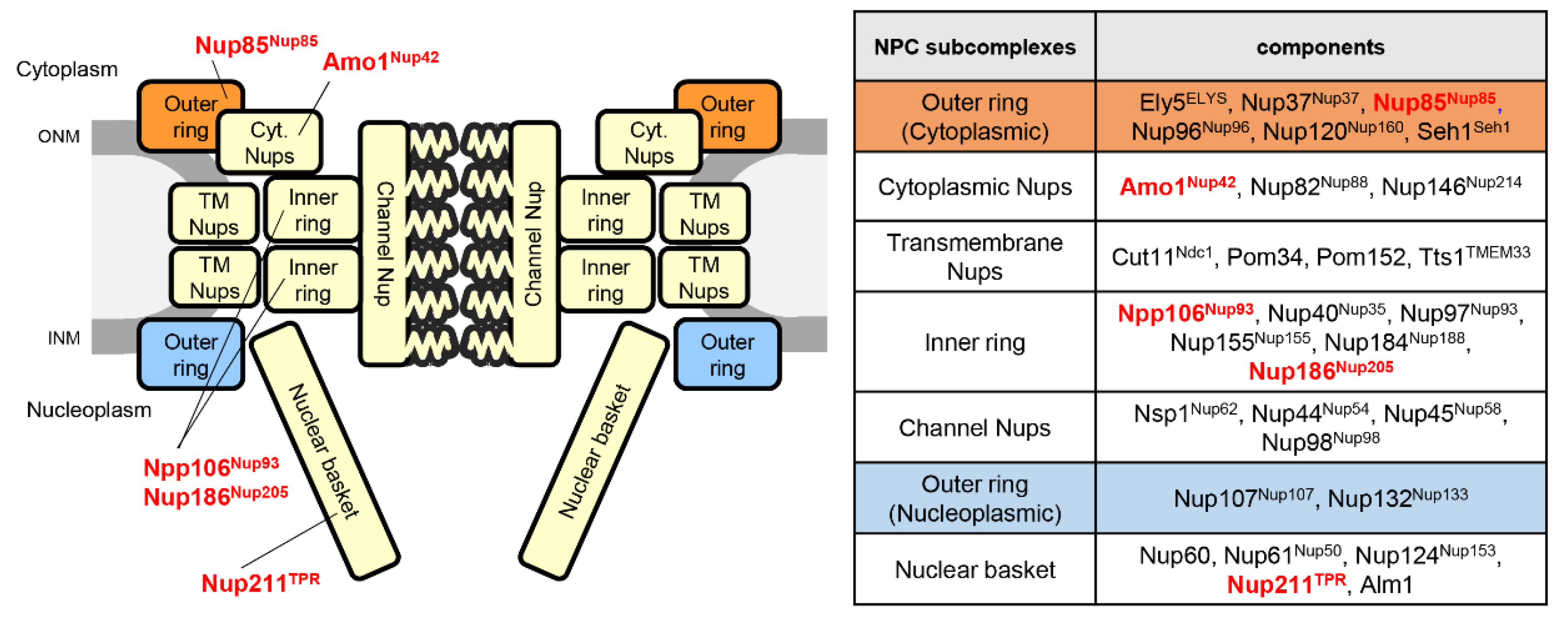
The NPC is composed of several subcomplexes, namely, the outer ring, inner ring, central channel, nuclear basket, and the cytoplasmic filament (Figure 7). These subcomplexes associate with the nuclear membrane by interacting with the transmembrane Nups to form cylindrical structures connecting the nucleoplasm and cytoplasm [19,22,23,24,25,26,27,28,29,154,155]. This typical organization of NPC is conserved among eukaryotes, although it exhibits species-specific variations in S. pombe [23,156]. Nups also contribute to chromatin organization in the nucleus. In S. pombe, some of the Nups are reported to be involved in gene silencing [31,32].
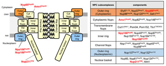
Figure 7.
Subcomplex structure of the S. pombe NPC. (Left) Schematic drawing of the NPC. Nups described in the text are shown in red; human orthologs are written in superscript. TM Nup, transmembrane Nup; ONM, outer nuclear membrane; INM, inner nuclear membrane. (Right) NPC subcomplexes and their components.
6.1. Nucleoporins Modulate Gene Silencing
Dcr1, a key regulator of RNAi-mediated gene silencing, has been shown to be localized at the NPC; importantly, this localization is essential for pericentromeric heterochromatin formation [34].
Moreover, Dcr1 and Nup85 (one of the outer ring Nups) are enriched in the promoter regions of stress response genes [157]. This implies that the transcription of stress response genes at the NPC is repressed by Dcr1; however, the relevance of NPC localization of Dcr1 to a functional RNAi-dependent gene silencing pathway remains elusive. In addition, the interaction of a CLRC complex component Raf1/Clr8 with a gene product of nup189+ was observed in yeast two-hybrid assay [158]. Proteome analysis of immunoprecipitated Swi6 fraction also identified several Nups, including inner ring Nups, Npp106, and Nup186 [32]. Npp106 and Nup211 (a nuclear basket Nup) accumulated at the pericentromeric regions [32]; Npp106 mutant showed a defect in gene silencing at pericentromeric heterochromatin [32]. It has also been reported that most of the tRNA loci, including the boundary element of pericentromeric heterochromatin, associate with Nup85 [117]. Moreover, the functional role of Drosophila Nup93, an inner ring Nups, in the silencing of Polycomb target genes was recently reported [159]. These cumulative pieces of evidence strongly suggest the involvement of NPC in the regulation of gene silencing in heterochromatic regions.
6.2. Nucleoporin Amo1 Sequesters Heterochromatin to the NE
Amo1 has been identified as a factor affecting cytoplasmic microtubule organization [160] and is localized at the cytoplasmic side of NPC [156]. Interestingly, Amo1 was recently reported to participate in heterochromatin formation at mat locus and pericentromeric regions [31]. Amo1 can bind to Rix1, a component of an RNA processing complex (RIX complex), which localizes to heterochromatin regions. Through interactions with Rix1, Amo1 can recruit the mat locus to the nuclear periphery. Importantly, this interaction is required for peripheral localization and silencing of ectopically induced heterochromatin. Amo1 also interacts with the FACT histone chaperon complex that binds to Swi6 and is required for heterochromatin formation [161,162]. Therefore, Amo1 is thought to facilitate the efficient loading of FACT onto Swi6-bound heterochromatin to suppress histone turnover and maintain the epigenetic state of the heterochromatin [31]. It is also suggested that Amo1, which localizes at a nuclear periphery different from NPC, may function in the regulation of heterochromatin maintenance [31].
7. Perspectives
In this review, we have described NE proteins that are involved in heterochromatin formation and functions in S. pombe. An important role for the NE is to provide a physicochemical platform for chromosomes, and tethering chromatin to their vicinity evokes chromatin potential to modulate its genetic activities. For example, FG repeat-containing proteins in NPCs produce an amphiphilic field by forming hydrogel or liquid droplets [163]. Thus, an intriguing possibility is that FG repeat proteins and Swi6, which also form liquid droplets [80], create a peripheral compartment through phase separation, which is required for the silencing of heterochromatin. Recently, other interesting studies have demonstrated that lipid metabolism is involved in modulating heterochromatin states and chromatin functions [142,164]. The involvement of the lipid components of NE and ER in chromatin functions need to be elucidated. Considering that the very-long-chain fatty acid elongase Elo2 could compensate for the loss of Lem2 and its interaction with NE proteins, one could speculate that a specific lipid generated by a membrane protein network focusing on Lem2 could have a yet unknown role in heterochromatin formation and functions.
Funding
This research was funded by KAKENHI grants: JP19K06489 to YHirano; JP19K06660 to HA; JP18H05528 to TH; JP18H05533, JP20H00454, and JP19K22389 to YHiraoka.
Conflicts of Interest
The authors declare no conflict of interest. The funders had no role in the design of the study; in the collection, analyses, or interpretation of data; in the writing of the manuscript, or in the decision to publish the results.
References
- Briand, N.; Collas, P. Lamina-associated domains: Peripheral matters and internal affairs. Genome Biol. 2020, 21, 85. [Google Scholar] [CrossRef]
- Iwamoto, M.; Hiraoka, Y.; Haraguchi, T. Uniquely designed nuclear structures of lower eukaryotes. Curr Opin. Cell Biol. 2016, 40, 66–73. [Google Scholar] [CrossRef]
- Mans, B.J.; Anantharaman, V.; Aravind, L.; Koonin, E.V. Comparative genomics, evolution and origins of the nuclear envelope and nuclear pore complex. Cell Cycle 2004, 3, 1612–1637. [Google Scholar] [CrossRef] [PubMed]
- Worman, H.J.; Yuan, J.; Blobel, G.; Georgatos, S.D. A lamin B receptor in the nuclear envelope. Proc. Natl. Acad. Sci. USA 1988, 85, 8531–8534. [Google Scholar] [CrossRef] [PubMed]
- Hirano, Y.; Hizume, K.; Kimura, H.; Takeyasu, K.; Haraguchi, T.; Hiraoka, Y. Lamin B receptor recognizes specific modifications of histone H4 in heterochromatin formation. J. Biol. Chem. 2012, 287, 42654–42663. [Google Scholar] [CrossRef] [PubMed]
- Solovei, I.; Kreysing, M.; Lanctot, C.; Kosem, S.; Peichl, L.; Cremer, T.; Guck, J.; Joffe, B. Nuclear architecture of rod photoreceptor cells adapts to vision in mammalian evolution. Cell 2009, 137, 356–368. [Google Scholar] [CrossRef] [PubMed]
- Solovei, I.; Wang, A.S.; Thanisch, K.; Schmidt, C.S.; Krebs, S.; Zwerger, M.; Cohen, T.V.; Devys, D.; Foisner, R.; Peichl, L.; et al. LBR and lamin A/C sequentially tether peripheral heterochromatin and inversely regulate differentiation. Cell 2013, 152, 584–598. [Google Scholar] [CrossRef] [PubMed]
- Dechat, T.; Vlcek, S.; Foisner, R. Review: Lamina-associated polypeptide 2 isoforms and related proteins in cell cycle-dependent nuclear structure dynamics. J. Struct Biol. 2000, 129, 335–345. [Google Scholar] [CrossRef]
- Gruenbaum, Y.; Margalit, A.; Goldman, R.D.; Shumaker, D.K.; Wilson, K.L. The nuclear lamina comes of age. Nat. Rev. Mol. Cell Biol. 2005, 6, 21–31. [Google Scholar] [CrossRef]
- Lee, K.K.; Wilson, K.L. All in the family: Evidence for four new LEM-domain proteins Lem2 (NET-25), Lem3, Lem4 and Lem5 in the human genome. Symp. Soc. Exp. Biol. 2004, 329–339. [Google Scholar]
- Brachner, A.; Foisner, R. Evolvement of LEM proteins as chromatin tethers at the nuclear periphery. Biochem. Soc. Trans. 2011, 39, 1735–1741. [Google Scholar] [CrossRef] [PubMed]
- Wagner, N.; Krohne, G. LEM-Domain proteins: New insights into lamin-interacting proteins. Int Rev. Cytol. 2007, 261, 1–46. [Google Scholar] [CrossRef] [PubMed]
- Iwamoto, M.; Fukuda, Y.; Osakada, H.; Mori, C.; Hiraoka, Y.; Haraguchi, T. Identification of the evolutionarily conserved nuclear envelope proteins Lem2 and MicLem2 in Tetrahymena thermophila. Gene X 2019, 1, 100006. [Google Scholar] [CrossRef] [PubMed]
- Korfali, N.; Wilkie, G.S.; Swanson, S.K.; Srsen, V.; de Las Heras, J.; Batrakou, D.G.; Malik, P.; Zuleger, N.; Kerr, A.R.; Florens, L.; et al. The nuclear envelope proteome differs notably between tissues. Nucleus 2012, 3, 552–564. [Google Scholar] [CrossRef] [PubMed]
- Bui, K.H.; von Appen, A.; DiGuilio, A.L.; Ori, A.; Sparks, L.; Mackmull, M.T.; Bock, T.; Hagen, W.; Andres-Pons, A.; Glavy, J.S.; et al. Integrated structural analysis of the human nuclear pore complex scaffold. Cell 2013, 155, 1233–1243. [Google Scholar] [CrossRef]
- Kosinski, J.; Mosalaganti, S.; von Appen, A.; Teimer, R.; DiGuilio, A.L.; Wan, W.; Bui, K.H.; Hagen, W.J.; Briggs, J.A.; Glavy, J.S.; et al. Molecular architecture of the inner ring scaffold of the human nuclear pore complex. Science 2016, 352, 363–365. [Google Scholar] [CrossRef]
- Lin, D.H.; Stuwe, T.; Schilbach, S.; Rundlet, E.J.; Perriches, T.; Mobbs, G.; Fan, Y.; Thierbach, K.; Huber, F.M.; Collins, L.N.; et al. Architecture of the symmetric core of the nuclear pore. Science 2016, 352, aaf1015. [Google Scholar] [CrossRef]
- Von Appen, A.; Kosinski, J.; Sparks, L.; Ori, A.; DiGuilio, A.L.; Vollmer, B.; Mackmull, M.T.; Banterle, N.; Parca, L.; Kastritis, P.; et al. In situ structural analysis of the human nuclear pore complex. Nature 2015, 526, 140–143. [Google Scholar] [CrossRef]
- Alber, F.; Dokudovskaya, S.; Veenhoff, L.M.; Zhang, W.; Kipper, J.; Devos, D.; Suprapto, A.; Karni-Schmidt, O.; Williams, R.; Chait, B.T.; et al. The molecular architecture of the nuclear pore complex. Nature 2007, 450, 695–701. [Google Scholar] [CrossRef]
- Eisenhardt, N.; Redolfi, J.; Antonin, W. Interaction of Nup53 with Ndc1 and Nup155 is required for nuclear pore complex assembly. J. Cell Sci. 2014, 127, 908–921. [Google Scholar] [CrossRef]
- Onischenko, E.; Stanton, L.H.; Madrid, A.S.; Kieselbach, T.; Weis, K. Role of the Ndc1 interaction network in yeast nuclear pore complex assembly and maintenance. J. Cell Biol. 2009, 185, 475–491. [Google Scholar] [CrossRef] [PubMed]
- Amlacher, S.; Sarges, P.; Flemming, D.; van Noort, V.; Kunze, R.; Devos, D.P.; Arumugam, M.; Bork, P.; Hurt, E. Insight into structure and assembly of the nuclear pore complex by utilizing the genome of a eukaryotic thermophile. Cell 2011, 146, 277–289. [Google Scholar] [CrossRef] [PubMed]
- Asakawa, H.; Hiraoka, Y.; Haraguchi, T. A method of correlative light and electron microscopy for yeast cells. Micron 2014, 61, 53–61. [Google Scholar] [CrossRef] [PubMed]
- Cronshaw, J.M.; Krutchinsky, A.N.; Zhang, W.; Chait, B.T.; Matunis, M.J. Proteomic analysis of the mammalian nuclear pore complex. J. Cell Biol. 2002, 158, 915–927. [Google Scholar] [CrossRef]
- DeGrasse, J.A.; DuBois, K.N.; Devos, D.; Siegel, T.N.; Sali, A.; Field, M.C.; Rout, M.P.; Chait, B.T. Evidence for a shared nuclear pore complex architecture that is conserved from the last common eukaryotic ancestor. Mol. Cell Proteomics 2009, 8, 2119–2130. [Google Scholar] [CrossRef]
- Iwamoto, M.; Osakada, H.; Mori, C.; Fukuda, Y.; Nagao, K.; Obuse, C.; Hiraoka, Y.; Haraguchi, T. Compositionally distinct nuclear pore complexes of functionally distinct dimorphic nuclei in the ciliate Tetrahymena. J. Cell Sci. 2017, 130, 1822–1834. [Google Scholar] [CrossRef]
- Obado, S.O.; Brillantes, M.; Uryu, K.; Zhang, W.; Ketaren, N.E.; Chait, B.T.; Field, M.C.; Rout, M.P. Interactome Mapping Reveals the Evolutionary History of the Nuclear Pore Complex. PLoS Biol. 2016, 14, e1002365. [Google Scholar] [CrossRef]
- Rout, M.P.; Aitchison, J.D.; Suprapto, A.; Hjertaas, K.; Zhao, Y.; Chait, B.T. The yeast nuclear pore complex: Composition, architecture, and transport mechanism. J. Cell Biol. 2000, 148, 635–651. [Google Scholar] [CrossRef]
- Tamura, K.; Fukao, Y.; Iwamoto, M.; Haraguchi, T.; Hara-Nishimura, I. Identification and characterization of nuclear pore complex components in Arabidopsis thaliana. Plant. Cell 2010, 22, 4084–4097. [Google Scholar] [CrossRef]
- Knockenhauer, K.E.; Schwartz, T.U. The Nuclear Pore Complex as a Flexible and Dynamic Gate. Cell 2016, 164, 1162–1171. [Google Scholar] [CrossRef]
- Holla, S.; Dhakshnamoorthy, J.; Folco, H.D.; Balachandran, V.; Xiao, H.; Sun, L.L.; Wheeler, D.; Zofall, M.; Grewal, S.I.S. Positioning Heterochromatin at the Nuclear Periphery Suppresses Histone Turnover to Promote Epigenetic Inheritance. Cell 2020, 180, 150–164 e115. [Google Scholar] [CrossRef] [PubMed]
- Iglesias, N.; Paulo, J.A.; Tatarakis, A.; Wang, X.; Edwards, A.L.; Bhanu, N.V.; Garcia, B.A.; Haas, W.; Gygi, S.P.; Moazed, D. Native Chromatin Proteomics Reveals a Role for Specific Nucleoporins in Heterochromatin Organization and Maintenance. Mol. Cell 2020, 77, 51–66 e58. [Google Scholar] [CrossRef] [PubMed]
- Barrales, R.R.; Forn, M.; Georgescu, P.R.; Sarkadi, Z.; Braun, S. Control of heterochromatin localization and silencing by the nuclear membrane protein Lem2. Genes Dev. 2016, 30, 133–148. [Google Scholar] [CrossRef] [PubMed]
- Emmerth, S.; Schober, H.; Gaidatzis, D.; Roloff, T.; Jacobeit, K.; Buhler, M. Nuclear retention of fission yeast dicer is a prerequisite for RNAi-mediated heterochromatin assembly. Dev. Cell 2010, 18, 102–113. [Google Scholar] [CrossRef] [PubMed]
- Tange, Y.; Chikashige, Y.; Takahata, S.; Kawakami, K.; Higashi, M.; Mori, C.; Kojidani, T.; Hirano, Y.; Asakawa, H.; Murakami, Y.; et al. Inner nuclear membrane protein Lem2 augments heterochromatin formation in response to nutritional conditions. Genes Cells 2016, 21, 812–832. [Google Scholar] [CrossRef]
- Wood, V.; Gwilliam, R.; Rajandream, M.A.; Lyne, M.; Lyne, R.; Stewart, A.; Sgouros, J.; Peat, N.; Hayles, J.; Baker, S.; et al. The genome sequence of Schizosaccharomyces pombe. Nature 2002, 415, 871–880. [Google Scholar] [CrossRef]
- Chikashige, Y.; Ding, D.Q.; Funabiki, H.; Haraguchi, T.; Mashiko, S.; Yanagida, M.; Hiraoka, Y. Telomere-led premeiotic chromosome movement in fission yeast. Science 1994, 264, 270–273. [Google Scholar] [CrossRef]
- Funabiki, H.; Hagan, I.; Uzawa, S.; Yanagida, M. Cell cycle-dependent specific positioning and clustering of centromeres and telomeres in fission yeast. J. Cell Biol. 1993, 121, 961–976. [Google Scholar] [CrossRef]
- Matsuda, A.; Asakawa, H.; Haraguchi, T.; Hiraoka, Y. Spatial organization of the Schizosaccharomyces pombe genome within the nucleus. Yeast 2017, 34, 55–66. [Google Scholar] [CrossRef]
- Tanaka, K.; Kanbe, T. Mitosis in the fission yeast Schizosaccharomyces pombe as revealed by freeze-substitution electron microscopy. J. Cell Sci. 1986, 80, 253–268. [Google Scholar]
- Toda, T.; Yamamoto, M.; Yanagida, M. Sequential alterations in the nuclear chromatin region during mitosis of the fission yeast Schizosaccharomyces pombe: Video fluorescence microscopy of synchronously growing wild-type and cold-sensitive cdc mutants by using a DNA-binding fluorescent probe. J. Cell Sci. 1981, 52, 271–287. [Google Scholar] [PubMed]
- Uzawa, S.; Yanagida, M. Visualization of centromeric and nucleolar DNA in fission yeast by fluorescence in situ hybridization. J. Cell Sci. 1992, 101 Pt 2, 267–275. [Google Scholar]
- Yanagida, M.; Hiraoka, Y. [Dynamic structures of DNA, chromatin and chromosomes studied by video fluorescence microscopy]. Tanpakushitsu Kakusan Koso 1984, 29, 329–343. [Google Scholar] [PubMed]
- Fedorova, E.; Zink, D. Nuclear architecture and gene regulation. Biochim. Biophys. Acta 2008, 1783, 2174–2184. [Google Scholar] [CrossRef] [PubMed]
- Allshire, R.C.; Ekwall, K. Epigenetic Regulation of Chromatin States in Schizosaccharomyces pombe. Cold Spring Harb Perspect Biol. 2015, 7, a018770. [Google Scholar] [CrossRef]
- Grewal, S.I.; Jia, S. Heterochromatin revisited. Nat. Rev. Genet. 2007, 8, 35–46. [Google Scholar] [CrossRef]
- Martienssen, R.; Moazed, D. RNAi and heterochromatin assembly. Cold Spring Harb. Perspect. Biol. 2015, 7, a019323. [Google Scholar] [CrossRef]
- Matsuda, A.; Chikashige, Y.; Ding, D.Q.; Ohtsuki, C.; Mori, C.; Asakawa, H.; Kimura, H.; Haraguchi, T.; Hiraoka, Y. Highly condensed chromatins are formed adjacent to subtelomeric and decondensed silent chromatin in fission yeast. Nat. Commun. 2015, 6, 7753. [Google Scholar] [CrossRef]
- Egel, R.; Beach, D.H.; Klar, A.J. Genes required for initiation and resolution steps of mating-type switching in fission yeast. Proc. Natl. Acad. Sci. USA 1984, 81, 3481–3485. [Google Scholar] [CrossRef]
- Klar, A.J. The developmental fate of fission yeast cells is determined by the pattern of inheritance of parental and grandparental DNA strands. EMBO J. 1990, 9, 1407–1415. [Google Scholar] [CrossRef]
- Klar, A.J.; Bonaduce, M.J. swi6, a gene required for mating-type switching, prohibits meiotic recombination in the mat2-mat3 “cold spot” of fission yeast. Genetics 1991, 129, 1033–1042. [Google Scholar] [PubMed]
- Lorentz, A.; Ostermann, K.; Fleck, O.; Schmidt, H. Switching gene swi6, involved in repression of silent mating-type loci in fission yeast, encodes a homologue of chromatin-associated proteins from Drosophila and mammals. Gene 1994, 143, 139–143. [Google Scholar] [CrossRef]
- Folco, H.D.; Chalamcharla, V.R.; Sugiyama, T.; Thillainadesan, G.; Zofall, M.; Balachandran, V.; Dhakshnamoorthy, J.; Mizuguchi, T.; Grewal, S.I. Untimely expression of gametogenic genes in vegetative cells causes uniparental disomy. Nature 2017, 543, 126–130. [Google Scholar] [CrossRef] [PubMed]
- Roche, B.; Arcangioli, B.; Martienssen, R.A. RNA interference is essential for cellular quiescence. Science 2016, 354. [Google Scholar] [CrossRef] [PubMed]
- Hannon, G.J. RNA interference. Nature 2002, 418, 244–251. [Google Scholar] [CrossRef]
- Mello, C.C.; Conte, D., Jr. Revealing the world of RNA interference. Nature 2004, 431, 338–342. [Google Scholar] [CrossRef]
- Volpe, T.A.; Kidner, C.; Hall, I.M.; Teng, G.; Grewal, S.I.; Martienssen, R.A. Regulation of heterochromatic silencing and histone H3 lysine-9 methylation by RNAi. Science 2002, 297, 1833–1837. [Google Scholar] [CrossRef]
- Grewal, S.I. RNAi-dependent formation of heterochromatin and its diverse functions. Curr. Opin. Genet. Dev. 2010, 20, 134–141. [Google Scholar] [CrossRef]
- Cam, H.P.; Sugiyama, T.; Chen, E.S.; Chen, X.; FitzGerald, P.C.; Grewal, S.I. Comprehensive analysis of heterochromatin- and RNAi-mediated epigenetic control of the fission yeast genome. Nat. Genet. 2005, 37, 809–819. [Google Scholar] [CrossRef]
- Djupedal, I.; Portoso, M.; Spahr, H.; Bonilla, C.; Gustafsson, C.M.; Allshire, R.C.; Ekwall, K. RNA Pol II subunit Rpb7 promotes centromeric transcription and RNAi-directed chromatin silencing. Genes Dev. 2005, 19, 2301–2306. [Google Scholar] [CrossRef]
- Kato, H.; Goto, D.B.; Martienssen, R.A.; Urano, T.; Furukawa, K.; Murakami, Y. RNA polymerase II is required for RNAi-dependent heterochromatin assembly. Science 2005, 309, 467–469. [Google Scholar] [CrossRef] [PubMed]
- Verdel, A.; Jia, S.; Gerber, S.; Sugiyama, T.; Gygi, S.; Grewal, S.I.; Moazed, D. RNAi-mediated targeting of heterochromatin by the RITS complex. Science 2004, 303, 672–676. [Google Scholar] [CrossRef] [PubMed]
- Iida, T.; Nakayama, J.; Moazed, D. siRNA-mediated heterochromatin establishment requires HP1 and is associated with antisense transcription. Mol. Cell 2008, 31, 178–189. [Google Scholar] [CrossRef] [PubMed]
- Motamedi, M.R.; Verdel, A.; Colmenares, S.U.; Gerber, S.A.; Gygi, S.P.; Moazed, D. Two RNAi complexes, RITS and RDRC, physically interact and localize to noncoding centromeric RNAs. Cell 2004, 119, 789–802. [Google Scholar] [CrossRef]
- Bayne, E.H.; White, S.A.; Kagansky, A.; Bijos, D.A.; Sanchez-Pulido, L.; Hoe, K.L.; Kim, D.U.; Park, H.O.; Ponting, C.P.; Rappsilber, J.; et al. Stc1: A critical link between RNAi and chromatin modification required for heterochromatin integrity. Cell 2010, 140, 666–677. [Google Scholar] [CrossRef]
- Nakayama, J.; Rice, J.C.; Strahl, B.D.; Allis, C.D.; Grewal, S.I. Role of histone H3 lysine 9 methylation in epigenetic control of heterochromatin assembly. Science 2001, 292, 110–113. [Google Scholar] [CrossRef]
- Rea, S.; Eisenhaber, F.; O’Carroll, D.; Strahl, B.D.; Sun, Z.W.; Schmid, M.; Opravil, S.; Mechtler, K.; Ponting, C.P.; Allis, C.D.; et al. Regulation of chromatin structure by site-specific histone H3 methyltransferases. Nature 2000, 406, 593–599. [Google Scholar] [CrossRef]
- Goto, D.B.; Nakayama, J. RNA and epigenetic silencing: Insight from fission yeast. Dev. Growth Differ. 2012, 54, 129–141. [Google Scholar] [CrossRef]
- Zhang, K.; Mosch, K.; Fischle, W.; Grewal, S.I. Roles of the Clr4 methyltransferase complex in nucleation, spreading and maintenance of heterochromatin. Nat. Struct. Mol. Biol. 2008, 15, 381–388. [Google Scholar] [CrossRef]
- Conte, D., Jr.; Mello, C.C. Primal RNAs: The end of the beginning? Cell 2010, 140, 452–454. [Google Scholar] [CrossRef][Green Version]
- Bühler, M.; Spies, N.; Bartel, D.P.; Moazed, D. TRAMP-mediated RNA surveillance prevents spurious entry of RNAs into the Schizosaccharomyces pombe siRNA pathway. Nat. Struct Mol. Biol. 2008, 15, 1015–1023. [Google Scholar] [CrossRef] [PubMed]
- Chalamcharla, V.R.; Folco, H.D.; Dhakshnamoorthy, J.; Grewal, S.I. Conserved factor Dhp1/Rat1/Xrn2 triggers premature transcription termination and nucleates heterochromatin to promote gene silencing. Proc. Natl. Acad. Sci. USA 2015, 112, 15548–15555. [Google Scholar] [CrossRef] [PubMed]
- Marina, D.B.; Shankar, S.; Natarajan, P.; Finn, K.J.; Madhani, H.D. A conserved ncRNA-binding protein recruits silencing factors to heterochromatin through an RNAi-independent mechanism. Genes Dev. 2013, 27, 1851–1856. [Google Scholar] [CrossRef] [PubMed]
- Parsa, J.Y.; Boudoukha, S.; Burke, J.; Homer, C.; Madhani, H.D. Polymerase pausing induced by sequence-specific RNA-binding protein drives heterochromatin assembly. Genes Dev. 2018, 32, 953–964. [Google Scholar] [CrossRef] [PubMed]
- Reyes-Turcu, F.E.; Zhang, K.; Zofall, M.; Chen, E.; Grewal, S.I. Defects in RNA quality control factors reveal RNAi-independent nucleation of heterochromatin. Nat. Struct. Mol. Biol. 2011, 18, 1132–1138. [Google Scholar] [CrossRef] [PubMed]
- Tucker, J.F.; Ohle, C.; Schermann, G.; Bendrin, K.; Zhang, W.; Fischer, T.; Zhang, K. A Novel Epigenetic Silencing Pathway Involving the Highly Conserved 5’-3’ Exoribonuclease Dhp1/Rat1/Xrn2 in Schizosaccharomyces pombe. PLoS Genet. 2016, 12, e1005873. [Google Scholar] [CrossRef]
- Vo, T.V.; Dhakshnamoorthy, J.; Larkin, M.; Zofall, M.; Thillainadesan, G.; Balachandran, V.; Holla, S.; Wheeler, D.; Grewal, S.I.S. CPF Recruitment to Non-canonical Transcription Termination Sites Triggers Heterochromatin Assembly and Gene Silencing. Cell Rep. 2019, 28, 267–281 e265. [Google Scholar] [CrossRef]
- Sugiyama, T.; Cam, H.P.; Sugiyama, R.; Noma, K.; Zofall, M.; Kobayashi, R.; Grewal, S.I. SHREC, an effector complex for heterochromatic transcriptional silencing. Cell 2007, 128, 491–504. [Google Scholar] [CrossRef]
- Nicolas, E.; Yamada, T.; Cam, H.P.; Fitzgerald, P.C.; Kobayashi, R.; Grewal, S.I. Distinct roles of HDAC complexes in promoter silencing, antisense suppression and DNA damage protection. Nat. Struct. Mol. Biol. 2007, 14, 372–380. [Google Scholar] [CrossRef]
- Sanulli, S.; Trnka, M.J.; Dharmarajan, V.; Tibble, R.W.; Pascal, B.D.; Burlingame, A.L.; Griffin, P.R.; Gross, J.D.; Narlikar, G.J. HP1 reshapes nucleosome core to promote phase separation of heterochromatin. Nature 2019, 575, 390–394. [Google Scholar] [CrossRef]
- Aguilar-Arnal, L.; Marsellach, F.X.; Azorin, F. The fission yeast homologue of CENP-B, Abp1, regulates directionality of mating-type switching. EMBO J. 2008, 27, 1029–1038. [Google Scholar] [CrossRef] [PubMed]
- Jia, S.; Noma, K.; Grewal, S.I. RNAi-independent heterochromatin nucleation by the stress-activated ATF/CREB family proteins. Science 2004, 304, 1971–1976. [Google Scholar] [CrossRef] [PubMed]
- Kim, H.S.; Choi, E.S.; Shin, J.A.; Jang, Y.K.; Park, S.D. Regulation of Swi6/HP1-dependent heterochromatin assembly by cooperation of components of the mitogen-activated protein kinase pathway and a histone deacetylase Clr6. J. Biol. Chem. 2004, 279, 42850–42859. [Google Scholar] [CrossRef]
- Buscaino, A.; White, S.A.; Houston, D.R.; Lejeune, E.; Simmer, F.; de Lima Alves, F.; Diyora, P.T.; Urano, T.; Bayne, E.H.; Rappsilber, J.; et al. Raf1 Is a DCAF for the Rik1 DDB1-like protein and has separable roles in siRNA generation and chromatin modification. PLoS Genet. 2012, 8, e1002499. [Google Scholar] [CrossRef] [PubMed]
- Jackson, S.; Xiong, Y. CRL4s: The CUL4-RING E3 ubiquitin ligases. Trends Biochem. Sci. 2009, 34, 562–570. [Google Scholar] [CrossRef]
- Oya, E.; Nakagawa, R.; Yoshimura, Y.; Tanaka, M.; Nishibuchi, G.; Machida, S.; Shirai, A.; Ekwall, K.; Kurumizaka, H.; Tagami, H.; et al. H3K14 ubiquitylation promotes H3K9 methylation for heterochromatin assembly. EMBO Rep. 2019, 20, e48111. [Google Scholar] [CrossRef]
- Noma, K.; Allis, C.D.; Grewal, S.I. Transitions in distinct histone H3 methylation patterns at the heterochromatin domain boundaries. Science 2001, 293, 1150–1155. [Google Scholar] [CrossRef]
- Thon, G.; Bjerling, P.; Bunner, C.M.; Verhein-Hansen, J. Expression-state boundaries in the mating-type region of fission yeast. Genetics 2002, 161, 611–622. [Google Scholar]
- Noma, K.; Cam, H.P.; Maraia, R.J.; Grewal, S.I. A role for TFIIIC transcription factor complex in genome organization. Cell 2006, 125, 859–872. [Google Scholar] [CrossRef]
- Ayoub, N.; Noma, K.; Isaac, S.; Kahan, T.; Grewal, S.I.; Cohen, A. A novel jmjC domain protein modulates heterochromatization in fission yeast. Mol. Cell Biol. 2003, 23, 4356–4370. [Google Scholar] [CrossRef]
- Zofall, M.; Grewal, S.I. Swi6/HP1 recruits a JmjC domain protein to facilitate transcription of heterochromatic repeats. Mol. Cell 2006, 22, 681–692. [Google Scholar] [CrossRef] [PubMed]
- Trewick, S.C.; Minc, E.; Antonelli, R.; Urano, T.; Allshire, R.C. The JmjC domain protein Epe1 prevents unregulated assembly and disassembly of heterochromatin. EMBO J. 2007, 26, 4670–4682. [Google Scholar] [CrossRef] [PubMed]
- Tsukada, Y.; Fang, J.; Erdjument-Bromage, H.; Warren, M.E.; Borchers, C.H.; Tempst, P.; Zhang, Y. Histone demethylation by a family of JmjC domain-containing proteins. Nature 2006, 439, 811–816. [Google Scholar] [CrossRef] [PubMed]
- Isaac, S.; Walfridsson, J.; Zohar, T.; Lazar, D.; Kahan, T.; Ekwall, K.; Cohen, A. Interaction of Epe1 with the heterochromatin assembly pathway in Schizosaccharomyces pombe. Genetics 2007, 175, 1549–1560. [Google Scholar] [CrossRef] [PubMed]
- Braun, S.; Garcia, J.F.; Rowley, M.; Rougemaille, M.; Shankar, S.; Madhani, H.D. The Cul4-Ddb1(Cdt)(2) ubiquitin ligase inhibits invasion of a boundary-associated antisilencing factor into heterochromatin. Cell 2011, 144, 41–54. [Google Scholar] [CrossRef]
- Audergon, P.N.; Catania, S.; Kagansky, A.; Tong, P.; Shukla, M.; Pidoux, A.L.; Allshire, R.C. Epigenetics. Restricted epigenetic inheritance of H3K9 methylation. Science 2015, 348, 132–135. [Google Scholar] [CrossRef]
- Ragunathan, K.; Jih, G.; Moazed, D. Epigenetics. Epigenetic inheritance uncoupled from sequence-specific recruitment. Science 2015, 348, 1258699. [Google Scholar] [CrossRef]
- Shimada, A.; Dohke, K.; Sadaie, M.; Shinmyozu, K.; Nakayama, J.; Urano, T.; Murakami, Y. Phosphorylation of Swi6/HP1 regulates transcriptional gene silencing at heterochromatin. Genes Dev. 2009, 23, 18–23. [Google Scholar] [CrossRef]
- Bao, K.; Shan, C.M.; Moresco, J.; Yates, J., 3rd; Jia, S. Anti-silencing factor Epe1 associates with SAGA to regulate transcription within heterochromatin. Genes Dev. 2019, 33, 116–126. [Google Scholar] [CrossRef]
- Garcia, J.F.; Al-Sady, B.; Madhani, H.D. Intrinsic Toxicity of Unchecked Heterochromatin Spread Is Suppressed by Redundant Chromatin Boundary Functions in Schizosacchromyces pombe. G3 (Bethesda) 2015, 5, 1453–1461. [Google Scholar] [CrossRef]
- Buchanan, L.; Durand-Dubief, M.; Roguev, A.; Sakalar, C.; Wilhelm, B.; Stralfors, A.; Shevchenko, A.; Aasland, R.; Shevchenko, A.; Ekwall, K.; et al. The Schizosaccharomyces pombe JmjC-protein, Msc1, prevents H2A.Z localization in centromeric and subtelomeric chromatin domains. PLoS Genet. 2009, 5, e1000726. [Google Scholar] [CrossRef] [PubMed]
- Tashiro, S.; Handa, T.; Matsuda, A.; Ban, T.; Takigawa, T.; Miyasato, K.; Ishii, K.; Kugou, K.; Ohta, K.; Hiraoka, Y.; et al. Shugoshin forms a specialized chromatin domain at subtelomeres that regulates transcription and replication timing. Nat. Commun. 2016, 7, 10393. [Google Scholar] [CrossRef] [PubMed]
- Murawska, M.; Schauer, T.; Matsuda, A.; Wilson, M.D.; Pysik, T.; Wojcik, F.; Muir, T.W.; Hiraoka, Y.; Straub, T.; Ladurner, A.G. The Chaperone FACT and Histone H2B Ubiquitination Maintain S. pombe Genome Architecture through Genic and Subtelomeric Functions. Mol. Cell 2020, 77, 501–513 e507. [Google Scholar] [CrossRef] [PubMed]
- Mata, J.; Lyne, R.; Burns, G.; Bahler, J. The transcriptional program of meiosis and sporulation in fission yeast. Nat. Genet. 2002, 32, 143–147. [Google Scholar] [CrossRef]
- Ishii, K.; Ogiyama, Y.; Chikashige, Y.; Soejima, S.; Masuda, F.; Kakuma, T.; Hiraoka, Y.; Takahashi, K. Heterochromatin integrity affects chromosome reorganization after centromere dysfunction. Science 2008, 321, 1088–1091. [Google Scholar] [CrossRef]
- Gallardo, P.; Barrales, R.R.; Daga, R.R.; Salas-Pino, S. Nuclear Mechanics in the Fission Yeast. Cells 2019, 8, 1285. [Google Scholar] [CrossRef]
- Hagan, I.; Yanagida, M. The product of the spindle formation gene sad1+ associates with the fission yeast spindle pole body and is essential for viability. J. Cell Biol. 1995, 129, 1033–1047. [Google Scholar] [CrossRef]
- Hou, H.; Kallgren, S.P.; Jia, S. Csi1 illuminates the mechanism and function of Rabl configuration. Nucleus 2013, 4, 176–181. [Google Scholar] [CrossRef]
- Hou, H.; Zhou, Z.; Wang, Y.; Wang, J.; Kallgren, S.P.; Kurchuk, T.; Miller, E.A.; Chang, F.; Jia, S. Csi1 links centromeres to the nuclear envelope for centromere clustering. J. Cell Biol. 2012, 199, 735–744. [Google Scholar] [CrossRef]
- Crisp, M.; Liu, Q.; Roux, K.; Rattner, J.B.; Shanahan, C.; Burke, B.; Stahl, P.D.; Hodzic, D. Coupling of the nucleus and cytoplasm: Role of the LINC complex. J. Cell Biol. 2006, 172, 41–53. [Google Scholar] [CrossRef]
- Kim, D.I.; Birendra, K.C.; Roux, K.J. Making the LINC: SUN and KASH protein interactions. Biol. Chem. 2015, 396, 295–310. [Google Scholar] [CrossRef] [PubMed]
- Starr, D.A. KASH and SUN proteins. Curr. Biol. 2011, 21, R414–R415. [Google Scholar] [CrossRef] [PubMed]
- Tapley, E.C.; Starr, D.A. Connecting the nucleus to the cytoskeleton by SUN-KASH bridges across the nuclear envelope. Curr. Opin. Cell Biol. 2013, 25, 57–62. [Google Scholar] [CrossRef] [PubMed]
- Fernandez-Alvarez, A.; Cooper, J.P. The functionally elusive RabI chromosome configuration directly regulates nuclear membrane remodeling at mitotic onset. Cell Cycle 2017, 16, 1392–1396. [Google Scholar] [CrossRef]
- Chikashige, Y.; Yamane, M.; Okamasa, K.; Tsutsumi, C.; Kojidani, T.; Sato, M.; Haraguchi, T.; Hiraoka, Y. Membrane proteins Bqt3 and -4 anchor telomeres to the nuclear envelope to ensure chromosomal bouquet formation. J. Cell Biol. 2009, 187, 413–427. [Google Scholar] [CrossRef]
- Gonzalez, Y.; Saito, A.; Sazer, S. Fission yeast Lem2 and Man1 perform fundamental functions of the animal cell nuclear lamina. Nucleus 2012, 3, 60–76. [Google Scholar] [CrossRef]
- Steglich, B.; Stralfors, A.; Khorosjutina, O.; Persson, J.; Smialowska, A.; Javerzat, J.P.; Ekwall, K. The Fun30 chromatin remodeler Fft3 controls nuclear organization and chromatin structure of insulators and subtelomeres in fission yeast. PLoS Genet. 2015, 11, e1005101. [Google Scholar] [CrossRef] [PubMed]
- Chikashige, Y.; Tsutsumi, C.; Yamane, M.; Okamasa, K.; Haraguchi, T.; Hiraoka, Y. Meiotic proteins bqt1 and bqt2 tether telomeres to form the bouquet arrangement of chromosomes. Cell 2006, 125, 59–69. [Google Scholar] [CrossRef]
- Chikashige, Y.; Haraguchi, T.; Hiraoka, Y. Another way to move chromosomes. Chromosoma 2007, 116, 497–505. [Google Scholar] [CrossRef]
- Hiraoka, Y.; Dernburg, A.F. The SUN rises on meiotic chromosome dynamics. Dev. Cell 2009, 17, 598–605. [Google Scholar] [CrossRef]
- Brachner, A.; Reipert, S.; Foisner, R.; Gotzmann, J. LEM2 is a novel MAN1-related inner nuclear membrane protein associated with A-type lamins. J. Cell Sci. 2005, 118, 5797–5810. [Google Scholar] [CrossRef] [PubMed]
- Batsios, P.; Graf, R.; Koonce, M.P.; Larochelle, D.A.; Meyer, I. Nuclear envelope organization in Dictyostelium discoideum. Int. J. Dev. Biol. 2019, 63, 509–519. [Google Scholar] [CrossRef] [PubMed]
- Batsios, P.; Ren, X.; Baumann, O.; Larochelle, D.A.; Graf, R. Src1 is a Protein of the Inner Nuclear Membrane Interacting with the Dictyostelium Lamin NE81. Cells 2016, 5, 13. [Google Scholar] [CrossRef] [PubMed]
- Banday, S.; Farooq, Z.; Rashid, R.; Abdullah, E.; Altaf, M. Role of Inner Nuclear Membrane Protein Complex Lem2-Nur1 in Heterochromatic Gene Silencing. J. Biol. Chem. 2016, 291, 20021–20029. [Google Scholar] [CrossRef] [PubMed]
- Pidoux, A.L.; Allshire, R.C. Kinetochore and heterochromatin domains of the fission yeast centromere. Chromosome Res. 2004, 12, 521–534. [Google Scholar] [CrossRef] [PubMed]
- Takahashi, K.; Chen, E.S.; Yanagida, M. Requirement of Mis6 centromere connector for localizing a CENP-A-like protein in fission yeast. Science 2000, 288, 2215–2219. [Google Scholar] [CrossRef]
- Chikashige, Y.; Kinoshita, N.; Nakaseko, Y.; Matsumoto, T.; Murakami, S.; Niwa, O.; Yanagida, M. Composite motifs and repeat symmetry in S. pombe centromeres: Direct analysis by integration of NotI restriction sites. Cell 1989, 57, 739–751. [Google Scholar] [CrossRef]
- Hirano, Y.; Kinugasa, Y.; Asakawa, H.; Chikashige, Y.; Obuse, C.; Haraguchi, T.; Hiraoka, Y. Lem2 is retained at the nuclear envelope through its interaction with Bqt4 in fission yeast. Genes Cells 2018, 23, 122–135. [Google Scholar] [CrossRef]
- Ebrahimi, H.; Masuda, H.; Jain, D.; Cooper, J.P. Distinct ‘safe zones’ at the nuclear envelope ensure robust replication of heterochromatic chromosome regions. eLife 2018, 7. [Google Scholar] [CrossRef]
- Finnegan, D.J. Retrotransposons. Curr. Biol. 2012, 22, R432–R437. [Google Scholar] [CrossRef]
- Cam, H.P.; Noma, K.; Ebina, H.; Levin, H.L.; Grewal, S.I. Host genome surveillance for retrotransposons by transposon-derived proteins. Nature 2008, 451, 431–436. [Google Scholar] [CrossRef] [PubMed]
- Zaratiegui, M.; Vaughn, M.W.; Irvine, D.V.; Goto, D.; Watt, S.; Bahler, J.; Arcangioli, B.; Martienssen, R.A. CENP-B preserves genome integrity at replication forks paused by retrotransposon LTR. Nature 2011, 469, 112–115. [Google Scholar] [CrossRef] [PubMed]
- Hu, C.; Inoue, H.; Sun, W.; Takeshita, Y.; Huang, Y.; Xu, Y.; Kanoh, J.; Chen, Y. The Inner Nuclear Membrane Protein Bqt4 in Fission Yeast Contains a DNA-Binding Domain Essential for Telomere Association with the Nuclear Envelope. Structure 2019, 27, 335–343. [Google Scholar] [CrossRef] [PubMed]
- Hu, C.; Inoue, H.; Sun, W.; Takeshita, Y.; Huang, Y.; Xu, Y.; Kanoh, J.; Chen, Y. Structural insights into chromosome attachment to the nuclear envelope by an inner nuclear membrane protein Bqt4 in fission yeast. Nucleic Acids Res. 2019, 47, 1573–1584. [Google Scholar] [CrossRef]
- Pieper, G.H.; Sprenger, S.; Teis, D.; Oliferenko, S. ESCRT-III/Vps4 Controls Heterochromatin-Nuclear Envelope Attachments. Dev. Cell 2020. [Google Scholar] [CrossRef]
- Hiraoka, Y.; Maekawa, H.; Asakawa, H.; Chikashige, Y.; Kojidani, T.; Osakada, H.; Matsuda, A.; Haraguchi, T. Inner nuclear membrane protein Ima1 is dispensable for intranuclear positioning of centromeres. Genes Cells 2011, 16, 1000–1011. [Google Scholar] [CrossRef]
- Chen, S.; Desai, T.; McNew, J.A.; Gerard, P.; Novick, P.J.; Ferro-Novick, S. Lunapark stabilizes nascent three-way junctions in the endoplasmic reticulum. Proc. Natl. Acad. Sci. USA 2015, 112, 418–423. [Google Scholar] [CrossRef]
- Chen, S.; Novick, P.; Ferro-Novick, S. ER network formation requires a balance of the dynamin-like GTPase Sey1p and the Lunapark family member Lnp1p. Nat. Cell Biol. 2012, 14, 707–716. [Google Scholar] [CrossRef]
- Wang, S.; Powers, R.E.; Gold, V.A.; Rapoport, T.A. The ER morphology-regulating lunapark protein induces the formation of stacked bilayer discs. Life Sci. Alliance 2018, 1, e201700014. [Google Scholar] [CrossRef]
- Wang, S.; Tukachinsky, H.; Romano, F.B.; Rapoport, T.A. Cooperation of the ER-shaping proteins atlastin, lunapark, and reticulons to generate a tubular membrane network. eLife 2016, 5. [Google Scholar] [CrossRef]
- Zhao, Y.; Zhang, T.; Huo, H.; Ye, Y.; Liu, Y. Lunapark Is a Component of a Ubiquitin Ligase Complex Localized to the Endoplasmic Reticulum Three-way Junctions. J. Biol. Chem. 2016, 291, 18252–18262. [Google Scholar] [CrossRef] [PubMed]
- Hirano, Y.; Kinugasa, Y.; Osakada, H.; Shindo, T.; Kubota, Y.; Shibata, S.; Haraguchi, T.; Hiraoka, Y. Lem2 and Lnp1 maintain the membrane boundary between the nuclear envelope and endoplasmic reticulum. Commun. Biol. 2020, 3, 276. [Google Scholar] [CrossRef] [PubMed]
- Kume, K.; Cantwell, H.; Burrell, A.; Nurse, P. Nuclear membrane protein Lem2 regulates nuclear size through membrane flow. Nat. Commun. 2019, 10, 1871. [Google Scholar] [CrossRef] [PubMed]
- Kinugasa, Y.; Hirano, Y.; Sawai, M.; Ohno, Y.; Shindo, T.; Asakawa, H.; Chikashige, Y.; Shibata, S.; Kihara, A.; Haraguchi, T.; et al. The very-long-chain fatty acid elongase Elo2 rescues lethal defects associated with loss of the nuclear barrier function in fission yeast cells. J. Cell Sci. 2019, 132. [Google Scholar] [CrossRef] [PubMed]
- Imgrund, S.; Hartmann, D.; Farwanah, H.; Eckhardt, M.; Sandhoff, R.; Degen, J.; Gieselmann, V.; Sandhoff, K.; Willecke, K. Adult ceramide synthase 2 (CERS2)-deficient mice exhibit myelin sheath defects, cerebellar degeneration, and hepatocarcinomas. J. Biol. Chem. 2009, 284, 33549–33560. [Google Scholar] [CrossRef] [PubMed]
- Kihara, A. Synthesis and degradation pathways, functions, and pathology of ceramides and epidermal acylceramides. Prog. Lipid Res. 2016, 63, 50–69. [Google Scholar] [CrossRef] [PubMed]
- Mizutani, Y.; Mitsutake, S.; Tsuji, K.; Kihara, A.; Igarashi, Y. Ceramide biosynthesis in keratinocyte and its role in skin function. Biochimie 2009, 91, 784–790. [Google Scholar] [CrossRef]
- Gu, M.; LaJoie, D.; Chen, O.S.; von Appen, A.; Ladinsky, M.S.; Redd, M.J.; Nikolova, L.; Bjorkman, P.J.; Sundquist, W.I.; Ullman, K.S.; et al. LEM2 recruits CHMP7 for ESCRT-mediated nuclear envelope closure in fission yeast and human cells. Proc. Natl. Acad. Sci. USA 2017, 114, E2166–E2175. [Google Scholar] [CrossRef]
- Olmos, Y.; Hodgson, L.; Mantell, J.; Verkade, P.; Carlton, J.G. ESCRT-III controls nuclear envelope reformation. Nature 2015, 522, 236–239. [Google Scholar] [CrossRef]
- Vietri, M.; Schink, K.O.; Campsteijn, C.; Wegner, C.S.; Schultz, S.W.; Christ, L.; Thoresen, S.B.; Brech, A.; Raiborg, C.; Stenmark, H. Spastin and ESCRT-III coordinate mitotic spindle disassembly and nuclear envelope sealing. Nature 2015, 522, 231–235. [Google Scholar] [CrossRef]
- von Appen, A.; LaJoie, D.; Johnson, I.E.; Trnka, M.J.; Pick, S.M.; Burlingame, A.L.; Ullman, K.S.; Frost, A. LEM2 phase separation promotes ESCRT-mediated nuclear envelope reformation. Nature 2020, 582, 115–118. [Google Scholar] [CrossRef] [PubMed]
- Willan, J.; Cleasby, A.J.; Flores-Rodriguez, N.; Stefani, F.; Rinaldo, C.; Pisciottani, A.; Grant, E.; Woodman, P.; Bryant, H.E.; Ciani, B. ESCRT-III is necessary for the integrity of the nuclear envelope in micronuclei but is aberrant at ruptured micronuclear envelopes generating damage. Oncogenesis 2019, 8, 29. [Google Scholar] [CrossRef] [PubMed]
- Xu, Y.J. Inner nuclear membrane protein Lem2 facilitates Rad3-mediated checkpoint signaling under replication stress induced by nucleotide depletion in fission yeast. Cell Signal. 2016, 28, 235–245. [Google Scholar] [CrossRef] [PubMed]
- Kim, S.J.; Fernandez-Martinez, J.; Nudelman, I.; Shi, Y.; Zhang, W.; Raveh, B.; Herricks, T.; Slaughter, B.D.; Hogan, J.A.; Upla, P.; et al. Integrative structure and functional anatomy of a nuclear pore complex. Nature 2018, 555, 475–482. [Google Scholar] [CrossRef] [PubMed]
- Mosalaganti, S.; Kosinski, J.; Albert, S.; Schaffer, M.; Strenkert, D.; Salome, P.A.; Merchant, S.S.; Plitzko, J.M.; Baumeister, W.; Engel, B.D.; et al. In situ architecture of the algal nuclear pore complex. Nat. Commun. 2018, 9, 2361. [Google Scholar] [CrossRef] [PubMed]
- Asakawa, H.; Kojidani, T.; Yang, H.J.; Ohtsuki, C.; Osakada, H.; Matsuda, A.; Iwamoto, M.; Chikashige, Y.; Nagao, K.; Obuse, C.; et al. Asymmetrical localization of Nup107-160 subcomplex components within the nuclear pore complex in fission yeast. PLoS Genet. 2019, 15, e1008061. [Google Scholar] [CrossRef]
- Woolcock, K.J.; Stunnenberg, R.; Gaidatzis, D.; Hotz, H.R.; Emmerth, S.; Barraud, P.; Buhler, M. RNAi keeps Atf1-bound stress response genes in check at nuclear pores. Genes Dev. 2012, 26, 683–692. [Google Scholar] [CrossRef][Green Version]
- Thon, G.; Hansen, K.R.; Altes, S.P.; Sidhu, D.; Singh, G.; Verhein-Hansen, J.; Bonaduce, M.J.; Klar, A.J. The Clr7 and Clr8 directionality factors and the Pcu4 cullin mediate heterochromatin formation in the fission yeast Schizosaccharomyces pombe. Genetics 2005, 171, 1583–1595. [Google Scholar] [CrossRef]
- Gozalo, A.; Duke, A.; Lan, Y.; Pascual-Garcia, P.; Talamas, J.A.; Nguyen, S.C.; Shah, P.P.; Jain, R.; Joyce, E.F.; Capelson, M. Core Components of the Nuclear Pore Bind Distinct States of Chromatin and Contribute to Polycomb Repression. Mol. Cell 2020, 77, 67–81 e67. [Google Scholar] [CrossRef]
- Pardo, M.; Nurse, P. The nuclear rim protein Amo1 is required for proper microtubule cytoskeleton organisation in fission yeast. J. Cell Sci. 2005, 118, 1705–1714. [Google Scholar] [CrossRef]
- Lejeune, E.; Bortfeld, M.; White, S.A.; Pidoux, A.L.; Ekwall, K.; Allshire, R.C.; Ladurner, A.G. The chromatin-remodeling factor FACT contributes to centromeric heterochromatin independently of RNAi. Curr. Biol. 2007, 17, 1219–1224. [Google Scholar] [CrossRef] [PubMed]
- Motamedi, M.R.; Hong, E.J.; Li, X.; Gerber, S.; Denison, C.; Gygi, S.; Moazed, D. HP1 proteins form distinct complexes and mediate heterochromatic gene silencing by nonoverlapping mechanisms. Mol. Cell 2008, 32, 778–790. [Google Scholar] [CrossRef] [PubMed]
- Frey, S.; Richter, R.P.; Gorlich, D. FG-rich repeats of nuclear pore proteins form a three-dimensional meshwork with hydrogel-like properties. Science 2006, 314, 815–817. [Google Scholar] [CrossRef] [PubMed]
- Yang, H.J.; Iwamoto, M.; Hiraoka, Y.; Haraguchi, T. Function of nuclear membrane proteins in shaping the nuclear envelope integrity during closed mitosis. J. Biochem. 2017, 161, 471–477. [Google Scholar] [CrossRef]
© 2020 by the authors. Licensee MDPI, Basel, Switzerland. This article is an open access article distributed under the terms and conditions of the Creative Commons Attribution (CC BY) license (http://creativecommons.org/licenses/by/4.0/).

