Article

Comparison of Enterococcus faecalis Biofilm Removal Efficiency among Bacteriophage PBEF129, Its Endolysin, and Cefotaxime

1
Department of Bioscience and Biotechnology, Hankuk University of Foreign Studies, Gyung-Gi Do 17035, Korea
2
LyseNTech Co. Ltd., Gyung-Gi Do 17035, Korea
3
Bacteriophage Bank of Korea, Yong-In, Mo-Hyun, Gyung-Gi Do 17035, Korea
*
Author to whom correspondence should be addressed.
These two authors contributed equally to this work.
Viruses 2021, 13(3), 426; https://doi.org/10.3390/v13030426
Submission received: 17 December 2020 / Revised: 1 March 2021 / Accepted: 3 March 2021 / Published: 7 March 2021
(This article belongs to the Section Bacterial Viruses)

Abstract

:
Enterococcus faecalis is a Gram-positive pathogen which colonizes human intestinal surfaces, forming biofilms, and demonstrates a high resistance to many antibiotics. Especially, antibiotics are less effective for eradicating biofilms and better alternatives are needed. In this study, we have isolated and characterized a bacteriophage, PBEF129, infecting E. faecalis. PBEF129 infected a variety of strains of E. faecalis, including those exhibiting antibiotic resistance. Its genome is a linear double-stranded DNA, 144,230 base pairs in length. Its GC content is 35.9%. The closest genomic DNA sequence was found in Enterococcus phage vB_EfaM_Ef2.3, with a sequence identity of 99.06% over 95% query coverage. Furthermore, 75 open reading frames (ORFs) were functionally annotated and five tRNA-encoding genes were found. ORF 6 was annotated as a phage endolysin having an L-acetylmuramoyl-l-alanine amidase activity. We purified the enzyme as a recombinant protein and confirmed its enzymatic activity. The endolysin’s host range was observed to be wider than its parent phage PBEF129. When applied to bacterial biofilm on the surface of in vitro cultured human intestinal cells, it demonstrated a removal efficacy of the same degree as cefotaxime, but much lower than its parent bacteriophage.

1. Introduction

Enterococcus faecalis is a Gram-positive bacterium found in the intestinal tracts of mammals. E. faecalis is an opportunistic pathogen which has seen an increasing number of cases of antibiotic resistance being reported [1,2,3]. In immunocompromised patients, Enterococcal biofilm is frequently found in the gut and it is associated with hemorrhagic enteritis, food poisoning, and urinary tract infections [4,5,6]. The biofilm is also found in medical devices [7]. Bacteria inside a biofilm frequently secrete complex extracellular polymeric substances (EPSs), and interaction between these EPSs results in different characteristics of the bacteria than planktonic ones. Especially, antibiotics are not efficient for killing bacteria inside the biofilm [8]. Bacteriophages are viruses infecting bacteria which are useful as alternatives to antibiotics, especially for multidrug-resistant (MDR) pathogens. Many Enterococcal phages have been reported and used for the control of bacterial infections and biofilms [9,10,11,12].
Cefotaxime is a third-generation cephalosporin antibiotic active against Gram-positive and Gram-negative bacteria [13]. It is reported that the antibiotic showed a limited efficacy against E. faecalis [14], which makes it a good candidate for showing additive effects when used in combination with other modalities such as phages and endolysins.
Lytic phage has a gene encoding endolysin which is an essential component for phage burst from inside the host bacterium [15,16,17]. The enzyme’s role is to degrade the cell wall so that assembled phages can pass through them. In Gram-positive bacteria, the cell wall is the outermost component and purified endolysins can instantly kill the bacteria upon contact when added to the culture. Endolysins from E. faecalis phages have been previously purified and characterized a number of times [18,19,20,21]. Biochemical activity, as well as in vivo efficacy [18], was observed. In addition, some endolysins from phages of different hosts than E. faecalis were shown to be active against E. faecalis [22,23]. A synergistic effect of antibiotics and phage endolysin was also reported [24,25].
In this study, we compare the efficacy of the bacteriophage, its endolysin, and an antibiotic for the removal of E. faecalis biofilm on human intestinal cells cultured in vitro.

2. Materials and Methods

2.1. Bacterial Strains and Bacteriophage

The host bacterium used was Enterococcus faecalis CCARM5520 obtained from the Culture Collection of Antibiotic-Resistant Microorganisms in Korea [26]. Using this strain, phages were screened for infectivity from water samples obtained from the Opo waste water facility in Gwang-Ju, Gyung-Gi Do, Korea. Briefly, 4 ml of water sample was added to 100 μL of fresh cultured bacteria (OD600 = 0.5) and the mixture was grown using a soft agar overlay technique on an LB plate. Plaques were isolated after overnight incubation. One phage was selected, named PBEF129, and further characterized. For host range tests of the purified endolysin, nine E. faecalis strains, namely CCARM 5511, 5518, 5520, 5526, 5537, 5539, 5548, 5568, and 5569, from the Culture Collection of Antibiotic-Resistant Microorganisms in Korea and ATCC 19433 were used. The antibiotic resistance profiles of these strains are provided in Table 1. The E. faecalis strain expressing green fluorescence protein (E. faecalis OG1RF harboring pMV158GFP) was a generous gift from professor SS Yoon [27].

2.2. Bacteriophage Propagation and Purification

Standard phage techniques were used as previously described [28]. Briefly, dilutions of phage suspension were mixed with host bacteria in a dilute, molten agar matrix (the “top agar” or “overlay”) containing 0.7% (w/v) agar in LB broth (Duchefa, Haarlem, Netherlands) which was distributed evenly to solidify on a standard agar plate (the “bottom agar” or “underlay”). After overnight incubation, plaques were visualized. The phages were purified using centrifugation and a glycerol gradient method, as described elsewhere [29].

2.3. Genomic Analysis of Bacteriophage PBEF129

Genomic DNA was isolated from purified phage PBEF129 particles (300 μL of 1 × 10⁸ PFU/mL) using the Phage DNA Isolation Kit (Norgen, Thorold, ON, Canada). Isolated genomic DNA was subjected to whole-genome sequencing using Illumina MiSeq (LAS, Gyung-Gi Do, Korea). The genomic library was prepared using the Truseq® Nano DNA Sample Preparation kit and sequenced to yield 150 bp paired-end DNA reads. Preprocessing to obtain trustworthy reads was performed by Skewer [30]. Then, the sequencing platform-derived errors were corrected by Karect [31] with the option -matchtype=hamming, which dealt with substitution errors only for the Illumina dataset. Viral genome assembly and annotation reconstruction of viral genomes were performed by SAVAGE [32], which performed viral quasispecies assembly using the overlap graph assembly paradigm. The assembled genome was remapped with the whole-genome sequence reads before conducting manual curation. Finally, annotation of protein coding genes was carried out using BLAST+ [33] against COG, SwissProt, and Pfam. Open reading frames (ORFs) were identified using PATRIC [34]. CLC genomics workbench 11 was used for phage DNA plotting. For domain searches of the phage endolysin, the Conserved Domain Search Service (CD Search) tool (https://www.ncbi.nlm.nih.gov/Structure/cdd/wrpsb.cgi, 15 July 2020) was used. A phylogenetic tree among related phages was drawn on the basis of the genes encoding phage tail fibers using Mega X (version 10.5). MAUVE was used to compare the genomic DNA from closely related phages.

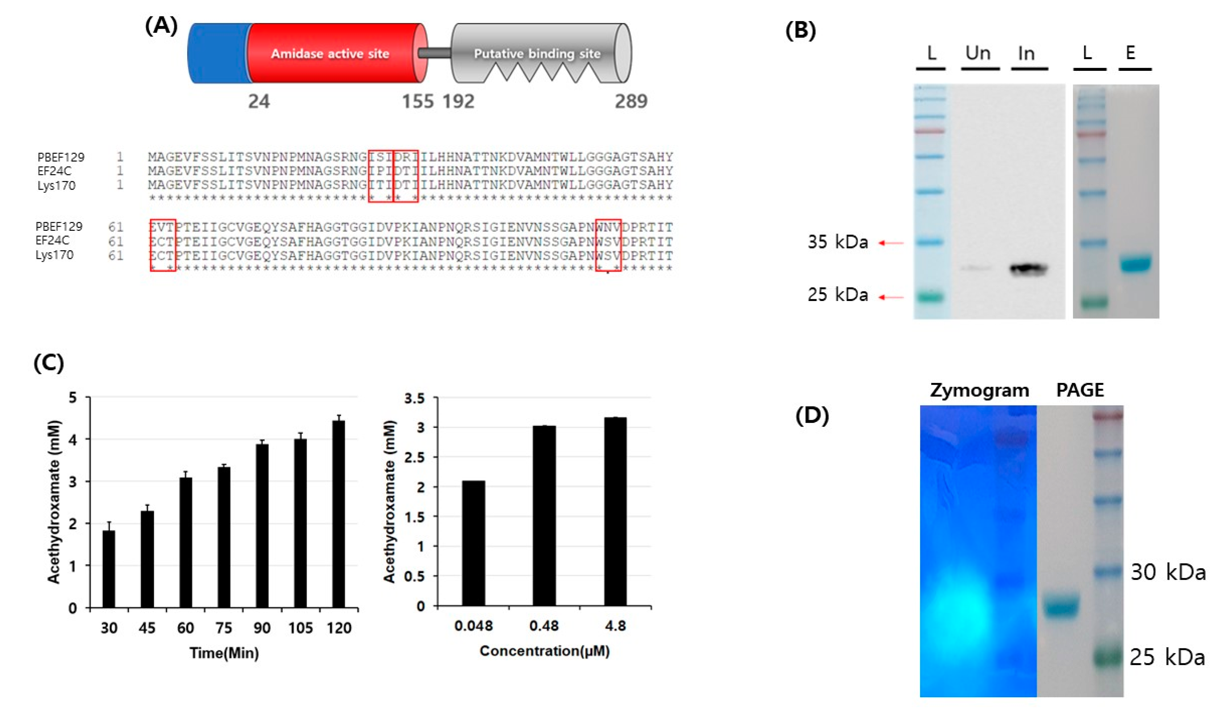
2.4. Cloning and Purification of Recombinant Phage Endolysin

The gene encoding ORF 6, which was annotated as a phage endolysin, was PCR-amplified with the primers Forward: 5’ GCGGCGCATATGGCAGGAGAAGTATTTAGT 3’ and Reverse: 5’ GCGGCGCTCGAGAGATTTTTTAGTAATACC 3’. The PCR conditions were as follows: initial activation of pfu DNA polymerase at 94 °C for 2 min, followed by 30 cycles of denaturation at 94 °C for 20 s, annealing at 55 °C for 2 min, and extension at 72 °C for 5 min. The PCR product was cloned in an expression vector of pET21-a(+) using restriction enzyme sites NdeI and XhoI. The resulting recombinant plasmid was used for transformation into an expression strain, Escherichia coli BL21 (DE3)pLyss (Promega, Madison, Wisconsin, USA). Overexpressed N-terminal 6X histidine-tagged recombinant protein was purified using a Ni-NTA column (Qiagen, Hilden, Germany) according to the manufacturer’s instructions. The recombinant protein was eluted in 200 mM imidazole, pH 7.5, and the final eluate was dialyzed against 20 mM tris-Cl, pH 7.5.

2.5. Biochemical Activity (amidase) Test of Purified Endolysin

For biochemical enzymatic activity, since ORF 6 was annotated as an amidase, an amidase assay was performed by means previously indicated [35]. Briefly, purified endolysin was incubated in 50 mM sodium phosphate buffer, pH 7.2, containing 100 mM acetamide solution and 500 mM hydroxylamine solution at 37 °C for one hour. Then, a color reagent solution containing 6% (w/v) ferric chloride in 5.7% (v/v) hydrochloric acid was added, forming acethydroxamate, which was measured by colorimetric detection at 500 nm. Reagents were purchased from Sigma-Aldrich, USA. A standard curve was plotted before the actual amidase assay of the purified protein according to the protocol [35]. Endolysin activity was also visualized by using zymography. Freshly grown E. faecalis was added to a 15% acrylamide gel. Purified endolysin was loaded onto the gel and electrophoresis was performed in a non-denaturing condition at the maximal power of 130 V for 3 h and stained with Coomassie Brilliant Blue. The gel was washed using distilled water at room temperature for 20 min. A second wash was performed with 0.1% triton X-100 in PBS for 1 h. Finally, a third wash was performed with PBS.

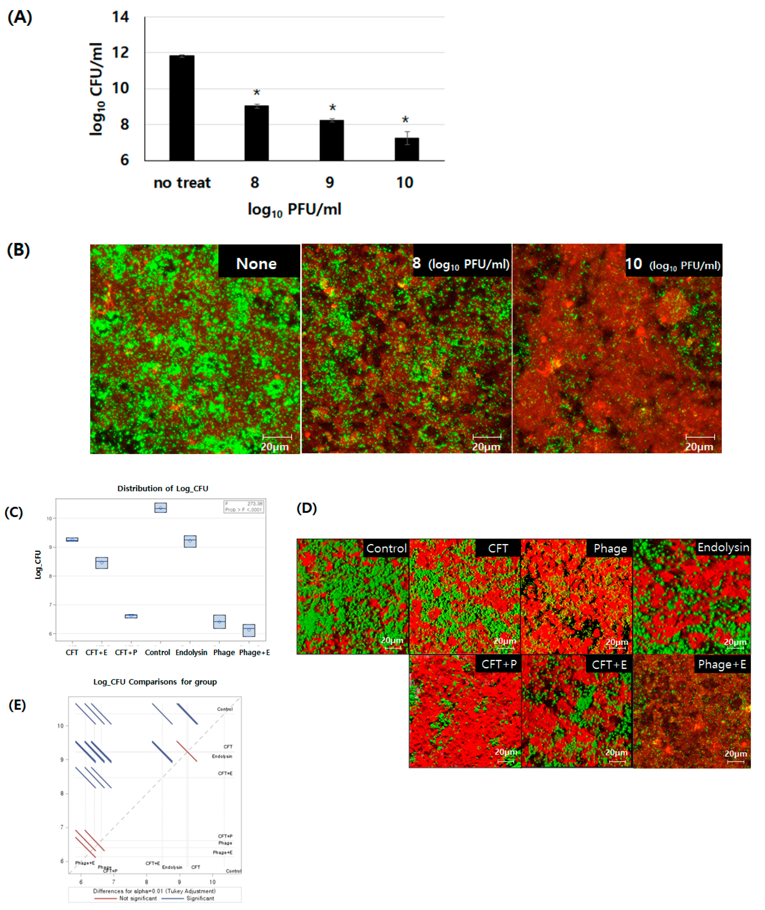
2.6. Antibacterial Activity Test of Purified Endolysin on Live Bacterial Cells

Time and dose dependency of antibacterial activity were observed on live bacterial cells. The standard reaction buffer used was 20 mM Tris-Cl, pH 7.5. When freshly grown E. faecalis CCARM 5520 in a brain heart infusion (BHI) reached a cell density of 8.8 × 10⁸ CFU/mL as measured using hemocytometer, 1 ml cells were harvested using low-speed centrifugation (1000× g for 2 min) with the supernatant being discarded. The pellet was resuspended in the standard reaction buffer with the addition of purified endolysin and the mixture was incubated at 37 °C for six hours. The number of surviving cells was counted on a BHI plate. The minimum inhibitory concentration of the endolysin on E. faecalis CCARM 5520 was determined using the methods suggested by the Clinical and Laboratory Standards Institute (CLSI) [36].

2.7. Human Cell Line and Formation of a Bacterial Biofilm In Vitro

A human intestinal cell line, SNU-C4, was obtained from the Korean Cell Line Bank at Seoul National University, Korea. Briefly, 3 × 10⁶ cells were seeded in a 6-well plate containing RPMI 1640 (Fischer Scientific, Rockingham County, New Hampshire, USA) supplemented with 10% fetal bovine serum (FBS, Gibco, Dún Laoghaire, Dublin, Ireland), and the cells were allowed to grow at 37 °C in a CO2 incubator set at 5% CO2 until confluency was observed. An exponentially grown culture of E. faecalis expressing GFP—E. faecalis OG1RF harboring pMV158GFP (EF/green)—was harvested and resuspended in RPMI 1640 media, and 1 × 10⁹ CFU were added to each well containing confluently grown intestinal cells. The mixture was incubated for 2 h for the bacteria to adhere to the surface of the intestinal cells, and then the mixture was washed with phosphate-buffered saline to discard any unbound bacteria. Finally, RPMI 1640 and FBS were added to the mixture and it was further incubated for 24 h for the biofilm to grow. Then, phage PBEF129 (1 × 10¹⁰ PFU per well), its endolysin (4.8 µM), cefotaxime (32 μg/mL, which was the minimum inhibitory concentration for the strain), or various combinations were added to the culture to observe the efficacy of biofilm removal. The mixture was incubated for 2 h. Then, the supernatant of each well was discarded and the wells were washed with PBS to remove all planktonic cells, followed by treatment with 1% triton X-100 (Sigma-Aldrich, Saint Louis, MO, USA) for 1 min. Then, the remaining bacteria were viewed under a laser scanning confocal microscope (Zeiss, Oberkochen, Germany). They were also counted on a solid agar medium as colony-forming units after recovery from the mixture. F-actin of human intestinal cells was immunostained with Phalloidin-iFluor 594 reagent (Abcam, Cambridge, United Kingdom cat. #176757).

2.8. Statistical Analysis

All the data are presented as the mean ± standard deviation (SD) of 3 independent experiments. Statistical significance was determined using Student’s t-test or Tukey’s test using proc glm in SAS (SAS Institute), with p-values < 0.05 representing significance.

3. Results

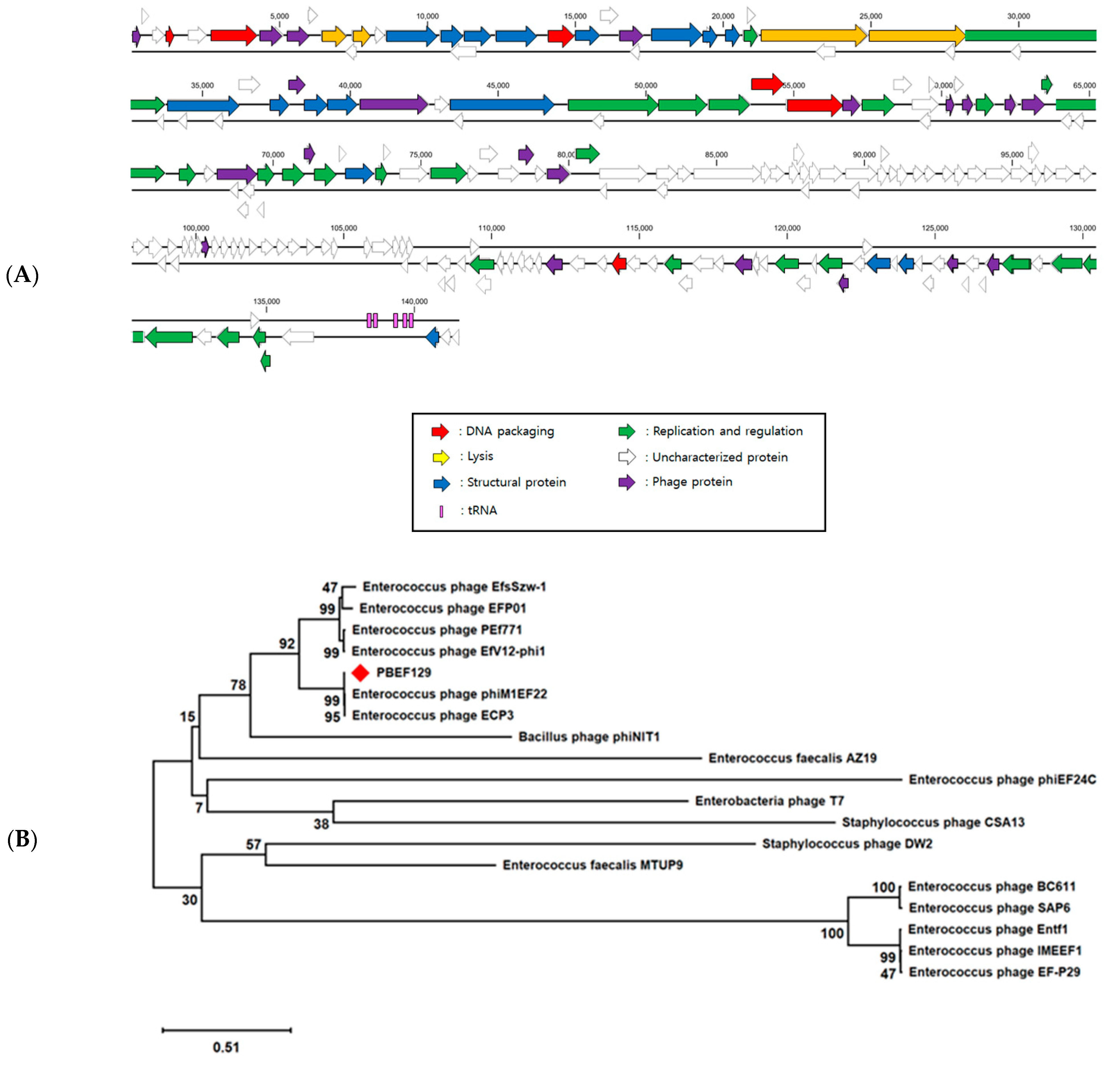
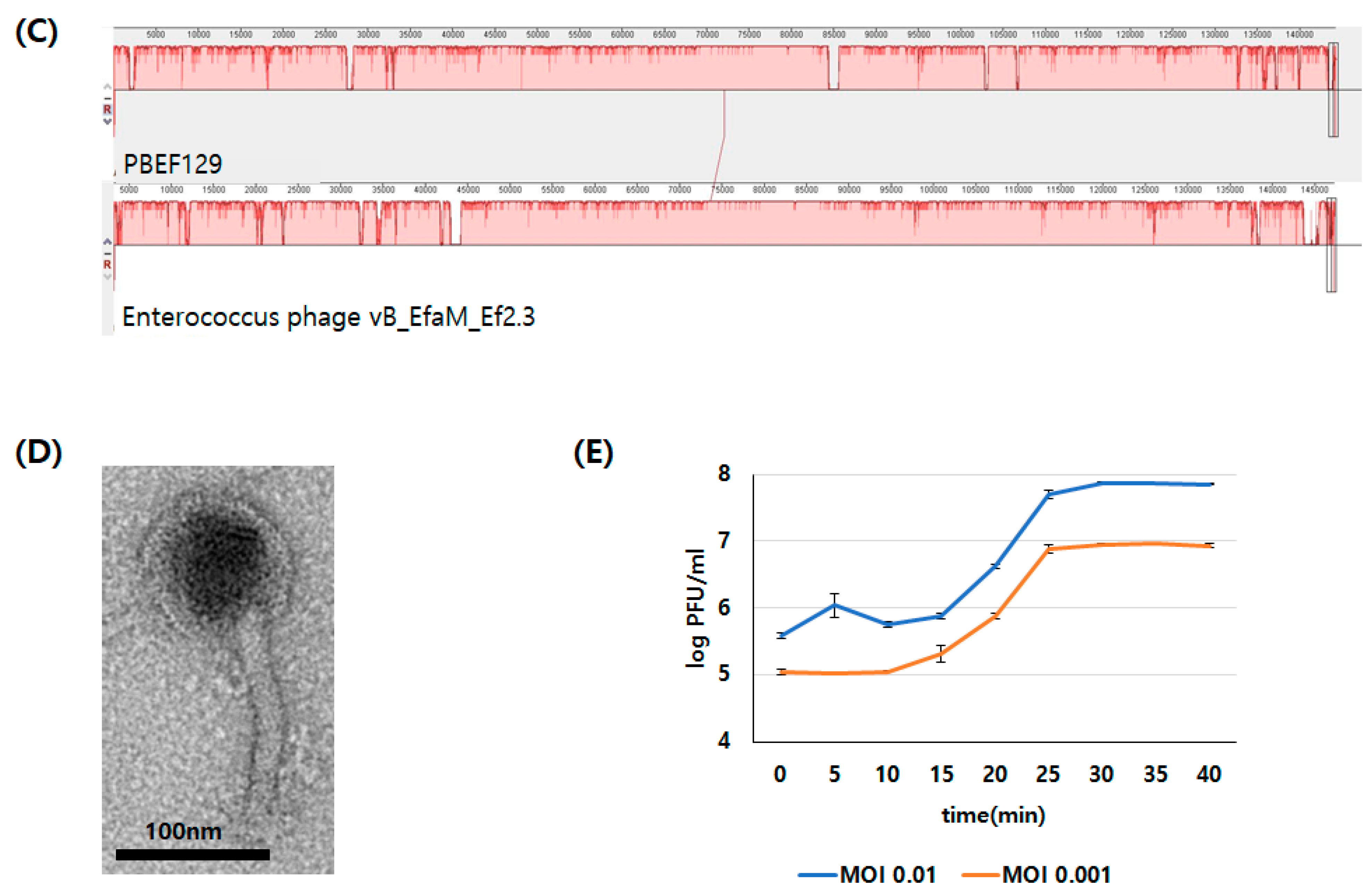
3.1. Genomic Analysis of Bacteriopahge PBEF129 Infecting E. feacalis

The genome of phage PBEF129 is a linear, double-stranded DNA, 144,230 base pairs in length (GenBank accession number MN854830.2). Its GC content is 35.9%. The closest genomic DNA sequence was found in Enterococcus phage vB_EfaM_Ef2.3, belonging to Herelleviridae, with a sequence identity of 99.06% over 95% query coverage from a BLASTN search. A total of 209 open reading frames (ORFs) were found and 75 were functionally annotated (Supplementary Table S1). Six were related to DNA packaging, four were related to lysis, 27 were related to replication and regulation, 17 were related to structural proteins, and 21 were annotated as phage proteins with unspecified functions. Five tRNA-encoding genes were also found. It was also verified using tRNAScan-SE [37]. The ORF map is shown in Figure 1A. Based on genes encoding tail fibers of related phages, a tree showing their relatedness was drawn (Figure 1B). Many Enterococcus phages were found to be closely related, but phages infecting Bacillus, Enterobacteria, or Staphylococcus were found to be distantly related. When comparing the genomes of phage PBEF129 and the closest phage vB_EfaM_Ef2.3 (Figure 1C), the arrangement of genes was linearly correlated, suggesting that the two phages were divergent due to the accumulation of point mutations over a long time rather than due to horizontal genetic exchange (mosaicism) [38]. Based on the morphology seen with the transmission electron microscope (TEM), phage PBEF129 belonged to the Siphovirus morphotype (Figure 1D). One-step multiplication of the phage showed a lytic cycle of 30 min and a burst size of 83 (Figure 1E).

3.2. Biochemical Characterization of Enzymatic Activity of Putative Endolysin from Phage PBEF129

Of the 75 ORFs annotated, ORF 6 (GenBank accession no. QHJ73506.1) was annotated as an N-acetylmuramoyl-L-alanine amidase (endolysin) (Supplementary Table S1). The length of the protein is 289 amino acids and the closest related protein is the putative endolysin in Enterococcus phage vB_EfaM_Ef2.3 (GenBank accession no. MK721192.1), with a sequence identity of 99.31% over 100% query coverage from a BLAST search. An enzymatic active domain (EAD) was found at the N-terminus while a cell wall binding domain (CBD) was found at the C-terminus using BLAST domain searches (Figure 2A). When compared to two closely related endolysins, EF24C (GenBank accession no. BAF81277) and Lys170 (GenBank accession no. YP_001504118) [17], amino acid residues 27, 30, 62, and 113 were different in the amidase active domain. To determine its enzymatic activity, the gene encoding ORF 2 was PCR-amplified and cloned to an expression vector. A 6XHis-tagged form of the protein was overexpressed and purified to near-homogeneity using affinity chromatography (Figure 2B). Since the assay method for enzymatic activity of amidase is performed by measuring the amount of reaction product, acethydroxamate, using optical density at 500 nm, we first prepared a standard curve (Supplementary Figure S1). Then, we could observe amidase activity from the purified protein both in time- and concentration-dependent manners (Figure 2C). The amidase activity was also confirmed using zymography (Figure 2D).

3.3. Characterization of Antibacterial Activity on Live Bacterial Cells

We first checked what the optimal pH was for antibacterial activity of the endolysin. A range of pH values were tested from 5.5 to 9.5, and stronger activities were observed at lower pH (Figure 3A). To confirm the specificity of the antibacterial activity, we next performed a series of tests with different concentrations of endolysin for different periods of incubation time. As seen in Figure 3B, the antibacterial activity was shown to be both dose- and time-dependent, suggesting that the activity was specific for the target bacteria. Treatment of bacterial cells with the endolysin at the final concentration of 5 M for 4 h led to a 4 log reduction. The purified protein was active against an array of E. faecalis strains, including antibiotic-resistant strains, some of which the parent phage could not infect (Table 2). The minimum inhibitory concentration of the endolysin was >128 μg/mL, as measured using the standard protocol from the Clinical and Laboratory Standards Institute (CLSI) guidelines.

3.4. Comparison of Removal Efficiency Among Cefotaxime, Bacteriophage PBEF129, Its Endolysin, and their Combinations on E. faecalis Biofilm Formed on Human Intestinal Cells Grown In Vitro

Intestinal colonization of E. faecalis is known to be associated with biofilm formation [39,40,41]. To eradicate the bacterial colonization, we need a modality which is effective for the removal of biofilms. Although the actual intestinal surface is covered with a mucin layer, chronic colonization of pathogenic bacteria would build a biofilm on top of the epithelial cells, leading to establishing an in vitro model of biofilm on top of cultured epithelial cells in this experiment. The efficacy of a less effective antibiotic, cefotaxime, was compared with alternatives to antibiotics, i.e., a bacteriophage, its endolysin, or their combination, in biofilm removal by counting residual bacteria after each treatment (Figure 4). First, we confirmed that phage treatment of a bacterial biofilm on an animal cell culture reduced bacterial load in a dose-dependent manner (Figure 4A,B). Treatment of the biofilm with cefotaxime or phage endolysin resulted in a limited reduction (~1 log) in bacterial load from the biofilm (Figure 4C). However, phage treatment was shown to be much more effective, with a 4 log reduction in bacteria. Combination treatment of cefotaxime and endolysin (CFT + E) showed a synergistic effect, while other combinations showed no synergistic effect. We also observed whether there was any morphological change in biofilm after each treatment under a confocal laser scanning fluorescence microscope (Figure 4D). On top of the confluently cultured human intestinal cells (red), E. faecalis (green) was seeded and incubated until a biofilm was formed. Then, each antibacterial agent or a combination of them was applied and removal of the biofilm was observed. Consistent with the results from the bacterial counting experiments, cefotaxime or phage endolysin removed bacteria to a limited extent, while application of phage PBEF129 to the biofilm resulted in a significant reduction in bacterial load.

4. Discussion

E. faecalis infection often leads to intestinal colonization, and it is involved in biofilm formation. In addition, multidrug-resistant pathogenic strains are frequently isolated in clinical settings. Vancomycin-resistant Enterococci (VRE) are considered one of the major threats in pathogenic microbiology. Thus, new modalities other than current antibiotics are urgently needed and bacteriophages are one such candidate. A newly isolated phage PBEF129 infects a series of drug-resistant E. faecalis strains. Although PBEF129 is closely related to a previously reported Enterococcus phage, vB_EfaM_Ef2.3 (Herelleviridae) [42], in this study, we focused on the characterization of a putative endolysin encoded by phage PBEF129.
Usually, an endolysin belongs to one of four different classes of enzymatic activity in cell wall degradation: amidase, endopeptidase, lytic transglycosylase (lysozyme-like muramidase), or glucosaminidase [43,44]. From the amino acid sequence of the gene product of ORF 2, it was suggested that the candidate was an amidase which cleaved the chemical bond between N-acetyl muramic acid (NAM) and the first amino acid of the peptide chain. We experimentally confirmed its amidase activity using biochemical methods. Its antibacterial activity was confirmed in vitro against a panel of E. faecalis strains. It is not unusual that an endolysin shows a broader spectrum of target bacteria than its parent phage [22,44]. A phage’s host range is mainly determined by the presence of the receptor on the bacterial surface and myriad other factors which can also affect the host range. However, an endolysin only needs to recognize the cell wall before enzymatic degradation, so the presence of a specific receptor is not required. Accordingly, we also confirmed a broader host range for the endolysin than its parent phage, PBEF129. The fact that the endolysin was more effective against target bacteria at lower pH may reflect a favorable surface charge alteration of target bacteria for the endolysin. The anionic nature of teichoic acids and wall-associated proteins on cell walls would change as pH changes, possibly leading to a stronger ionic interaction between the cell wall and the endolysin. There are several reports demonstrating higher activities of endolysins at lower pH levels [45,46,47].
In terms of antibacterial killing efficacy, this endolysin showed a relatively weak activity compared to other E. faecalis phage endolysins reported previously. For example, the LysEF-P10 endolysin showed a 6.5 log reduction in bacterial count (CFU/mL) in an hour [16], while this endolysin showed a 3 log reduction in two hours for cultured bacterial cells. In addition, the MIC could not be obtained using this endolysin.
Combination treatment using a lytic phage and an antibiotic was reported to show a synergistic effect [48]. Combination treatments of phage vB_AbaP_AGC01 and ciprofloxacin or gentamycin decreased Acinetobacter count 10 times more than separate treatments. This was not the case for phage PBEF129 in this study. Nonetheless, a combination treatment using the endolysin and an antibiotic showed a synergistic effect and it was demonstrated in a previous report [49]. An in vivo study using a Pneumococcal mouse model showed a higher survival rate (100%) with the combination of the endolysin Cpl-711 and cefotaxime than separate treatments (58%). A synergistic effect was also shown for the endolysin reported in this study, suggesting that the weak activity of an endolysin can be complemented by the addition of an appropriate antibiotic.
There are several reports describing various phage endolysins’ efficacy against bacterial biofilms where only limited activities were reported [50,51,52,53]. One report described an endolysin against Staphylococcus aureus biofilm on the surface of a medical device which showed no apparent efficacy [54]. In a case where an endolysin and a polysaccharide depolymerase from a phage against S. aureus were used simultaneously, biofilm was effectively removed [55]. Antibiotics are usually considered much less effective at removing bacterial biofilm than bacteriophages [56,57]. Since many phages, if not all, have polysaccharide depolymerases in their virion, they have more chance to confront bacterial cells inside extracellular polymers surrounding bacteria [29]. Antibiotics or endolysins do not harbor depolymerase activity, resulting in poor efficacy against biofilms. We could expect additive effects of combination treatment using bacteriophages and endolysins over each treatment individually, provided that bacteriophages armed with stronger depolymerase initially destroy extracellular polymer in biofilms, and endolysin then destroys the bacterial cells more efficiently than an antibiotic.
Endolysins are also reported to inhibit the formation of biofilms [50]. An endolysin with amidase activity against Listeria monocytogenes prevented biofilm formation when co-incubated with the bacteria for four days. Thus, endolysins could be applied to prevent biofilm formation on the surfaces of medical devices, e.g., catheters. Endolysins were also effective against tooth infections, which was another example of efficacy against biofilm [6,58]. Endolysins could effectively remove E. faecalis from human teeth or dentin slices.
Currently, four independent clinical trials of endolysins are underway (www.clinicaltrials.gov, 24 February 2021, identifiers NCT03163446, NCT01746654, NCT03089697, and NCT02840955), in which endolysins against Staphylococcus aureus are being tested from skin infections to bacteremia. Many other Gram (+) endolysins such as the one given in this paper and Gram (−) endolysins are being studied and may provide novel approaches for treating antibiotic-resistant bacteria.

Supplementary Materials

The following are available online at https://www.mdpi.com/1999-4915/13/3/426/s1, Figure S1: Standard curve of amidase enzymatic assay, Table S1: Functionally annotated open reading frames (ORFs) of phage PBEF129.

Author Contributions

Study conception and design: H.M.; acquisition of data: H.K.O., Y.J.H., and H.W.H.; drafting of manuscript: H.M. All authors have read and agreed to the published version of the manuscript.

Funding

This work was supported by the ATC Program 10076996 from the Ministry of Trade, Industry, and Energy of Korea; NRF grants 2017M3A9B8069292 and 2019M3E5D5066666 from the National Research Foundation of Korea, and the HUFS Research Grant of 2021.

Data Availability Statement

The data presented in this study are available on request from the corresponding author.

Conflicts of Interest

The authors declare no conflict of interest.

References

  1. Shiadeh, S.M.J.; Pormohammad, A.; Hashemi, A.; Lak, P. Global prevalence of antibiotic resistance in blood-isolated Enterococcus faecalis and Enterococcus faecium: A systematic review and meta-analysis. Infect. Drug Resist. 2019, 12, 2713–2725. [Google Scholar] [CrossRef] [PubMed] [Green Version]
  2. Beganovic, M.; Luther, M.K.; Rice, L.B.; Arias, C.A.; Rybak, M.J.; LaPlante, K.L. A Review of combination antimicrobial therapy for enterococcus faecalis bloodstream infections and infective endocarditis. Clin. Infect. Dis. 2018, 67, 303–309. [Google Scholar] [CrossRef] [Green Version]
  3. Gao, W.; Howden, B.P.; Stinear, T.P. Evolution of virulence in Enterococcus faecium, a hospital-adapted opportunistic pathogen. Curr. Opin. Microbiol. 2018, 41, 76–82. [Google Scholar] [CrossRef]
  4. Høiby, N.; Bjarnsholt, T.; Givskov, M.; Molin, S.; Ciofu, O. Antibiotic resistance of bacterial biofilms. Int. J. Antimicrob. Agents 2010, 35, 322–332. [Google Scholar] [CrossRef] [PubMed] [Green Version]
  5. Zheng, J.-X.; Bai, B.; Lin, Z.-W.; Pu, Z.-Y.; Yao, W.-M.; Chen, Z.; Li, D.-Y.; Deng, X.-B.; Deng, Q.-W.; Yu, Z.-J. Characterization of biofilm formation by Enterococcus faecalis isolates derived from urinary tract infections in China. J. Med. Microbiol. 2018, 67, 60–67. [Google Scholar] [CrossRef] [PubMed]
  6. Khalifa, L.; Brosh, Y.; Gelman, D.; Coppenhagen-Glazer, S.; Beyth, S.; Poradosu-Cohen, R.; Que, Y.-A.; Beyth, N.; Hazan, R. Targeting enterococcus faecalis biofilms with phage therapy. Appl. Environ. Microbiol. 2015, 81, 2696–2705. [Google Scholar] [CrossRef] [Green Version]
  7. Khatoon, Z.; McTiernan, C.D.; Suuronen, E.J.; Mah, T.-F.; Alarcon, E.I. Bacterial biofilm formation on implantable devices and approaches to its treatment and prevention. Heliyon 2018, 4, e01067. [Google Scholar] [CrossRef] [PubMed] [Green Version]
  8. Sharma, D.; Misba, L.; Khan, A.U. Antibiotics versus biofilm: An emerging battleground in microbial communities. Antimicrob. Resist. Infect. Control. 2019, 8, 76. [Google Scholar] [CrossRef] [PubMed]
  9. Bolocan, A.S.; Upadrasta, A.; de Almeida Bettio, P.H.; Clooney, A.G.; Draper, L.A.; Ross, R.P.; Hill, C. Evaluation of phage therapy in the context of enterococcus faecalis and its associated diseases. Viruses 2019, 11, 366. [Google Scholar] [CrossRef] [Green Version]
  10. Chatterjee, A.; Johnson, C.N.; Luong, P.; Hullahalli, K.; McBride, S.W.; Schubert, A.M.; Palmer, K.L.; Carlson, P.E.; Duerkop, B.A. Bacteriophage resistance alters antibiotic-mediated intestinal expansion of enterococci. Infect. Immun. 2019, 87, e00085-19. [Google Scholar] [CrossRef] [PubMed] [Green Version]
  11. Khalifa, L.; Shlezinger, M.; Beyth, S.; Houri-Haddad, Y.; Coppenhagen-Glazer, S.; Beyth, N.; Hazan, R. Phage therapy against enterococcus faecalis in dental root canals. J. Oral Microbiol. 2016, 8, 32157. [Google Scholar] [CrossRef] [PubMed] [Green Version]
  12. Tagliaferri, T.L.; Jansen, M.; Horz, H.-P. Fighting pathogenic bacteria on two fronts: Phages and antibiotics as combined strategy. Front. Cell. Infect. Microbiol. 2019, 9, 22. [Google Scholar] [CrossRef] [PubMed]
  13. Carmine, A.A.; Brogden, R.N.; Heel, R.C.; Speight, T.M.; Avery, G.S. Cefotaxime A review of its antibacterial activity, pharmacological properties and therapeutic use. Drugs 1983, 25, 223–289. [Google Scholar] [CrossRef]
  14. Moellering, R.C., Jr.; Eliopoulos, G.M. Activity of cefotaxime against Enterococci. Diagn. Microbiol. Infect. Dis. 1984, 2, 85S–90S. [Google Scholar]
  15. Gondil, V.S.; Harjai, K.; Chhibber, S. Endolysins as emerging alternative therapeutic agents to counter drug-resistant infections. Int. J. Antimicrob. Agents 2020, 55, 105844. [Google Scholar] [CrossRef]
  16. Abdelkader, K.; Gerstmans, H.; Saafan, A.; Dishisha, T.; Briers, Y. The preclinical and clinical progress of bacteriophages and their lytic enzymes: The parts are easier than the whole. Viruses 2019, 11, 96. [Google Scholar] [CrossRef] [PubMed] [Green Version]
  17. Oliveira, H.; São-José, C.; Azeredo, J. Phage-derived peptidoglycan degrading enzymes: Challenges and future prospects for in vivo therapy. Viruses 2018, 10, 292. [Google Scholar] [CrossRef] [PubMed] [Green Version]
  18. Cheng, M.; Zhang, Y.; Li, X.; Liang, J.; Hu, L.; Gong, P.; Zhang, L.; Cai, R.; Zhang, H.; Ge, J. Endolysin LysEF-P10 shows potential as an alternative treatment strategy for multidrug-resistant enterococcus faecalis infections. Sci. Rep. 2017, 7, 10164. [Google Scholar] [CrossRef] [Green Version]
  19. Proença, D.; Fernandes, S.; Leandro, C.; Silva, F.A.; Santos, S.; Lopes, F.; Mato, R.; Cavaco-Silva, P.; Pimentel, M.; São-José, C. Phage endolysins with broad antimicrobial activity against enterococcus faecalis clinical strains. Microb. Drug Resist. 2012, 18, 322–332. [Google Scholar] [CrossRef]
  20. Serrano-Maldonado, C.E.; García-Cano, I.; González-Canto, A.; Ruiz-May, E.; Elizalde-Contreras, J.M.; Quirasco, M. Cloning and characterization of a novel N-acetylglucosaminidase (AtlD) from Enterococcus faecalis. J. Mol. Microbiol. Biotechnol. 2018, 28, 14–27. [Google Scholar] [CrossRef]
  21. Stevens, R.H.; Zhang, H.; Sedgley, C.; Bergman, A.; Manda, A.R. The prevalence and impact of lysogeny among oral isolates of Enterococcus faecalis. J. Oral Microbiol. 2019, 11, 1643207. [Google Scholar] [CrossRef] [PubMed] [Green Version]
  22. Gong, P.; Cheng, M.; Li, X.; Jiang, H.; Yu, C.; Kahaer, N.; Li, J.; Zhang, L.; Xia, F.; Hu, L.; et al. Characterization of Enterococcus faecium bacteriophage IME-EFm5 and its endolysin LysEFm5. Virology 2016, 492, 11–20. [Google Scholar] [CrossRef]
  23. Lu, N.; Sun, Y.; Wang, Q.; Qiu, Y.; Chen, Z.; Wen, Y.; Wang, S.; Song, Y. Cloning and characterization of endolysin and holin from Streptomyces avermitilis bacteriophage phiSASD1 as potential novel antibiotic candidates. Int. J. Biol. Macromol. 2020, 147, 980–989. [Google Scholar] [CrossRef]
  24. Kim, N.-H.; Park, W.B.; Cho, J.E.; Choi, Y.J.; Choi, S.J.; Jun, S.Y.; Kang, C.K.; Song, K.-H.; Choe, P.G.; Bang, J.-H.; et al. Effects of phage endolysin SAL200 combined with antibiotics on staphylococcus aureus infection. Antimicrob. Agents Chemother. 2018, 62, e00731-18. [Google Scholar] [CrossRef] [Green Version]
  25. Chang, Y. Bacteriophage-derived endolysins applied as potent biocontrol agents to enhance food safety. Microorganisms 2020, 8, 724. [Google Scholar] [CrossRef] [PubMed]
  26. Korea National Research Resources Center. Available online: http://knrrc.swu.ac.kr/sale/sale.jsp (accessed on 3 March 2021).
  27. Lee, K.; Lee, K.-M.; Kim, D.; Yoon, S.S. Molecular determinants of the thickened matrix in a dual-species pseudomonas aeruginosa and enterococcus faecalis biofilm. Appl. Environ. Microbiol. 2017, 83, e01182-17. [Google Scholar] [CrossRef] [Green Version]
  28. Kropinski, A.M.; Mazzocco, A.; Waddell, T.E.; Lingohr, E.; Johnson, R.P. Enumeration of bacteriophages by double agar overlay plaque assay. In NMR-Based Metabolomics; Springer: Berlin/Heidelberg, Germany, 2009; Volume 501, pp. 69–76. [Google Scholar]
  29. Kim, M.S.; Kim, Y.D.; Hong, S.S.; Park, K.; Ko, K.S.; Myung, H. Phage-encoded colanic acid-degrading enzyme permits lytic phage infection of a capsule-forming resistant mutant escherichia coli strain. Appl. Environ. Microbiol. 2015, 81, 900–909. [Google Scholar] [CrossRef] [PubMed] [Green Version]
  30. Jiang, H.; Lei, R.; Ding, S.-W.; Zhu, S. Skewer: A fast and accurate adapter trimmer for next-generation sequencing paired-end reads. BMC Bioinform. 2014, 15, 182. [Google Scholar] [CrossRef] [PubMed]
  31. Allam, A.; Kalnis, P.; Solovyev, V. Karect: Accurate correction of substitution, insertion and deletion errors for next-generation sequencing data. Bioinformatics 2015, 31, 3421–3428. Available online: https://github.com/aminallam/karect (accessed on 3 March 2021). [CrossRef] [Green Version]
  32. Baaijens, J.A.; El Aabidine, A.Z.; Rivals, E.; Schönhuth, A. De novo assembly of viral quasispecies using overlap graphs. Genome Res. 2017, 27, 835–848. [Google Scholar] [CrossRef] [Green Version]
  33. Altschul, S.F.; Gish, W.; Miller, W.; Myers, E.W.; Lipman, D.J. Basic local alignment search tool. J. Mol. Biol. 1990, 215, 403–410. [Google Scholar] [CrossRef]
  34. Pathosystems Resource Integration Center. Available online: https://www.patricbrc.org/ (accessed on 3 March 2021).
  35. Enzymatic Assay of Amidase (EC3.5.1.4). Available online: https://www.sigmaaldrich.com/content/dam/sigma-aldrich/docs/Sigma/General_Information/2/amidase.pdf (accessed on 3 March 2021).
  36. CLSI. M07: Methods for Dilution Antimicrobial Susceptibility Tests for Bacteria That Grow Aerobically, 11th ed.; CLSI: Wayne, PA, USA, 2018. [Google Scholar]
  37. Lowe, T.M.; Chan, P.P. tRNAscan-SE On-line: Search and contextual analysis of transfer RNA genes. Nucl. Acids Res. 2016, 44, W54–W57. [Google Scholar] [CrossRef]
  38. Hatfull, G.F.; Hendrix, R.W. Bacteriophages and their genomes. Curr. Opin. Virol. 2011, 1, 298–303. [Google Scholar] [CrossRef] [PubMed] [Green Version]
  39. McKenney, P.T.; Yan, J.; Vaubourgeix, J.; Becattini, S.; Lampen, N.; Motzer, A.; Larson, P.J.; Dannaoui, D.; Fujisawa, S.; Xavier, J.B.; et al. Intestinal bile acids induce a morphotype switch in vancomycin-resistant enterococcus that facilitates intestinal colonization. Cell Host Microbe 2019, 25, 695–705.e5. [Google Scholar] [CrossRef]
  40. Ghosh, A.; Borst, L.; Stauffer, S.H.; Suyemoto, M.; Moisan, P.; Zurek, L.; Gookin, J.L. Mortality in kittens is associated with a shift in ileum mucosa-associated enterococci from enterococcus hirae to biofilm-forming enterococcus faecalis and adherent escherichia coli. J. Clin. Microbiol. 2013, 51, 3567–3578. [Google Scholar] [CrossRef] [Green Version]
  41. Shridhar, S.; Dhanashree, B. Antibiotic susceptibility pattern and biofilm formation in clinical isolates of enterococcus spp. Interdiscip. Perspect. Infect. Dis. 2019, 2019, 7854968. [Google Scholar] [CrossRef] [Green Version]
  42. Duan, Y.; Llorente, C.; Lang, S.; Brandl, K.; Chu, H.; Jiang, L.; White, R.C.; Clarke, T.H.; Nguyen, K.; Torralba, M.; et al. Bacteriophage targeting of gut bacterium attenuates alcoholic liver disease. Nature 2019, 575, 505–511. [Google Scholar] [CrossRef] [PubMed]
  43. São-José, C. Engineering of phage-derived lytic enzymes: Improving their potential as antimicrobials. Antibiotics 2018, 7, 29. [Google Scholar] [CrossRef] [Green Version]
  44. Schmelcher, M.; Donovan, D.M.; Loessner, M.J. Bacteriophage endolysins as novel antimicrobials. Futur. Microbiol. 2012, 7, 1147–1171. [Google Scholar] [CrossRef] [Green Version]
  45. Ding, Y.; Zhang, Y.; Huang, C.; Wang, J.; Wang, X. An endolysin LysSE24 by bacteriophage LPSE1 confers specific bactericidal activity against multidrug-resistant salmonella strains. Microorganisms 2020, 8, 737. [Google Scholar] [CrossRef]
  46. Swift, S.M.; Rowley, D.T.; Young, C.; Franks, A.; Hyman, P.; Donovan, D.M. The endolysin from the Enterococcus faecalis bacteriophage VD13 and conditions stimulating its lytic activity. FEMS Microbiol. Lett. 2016, 363, fnw216. [Google Scholar] [CrossRef] [Green Version]
  47. Lu, S.-H.; Bischoff, K.M.; Rich, J.O.; Liu, S.; Skory, C.D. Recombinant bacteriophage LysKB317 endolysin mitigates Lactobacillus infection of corn mash fermentations. Biotechnol. Biofuels 2020, 13, 157. [Google Scholar] [CrossRef]
  48. Grygorcewicz, B.; Roszak, M.; Golec, P.; Śleboda-Taront, D.; Łubowska, N.; Górska, M.; Jursa-Kulesza, J.; Rakoczy, R.; Wojciuk, B.; Dołęgowska, B. Antibiotics act with vB_AbaP_AGC01 phage against acinetobacter baumannii in human heat-inactivated plasma blood and galleria mellonella models. Int. J. Mol. Sci. 2020, 21, 4390. [Google Scholar] [CrossRef]
  49. Letrado, P.; Corsini, B.; Díez-Martínez, R.; Bustamante, N.; Yuste, J.E.; García, P. Bactericidal synergism between antibiotics and phage endolysin Cpl-711 to kill multidrug-resistant pneumococcus. Futur. Microbiol. 2018, 13, 1215–1223. [Google Scholar] [CrossRef] [PubMed] [Green Version]
  50. Cha, Y.; Son, B.; Ryu, S. Effective removal of staphylococcal biofilms on various food contact surfaces by Staphylococcus aureus phage endolysin LysCSA13. Food Microbiol. 2019, 84, 103245. [Google Scholar] [CrossRef] [PubMed]
  51. Pennone, V.; Sanz-Gaitero, M.; O’Connor, P.; Coffey, A.; Jordan, K.; Van Raaij, M.J.; McAuliffe, O.; Gaitero, S.-; Connor, O.; Raaij, V. Inhibition of L. monocytogenes biofilm formation by the amidase domain of the phage vB_LmoS_293 endolysin. Viruses 2019, 11, 722. [Google Scholar] [CrossRef] [Green Version]
  52. Zhang, H.; Buttaro, B.A.; Fouts, D.E.; Sanjari, S.; Evans, B.S.; Stevens, R.H. Bacteriophage φEf11 ORF28 endolysin, a multifunctional lytic enzyme with properties distinct from all other identified enterococcus faecalis phage endolysins. Appl. Environ. Microbiol. 2019, 85. [Google Scholar] [CrossRef] [PubMed] [Green Version]
  53. Melo, L.D.R.; Brandão, A.; Akturk, E.; Santos, S.B.; Azeredo, J. Characterization of a new staphylococcus aureus kayvirus harboring a lysin active against biofilms. Viruses 2018, 10, 182. [Google Scholar] [CrossRef] [PubMed] [Green Version]
  54. Idelevich, E.A.; Knaack, D.; Nugroho, N.T.; Peters, G.; Bisdas, T.; Molinaro, S.; Torsello, G.B.; Becker, K.; Herten, M. Comparative in vitro activity of bacteriophage endolysin HY-133 against Staphylococcus aureus attached to vascular graft surface. Med. Microbiol. Immunol. 2020, 209, 51–57. [Google Scholar] [CrossRef]
  55. Olsen, N.M.C.; Thiran, E.; Hasler, T.; Vanzieleghem, T.; Belibasakis, G.N.; Mahillon, J.; Loessner, M.J.; Schmelcher, M. Synergistic removal of static and dynamic staphylococcus aureus biofilms by combined treatment with a bacteriophage endolysin and a polysaccharide depolymerase. Viruses 2018, 10, 438. [Google Scholar] [CrossRef] [Green Version]
  56. Yan, Z.; Huang, M.; Melander, C.; Kjellerup, B.V. Dispersal and inhibition of biofilms associated with infections. J. Appl. Microbiol. 2019, 128, 1279–1288. [Google Scholar] [CrossRef] [PubMed] [Green Version]
  57. Abedon, S.T. Phage-antibiotic combination treatments: Antagonistic impacts of antibiotics on the pharmacodynamics of phage therapy? Antibiotics 2019, 8, 182. [Google Scholar] [CrossRef] [PubMed] [Green Version]
  58. Lee, D.; Im, J.; Na, H.; Ryu, S.; Yun, C.-H.; Han, S.H. The novel enterococcus phage vB_EfaS_HEf13 has broad lytic activity against clinical isolates of enterococcus faecalis. Front. Microbiol. 2019, 10, 2877. [Google Scholar] [CrossRef] [PubMed]
Figure 1. Genomic analysis of the bacteriophage PBEF129. (A) Open reading frame (ORF) map. Each arrow represents a functionally annotated ORF color coded according to its function. Blue, structural protein; green, replication and regulation; yellow, lysis; red, DNA packaging; violet, unspecified phage protein; white, uncharacterized. (B) Genomic tree showing distances among related phages. Red rhombus indicates PBEF129. The tree was drawn based on the genes encoding tail fibers of each phage. Bootstrap values are shown. (C) Alignment of whole genomes of phage PBEF129 and the closest phage vB_EfaM_Ef2 using MAUVE. (D) TEM image of phage PBEF129 shown with a scale bar. (E) One-step multiplication of phage PBEF129.
Viruses 13 00426 g001aViruses 13 00426 g001b
Figure 2. Purification and enzymatic activity of endolysin. (A) Suggested domain structure of the endolysin of phage PBEF129 from BLAST domain search. The total length of the endolysin is 289 amino acids. The putative amidase active domain (red) is from 24 to 155 amino acids and the putative peptidoglycan binding domain (gray) is 192 to 289 amino acids. Amino acids’ sequence alignment among related endolysins, PBEF129, EF24C and Lys170 [17], is seen under the domain diagram. Different amino acid residues in sequences are shown in the box. Only four amino acid residues differ in the enzymatically active domain (EAD). (B) Purification of the recombinant endolysin. Right panel, purification of putative endolysin. L, pre-stained protein ladder; E, eluted fraction. Left panel, Western blot analysis of the purified protein using anti-6XHis antibody. L, pre-stained protein ladder; Un, un-induced culture lysate; In, induced culture lysate. (C) Left: confirmation of amidase activity of the purified protein with various incubation times; 4.8 μM of protein was used for each time point. Right: confirmation of amidase activity of the purified protein with various amounts of protein. One-hour incubation was used for each enzyme concentration. (D) Left: zymogram of purified endolysin run on a non-denaturing PAGE containing the target bacteria. Right: PAGE analysis of the endolysin followed by Coomassie Brilliant Blue staining.
Viruses 13 00426 g002
Figure 3. Characterization of antibacterial activity of the endolysin on live bacterial cells. (A) Optimal pH of the antibacterial activity of the endolysin. Buffer containing each pH was used for incubation of the antibacterial reaction with 4.8 μM of protein and 1-hour incubation. (B) Antibacterial efficacy was shown in a dose-dependent manner. Log reduction of bacterial count is shown with the amount of protein used indicated. Assay mixture at pH 7.5 was incubated at 37 °C for 2 or 4 hours.
Viruses 13 00426 g003
Figure 4. Biofilm removal efficacy of each antibacterial agent on in vitro cultured human intestinal cells. (A) After treatment with various concentrations of phages, the remaining bacterial load was recovered and counted as colony-forming units (CFUs). * p < 0.05. (B) Observation of remaining bacterial load after phage treatment on biofilm formed on top of in vitro cultured human intestinal cells. E. faecalis expressing green fluorescent protein (GFP) is shown in green, while human intestinal cells immunostained with anti-F actin antibody are shown in red. (C) After treatment with each agent, the remaining bacterial load was recovered and counted as colony-forming units (CFUs). CFT, cefotaxime; E, endolysin; P, phage PBEF129. Control is where the biofilm was treated with no agent. The experiment was performed in triplicate. (D) Observation of remaining bacterial load after each treatment on biofilm formed on top of in vitro cultured human intestinal cells. E. faecalis expressing green fluorescent protein (GFP) is shown as green, while human intestinal cells immunostained with anti-F actin antibody are shown as red. (E) Analysis of statistical significance among each treatment. The diffogram shows the result of Tukey’s test. Blue lines mean p < 0.01.
Viruses 13 00426 g004
Table 1. Antibiotic resistance profiles of E. faecalis strains used in this study.
Antibiotic
(μg/mL)
Enterococcus faecalis Strains
CCARM 5511 *CCARM 5518CCARM 5520CCARM 5526CCARM 5537CCARM 5539CCARM 5548CCARM 5568CCARM 5569
Ampicillin4444444
Ciprofloxacin≥2≤1≥2≥2≤1≥2≥21≥2
Erythromycin ≥4≥4≥4≥4 ≥4
Gentamycin≥500≥500≥500≥500≥500≥500≥500≥500≥500
Levofloxacin≥42≥4≥4≤1≥4≥4≥2≥4
Linezolid≤2≤2≤2≤2≤2≤2≤2≤1≤1
Penicillin≥88≥888≥8≥88≥8
Rifampin≥2≤1≤1≤1≥2≥2≤1≤1≤1
Streptomycin≥1000≤1000≤1000≤1000≥1000≥1000≤1000≤1000≤1000
Quinupristin-
dalfopristin
≥2≥2≥2≥2≥2≥2≥2≥2≥2
Teicoplanin≤4≤4≤4≤4≤4≤4≤4≤1≤1
Tetracycline≥8≥8≥8≥8≥8≥8≥8≥8≥8
Vancomycin212222222
* CCARM number is for strains listed in the Culture Collection of Antibiotic-Resistant Microorganisms in Korea [26].
Table 2. Lysis of E. faecalis strain by phage vs. endolysin.
E. faecalis StrainPhage PBEF129Endolysin
CCARM5511+
CCARM5518++
CCARM5520++
CCARM5526++
CCARM5537+
CCARM5539++
CCARM5548++
CCARM5568++
CCARM5569++
CCARM5571++
ATCC19433++
On a lawn of each bacterium, 20 μL of phage (109 PFU/mL) or purified endolysin (1 mg/mL) was spotted and the formation of clear zone was observed.
Publisher’s Note: MDPI stays neutral with regard to jurisdictional claims in published maps and institutional affiliations.

