Article

RNAi-Mediated Silencing of Chitin Synthase 1 (CHS1) Disrupts Molting and Growth in Monochamus alternatus

1
State Key Laboratory of Agricultural and Forestry Biosecurity, College of Forestry, Fujian Agriculture and Forestry University, Fuzhou 350002, China
2
College of Forestry, Fujian Agriculture and Forestry University, Fuzhou 350002, China
3
Key Laboratory of Integrated Pest Management in Ecological Forests, Fujian Province University, Fujian Agriculture and Forestry University, Fuzhou 350002, China
4
Guizhou Institute of Biology, Guizhou Academy of Sciences, Guiyang 550009, China
5
Fujian Colleges and Universities Engineering Research Institute of Conservation and Utilization of Natural Bioresources, College of Forestry, Fujian Agriculture and Forestry University, Fuzhou 350002, China
*
Authors to whom correspondence should be addressed.
These authors contributed equally to this work.
Forests 2025, 16(6), 922; https://doi.org/10.3390/f16060922
Submission received: 14 April 2025 / Revised: 24 May 2025 / Accepted: 27 May 2025 / Published: 30 May 2025
(This article belongs to the Section Forest Health)

Abstract

:
Chitin synthase (CHS) plays a key role in chitin synthesis. CHS1 is ubiquitous in insects, and some studies have found that the RNA interference with CHS1 can hinder three types of molting processes (larva–larva, larva–pupa and pupa–adult). In the present study, the CHS1 of Monochamus alternatus was identified and characterized by a bioinformatics analysis. The developmental stage-specific expression of the MalCHS1 (Monochamus alternatus CHS1) gene was obtained by a RT-qPCR, and the corresponding dsRNA was designed for functional verification. The RNA interference experiment was conducted using the microinjection method, and the injection site was selected from the abdominal segments of fifth-instar larvae. The results showed that after silencing the CHS1 gene, the larvae of M. alternatus showed morphological abnormalities, such as the softening of the body wall, a transparent abdomen and the swelling of somites, indicating that MalCHS1 mediates the molting, growth and development of M. alternatus. RNAi-mediated MalCHS1 gene silencing may become a promising new biological pesticide that can provide a new target gene for pest control.

1. Introduction

Monochamus alternatus Hope is a crucial stem borer of Pinus plants [1,2]. It mainly feeds on Pinus massoniana Lamb., P. thunbergii Parlatore and P. tabulaeformis Carr. In China, M. alternatus also serves as a vector of the pinewood nemotade (PWN), which is responsible for pine wilt disease (PWD) [3]. This species plays a significant role in the pathogen’s infection cycle by contributing to its spread within pine stands. PWD is the most serious forest disease in Japan and East Asia and it is currently considered to be one of the most serious threats to pine forests worldwide [4]. PWD is known as the cancer of pine trees [5]. Since the discovery of the PWN in the Sun Zhong Shan mausoleum in Nanjing, China in 1982, and by 2023, pine wilt disease has spread to 19 provinces (including 701 counties and cities) [6].
According to the morphological characteristics and life habits of M. alternatus, its life cycle consists of four stages: egg, larva, pupa and adult. The larvae are 1–5 instars (L1, L2, L3, L4 and L5). The transmission harm of M. alternatus is mainly in the supplementary nutrition and oviposition period of adults [2]. M. alternatus overwinters as larvae. In the larval stage, they mainly feed on the bast of the trunk and then gradually bore into the xylem of the trunk as it develops, disrupting water and nutrient transport, ultimately killing the tree. In the adult stage, they invade pines by feeding on the shoots and laying their eggs on the tree and at the same time carrying PWNs. The PWN feeds on the epithelial cells of the resin ducts of the tree and its proliferation impairs water transport, causing the tree to WILT and die and ultimately to the massive death of the trees [2,7,8].
Current studies have shown that chitin is a key component of the insect synthetic cuticle, digestive tract and other parts involved in growth and molting processes [9,10]. Chitinase (CHS) is the important enzyme in chitin synthesis [11,12]. Compared with the CHS of different insects, CHS has three main domains: domain A, domain B and domain C. Domain A and C are, respectively, located at the N-terminal and C-terminal and contain one transmembrane helix and seven transmembrane helixes, respectively, while domain B is the active center of CHS [13]. Currently, most insect chitinases are divided into two groups: Chitin Synthase 1 (CHS1) and chitin synthase 2 (CHS2) [14]. CHS1 mainly mediates the synthesis of chitin in the outermost cuticle of the insect epidermis, trachea and foregut of insects, while CHS2 is mainly responsible for the synthesis of chitin in the periplasmic membrane [15,16]. CHS1 is only distributed in epidermal cells derived from the ectoderm, including the epidermis, trachea, salivary glands, foregut and hindgut cells. It is involved in the synthesis of chitin in these tissues and thus plays an essential role in insect growth and molting processes [15,17,18]. Currently, RNA interference (RNAi) technology has been widely applied to the functional analysis of genes, pest management and many other aspects [19]. For example, reducing heat shock protein (hsp) expression by RNAi in Dendroctonus spp. resulted in greater environmental sensitivity and mortality rates [20,21]. In Aphis gossypii Glover, silencing the CHS1 gene through RNAi can lead to an increased mortality, a shortened lifespan of adults and a decreased reproductive capacity [22]. In Tribolium castaneum, RNAi targeting CHS1 expression significantly reduced the insect resistance against fungal infestation [23]. The knockout of the CHS1 gene in Henosepilachna vigintioctopunctata can lead to impaired intestinal integrity, the reduced thickness of the stratum corneum and the delayed formation of the newly generated stratum corneum during molting. In addition, the absence of HvCHS1 inhibits the development of the tracheal system, resulting in the thinning of the tracheal band [24].
The destruction of the diseased trees and using insecticides has controlled pine wilt disease, but the public is now demanding environmentally friendly control methods. Research is focusing on finding alternative means of control, such as biological control agents for both nematodes and vectors, insect attractants and RNA pesticides with nanocarriers [3,25,26]. Based on the M. alternatus genome, the MalCHS1 (Monochamus alternatus CHS1) gene was identified. The expression level of the MalCHS1 gene at key developmental stages was obtained by a quantitative reverse transcription PCR (RT-qPCR), and then the corresponding dsRNA was designed for functional verification. Afterward, dsRNA was injected into M. alternatus by a microinjector, and its silencing effect was verified by an RT-qPCR. In this study, we will analyze the role of CHS1 in the growth and development of M. alternatus through molecular biology, RNAi technology and phenotypic observation to provide new target genes for pest control and a theoretical basis for novel biorational control methods, thus reducing the spread of pine wilt disease.

2. Materials and Methods

2.1. Samples and Feeding Conditions

Experimental insects were the first generation of larvae reared in our laboratory after being trapped by lamp lure method in the field (Fuzhou, Fujian Province 119°14′ 8.3616″ E, 26°5′ 27.8658″ N). The rearing conditions within environmental chambers were as follows: GHP-300E intelligent light and temperature incubator (Sanfa, Yangzhou, China), with temperature held at 23–26 °C, light cycle set to 12 h light: 12 h dark and relative humidity held at 70%–75%. Samples for expression level: 5th-instar larvae, male and female new pupae, male and female mature pupae and sexually mature male and female adults of M. alternatus were collected, with three biological replicates per group. They were placed in 1.5 mL centrifuge tubes, frozen in liquid nitrogen and stored at −80 °C. Samples for real-time quantitative PCR after RNAi: 5th-instar larvae of M. alternatus were collected, and the intestines of the control group and the treated group larvae were dissected and separated, with three replicates for each group.

2.2. Gene Cloning

RNA of M. alternatus was extracted using Trizol reagent (Ambion/Invitrogen, Waltham, MA, USA). The extracted RNA was quantitatively analyzed using a NanoDrop 2000 spectrophotometer (Thermo Fisher Science, Waltham, MA, USA). The first-strand cDNA was prepared using the Hifair® III First Strand cDNA Synthesis Kit (gDNA Eliminator Plus) (Yeasen, Gaithersburg, MD, USA). Primers for PCR amplification were designed based on the MalCHS1 sequence (Table 1).

2.3. Bioinformatics Characterization of MalCHS1 in M. alternatus

The full-length MalCHS1 gene sequence was analyzed by open reading frame analysis and protein sequence prediction (data source: https://www.ncbi.nlm.nih.gov/, accessed on 1 July 2024), identifying its coding region and potential functional domains. In NCBI, BLAST protein was used to analyze amino acid sequences, and sequences with high homology were downloaded for homology comparison. MEGA 12.0 was used to construct a phylogenetic tree, and the Neighbor-Joining Method (NJ) was selected to construct the identified CHS1 gene [27]. The gene domain was analyzed using Web CD Search Tool online tool [28], and gene protein sequence motifs were identified with MEME suite (http://meme-suite.org/tools/meme (accessed on 5 July 2024)).

2.4. Quantitative Real-Time PCR

The 5th-instar larvae, male and female new pupae and old mature pupa and sexually mature male and female larvae of M. alternatus were collected in 3 replicates per group. RNA was extracted after freezing with liquid nitrogen, and the expression levels of the MalCHS1 gene at key developmental stages were obtained by real-time quantitative PCR (RT-qPCR). β-actin (Table 1) was selected as the internal reference gene. All RT-qPCR primers were designed by National Online and further validated with Oligo 7 [29]. The RT-qPCR procedure is as follows: initial denaturation at 95 °C for 5 min, followed by 10 s at 95 °C and 30 s at 60 °C, for a total of 40 denaturation cycles.

2.5. RNAi of MalCHS1

To synthesize dsRNA via in vitro transcription, T7 promoter sequences were added to the 5′ ends of PCR primers during target gene cloning (Table 1). Synthesize the target gene dsRNA using T7 RNAi Transcription Kit TR 102 (Vazyme, Nanjing, China). Afterward, RNAi treatment was performed on the 5th-instar larvae using dsRNA-MalCHS1, with an injection volume of 2000 ng/uL. RNase-free ddH2O was injected as a negative control group, and dsGFP was injected as a positive control group. The syringe was washed three times with anhydrous ethanol and RNase-free ddH2O, with three replicates for each treatment. Observe the morphological changes in larvae after injection and record the time and quantity of death and pupation. To maintain the effectiveness of RNAi throughout the phenotypic observation period, test insects were re-injected with dsRNA every 48–72 h. Based on the close relationship between CHS1 and intestinal chitin synthesis, this study will dissect and isolate the gut of 5th-instar larvae 24 h after injection and detect the expression level of MalCHS1 gene in the gut of M. alternatus larvae after injection of dsRNA through RT-qPCR experiment.
Mortality =   The   number   of   dead   larvae Total   number   of   larvae   ×   100 %
Pupation   rate =   The   number   of   pupae Total   number   of   larvae   ×   100 %

2.6. Data Analysis

Each group of experiments was repeated three times independently, and the data were expressed as the mean ± standard deviation of three independent experiments. Data were analyzed using SPSS statistical software (IBM SPSS Statistics 27 software). Differences in expression at key developmental stages were analyzed by one-way ANOVA test, and differences between treatment and control groups in RNAi experiments were plotted by independent samples t-test and then plotted using Graphpad (https://www.graphpad.com/) and SPSS software [30,31].

3. Results

3.1. Bioinformatics Analysis of MalCHS1 in M. alternatus

The phylogenetic analysis shows that the MalCHS1 is clustered with Anthonomus grandis Boheman and closely related to H. vigintioctopunctata, suggesting its function may be conserved (Figure 1A). To further analyze the conservative features of MalCHS1, 10 conserved motifs (motifs 1–10, Figure 1B) were identified using the MEME online tool. The multiple sequence alignment shows that the CHS1 in most species has a highly consistent motif tissue pattern. The structural domain prediction analysis shows that the MalCHS1 contains the following functional domains (Figure 1C): the GH18_chitinase-like superfamily, which mediates physiological processes such as insect molting, embryonic development and allergic inflammation and the Glyco_tranf_GTA_type superfamily, which catalyzes glycosyltransfer reactions and mediates the biosynthesis of oligosaccharides, polysaccharides and sugar conjugates.

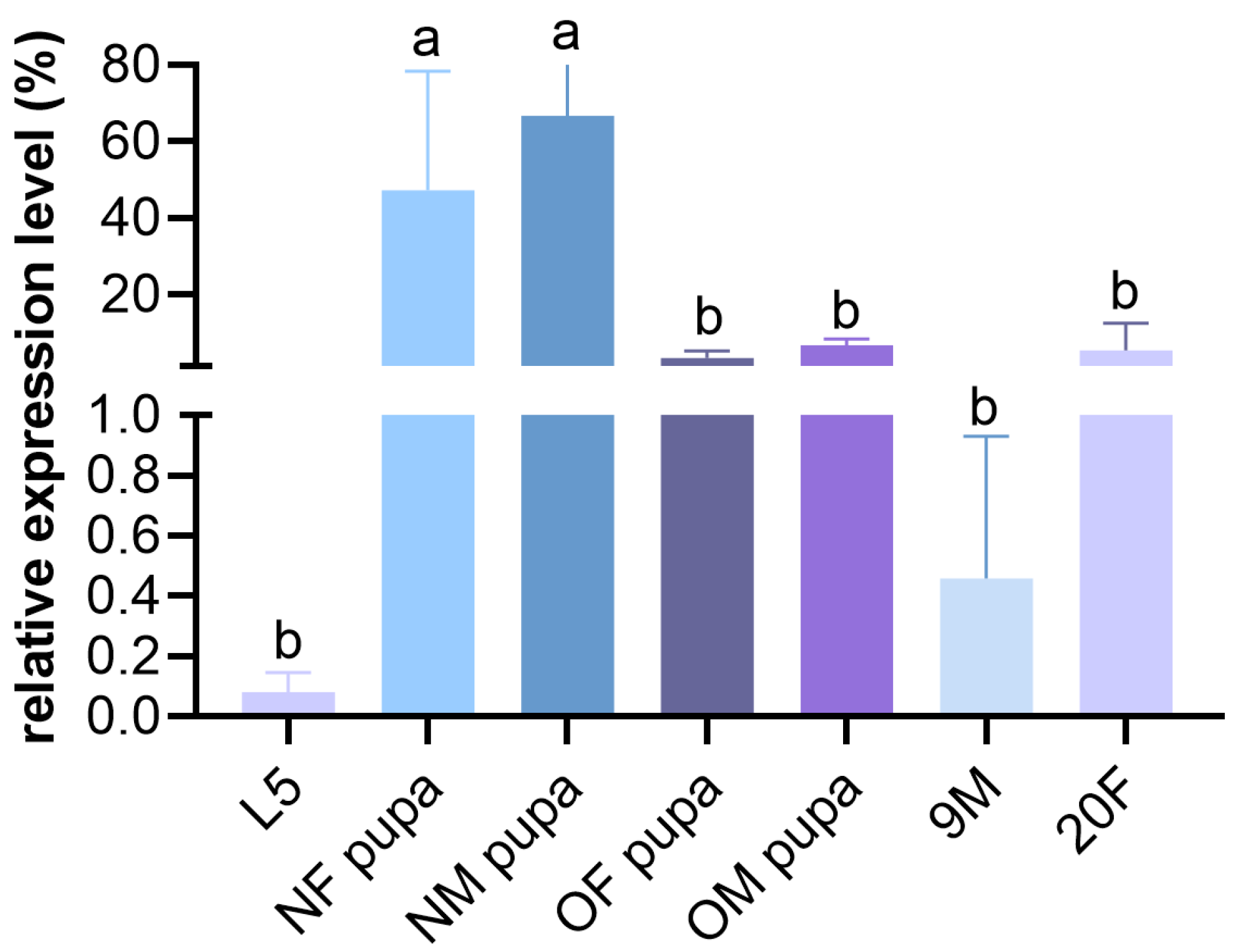
3.2. The Developmental Stage-Specific Expression of the MalCHS1 Gene

The developmental stage-specific expression analysis revealed that the MalCHS1 gene was expressed in all key stages of M. alternatus, with significantly lower expression levels in the fifth-instar larval stage than in other developmental stages. However, in the new male and female pupae stage, the expression level of the MalCHS1 gene was significantly upregulated (Figure 2). This stage-specific high expression pattern suggests that MalCHS1 may be involved in the degradation and synthesis of chitin from fifth-instar larvae to the pupal stage of M. alternatus, particularly in developmental processes such as new epidermal synthesis and old epidermal degradation.

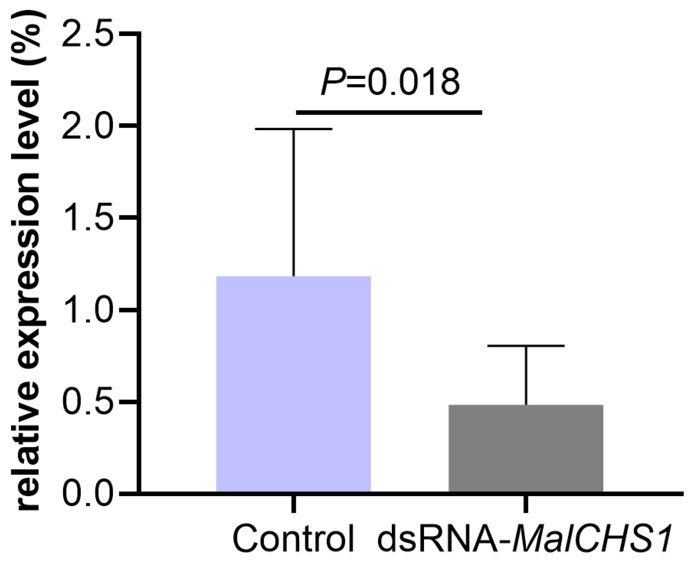
3.3. Changes in MalCHS1 Gene Expression in M. alternatus After RNAi

The RT-qPCR results showed that RNAi could effectively reduce the expression level of the CHS1 gene in the fifth-instar larvae of M. alternatus. As shown in Figure 3, the relative expression level of MalCHS1 in the intestine of larvae in the dsRNA-MalCHS1 treatment group was significantly reduced compared to the control group (injected with RNase-free water). This significant inhibitory effect not only indicates that RNAi is highly effective in targeting MalCHS1 transcripts but also provides a theoretical basis for the CHS1 gene as a feasible target for controlling M. alternatus.

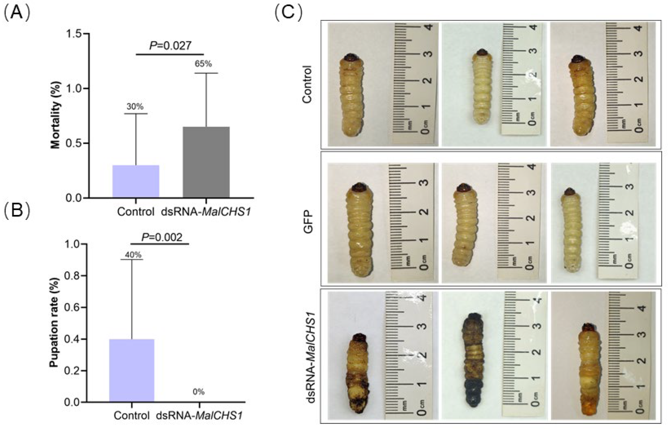
3.4. Phenotypic Changes in M. alternatus Larvae After RNAi

After the RNAi treatment, the pupation rate of M. alternatus larvae was 0% (Formula (2)), and the mortality was as high as 65% (Formula (1)), while the control group had a pupation rate of 40% (Formula (2)) and a mortality of 30% (Formula (1)) (Figure 4A,B). The dsRNA-MalCHS1 treatment group of larvae showed the phenomenon of larval death, with the body becoming soft, the abdomen becoming transparent or the tail turning black. The abdomen or chest appears swollen (Figure 4C), indicating that this gene may be involved in synthesizing the new epidermis and degrading the old epidermis in M. alternatus.

4. Discussion

In this study, we determined the role of CHS1 in the growth and development of M. alternatus through molecular biology and RNAi technology, combined with phenotypic observations. This study not only reveals the function of CHS1 in the M. alternatus, but also provides target genes for the subsequent control of M. alternatus using RNAi technology and provides a theoretical basis for novel biorational prevention and control tools [32].
Chitin Synthase 1 is an important enzyme in the formation of chitin in insects and widely regulates the development of the insect epidermis, trachea and other tissues [15]. Recent studies have shown that the silencing of this gene can significantly affect the physiological function of pests. For example, using RNAi against Panonychus citri McGregor also showed that dsRNA significantly inhibited the oviposition ability and egg hatching rate of P. citri [33]. The RNAi-mediated CHS1 gene knockout increased the formation of the cuticular appressorium in Nilaparvata lugens (Stal), thereby enhancing the sensitivity of N. lugens to Cordyceps sinensis, nitenpyram and nitenpyram [34]. Therefore, RNAi-mediated CHS1 gene silencing can provide theoretical support for the development of biorational and safe biopesticides and pest control target genes.
The experimental results showed that after the MalCHS1 gene silencing, the larva mortality during the critical period of pupation was greatly increased and the pupation rate was significantly reduced. At the same time, the larvae showed typical phenomena of chitin synthesis disorders, such as the softening of the body wall, a transparent abdomen, the localized swelling of the body and other morphological abnormalities. Similarly, after the RNAi treatment of the CHS1 gene of Culex pipiens, the larval survival rate decreased and molting disorders occurred [35]. In addition, the plant-mediated RNAi targeting of the CHS1 gene of M. avenae also resulted in significantly higher mortality and malformation rates [36]. Therefore, MalCHS1 may play an important role in the pupation process of M. alternatus, and its mechanism may be related to the synthesis and degradation of the epidermis. It may hinder larval growth and development by disrupting the metabolic processes of M. alternatus chitin, which in turn hinders larval growth and development and even leads to larval death.
Conventional chemical pesticides are prone to inducing pest resistance and causing environmental pollution [37]. Currently, dsRNA is becoming a new control method, which can efficiently and specifically interfere with the expression of target genes and, therefore, can specifically control pests [38]. Studies have shown that dsRNA pesticides for forest pests, including nanocarrier-based formulations, have broad applications and can be applied by foliar spraying or trunk injections, which is an environmentally friendly, residue-free and highly targeted insecticide compared with traditional chemical pesticides [39,40]. It not only improves the efficiency of insecticides but also conforms to the direction of the development of biorational prevention and control [25,41,42]. However, there are still challenges in developing RNA pesticides, including ensuring the effectiveness of the dsRNA under field conditions, determining the optimal dsRNA concentration for the target pest and avoiding off-target effects [40]. Therefore, this study not only provides target genes for pest control but also lays the theoretical foundation for the development of new biorational pesticides, such as nanocarrier RNA insecticides.

5. Conclusions

This study comprehensively investigated the functional role and expression dynamics of MalCHS1 in M. alternatus. The RNAi-mediated silencing of MalCHS1 induced severe developmental impairments, manifesting as a significantly reduced larval viability and the complete suppression of pupal transition. These phenotypic alterations unequivocally demonstrate the enzyme’s pivotal involvement in chitin biosynthesis and exoskeletal formation. The results of this study not only revealed the role of the CHS1 gene in M. alternatus but also that the current control methods for M. alternatus have problems such as poor targeting and environmental pollution, so the results of this study can provide new target genes for the control of M. alternatus and provide theoretical support for the biorational and safe prevention and control of pests.

Author Contributions

Conceptualization, W.Y. (Wanlin Ye) and M.W.; methodology, W.Y. (Wanlin Ye) and T.L.; software, T.L. and W.G.; validation, T.L., W.Y. (Wei Yu) and F.X.; formal analysis, M.W. and W.Y. (Wanlin Ye); investigation, T.L. and W.G.; data curation, W.Y. (Wanlin Ye); writing—original draft preparation, W.Y. (Wanlin Ye) and M.W.; writing—review and editing, T.L. and M.W.; visualization, W.Y. (Wanlin Ye); supervision, Y.G.; project administration, S.W. and Y.G.; funding acquisition, S.W. All authors have read and agreed to the published version of the manuscript.

Funding

This research was funded by the National Natural Science Foundation of China [grant numbers 32401582 and 32171805]; the Natural Science Foundation of Fujian Province, China [grant number 2024J08036]; and the Science and Technology Plan Project of Guizhou Province, China (grant number [2024]083).

Data Availability Statement

The data presented in this study are available on request from the corresponding author. The data are not publicly available due to privacy restrictions.

Conflicts of Interest

The authors declare no conflicts of interest.

References

  1. Li, H.; Zhao, X.; Qiao, H.; He, X.; Tan, J.; Hao, D. Comparative transcriptome analysis of the heat stress response in Monochamus alternatus Hope (Coleoptera: Cerambycidae). Front. Physiol. 2020, 10, 1568. [Google Scholar] [CrossRef] [PubMed]
  2. Zhao, B.G. Pine Wilt Disease in China. In Pine Wilt Disease; Zhao, B.G., Futai, K., Sutherland, J.R., Takeuchi, Y., Eds.; Springer: Tokyo, Japan, 2008. [Google Scholar] [CrossRef]
  3. Uchiyama, Y.; Futai, K. Assessing the role of asymptomatic infected trees in pine wilt disease spread in Japan—Insights from tree health monitoring. Forests 2025, 16, 583. [Google Scholar] [CrossRef]
  4. Linit, M.J. Nemtaode-vector relationships in the pine wilt disease system. J. Nematol. 1988, 20, 227–235. [Google Scholar]
  5. Wu, X.Q.; Yuan, W.M.; Tian, X.J.; Fan, B.; Fang, X.; Ye, J.R.; Ding, X.L. Specific and Functional Diversity of Endophytic Bacteria from Pine Wood Nematode Bursaphelenchus Xylophilus with Different Virulence. Int. J. Biol. Sci. 2013, 9, 34–44. [Google Scholar] [CrossRef]
  6. Wu, Q.; Chen, M.; Shi, H.; Yi, T.; Xu, G.; Wang, W.; Zhao, C.; Zhang, R. Algorithm for Detecting Trees Affected by Pine Wilt Disease in Complex Scenes Based on CNN-Transformer. Forests 2025, 16, 596. [Google Scholar] [CrossRef]
  7. Zhao, J.; Huang, J.; Yan, J.; Fang, G. Economic Loss of Pine Wood Nematode Disease in Mainland China from 1998 to 2017. Forests 2020, 11, 1042. [Google Scholar] [CrossRef]
  8. Chen, L.; Lu, W.; Lamont, B.A.O.; Liu, Y.; Wei, P.; Xue, W.; Xiong, Z.; Tang, L.; Wang, Y.A.O.; Wang, P.; et al. Modeling the distribution of pine wilt disease in China using the ensemble models MaxEnt and CLIMEX. Ecol. Evol. 2024, 14, e70277. [Google Scholar] [CrossRef]
  9. Wang, Z.; Yang, H.; Zhou, C.; Yang, W.J.; Jin, D.C.; Long, G.Y. Molecular cloning, expression, and functional analysis of the chitin synthase 1 gene and its two alternative splicing variants in the white-backed planthopper, Sogatella furcifera (Hemiptera: Delphacidae). Sci. Rep. 2019, 9, 1087. [Google Scholar] [CrossRef]
  10. Zhu, K.Y.; Merzendorfer, H.; Zhang, W.; Zhang, J.; Muthukrishnan, S. Biosynthesis, Turnover, and Functions of Chitin in Insects. Annu. Rev. Entomol. 2016, 61, 177–196. [Google Scholar] [CrossRef]
  11. Merzendorfer, H. The cellular basis of chitin synthesis in fungi and insects: Common principles and differences. Eur. J. Cell Biol. 2011, 90, 759–769. [Google Scholar] [CrossRef]
  12. Chen, D.D.; Wang, Z.B.; Wang, L.X.; Zhao, P.; Yun, C.H.; Bai, L. Structure, catalysis, chitin transport, and selective inhibition of chitin synthase. Nat. Commun. 2023, 14, 4776. [Google Scholar] [CrossRef] [PubMed]
  13. Tellam, R.L.; Eisemann, C. Chitin is only a minor component of the peritrophic matrix from larvae of Lucilia cuprina. Insect Biochem. Mol. Biol. 2000, 30, 1189–1201. [Google Scholar] [CrossRef] [PubMed]
  14. Li, L.; Wang, Y.Q.; Li, G.Y.; Song, Q.S.; Stanley, D.; Wei, S.J.; Zhu, J.Y. Genomic and transcriptomic analyses of chitin metabolism enzymes in Tenebrio molitor. Arch. Insect Biochem. Physiol. 2022, 111, e21950. [Google Scholar] [CrossRef] [PubMed]
  15. Arakane, Y.; Muthukrishnan, S.; Kramer, K.J.; Specht, C.A.; Tomoyasu, Y.; Lorenzen, M.D.; Kanost, M.; Beeman, R.W. The Tribolium chitin synthase genes TcCHS1 and TcCHS2 are specialized for synthesis of epidermal cuticle and midgut peritrophic matrix. Insect Mol. Biol. 2005, 14, 453–463. [Google Scholar] [CrossRef]
  16. Zimoch, L.; Merzendorfer, H. Immunolocalization of chitin synthase in the tobacco hornworm. Cell Tissue Res. 2002, 308, 287–297. [Google Scholar] [CrossRef]
  17. Arakane, Y.; Hogenkamp, D.G.; Zhu, Y.C.; Kramer, K.J.; Specht, C.A.; Beeman, R.W.; Kanost, M.R.; Muthukrishnan, S. Characterization of two chitin synthase genes of the red flour beetle, Tribolium castaneum, and alternate exon usage in one of the genes during development. Insect Biochem. Mol. Biol. 2004, 34, 291–304. [Google Scholar] [CrossRef]
  18. Merzendorfer, H.; Zimoch, L. Chitin metabolism in insects: Structure, function and regulation of chitin synthases and chitinases. J. Exp. Biol. 2003, 206, 4393–4412. [Google Scholar] [CrossRef]
  19. Scott, J.G.; Michel, K.; Bartholomay, L.C.; Siegfried, B.D.; Hunter, W.B.; Smagghe, G.; Zhu, K.Y.; Douglas, A.E. Towards the elements of successful insect RNAi. J. Insect Physiol. 2013, 59, 1212–1221. [Google Scholar] [CrossRef]
  20. Kyre, B.R.; Rieske, L.K. Using RNAi to silence heat shock protein has congeneric effects in North America’s Dendroctonus bark beetles. For. Ecol. Manag. 2022, 520, 120367. [Google Scholar] [CrossRef]
  21. Kyre, B.R.; Bentz, B.J.; Rieske, L.K. Susceptibility of mountain pine beetle (Dendroctonus ponderosae Hopkins) to gene silencing through RNAi provides potential as a novel management tool. For. Ecol. Manag. 2020, 473, 118322. [Google Scholar] [CrossRef]
  22. Ullah, F.; Gul, H.; Wang, X.; Ding, Q.; Said, F.; Gao, X.; Desneux, N.; Song, D. RNAi-Mediated Knockdown of Chitin Synthase 1 (CHS1) Gene Causes Mortality and Decreased Longevity and Fecundity in Aphis gossypii. Insects 2020, 11, 22. [Google Scholar] [CrossRef] [PubMed]
  23. Hayakawa, Y.; Kato, D.; Kamiya, K.; Minakuchi, C.; Miura, K. Chitin synthase 1 gene is crucial to antifungal host defense of the model beetle, Tribolium castaneum. J. Invertebr. Pathol. 2017, 143, 26–34. [Google Scholar] [CrossRef]
  24. Jiang, L.H.; Mu, L.L.; Jin, L.; Anjum, A.A.; Li, G.Q. RNAi for chitin synthase 1 rather than 2 causes growth delay and molting defect in Henosepilachna vigintioctopunctata. Pestic. Biochem. Physiol. 2021, 178, 104934. [Google Scholar] [CrossRef]
  25. Arjunan, N.; Thiruvengadam, V.; Sushil, S.N. Nanoparticle-mediated dsRNA delivery for precision insect pest control: A comprehensive review. Mol. Biol. Rep. 2024, 51, 355. [Google Scholar] [CrossRef]
  26. Back, M.A.; Bonifácio, L.; Inácio, M.L.; Mota, M.; Boa, E. Pine wilt disease: A global threat to forestry. Plant Pathol. 2024, 73, 1026–1041. [Google Scholar] [CrossRef]
  27. Tamura, K.; Nei, M.; Kumar, S. Prospects for inferring very large phylogenies by using the neighbor-joining method. Proc. Natl. Acad. Sci. USA 2004, 101, 11030–11035. [Google Scholar] [CrossRef]
  28. Chen, C.; Chen, H.; Zhang, Y.; Thomas, H.R.; Frank, M.H.; He, Y.; Xia, R. TBtools: An Integrative Toolkit Developed for Interactive Analyses of Big Biological Data. Mol. Plant 2020, 13, 1194–1202. [Google Scholar] [CrossRef]
  29. Rychlik, W. OLIGO 7 primer analysis software. Methods Mol. Biol. 2007, 402, 35–60. [Google Scholar] [CrossRef]
  30. Park, H.M. Comparing Group Means: T-Tests and One-Way ANOVA Using STATA, SAS, R, and SPSS [Working Paper]. Indiana University, Center for Statistical and Mathematical Computing. 2009. Available online: https://hdl.handle.net/2022/19735 (accessed on 15 January 2024).
  31. Schmittgen, T.D.; Livak, K.J. Analyzing real-time PCR data by the comparative CT method. Nat. Protoc. 2008, 3, 1101–1108. [Google Scholar] [CrossRef]
  32. Dhandapani, R.K.; Gurusamy, D.; Duan, J.J.; Palli, S.R. RNAi for management of Asian long-horned beetle, Anoplophora glabripennis: Identification of target genes. J. Pest Sci. 2020, 93, 823–832. [Google Scholar] [CrossRef]
  33. Ali, M.W.; Khan, M.M.; Song, F.; Wu, L.; He, L.; Wang, Z.; Zhang, Z.y.; Zhang, H.; Jiang, Y. RNA Interference-Based Silencing of the Chitin Synthase 1 Gene for Reproductive and Developmental Disruptions in Panonychus citri. Insects 2020, 11, 786. [Google Scholar] [CrossRef] [PubMed]
  34. Sun, S.; Sun, M.; Du, P.; Niu, H.; Zhang, Z.; Zhao, D.; Liu, X.; Guo, H. Protective roles of chitin synthase gene 1 in Nilaparvata lugens against Cordyceps javanica and insecticides. Pestic. Biochem. Physiol. 2025, 209, 106324. [Google Scholar] [CrossRef] [PubMed]
  35. Yang, X.; Xu, Y.; Yin, Q.; Zhang, H.; Yin, H.; Sun, Y.; Ma, L.; Zhou, D.; Shen, B. Physiological characterization of chitin synthase A responsible for the biosynthesis of cuticle chitin in Culex pipiens pallens (Diptera: Culicidae). Parasites Vectors 2021, 14, 234. [Google Scholar] [CrossRef]
  36. Zhao, Y.; Sui, X.; Xu, L.; Liu, G.; Lu, L.; You, M.; Xie, C.; Li, B.; Ni, Z.; Liang, R. Plant-mediated RNAi of grain aphid CHS1 gene confers common wheat resistance against aphids. Pest Manag. Sci. 2018, 74, 2754–2760. [Google Scholar] [CrossRef]
  37. Hussain, T.; Aksoy, E.; Çalışkan, M.E.; Bakhsh, A. Transgenic potato lines expressing hairpin RNAi construct of molting-associated EcR gene exhibit enhanced resistance against Colorado potato beetle (Leptinotarsa decemlineata, Say). Transgenic Res. 2019, 28, 151–164. [Google Scholar] [CrossRef]
  38. Vogel, E.; Santos, D.; Mingels, L.; Verdonckt, T.W.; Broeck, J.V. RNA Interference in Insects: Protecting Beneficials and Controlling Pests. Front. Physiol. 2019, 9, 1912. [Google Scholar] [CrossRef]
  39. Joga, M.R.; Mogilicherla, K.; Smagghe, G.; Roy, A. RNA Interference-Based Forest Protection Products (FPPs) Against Wood-Boring Coleopterans: Hope or Hype? Front. Plant Sci. 2021, 12, 733608. [Google Scholar] [CrossRef]
  40. Singewar, K.; Fladung, M. Double-stranded RNA (dsRNA) technology to control forest insect pests and fungal pathogens: Challenges and opportunities. Funct. Integr. Genom. 2023, 23, 185. [Google Scholar] [CrossRef]
  41. Yan, S.A.O.; Ren, B.Y.; Shen, J.A.O. Nanoparticle-mediated double-stranded RNA delivery system: A promising approach for sustainable pest management. Insect Sci. 2021, 28, 21–34. [Google Scholar] [CrossRef]
  42. Hough, J.; Howard, J.D.; Brown, S.; Portwood, D.E.; Kilby, P.M.; Dickman, M.J. Strategies for the production of dsRNA biocontrols as alternatives to chemical pesticides. Front. Bioeng. Biotechnol. 2022, 10, 980592. [Google Scholar] [CrossRef]
Figure 1. Phylogenetic analysis, conserved motif analysis and domain analysis of chitin synthase gene (CHS) in Monochamus alternatus and related species. (A) Neighbor-Joining phylogenetic tree of CHS protein sequences from M. alternatus (highlighted in red) and other arthropods. Bootstrap values (>50%) are shown at nodes. GenBank accession numbers are listed for each sequence. (B) Motif architecture predicted by MEME, showing 10 conserved motifs (color-coded boxes) across 11 arthropod species. Motif positions are scaled to protein length (bottom axis: 0–1600 aa). (C) Schematic representation of conserved domains in CHS proteins. Glyco_transf_CTA_type (chitin synthase catalytic domain) and BcsA superfamily (transmembrane domain) are annotated. Scale bar indicates amino acid position (0–1600).
Forests 16 00922 g001
Figure 2. Expression patterns of CHS1 at different developmental stages in Monochamus alternatus. L5: fifth-instar larvae; NF: new female pupae; NM: new male pupae; OF: mature female pupae; OM: mature male pupae; 9M: sexually mature male insect; and 20F: sexually mature female. Standardized as β-actin (one-way ANOVA with Tukey’s post hoc test); a and b marked with different letters indicate statistically significant differences.
Forests 16 00922 g002
Figure 3. The effect of the dsRNA injection on the mRNA expression level of the CHS1 gene in Monochamus alternatus. Error bars represent SEM; asterisks indicate significance (the independent samples t-test).
Forests 16 00922 g003
Figure 4. The effect of the RNAi on the mortality, pupation rate and phenotypic changes in Monochamus alternatus larvae. (A) The mortality of the larvae after the injection of dsRNA-MalCHS1. (B) The pupation rate after the injection of dsRNA-MalCHS1. (C) Phenotypic changes in larvae after injections. Unlike the control and GFP groups, the treatment group showed morphological abnormalities, such as the softening of the body wall, the transparency of the abdomen and the swelling of the body segments. Asterisks indicate significance (independent samples t-test).
Forests 16 00922 g004
Table 1. Primers used in the present study.
PrimerSequence (5′-3′)
PCRMalCHS1-FCAAAGTATGGGAATGAACAG
MalCHS1-RTCTTTCTGTGTCTGATCTTC
RT-qPCRMalCHS1-FCAAACTTAACGCCAACCCTG
MalCHS1-RTTTCTCTGGGCCACATTGTT
β-actin-FGTTGCCCTCGACTTCGAACA
β-actin-RACGGATATCAACGTCGCACT
RNAiT7 promoterTAATACGACTCACTATAG
Disclaimer/Publisher’s Note: The statements, opinions and data contained in all publications are solely those of the individual author(s) and contributor(s) and not of MDPI and/or the editor(s). MDPI and/or the editor(s) disclaim responsibility for any injury to people or property resulting from any ideas, methods, instructions or products referred to in the content.

