Article

Unlocking Therapeutic Potential of Novel Thieno-Oxazepine Hybrids as Multi-Target Inhibitors of AChE/BChE and Evaluation Against Alzheimer’s Disease: In Vivo, In Vitro, Histopathological, and Docking Studies

by
Khulood H. Oudah
1,
Mazin A. A. Najm
1,
Triveena M. Ramsis
2,
Maha A. Ebrahim
3,
Nirvana A. Gohar
4,
Karema Abu-Elfotuh
5,6,
Ehsan Khedre Mohamed
7,
Ahmed M. E. Hamdan
8,
Amira M. Hamdan
9,
Reema Almotairi
10,
Shaimaa R. Abdelmohsen
11,
Khaled Ragab Abdelhakim
12,
Abdou Mohammed Ahmed Elsharkawy
13 and
Eman A. Fayed
3,*
1
Department of Pharmacy, Mazaya University College, Nasiriyah 64001, Iraq
2
Department of Pharmaceutical Chemistry, Faculty of Pharmacy, Sinai University, Kantara Branch, Ismailia 41636, Egypt
3
Department of Pharmaceutical Organic Chemistry, Faculty of Pharmacy (Girls), Al-Azhar University, Cairo 11754, Egypt
4
Department of Pharmaceutical Organic Chemistry, Faculty of Pharmacy, Modern University for Technology and Information (MTI), Cairo 11571, Egypt
5
Department of Clinical Pharmacy, Faculty of Pharmacy (Girls), Al-Azhar University, Cairo 11754, Egypt
6
College of Pharmacy, Al-Ayen Iraqi University (AUIQ), An Nasiriyah 64004, Iraq
7
Department of Biochemistry, Egyptian Drug Authority (EDA), Formerly National Organization of Drug Control and Research (NODCAR), Giza 12654, Egypt
8
Department of Pharmacy Practice, Faculty of Pharmacy, University of Tabuk, Tabuk 71491, Saudi Arabia
9
Oceanography Department, Faculty of Science, Alexandria University, Alexandria 21515, Egypt
10
Department of Medical Laboratory Technology, Faculty of Applied Medical Sciences, University of Tabuk, Tabuk 71491, Saudi Arabia
11
Department of Anatomy and Embryology, Faculty of Medicine, Al-Azhar University, Cairo 11754, Egypt
12
Department of Histology, Misr University for Science and Technology, Giza 12566, Egypt
13
Anatomy Department, Faculty of Medicine, Al-Azhar University, Cairo 11754, Egypt
*
Author to whom correspondence should be addressed.
Pharmaceuticals 2025, 18(8), 1214; https://doi.org/10.3390/ph18081214 (registering DOI)
Submission received: 18 July 2025 / Revised: 5 August 2025 / Accepted: 14 August 2025 / Published: 17 August 2025
(This article belongs to the Special Issue Heterocyclic Chemistry in Modern Drug Development)

Abstract

Background: Alzheimer’s disease (AD) is largely linked with oxidative stress, the accumulation of amyloid-β plaques, and hyperphosphorylated τ-protein aggregation. Alterations in dopaminergic and serotonergic neurotransmission have also been implicated in various AD-related symptoms. Methods: To explore new therapeutic agents, a series of bicyclic and tricyclic thieno-oxazepine derivatives were synthesized as potential acetylcholinesterase (AChE) and butyrylcholinesterase (BChE) inhibitors. The resultant compounds were purified via HPLC and characterized using spectral analysis techniques. Histopathological examinations, other antioxidants, and anti-inflammatory biomarkers were evaluated, and in silico ADMET calculations were performed for synthetic hybrids. Molecular docking was utilized to validate the new drugs’ binding mechanisms. Results: The most powerful AChE inhibitors were 14 and 16, with respective values of IC50 equal to 0.39 and 0.76 µM. Derivative 15 demonstrated remarkable BChE-inhibitory efficacy, on par with tacrine, with IC50 values of 0.70 µM. Hybrids 13 and 15 showed greater selectivity towards BChE, despite substantial inhibition of AChE. Compounds 13 and 15 reduced escape latency and raised residence time, with almost equal activity to donepezil. Conclusions: According to these findings, the designed hybrids constitute multipotent lead compounds that could be used in the creation of novel anti-AD medications.

Graphical Abstract

1. Introduction

Cholinergic and glutamatergic impairments are connected to a variety of mechanisms that contribute to cognitive loss in Alzheimer’s disease, a relatively complicated neurodegenerative illness. These elements include psychosis, dissatisfaction or violence, mood disorders (such as anxiety and depression), and deficiencies in facial expressions and emotional communication [1,2]. Previous biochemical studies have identified oxidative stress, the accumulation of hyperphosphorylated τ-protein, and amyloid-β (Aβ) formation as the key pathology of Alzheimer’s disease (AD) [3,4,5,6]. A decline in acetylcholine (ACh) levels within brain areas, accountable for memory and learning, results in the degeneration of cholinergic neurons, further driving the progression of AD [7,8,9,10,11,12]. ACh is increased in the synaptic cleft by AChE inhibitors, which prevent its breakdown [13,14,15,16,17,18,19].
Apoptotic cascades are regulated by the Bcl-2 family; although BAX stimulates apoptosis, Bcl-2 suppresses it [20,21,22,23,24]. A substantial change in the ratio of BAX (pro-apoptotic) to Bcl-2 (anti-apoptotic) is brought about by the overexpression of BAX and the decrease in Bcl-2 gene expression in AD [25,26,27,28,29]. The diffuse distribution of Aβ throughout the brain parenchyma causes oxidative stress, neuroinflammation, and neuronal apoptosis/necrosis [30,31,32]. Neurodegeneration is triggered by the stimulation of the NLRP3 inflammasome complex, which is initiated by an inflammatory cascade following the interaction of amyloid-β (Aβ) with Toll-like receptor 4 (TLR4) [33,34,35,36,37,38,39]. Treatment approaches that suppress GSK3β and/or increase Wnt/β-catenin signaling in neural cells can reduce the negative impact of Aβ and help neurogenesis in AD patients [40,41,42]. AChE inhibitor medications, such as donepezil, [43] rivastigmine, [44] memantine, [45] and galantamine, [46] are used to manage AD (Figure 1) [47].
Growing evidence suggests that BChE plays a critical role in regulating acetylcholine (ACh) in the brain throughout the progression of AD [48,49,50]. In advanced phases of AD, brain levels of BChE can increase to approximately 165% of normal, whereas AChE levels decline to about 55–67% of typical values [51]. Notably, BChE inhibition produces no adverse consequences. The creation of selective and powerful BChE inhibitors, which may increase ACh levels in the brain while drastically reducing adverse effects on the peripheral nervous system, represents a significant advancement in this regard [52,53,54,55].
Due to their abundant natural occurrence, heterocyclic compounds are used extensively in the domains of biology and synthesis [56,57,58,59,60]. The heterocyclic sulfur-containing moiety, thiophene, exhibits specific biological characteristics, including cytotoxic, antiviral, antibacterial, and antifungal effects [61,62,63,64,65,66,67]. Similarly, aryl-fused 1,4-oxazepines are structural elements of biologically active compounds that are utilized in medications such as analgesics, antihistaminics, psychoneurotics, and for the management of AD-related memory impairment [68,69].
As a therapeutic method to slow the progression of AD against AlCl3-induced AD, we implemented a rat model to investigate the impact of our newly synthesized compounds on behavioral parameters. This is because the pathological process of AD involves multiple mechanisms, all of which lead to increased inflammation and oxidative stress.
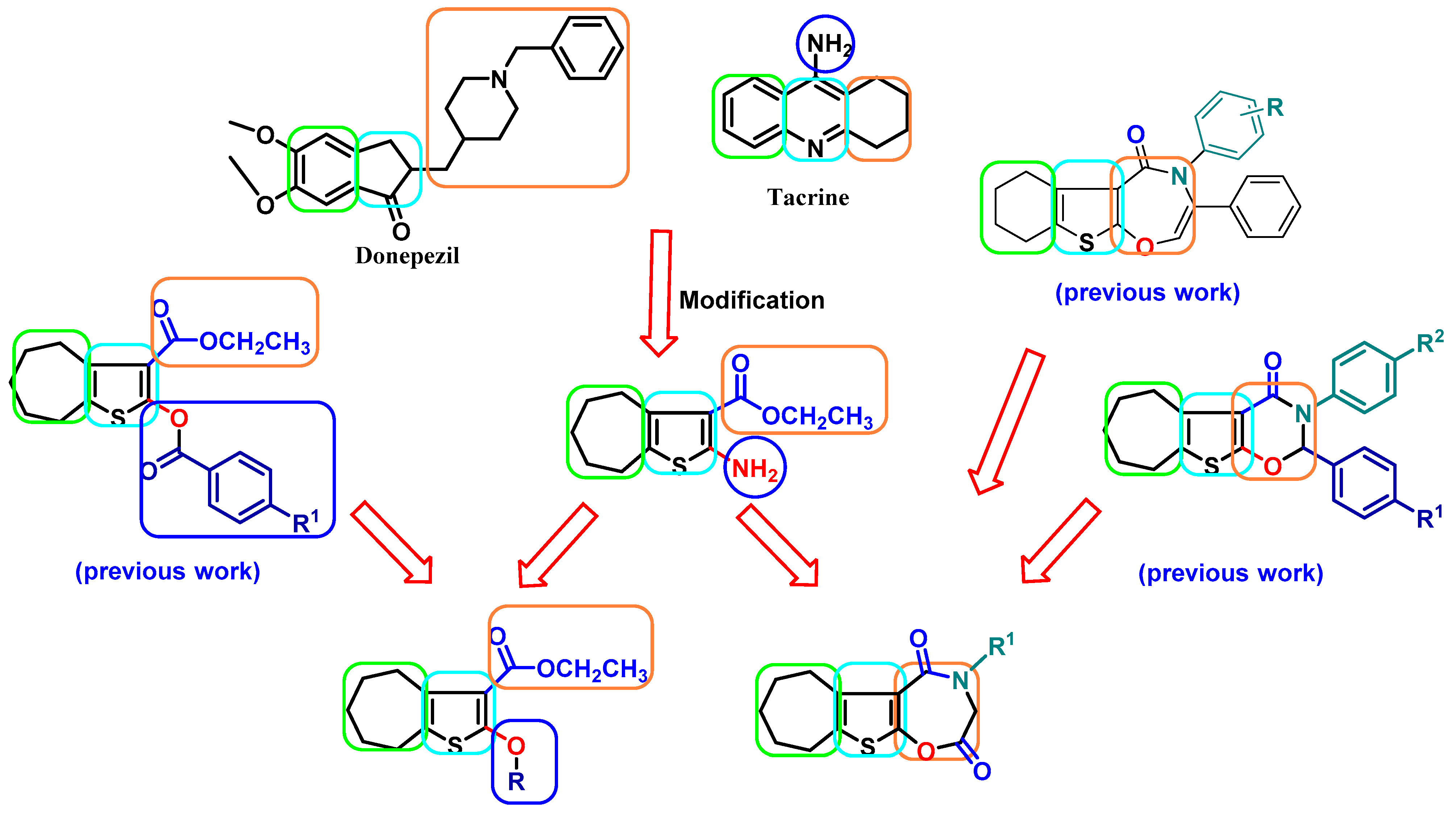
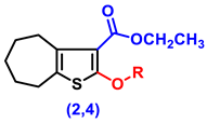
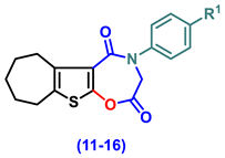
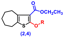
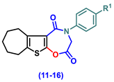
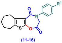
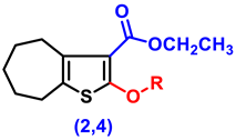
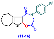
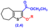
As an extension of our previous work on the creation of cyclohepta[b]thiophene-containing compounds, the aim of this article is to create new lead compounds that include cyclohepta-thieno-oxazepine hybrids that could be applied as AChE and BChE inhibitors to treat AD [70,71,72]. To discover novel candidates with dual inhibitory activity against AChE and BChE, we synthesized a new cyclohepta[b]thiophene derivatives which are bicyclic and tricyclic (Figure 2). These compounds were assessed for their effects on multiple pathways, including cognitive function (learning and memory), antioxidant capacity, suppression of neuroinflammation, reduction in β-amyloid accumulation, modulation of apoptotic markers, improvement of brain-derived neurotrophic factor (BDNF) levels, and regulation of BAX/Bcl-2 and Wnt/β-catenin signaling paths. The modes of action of inhibitors of AChE and BChE were also identified among the most effective hybrids. Additional lead optimization may be aided by the outcomes of the accomplished docking studies and the ADMET property predictions.

2. Results and Discussion

2.1. Chemistry

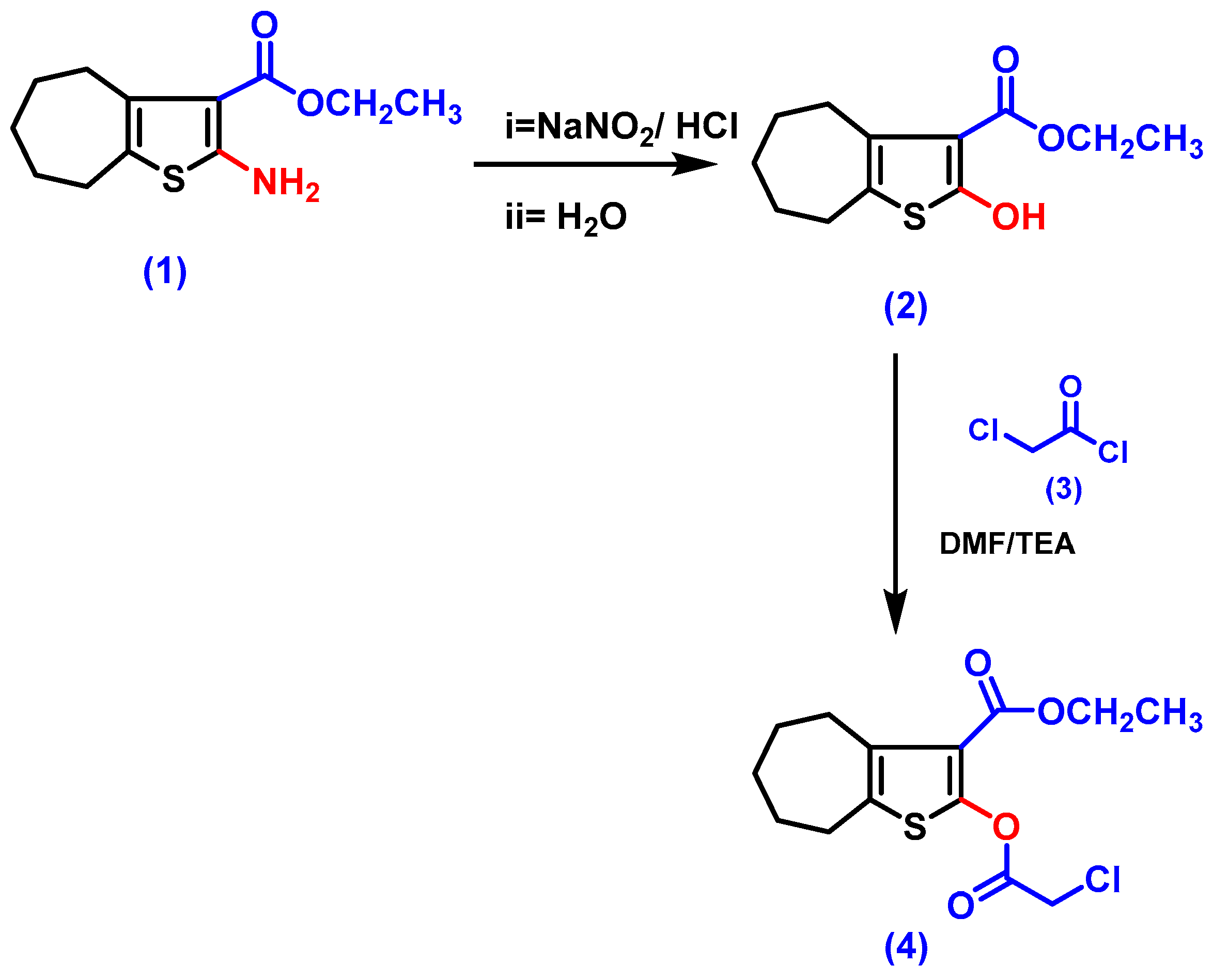
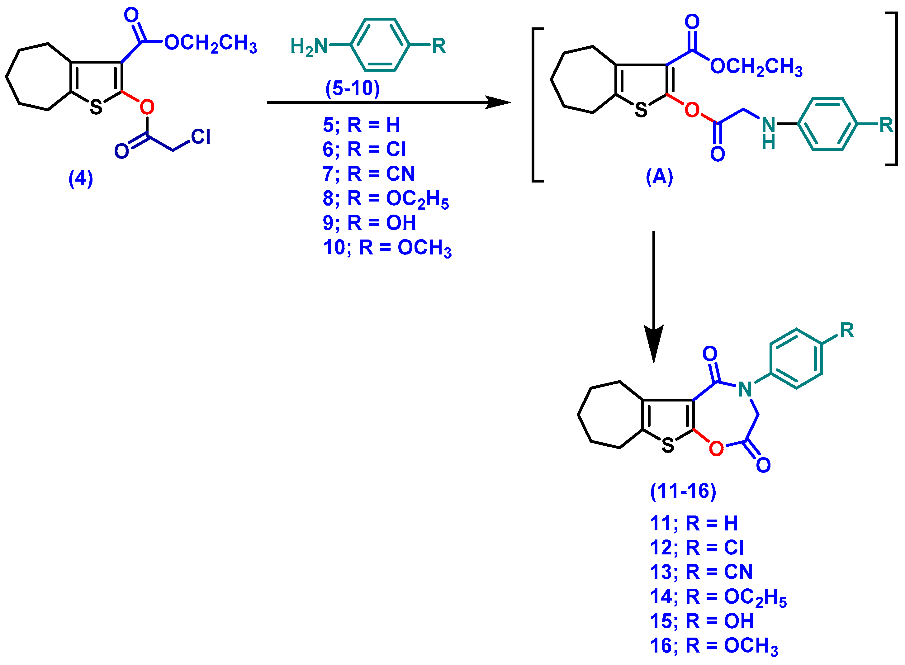
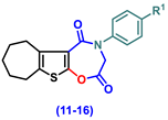
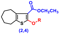
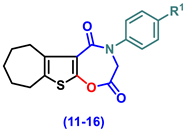
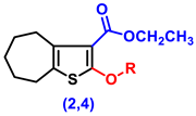
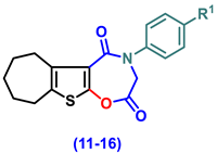
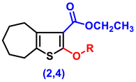
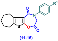
The study discusses newly made bi- and tricyclic cyclohepta[b]thiophene-oxazepine hybrids, shown in Scheme 1 and Scheme 2.
The target hybrids were synthesized following the outlined preparations. Derivative 1 was produced via the Gewald reaction [73]. Compound 2 was synthesized from 1 as reported [74].
Ester 4 was synthesized by reacting the 2-hydroxy thiophene derivative 2 with chloroacetylchloride [75,76].
The 1H NMR spectrum of hybrid 4 represented a quartet at δ 4.35 ppm due to 2Hs of -CH2-CH3) and a singlet at δ 4.25 ppm corresponding to 2Hs of CO-CH2-Cl, while the 13C NMR spectrum showed a signal at δ 166 ppm, indicating (CO-CH2-Cl), and another one at δ 163 ppm that indicates (CO-OCH2CH3).
Different aniline derivatives were allowed to react with thieno-chloroacetate 4, resulting in 1,4-oxazepine-2,5-diones 914 [77,78]. The structure of 11 was evidenced by the 1H NMR, which displayed a singlet at δ 4.7 ppm matching with 2Hs of oxazepine CH2 and multiplets at δ 7.0–7.3 ppm, indicating phenyl protons.
Furthermore, the 1H NMR spectrum of hybrid 12 demonstrated two doublets of the phenyl ring at δ 7.35 and 7.28 ppm and a singlet at δ 4.7 ppm due to the 2Hs of oxazepine CH2.
The 1H NMR of compound 13 revealed two doublets at δ 7.5 and 7.5 ppm relative to the para-substituted phenyl hydrogens and a singlet at δ 4.7 due to oxazine protons, while the 13C NMR spectrum displayed two signals at δ 47.0 and 118.0 ppm that refer to oxazine-CH and CN, respectively.
The 1H NMR spectrum of compound 14 displayed a singlet at δ 4.7 ppm, corresponding to oxazine protons; a quartet at δ 4.0 ppm, owing to -CH2-CH3; and a triplet at δ 2.8 ppm due to CH2-CH3. Meanwhile, the 13C NMR spectrum showed a signal at δ 47.00 ppm, which refers to oxazine CH2, and signals at 63.6 and 14.6 ppm, indicating -OCH2CH3 carbons.
The 1H NMR spectrum of derivative 15 displayed a singlet at δ 7.75 ppm due to the OH proton, a doublet of doublet signals at δ 7.21 and 6.80 ppm due to the para-substituted phenyl Hs, and a singlet signal at δ 4.77 ppm due to the oxazine protons.
The 1H NMR spectra of derivative 16 revealed a singlet signal at δ 3.79 ppm due to the OCH3 proton, a doublet of doublet signals at δ 7.29 and 6.87 ppm due to the para-substituted phenyl hydrogens, and a singlet signal at δ 4.77 ppm due to the oxazine hydrogens. However, the 13C NMR spectrum showed two carbonyl carbon signals at δ 164 and 163 ppm and a singlet at δ 47.00 ppm, referring to oxazine CH2, and a signal at δ 54.30, corresponding to OCH3 carbon.

2.2. Biology

2.2.1. Impact on Behavioral Assessments in AlCl3-Induced AD

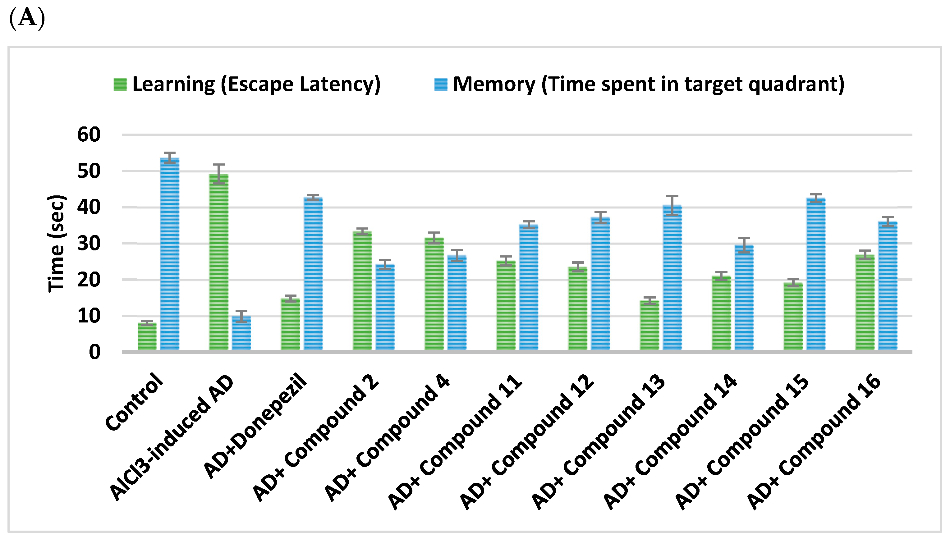
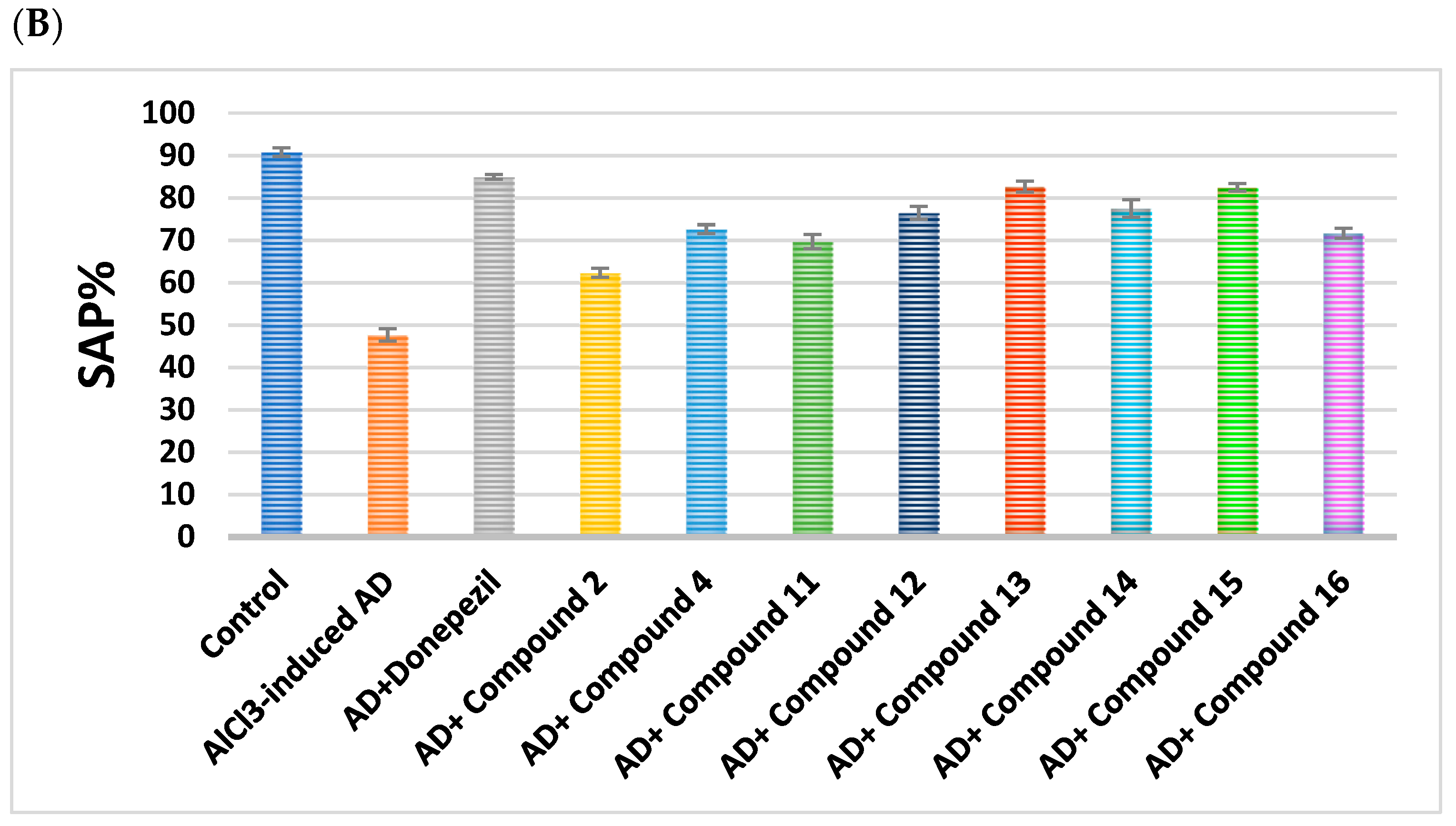
Donepezil served as a control medication for all behavioral tests (Figure 3). While compounds 11, 12, 14, and 16 produced moderate decreases in escape latency that fluctuated between 57.3% and 45.4%, compounds 13 and 15 displayed significant reductions in escape latency of 71.2% and 61%, respectively. Mild escape latency reductions of 32.2% and 35.9% were seen in hybrids 2 and 4, respectively.
While hybrids 11, 12, and 16 demonstrated a modest increase in quadrant resident duration by 3.5, 3.7, and 3.6 times, respectively, the delivery of hybrids 13 and 15 raised the residence time by about 4.1 and 4.3 times, displaying almost equal activity to donepezil.
Equipotency to donepezil was confirmed by the administration of conjugates 13 and 15, which displayed increases in SAP% of almost 35% and 34.8%, respectively. Furthermore, SAP% increased comparatively high in 4, 12, 14, and 16, ranging from 24% to 29.8%. A marginal rise in SAP% of 14.6% and 22% was noted by compounds 2 and 11, respectively (Table 1 and Table 2).

2.2.2. Effect on BACE1 and Amyloid-β

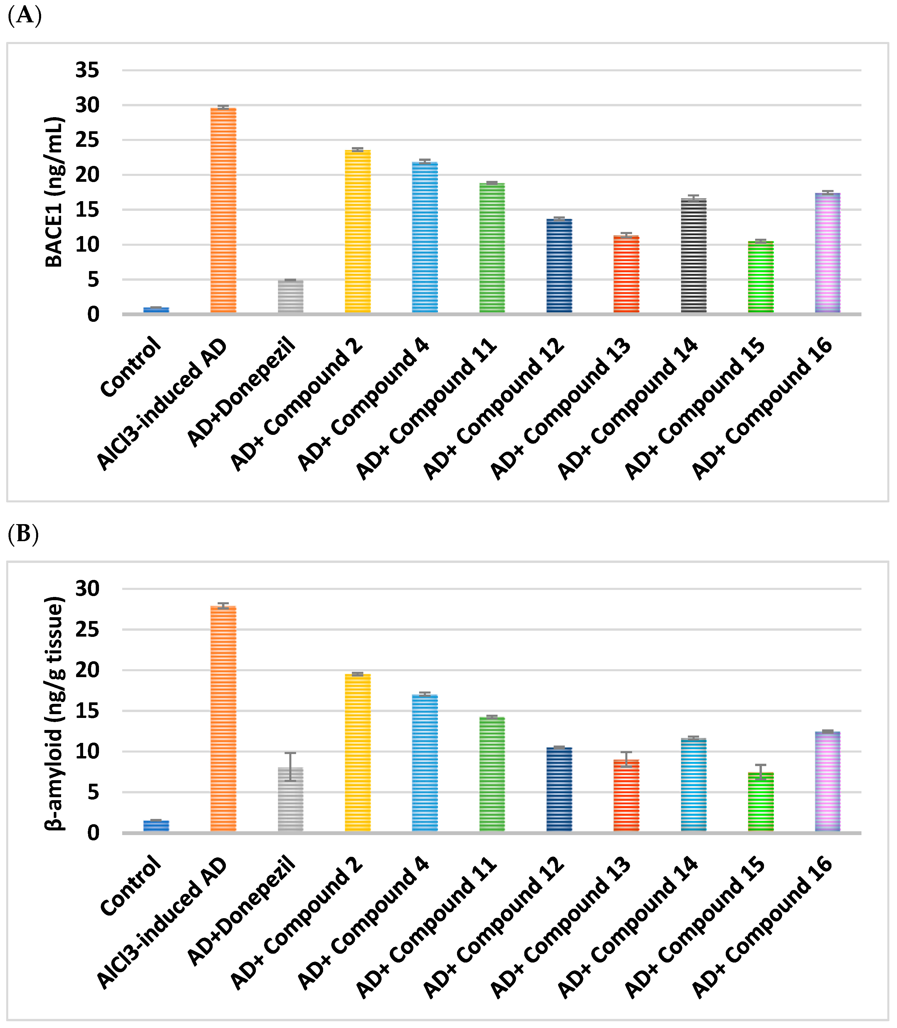
β-Amyloid levels were dramatically reduced by 73.2% after intervention with compound 15, demonstrating its superiority over donepezil (which reduced these levels by 70.9%). β-amyloid levels were significantly decreased by 62.3% and 67.6%, respectively, following administration of hybrids 12 and 13, fostering equipotency to donepezil. Comparing compounds 11, 16, and 14 to donepezil, the former illustrated a significant reduction in β-amyloid by 48.9%, 55.4%, and 58.4%, respectively, whereas compounds 2 and 4 displayed a moderate reduction in β-amyloid by 30.2% and 38.9%, respectively (Figure 4).
Simultaneously, hybrids 12, 13, and 15 disclosed a noteworthy drop in BACE1 levels of 53.9%, 61.7%, and 64.6%, respectively. BACE1 levels were found to be moderately reduced by 36.5%, 41.2%, and 43.8% after treatment with compounds 11, 5, and 14, whereas BACE1 levels were minimally reduced by 20.4% and 26.2% after treatment with compounds 2 and 4, respectively (Table 3 and Table 4).

2.2.3. Impact on Brain Neurotransmitters

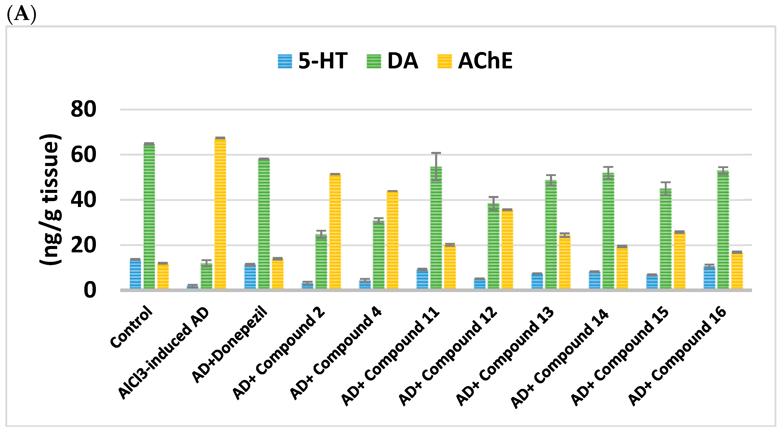
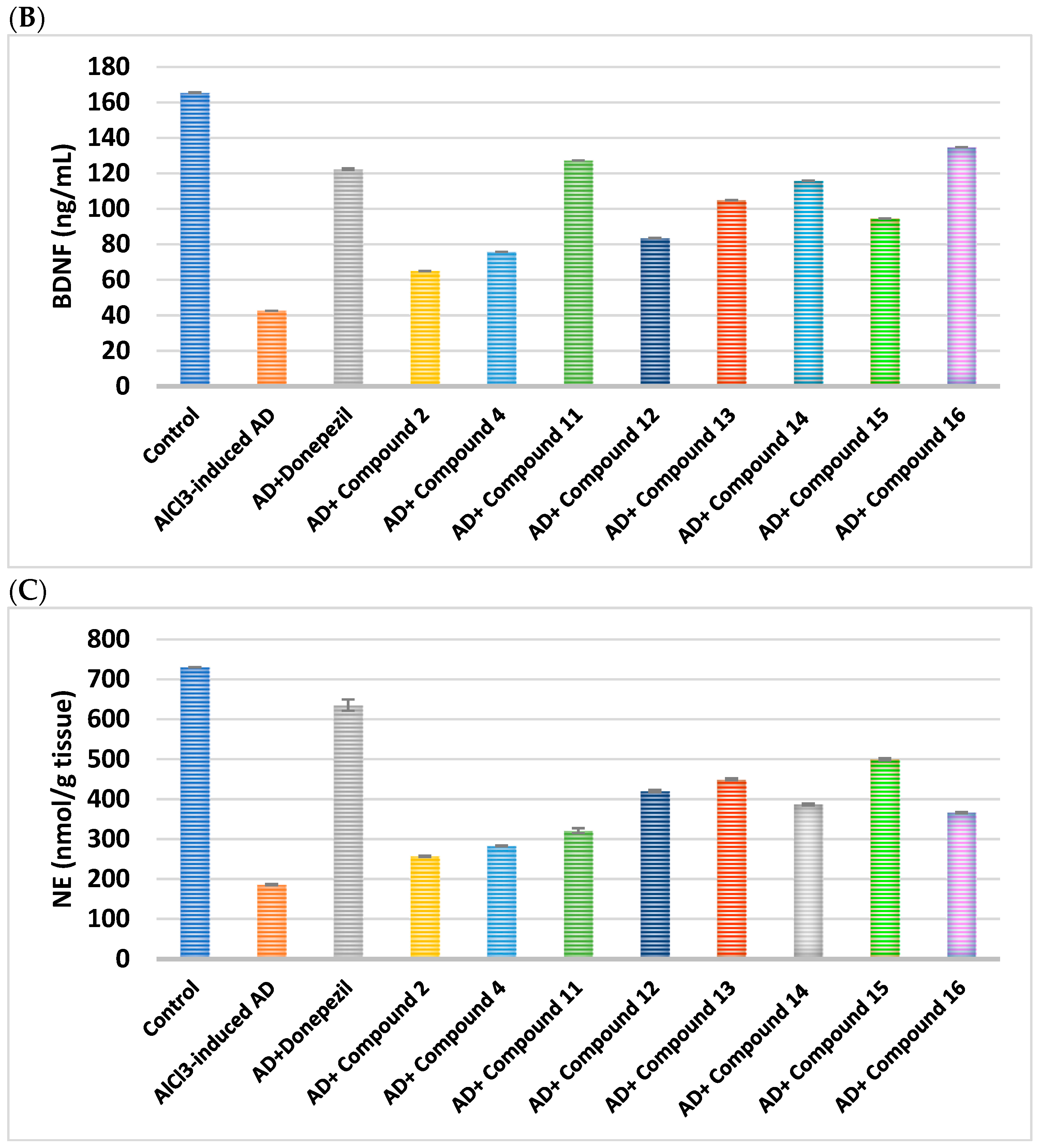
When compared to donepezil, compounds 12, 13, and 15 moderately raised DA levels by 3.2, 4.1, and 3.8-fold, while compounds 11, 14, and 16 produced an extremely high elevation of 4.6, 4.3, and 4.4-fold.
Additionally, compounds 11, 14, and 16 demonstrated a moderate rise in the level of NE by 1.7, 2.1, and 2 times, respectively, in contrast to donepezil; nevertheless, compounds 12, 13, and 15 effectively enhanced the amount of NE by 2.2, 2.4, and 2.7, respectively (Figure 5).
Conjugates 12, 13, and 15 elevated 5-HT levels moderately by 2.8-, 4.1-, and 3.7-fold, respectively, whereas hybrids 11, 14, and 16 markedly enhanced 5-HT levels by 5.0-, 4.5-, and 5.9-fold, respectively. Additionally, as compared to donepezil, hybrids 2 and 4 showed a slight improvement in 5-HT levels of 1.7- and 2.4-fold, respectively.
While compounds 13 and 14 comparatively raised BDNF levels by 2.4- and 2.7-fold, respectively, compounds 11 and 16 demonstrated greater elevation of BDNF levels compared to donepezil by 3- and 3.1-fold, respectively. When compared to donepezil, hybrids 12 and 15 demonstrated an acceptable increase of 2- and 2.2-fold, respectively. More importantly, a significant reduction in AChE was seen by 70.1%, 71.2%, and 75%, respectively, following the administration of compounds 11, 14, and 16. AChE was considerably reduced by compounds 12, 13, and 15 by 47%, 63.8%, and 61.8%, respectively. Compounds 2 and 4 showed a moderate reduction in AChE levels by 23.6% and 34.8%, respectively, in contrast to donepezil (AChE reduction of 79.3%) (Table 5 and Table 6).

2.2.4. Impact on Neuroinflammatory Biomarkers

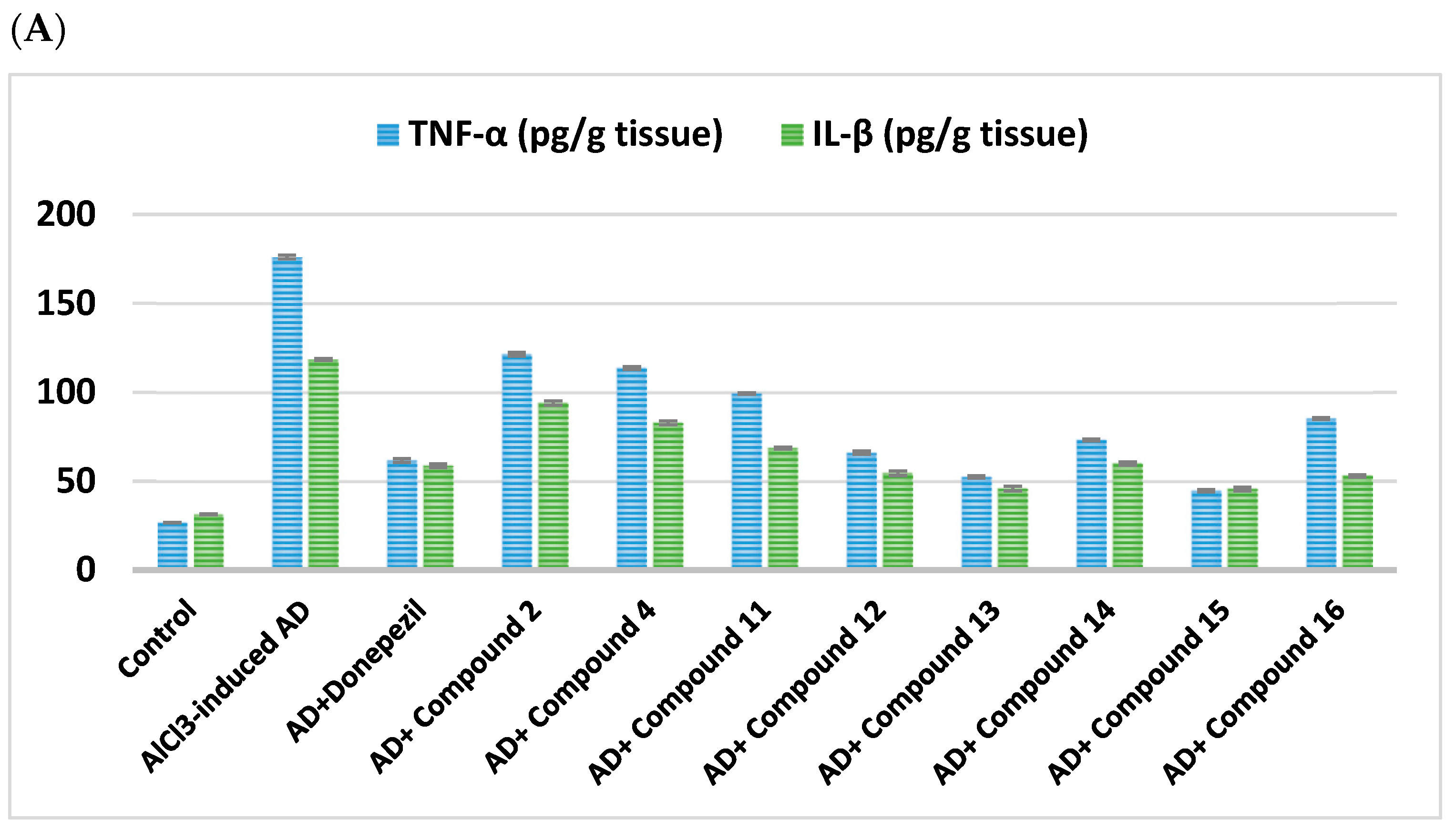
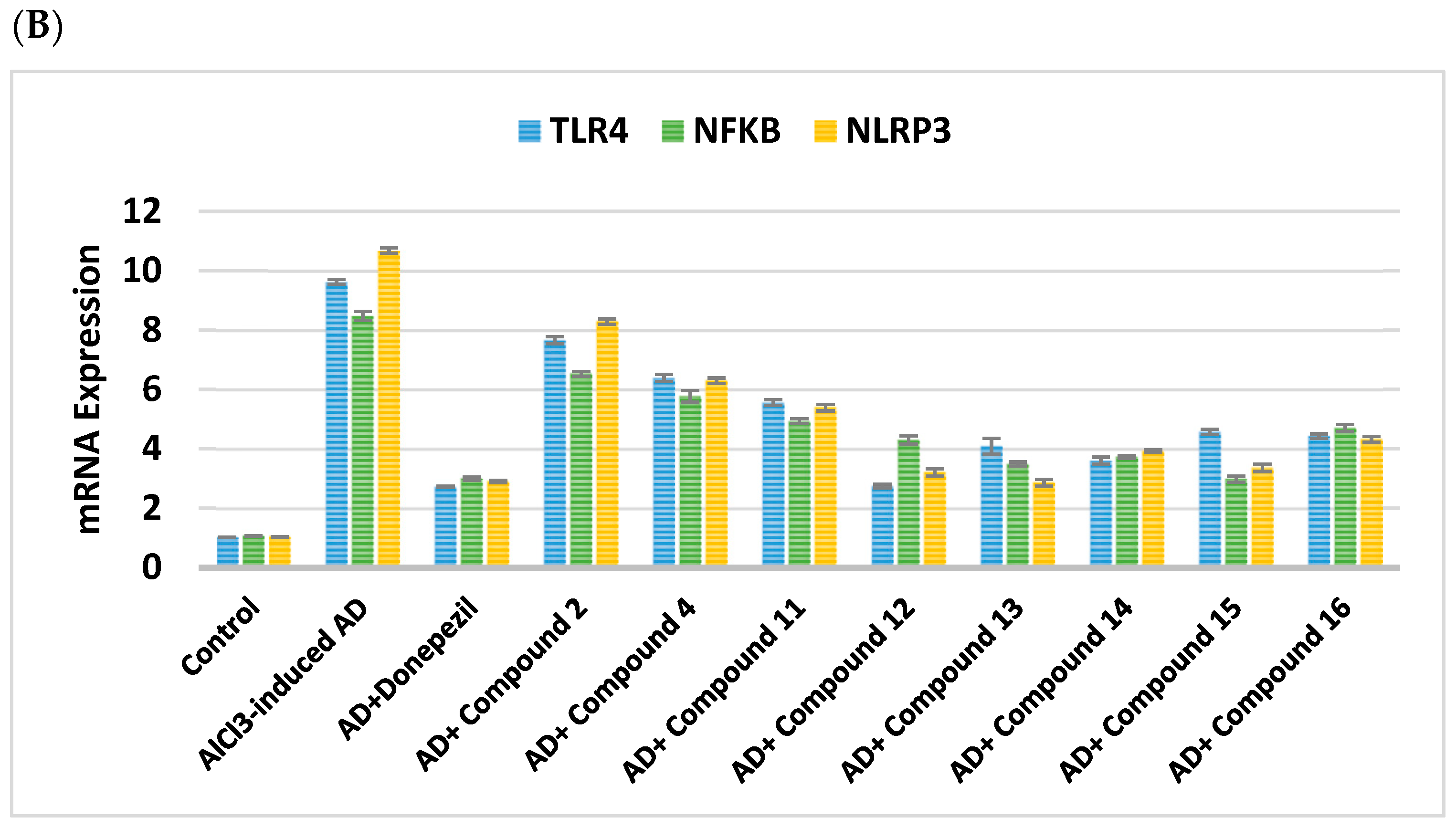
Examining the impact of the novel hybrids on IL-1β and TNF-α, along with mRNA expression levels of NFκβ, TLR4, and NLRP3, highlighted that administration of compounds 12, 13, 15, and 16 drastically lowered IL-1β levels by 53.9%, 61.3%, 61.1%, and 55.2%, respectively. This exceeds the pronounced 50.3% reduction in donepezil inhibition. Hybrids 11 and 14 showed a moderate reduction in IL-1β, by 42.1% and 49.3%, respectively, while compounds 2 and 4 only showed acceptable inhibitory activity, by 20.6% and 30%, respectively (Figure 6).
Concurrently, compounds 13 and 15 dramatically decreased TNF-α levels, outperforming the donepezil inhibitory effect by 70.2% and 74.6%, respectively. Hybrids 11, 12, 16, and 14 displayed a significant decline in TNF-α levels of 43.6%, 62.5%, 51.6%, and 58.4%, respectively, in contrast to hybrids 2 and 4, which displayed an average decline of 31% and 35.4% (Table 7 and Table 8).
mRNA expression levels of NFκβ upon treatment with hybrid 15 displayed paramount reduction by 64.8%, corresponding to the effect of donepezil. Excellent NFκβ reduction was achieved by hybrids 13 and 14 by 58.8% and 56%, respectively. Hybrids 11, 12, and 16 demonstrated notable NFκβ level reduction by 41.8%, 49.2%, and 44.5%, respectively.
mRNA expression levels of TLR4 upon administration of hybrid 12 displayed superior reduction by 71.4%, analogous to the impact of donepezil. Outstanding TLR4 reduction was achieved by hybrids 13 and 14 by 57.5% and 62.6%, respectively. Hybrids 11, 15, and 16 confirmed remarkable TLR4 level reduction by 42.2%, 52.5%, and 53.9%, respectively.
The expression levels of NLRP3 mRNA upon treatment with hybrid 13 displayed major reduction by 73.2%, equivalent to the impact of donepezil. Excellent NLRP3 reduction was achieved by hybrids 12, 14, and 15 by 69.9%, 63.2%, and 68.5%, respectively. Hybrids 11 and 16 established significant NLRP3 level reduction by 49.5% and 59.5%, respectively.

2.2.5. Effect on Caspase-1, BAX, and Bcl-2

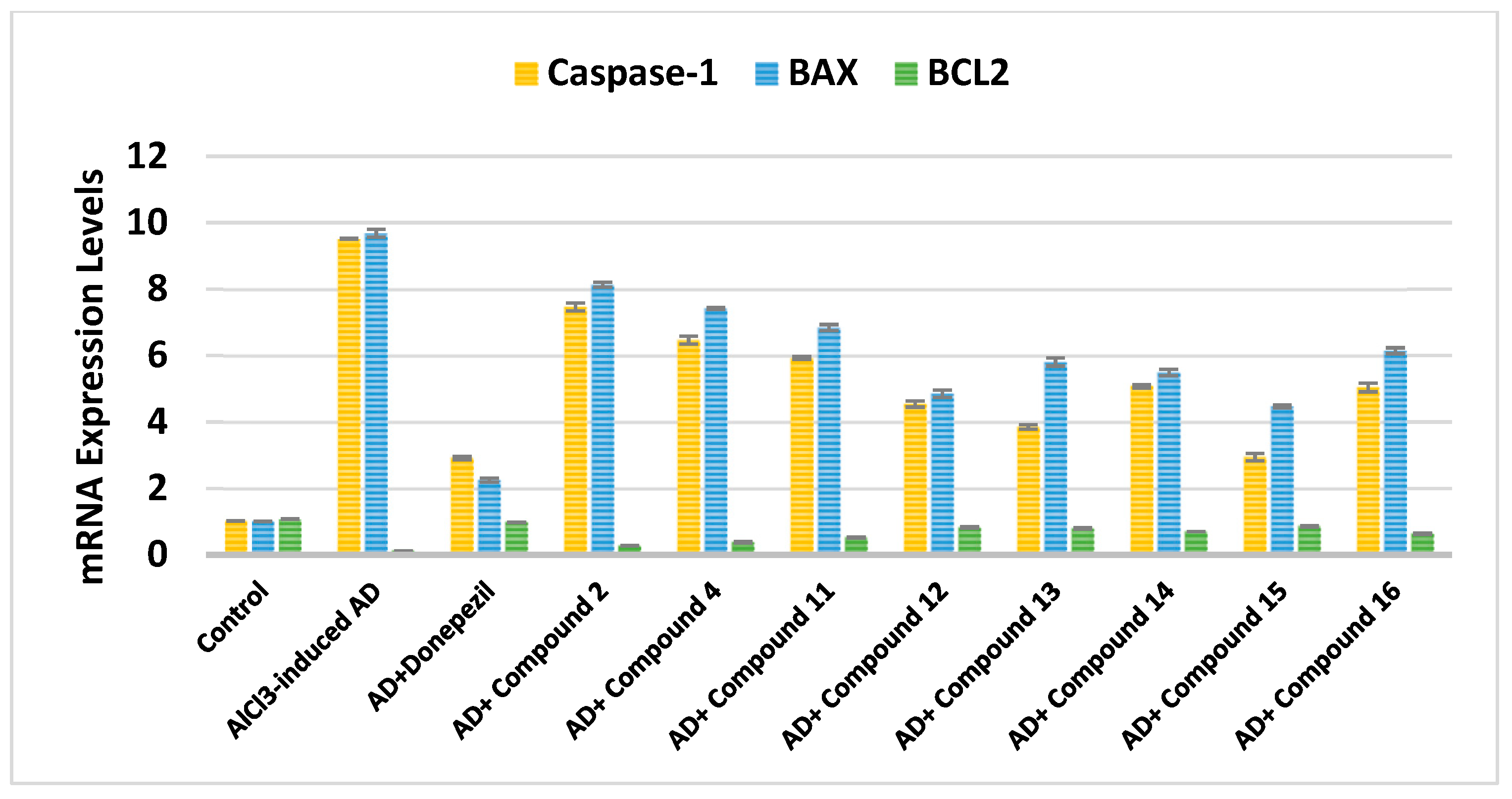
Hybrid 15 demonstrated superior downregulation of Caspase-1 levels by 69.1%, comparable to the pronounced effect of donepezil on Caspase-1 levels. Compounds 12 and 13, significantly suppressed Caspase-1 gene expression by 52.3% and 59.5%, respectively. Hybrids 11, 14, and 16 established moderate Caspase-1 level reductions of 37.8%, 46.6%, and 47%, respectively.
The BAX gene expression was dramatically suppressed by conjugates 12, 13, 14, and 15 by 40% to 53.8%. With corresponding values of 29.3% and 36.5%, compounds 11 and 16 showed a rather small BAX inhibitory activity. In contrast to donepezil, compounds 2 and 4 showed a slight downregulatory effect of 16% and 23.3%, respectively.
In contrast, hybrids 11 and 16 demonstrated substantial upregulation by 4.3 and 5.2 folds, whereas compounds 12, 13, 14, and 15 significantly elevated Bcl-2 gene expression by 6.8-, 6.5-, 5.8-, and 7.1-fold, respectively (Figure 7). Every synthesized drug exhibited a BAX/Bcl-2 ratio between 5.2 and 30.1, suggesting that they might increase brain cell proliferation by shifting the ratio to lower values (Table 9 and Table 10) [79].

2.2.6. Effect on WNT Signaling Pathway

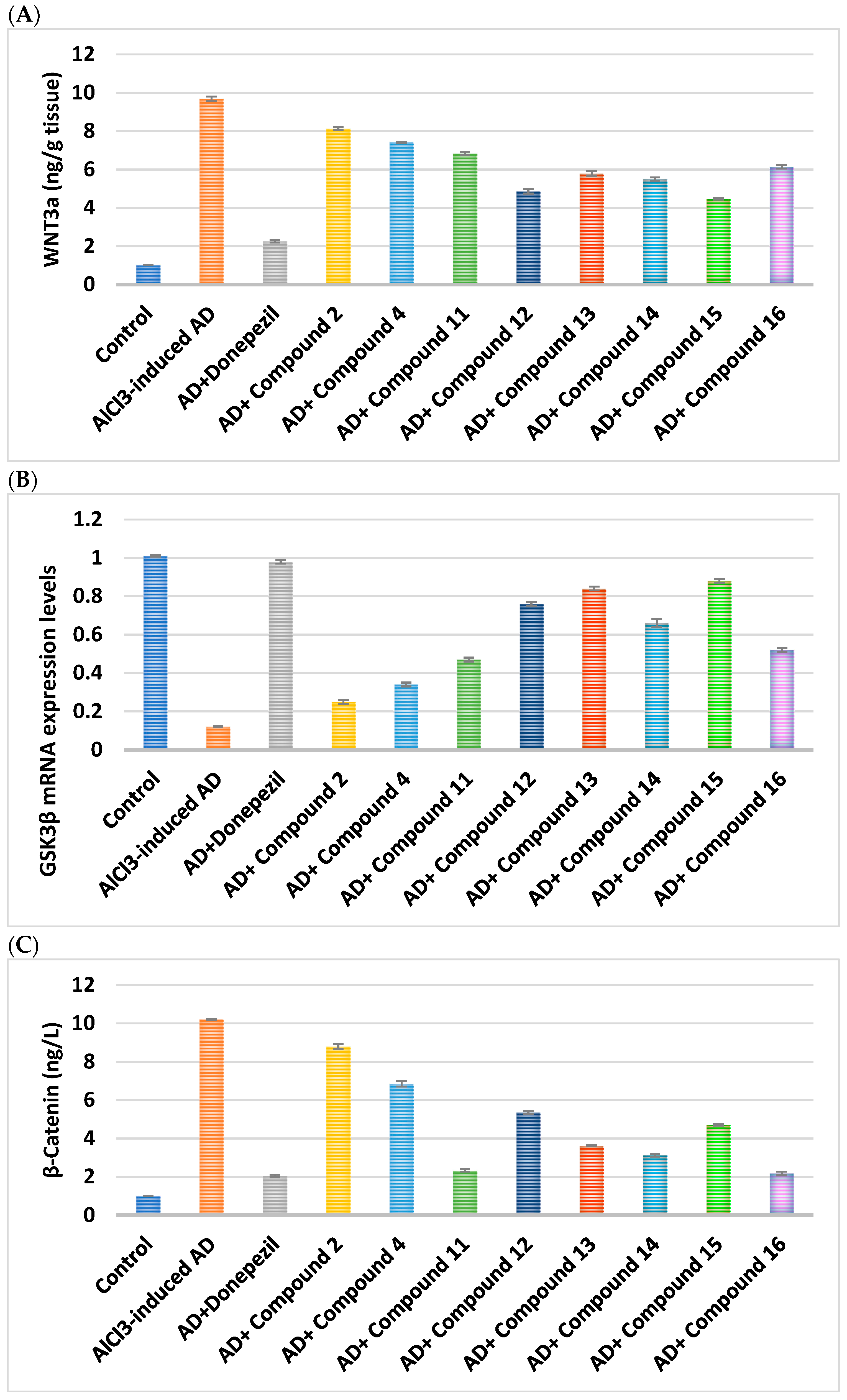
Examining the impact on the WNT signaling pathway (Figure 8), compounds 12, 13, and 15 showed strong elevation of Wnt3a levels by 6.3-, 7-, and 7.3-fold, respectively, whereas compounds 11, 16, and 14 demonstrated moderate elevation by 3.9-, 5.5-, and 4-fold, respectively. Wnt3a levels were slightly elevated by 2 and 2.8 fold in conjugates 2 and 4, respectively.
Furthermore, compared to donepezil, compound 15 dramatically enhanced the amount of β-catenin 8.8 times. The levels of β-catenin were elevated 7.7 times in compound 13. Meanwhile, compounds 2 and 4 displayed a slight rise in β-catenin levels by 2.7- and 3.9-fold, respectively, while compounds 11, 12, 16, and 14 demonstrated a moderate elevation in β-catenin levels by 4.8-, 6.3-, 4.6-, and 5.3-fold, respectively.
Conversely, compounds 11, 13, 16, and 14 significantly reduced GSK3β gene expression by 64.4% to 78.6%. The GSK3β gene expression was moderately downregulated by hybrids 12 and 15, by 47.4% and 53.7%, respectively, whereas compounds 2 and 4 showed the smallest degree of downregulation, by 13.7% and 32.7%, respectively (Table 11 and Table 12).

2.2.7. Impacts on Brain Oxidative Stress

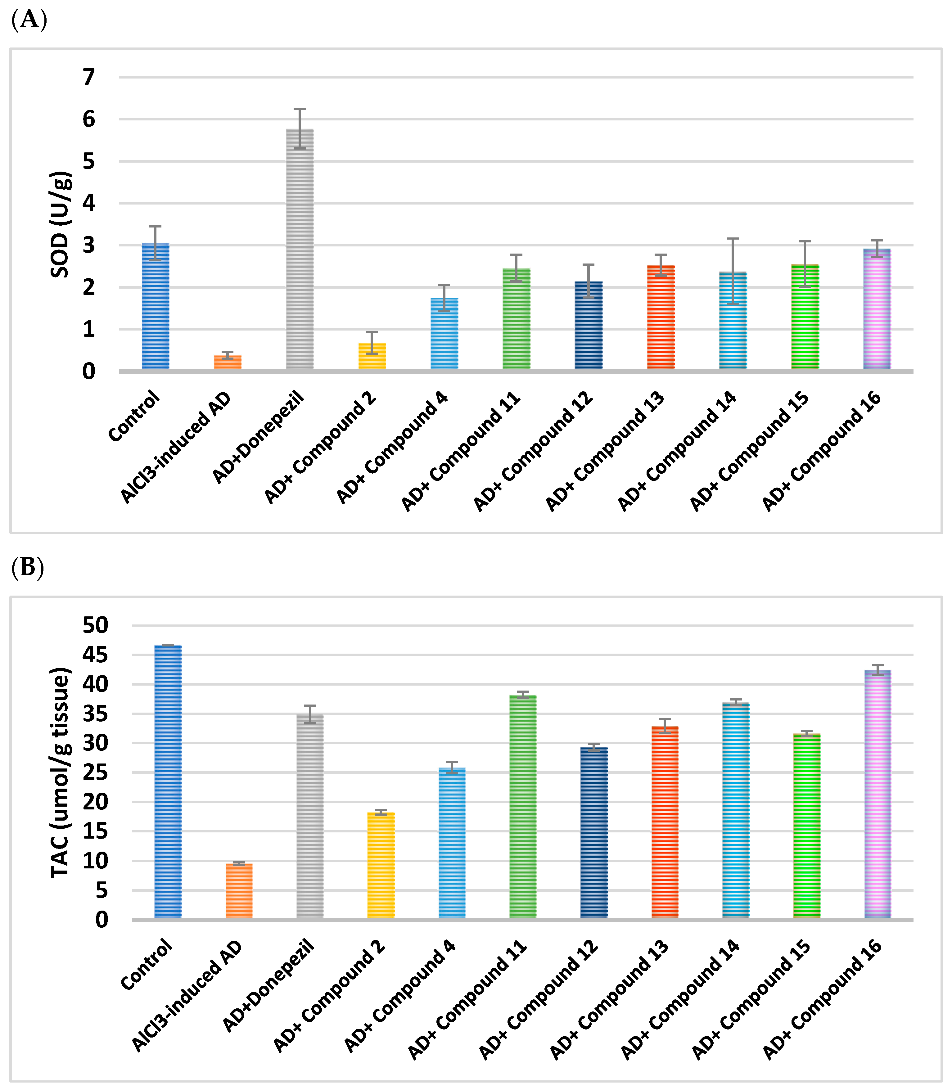
TAC levels improved by 4, 3.8, and 4.4 times, respectively, upon administering conjugates 11, 14, and 16, outperforming donepezil (Figure 9). In a manner comparable to donepezil, compounds 12, 13, and 15 showed improvements in TAC of approximately 3, 3.4, and 3.3 times, whereas compounds 2 and 4 showed only slight enhancements in TAC of 1.9 and 2.7 times, respectively.
Additionally, compounds 1116 exhibited a significant improvement in SOD activity levels, ranging from 2.15 to 2.92 U/g tissue, whereas compound 4 showed a little rise in SOD activity levels, reaching 1.75 U/g tissue.
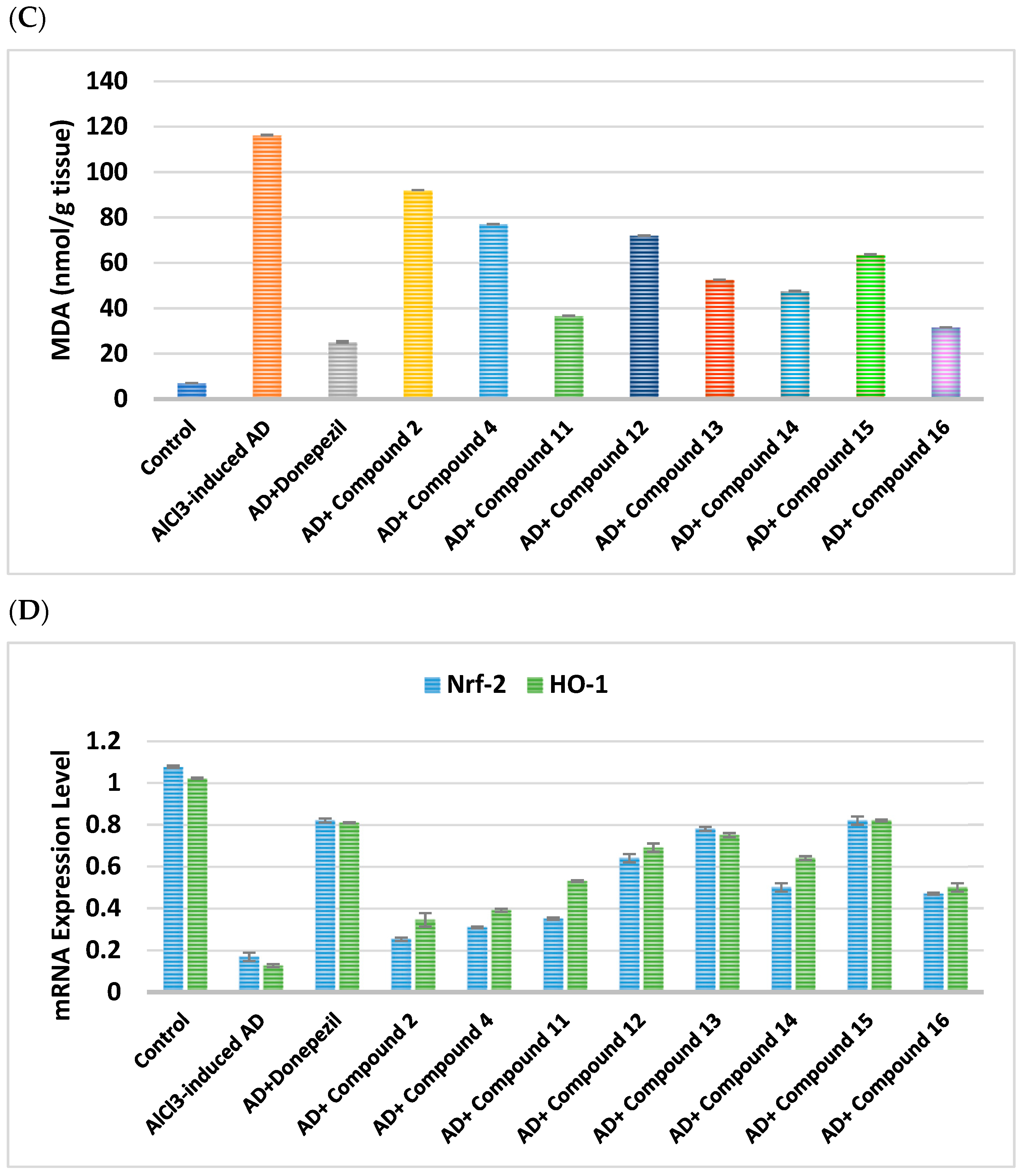
Furthermore, compounds 11, 13, 14, and 16 demonstrated an exceptional reduction in the MDA brain content, with respective values of 68.4%, 54.7%, 59%, and 72.8%. Following treatment with compounds 4, 12, and 15, there was a moderate decrease in MDA levels of 33.6%, 38%, and 45.2%, respectively (Table 13 and Table 14).
mRNA expression levels of HO-1 upon administration of hybrid 15 displayed superior elevation, by 6.7-fold, analogous to the impact of donepezil administration. Outstanding HO-1 elevation was achieved by hybrid 13, by 6.2-fold. Hybrids 12 and 14 confirmed remarkable HO-1 level elevation by 5.7- and 5.3-fold, respectively. Relatively good elevation of HO-1 level was demonstrated by 11 and 16, by 4.4- and 4.1-fold, respectively.
The expression levels of Nrf-2 mRNA upon treatment with hybrids 13 and 15 displayed major elevation by 4.8- and 5.1-fold, respectively, equivalent to the impact of donepezil. Moderate Nrf-2 elevation was achieved by hybrids 12, 14, and 16 by 4-, 3.1-, and 2.9-fold, respectively.

2.2.8. Histopathological Examination

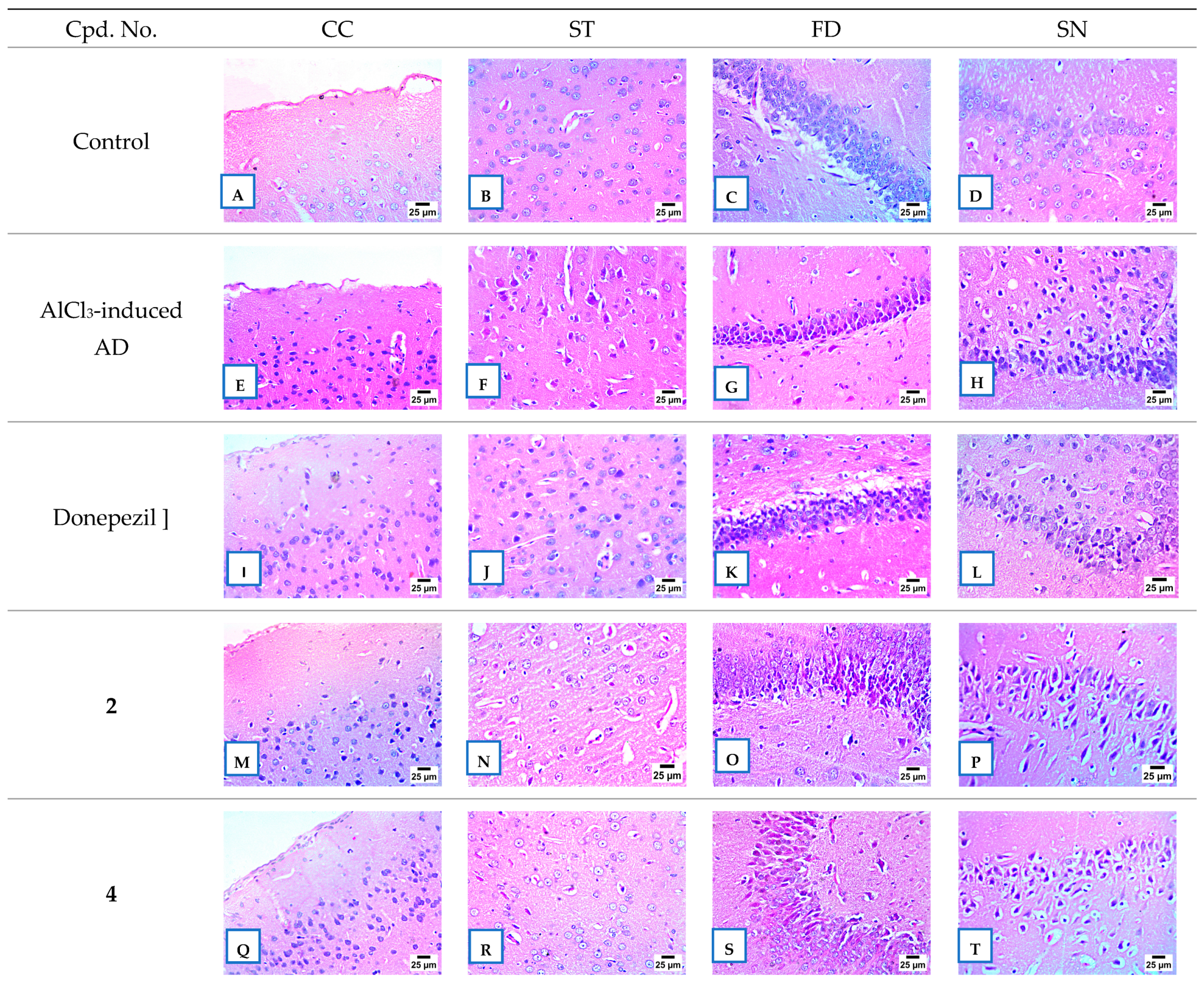
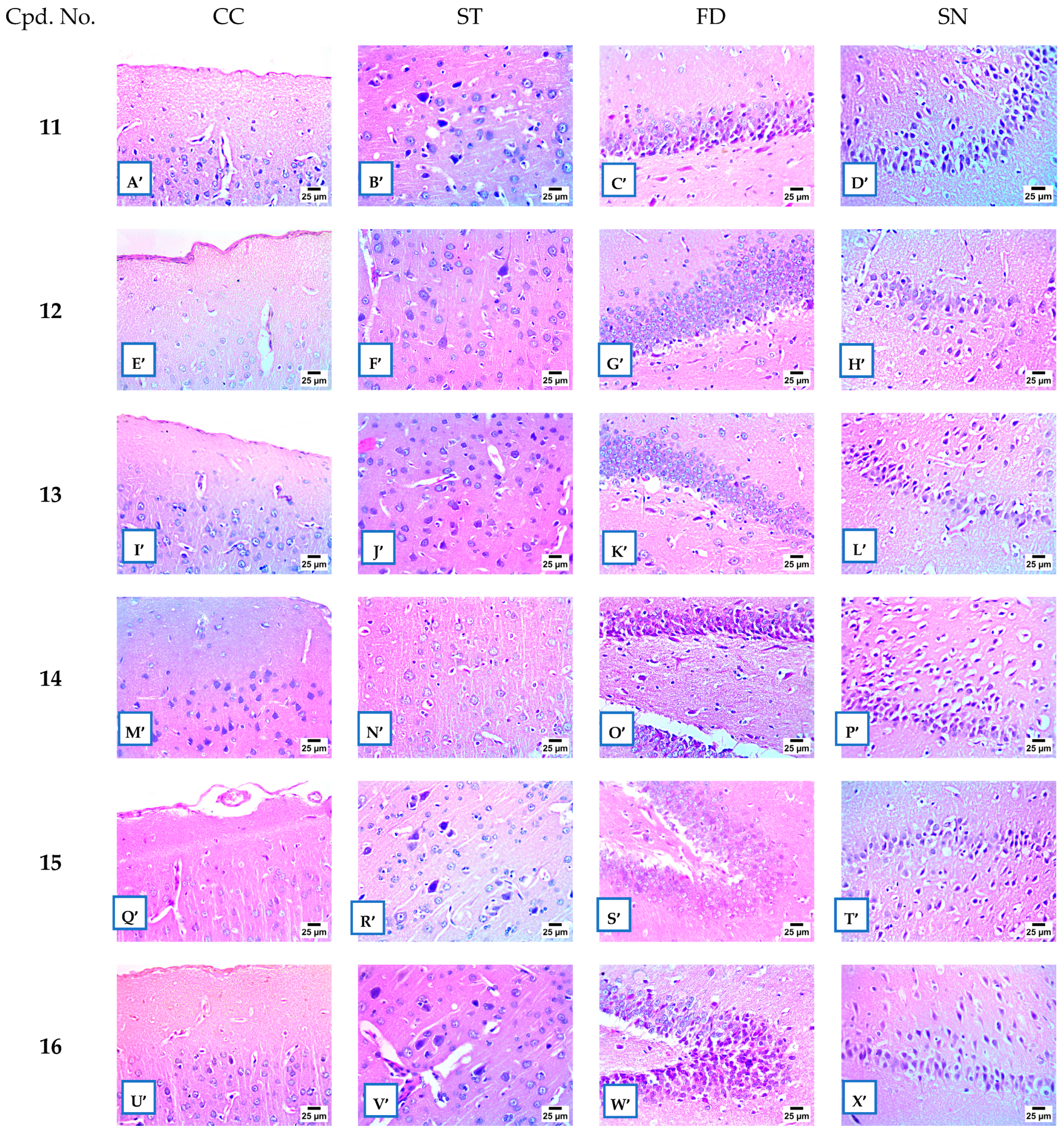
The brain tissues of rats given donepezil [70] experienced significant nuclear pyknosis (NO) in the cerebral cortex (CC), striatum (ST), fascia dentata (FD), and subiculum neurons (SN) (Figure 10I–L). Rats administered 2 and 4 additionally demonstrated significant NP in the CC, FD, and SN, whereas the striatal neurons’ histological structures remained normal (Figure 10M–T)). There was histologically minor NP in the CC and significant NP in FD, ST, and SN in hybrids 11 (Figure 11A′–D′) and 16 (Figure 11U′–X′)).
The CC neurons (Figure 11E′,I′,M′) and FD (Figure 11G′,K′,O′)) displayed normal histological structures following administration of hybrids 12, 13, and 15, respectively. Compounds 13 (Figure 11J′,N′) and 15 (Figure 11R′,T′) elicited moderate pyknosis in striatal and subicular neurons. In the ST and SN of hybrid 12, minimal pyknosis was seen (Figure 11F′,H′). The histological anatomy of neurons in the CC, FD, and SN evidenced significant pyknosis after treatment with hybrid 14 (Figure 11M′,O′,P′).

2.2.9. AChE and BChE Inhibition

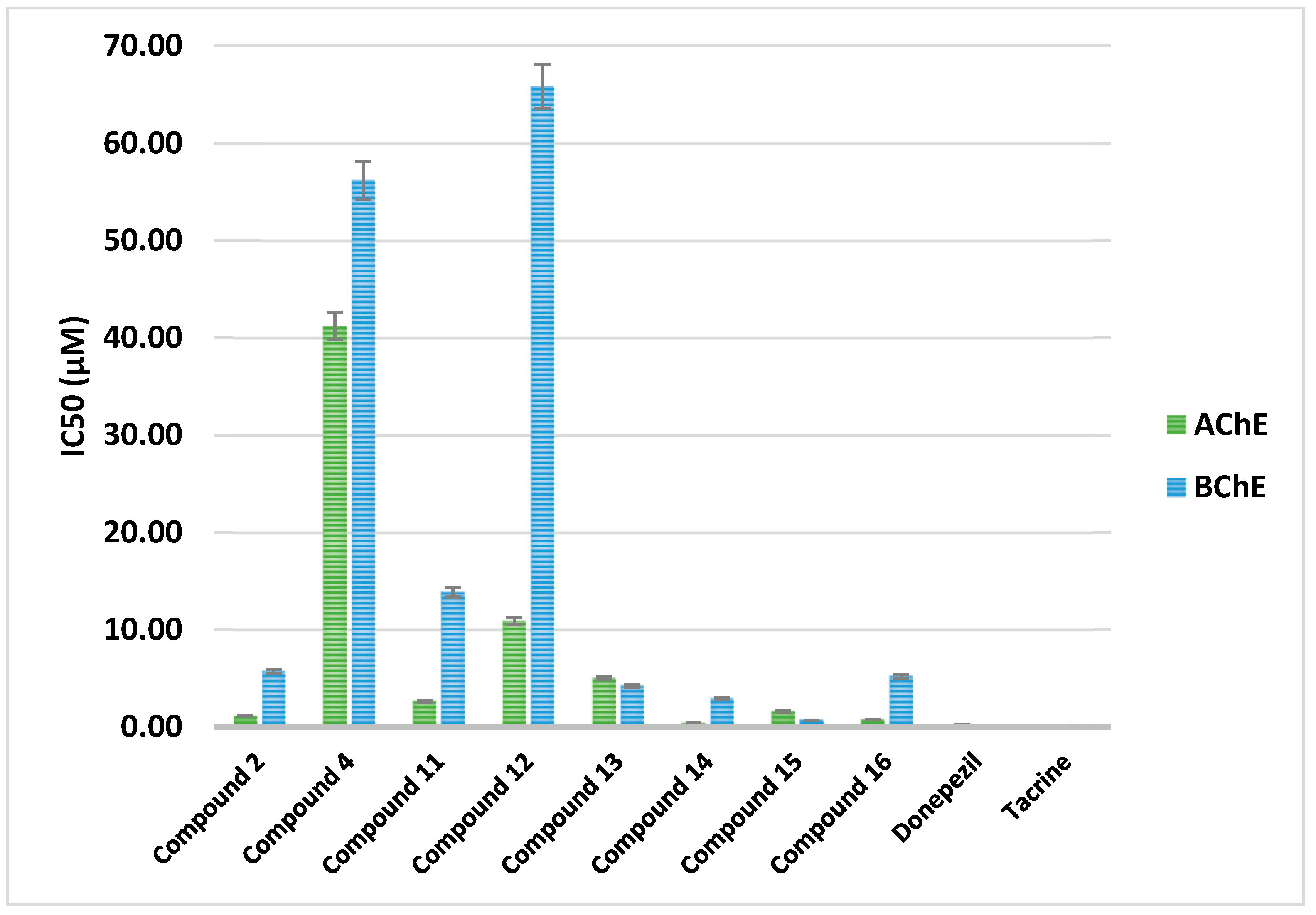
The potential inhibitory effect of all produced hybrids on AChE and BChE were assessed and compared to donepezil and tacrine, (Figure 12). Significant advancements have been achieved in the development of potent and specific BChE inhibitors able to raise brain ACh levels with few adverse peripheral effects. Based on their respective IC50 values of 1.10, 0.39, and 0.76 µM, compounds 2, 14, and 16 were found to be the most effective AChE inhibitors. Inhibitory activity against AChE was shown to be promising for compounds 11, 13, and 15, with corresponding IC50 values of 2.67, 5.04, and 1.59 µM. Comparable IC50 results of 10.90 µM indicated that compound 12 similarly exhibited considerable AChE inhibitory effect. On par with tacrine, compound 15 also demonstrated remarkable BChE inhibitory action, with an IC50 value of 0.70 µM. Additionally, with IC50 values ranging from 2.93 to 5.72 µM, compounds 2, 13, 14, and 16 show remarkable BChE inhibitory action. The IC50value of 13.85µM for compound 11 indicated substantial BChE inhibition.
The ratio of the IC50 values for BChE and AChE is used to compute the Selectivity Index (SI). When the result is less than 1, preferential inhibition of BChE is observed. Only the produced hybrids 13 and 15, with values of 0.84 and 0.44, respectively, showed greater selectivity towards BChE, despite the fact that the majority of the hits presented substantial inhibition of AChE, emphasizing the ability to increase ACh levels in the brain while drastically reducing adverse effects on the peripheral nervous system (Table 15 and Table 16).

2.3. SAR Analysis

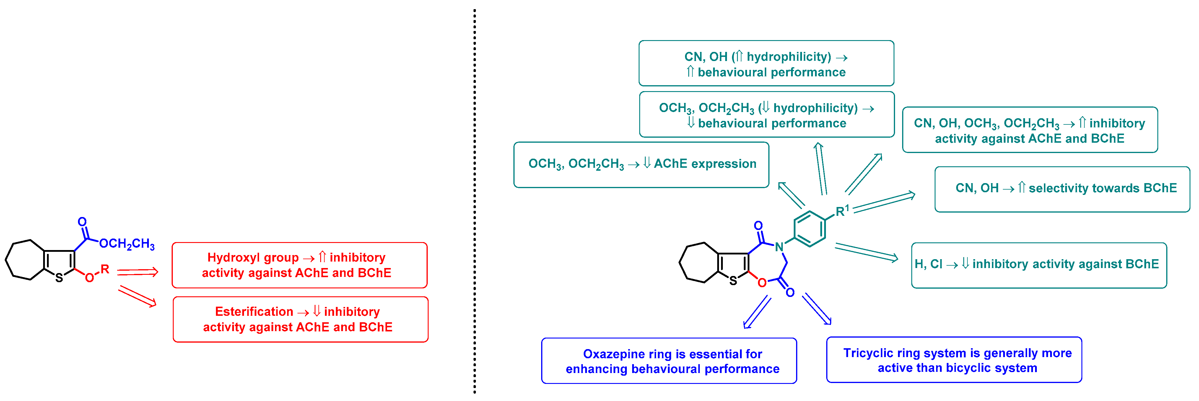
This investigation facilitated understanding of the structure–activity relationship through the biological effects of the novel derivatives (Figure 13). The significance of introducing the oxazepine ring can be witnessed by the remarkable learning, memory, and SAP% scores of compounds 1116 on behavioral tests, which suggest that the tricyclic ring system is generally better than the bicyclic ring system. It was verified by looking at the substitution on the N-phenyl ring that compounds 13 and 15, which have hydrophilic groups on the para position, performed the best in behavioral testing. This is further corroborated by tricyclic compounds 14 and 16, which possessed less hydrophilic para-alkoxy modification on the N-phenyl ring and performed well in behavioral testing, however slightly worse than compounds 2 and 4. Bicyclic hybrids are essentially less active than tricyclic compounds, as evidenced by the satisfactory, yet modest, performance displayed by compounds 2 and 4 in behavioral testing.
Similarly to behavioral performance, the tricyclic ring system actually takes precedence over the bicyclic ring system, as demonstrated by the influence of compounds 1116 on the suppression of AChE expression levels. Hybrids 14 and 16 displayed the greatest suppression of AChE expression levels, demonstrating the significance of the para-alkoxy group on the N-phenyl ring.
Further supporting the crucial need for a tricyclic ring system is the fact that tricyclic compounds (compounds 1116) outperform bicyclic compounds (compounds 2 and 4) in reducing the levels of inflammatory biomarkers. While switching to alkoxy groups typically reduced the inhibition of inflammatory indicators, tricyclic compounds 13 and 15, which contained para-hydroxy and para-cyano groups, showed the strongest anti-inflammatory action.
The inhibition effect of hybrids 14 and 16 against AChE exhibits a SAR, underscoring the significance of the alkoxy group in tricyclic hybrids. Regardless of the number of fused rings, the inhibitory activity against AChE is typically improved by the presence of an OH group.
The significance of having para-hydroxy or alkoxy groups, which result in maximal BChE inhibitory activity, was established by SAR contingent on the inhibitory activities of 2, 14, 15, and 16 against BChE. It was shown that BChE inhibition is decreased by unsubstituted or para-chloro-substituted phenyl rings.

2.4. In Silico ADME Evaluation

In silico drug-likeness, ADME, and physicochemical assessments were conducted on all hybrids in addition to the common reference drugs donepezil and tacrine. Various physicochemical characteristics, such as lipophilicity and rotating contacts, are noted in Table 17. Five of the new compounds featured four H-bond donor groups, exactly as observed with donepezil; nevertheless, all of them shared three to four H-bond acceptor groups. In contrast, hybrids 2 and 15 featured one H-bond donor group, which is analogous to tacrine.
The capacity of drugs to infiltrate the BBB is a crucial pharmacokinetic characteristic of AD treatments. For medications to easily pass across the blood–brain barrier, PSA ≤ 70 Å2 and log P = 2–5 are required [80,81]. MlogP values appeared over 3.0 for compounds 11, 12, and 14, and above 2.0 across all synthesized compounds. Large MlogP values ensure that the synthetic compounds can cross the BBB. Table 18 displays the values of the TPSA. SwissADME’s PAINS discovered that none of the compounds prompted any alerts [82]. All hybrids thrived better than donepezil and tacrine, with synthetic accessibility scores extending from 3.14 to 3.58, demonstrating easy wide scale synthesis (Table 17).
Each synthesized molecule is assigned a TPSA of less than 200 Å2. The range of the percentage ABS among all hits was between 81.11 to 89.06 percent, corresponding to donepezil and tacrine. This implies that these derivatives could possess the required bioavailability and permeability of cell membranes. Percent ABS, or absorption, was also computed using the formula % ABS = 109 − (0.345 × TPSA) (Table 18).
Table 19 lists the anticipated ADME and pharmacokinetic attributes of the hybrids under investigation. In line with the findings, all the substances that were evaluated indicated high gastrointestinal (GI) absorption and did not inhibit p-glycoprotein (P-gp).
The unit used to indicate the BBB penetration rate is cm/s. In contrast to BBB-molecules (Category 0), which exhibited log BBB ≤ −1, BBB+ molecules (Category 1) exhibited log BBB > −1. The output value, which ranges from 0 to 1, shows the probability that an input is BBB+. In comparison to donepezil, hybrids 11 and 12 were predicted to have greater BBB permeability, whereas hybrids 2 and 4 had a moderate probability of BBB permeability. Additionally, the entire examined Cytochrome P450 (CYP) isomers are inactivated by the newly generated oxazepine derivatives. Each of the compounds examined, with the exception of 15, 16, and 14, do not inhibit CYP2D6. Furthermore, every anticipated analog displayed CYP3A4 inhibition, with the exception of compounds 2, 4, and 12. The skin permeability coefficient values (log Kp), where Kp is expressed in cm/s, for the tested compounds were minimal. (Table 19).
The assorted drug-likeness rules, i.e., Lipinski [83], Veber [84], Ghose [85], Muegge [86], and Egan [87], were compelled to evaluate the molecule to be an influential drug candidate. The number of violations of the previously mentioned regulations and their bioavailability scores are displayed in Table 20. The Lipinski (Pfizer) strain is subject to the present setting rule-of-five (RO5), which asserts that every molecule under examination is drug-like. All compounds uphold the norm and have the potential to be medications, according to the predictions of the Ghose, Veber, Muegge, and Egan rules. For each of the compounds under examination, the bioavailability score was 0.55 (Table 20).
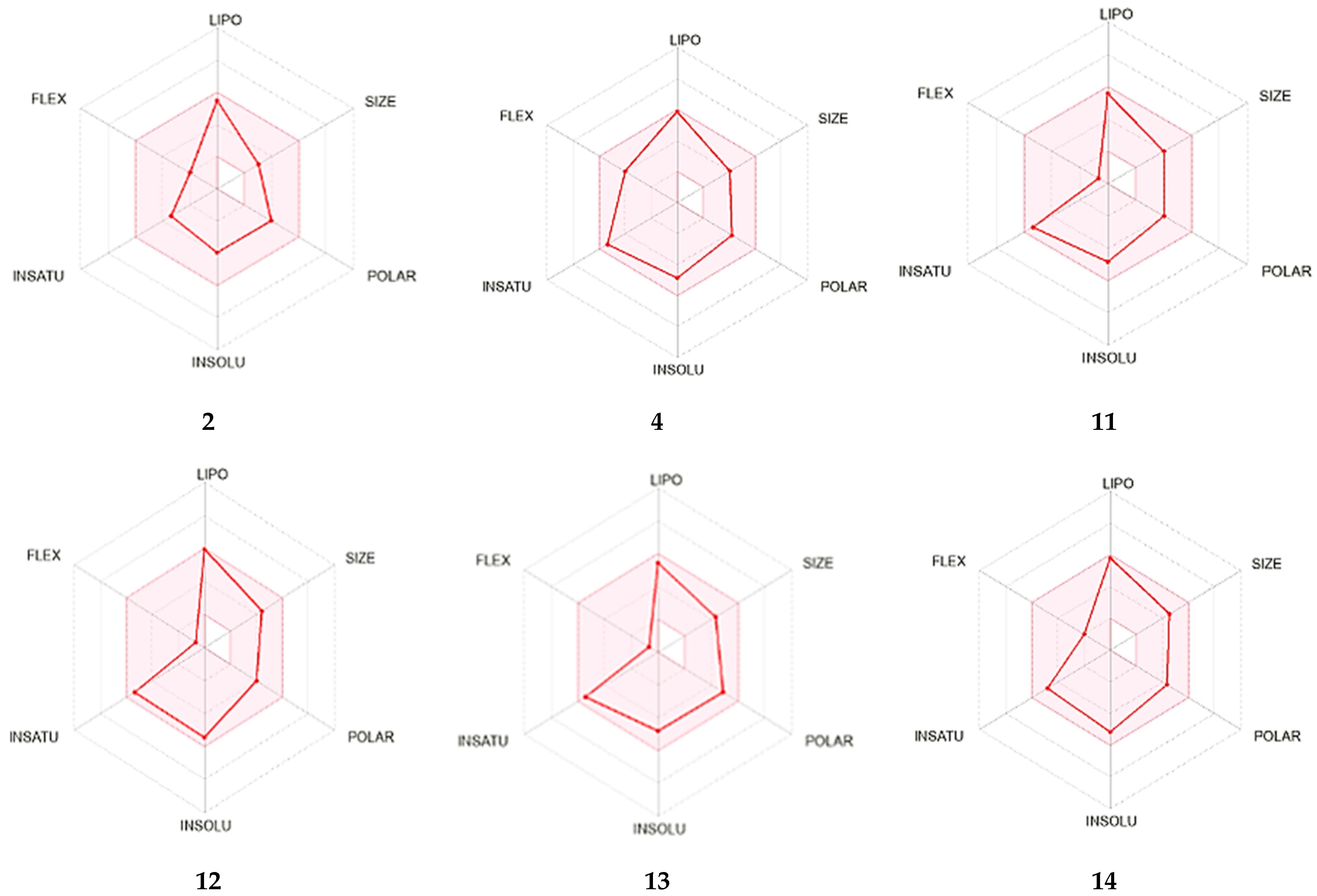
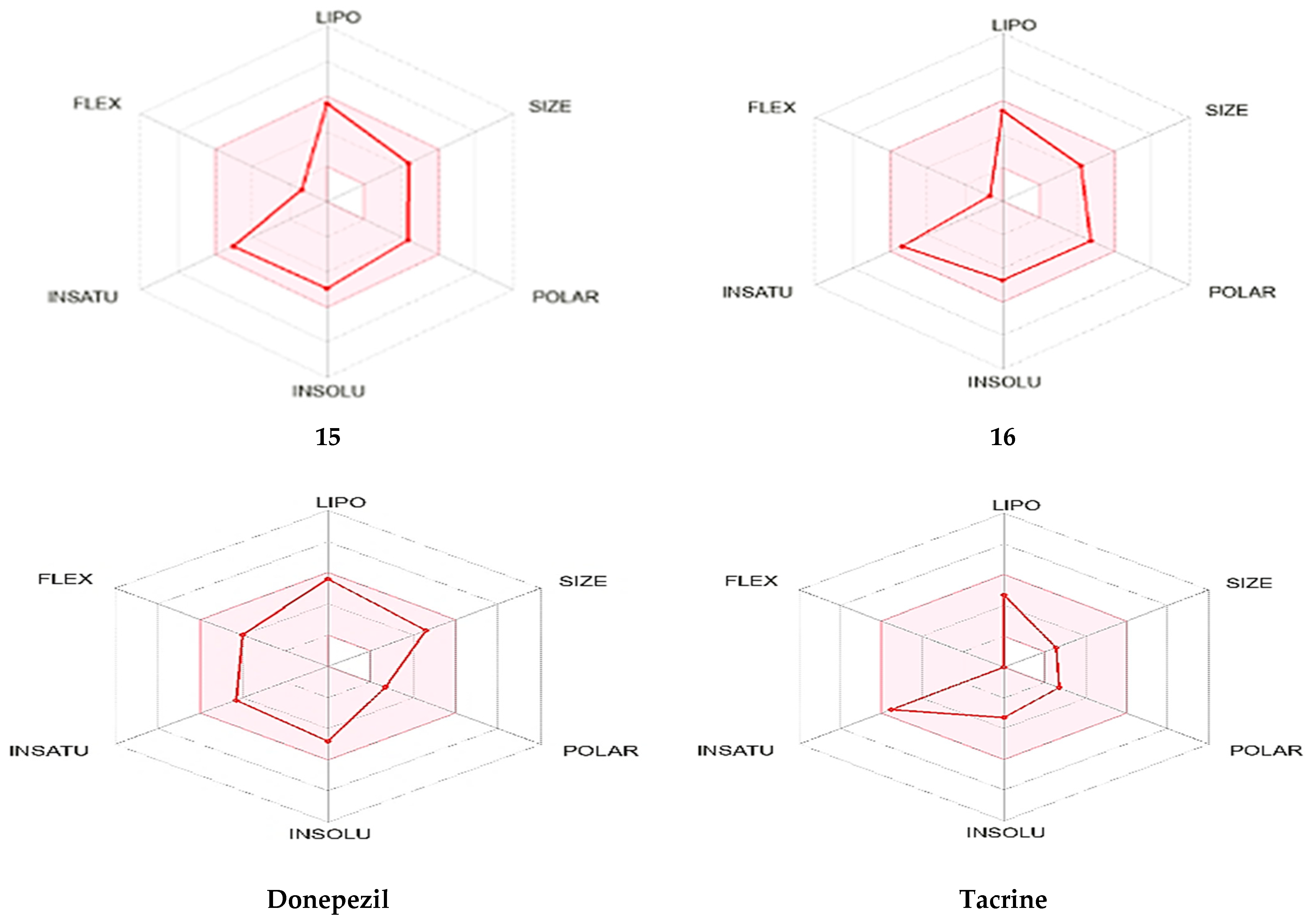
The following traits are most correctly represented by the pink surface of the bioavailability radar: flexibility, no more than six rotatable bonds; solubility; sp3 hybridization fraction, not less than 0.23; log S, not greater than 7; lipophilicity; XLOGP3 range between 4.79 and + 6.81; MW, 330–430 g/mol MW. Because the molecules are confined inside the pink surface, they are potentially bioavailable when taken orally (Figure 14).

2.5. In Silico Toxicity Evaluation

Based on online resources ProTox-II (https://tox-new.charite.de/protox_II, accessed on 1 February 2025) and pkCSM (http://biosig.unimelb.edu.au/pkcsm/prediction, accessed on 1 February 2025), Table 21 articulates the estimated toxicity of the most potent derivatives, 2, 14, and 16, alongside the two reference anti-AD medications, tacrine and donepezil [88,89]. The pkCSM online tools indicate that the only compounds that are safer than tacrine are compounds 2 and 14, which are comparable to donepezil in that they fail to demonstrate any signs of AMES toxicity. Additionally, the highest tolerated human dose value (0.668 log mg/kg/day) for active hit 2 was greater than that of tacrine and donepezil. Compared to donepezil and tacrine, 14 and 16 have higher oral rat acute toxicity (LD50) values. Additionally, all hits exhibited greater Oral Rat Chronic Toxicity (LOAEL) values than donepezil and tacrine. The proper repolarization of the pulse is controlled by the potassium channels expressed by the human Ether-à-go-go Related Gene (hERG). Serious cardiovascular disorders might arise from any blockage or dysfunction of these channels in heart cells. As a result, the pharmaceutical sector is highly concerned about drug-induced potassium channel blocking. It was anticipated that no active hybrids would inhibit hERG-I or hERG-II. Hepatotoxicity was examined using the pkCSM server, and it was found that none of the active hits were hepatotoxic. The only hybrid that displayed skin sensitivity was hybrid 2.
Tacrine was expected to fall into class two (GHS), while donepezil and all hybrids were predicted to fall into class four using the ProTox-II detection web tool (Table 21). All hits were found to display higher LD50 values than donepezil and tacrine. All active derivatives have been determined to be non-mutagenic and non-immunotoxic. Furthermore, conjugates 2 and 16, unlike donepezil, were expected to be non-cytotoxic.

2.6. Molecular Docking

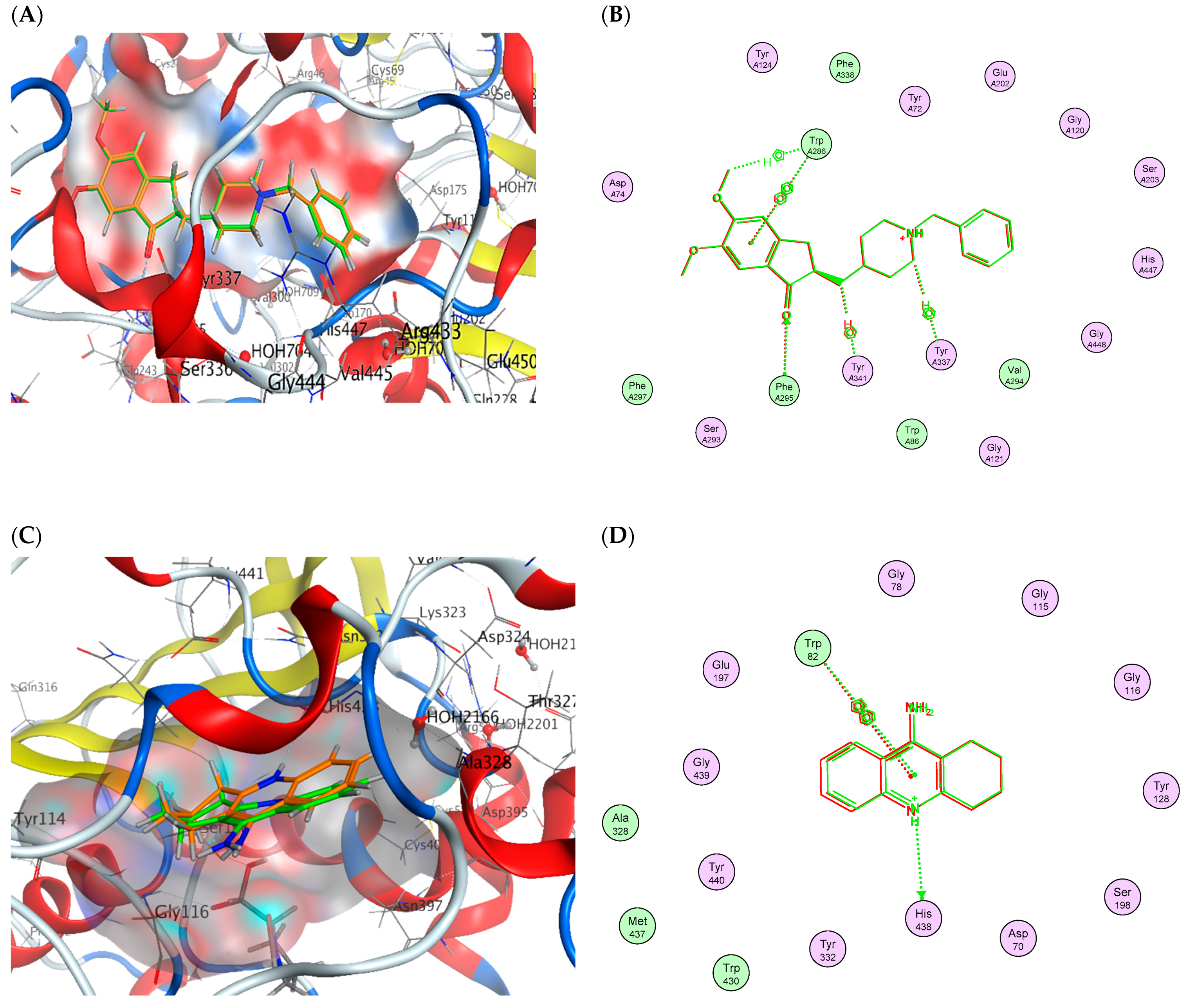
The reliability of docking was validated by self-docking the reference antagonists, tacrine and donepezil, into the active sites of AChE (PDB ID: 4EY7) and BChE (PDB ID: 4BDS), respectively. The validation was successful, with energy scores (S) of −17.710 kcal/mol and −10.080 kcal/mol, in addition to marginal RMSD values between the native ligands and the re-docked pose of 0.117 Å and 0.589 Å, respectively. Figure 15 shows that the re-docked location could superimpose the native ligand while also occupying crucial contacts produced by the bound ligand (Table 22) with the active sites of both AChE and BChE, supporting the reliability of the validation.
Phe295 was involved in one H-bond interaction between donepezil and the AChE pocket (PDB: 4EY7). The residues Trp286, Tyr341, and Tyr 337 in AChE were also observed to engage in three H–arene interactions with donepezil. Additionally, a single arene–arene interaction through Trp286 was revealed. Tacrine was shown to engage with the BChE pocket by forming an H-bond with a His438 residue (PDB: 4BDS). It was observed that tacrine and His438 produced one H–arene interaction and that the Trp82 residue produced one arene–arene contact (Table 22).
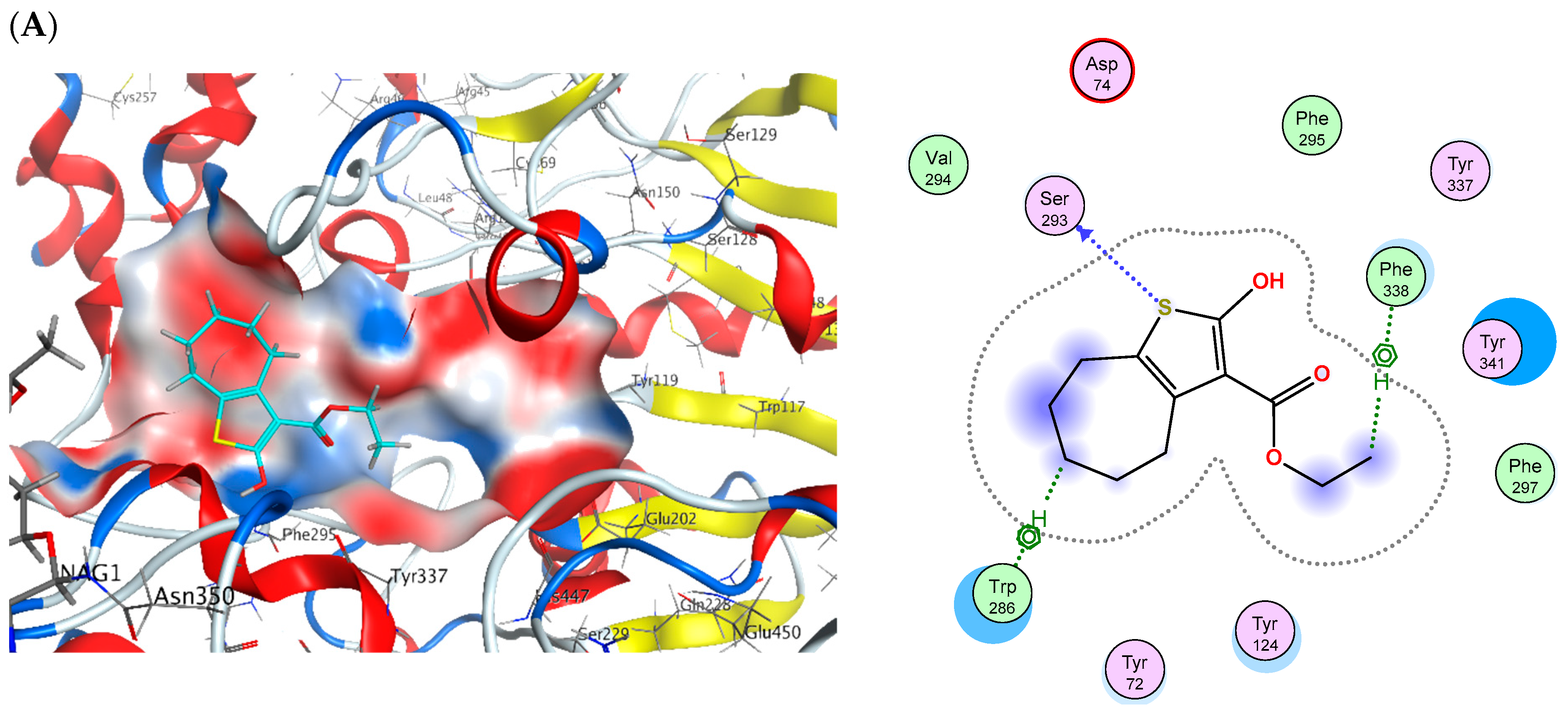
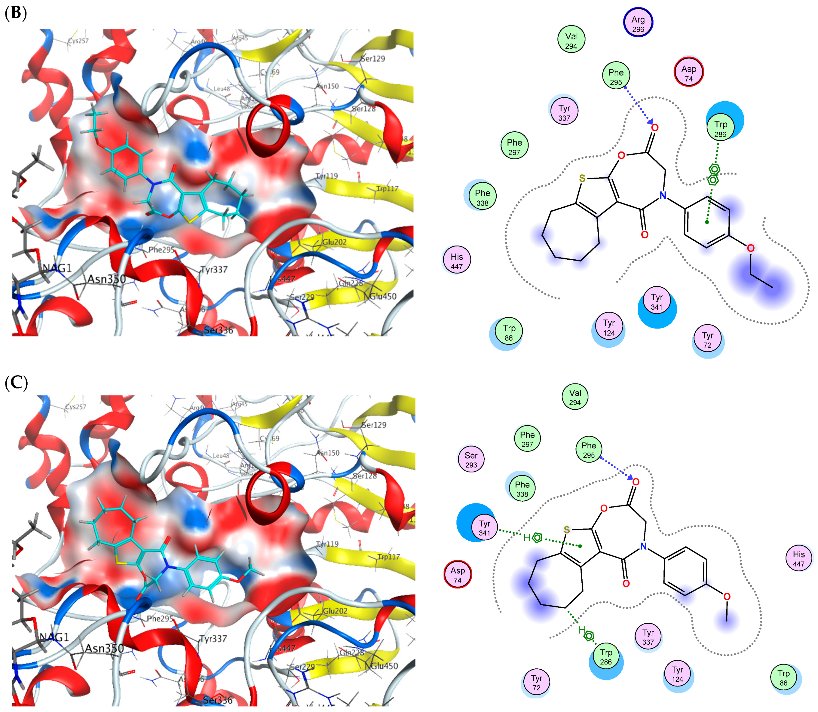
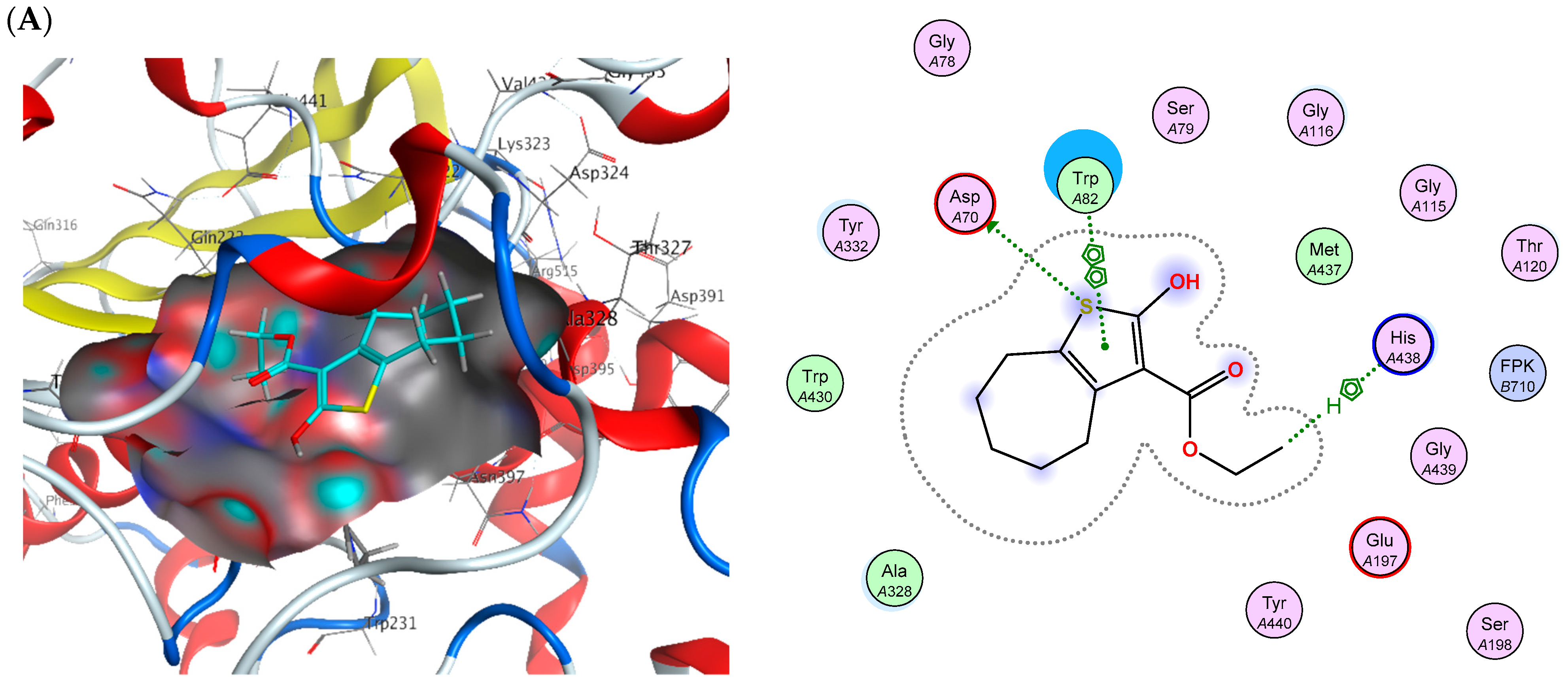
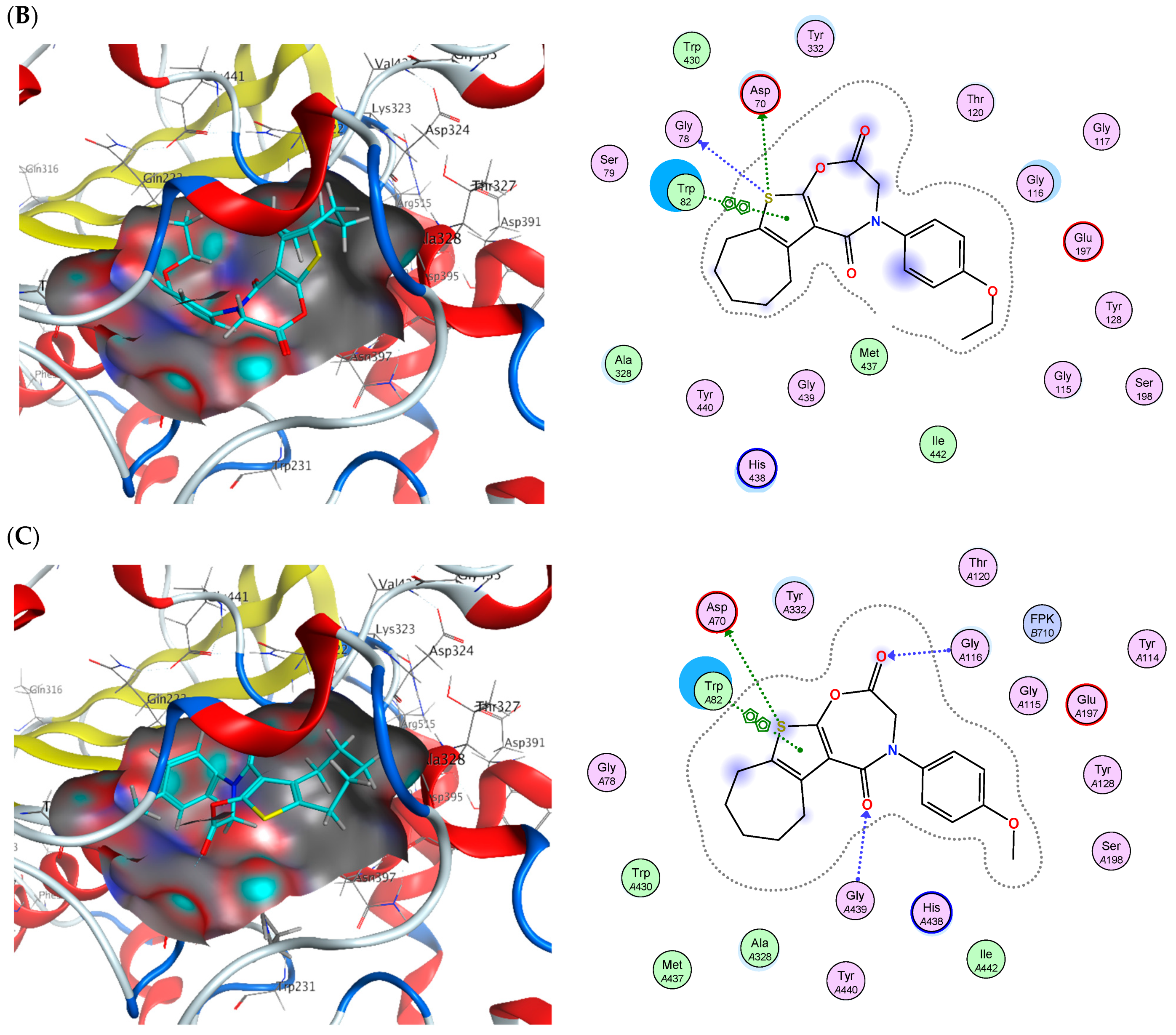
The binding energies along with interactions of the most potent derivatives—2, 14, and 16—with both AChE and BChE were inspected. With respective values of −14.45, −15.09, and −14.74 kCal/mol, the binding energies were found to be rather proximal to donepezil’s binding energy with AChE. In a comparable manner, it was discovered that the binding energies with BChE were very similar to those of tacrine, reporting −14.97, −16.41, and −18.10 kCal/mol, respectively. As they interact with the AChE pocket, the three most active hybrids were discovered to interact with the same amino acid residues—Phe295, Tyr341, and Trp286—that are implicated in donepezil interaction to the binding site (Figure 16). Both an additional H-bond with Ser293 and an additional H–arene contact with the Phe338 residue of AChE were observed by compound 2. Additionally, the three most active hits displayed binding interactions with BChE (Figure 17), including two amino acid residues that are also implicated in tacrine binding: His438 and Trp82. Asp70 additionally produced an additional H-bond with the three hits. Upon compound 14 binding, a second H-bond with Gly78 was discovered. Two further H-bonds with Gly439 and Gly116 were discovered upon binding of compound 16 (Table 23).

3. Materials and Methods

3.1. Chemistry

3.1.1. General Details

3.1.2. Synthesis

  • Ethyl 2-amino-5,6,7,8-tetrahydro-4H-cyclohepta[b]thiophene-3-carboxylate (1)
Created utilizing the documented process [73].
  • Ethyl 2-hydroxy-5,6,7,8-tetrahydro-4H-cyclohepta[b]thiophene-3-carboxylate (2)
Compound 1 (0.005 mol) was dissolved in 30 milliliters of water and 4.5 milliliters of 37% w/v hydrochloric acid. The diazonium salt was obtained by adding a dropwise, stirred cold solution of sodium nitrite (0.005 mol) in water (1 mL) to the cooled amine solution (0–5 °C). To guarantee a full response, stirring was maintained for fifteen minutes. Adding an equivalent amount of water and boiling it in a warm water bath at 50 °C for 30 min made hydrolysis simple. Once N2 evolution stopped, the precipitate that had formed was filtered, and crystallized from pure ethanol to produce (2) [73].
  • Ethyl 2-(2-chloroacetoxy)-5,6,7,8-tetrahydro-4H-cyclohepta[b]thiophene-3-carboxylate, (4)
A solution of compound 2 (0.01 mol) in N,N-dimethyl formamide (DMF) (20 mL) with five drops of triethylamine was refluxed for six hours before the addition of chloroacetyl chloride (0.01 mol). The mixture was introduced into ice-cold water once it cooled. Following a water rinse and a dry filter, the solid crystallized from the ethanol.
Yield: 92%; m.p.: 155–156 °C; IR (KBr, cm−1) = 3037 (sp2 C-H), 2930, 2840 (sp3 C-H), 1707, 1690 (2 C=O ester); 1H NMR (400 MHz, CDCl3): δ = 4.35 (q, J = 6.3 Hz, 2H, -CH2-CH3), 4.25 (s, 2H, CO-CH2-Cl), 2.96 (t, J = 8.2 Hz, 2H, cycloheptane-Hs), 2.68 (t, J = 7.3 Hz, 2H, cycloheptane-Hs), 1.73–1.51 (m, 6H, cycloheptane-Hs), 1.38 (t, J = 6.2 Hz, 3H, CH2-CH3); 13C NMR (100 MHz, CDCl3): δ = 165.97 (CO-CH2-Cl), 162.90 (CO-OCH2CH3), 160.04, 136.30, 125.65, 111.96, 59.81, 39.36, 31.59, 28.78, 28.40, 27.43, 25.93, 13.19.; MS (EI, 70 eV): m/z (%) = 318 [M. ++2] (9.14%), 316 [M. +] (10.11%), 42 (100%); Anal. Calc. for C14H17ClO4S (316.80): C, 53.08; H, 5.41; S, 10.12. Found: C, 53.10; H, 5.43; S, 10.13.
  • 4-(Aryl)-3,4,7,8,9,10-hexahydro-6H-cyclohepta[4,5]thieno[3,2-f][1,4]oxazepine-2,5-dione (11–16)
Typical operating procedure.
In ten milliliters of glacial acetic acid, the suitable amine (0.01 mol) and chloro-acetyl derivative 4 (0.01 mol) were refluxed for ten hours. After condensing the reaction mixture at reduced pressure, diethyl ether was employed to triturate it. To filter and crystallize the separate material, the proper solvent was utilized.
  • 4-Phenyl-3,4,7,8,9,10-hexahydro-6H-cyclohepta[4,5]thieno[3,2-f][1,4]oxazepine-2,5-dione, (11)
Compound 11 was yielded upon the reaction of compound 4 and aniline.
Crystallized from ethanol. Yield: 89%; m.p.: 189–191 °C; IR (KBr, cm−1) = 3078 (sp2 CH), 2910, 2860 (sp3 C-H), 1670 (2 C=O); 1H NMR (400 MHz, CDCl3): δ = 7.37 (dd, J = 7.4, 1.4 Hz, 2H, Ar-H), 7.30–7.26 (m, 2H, Ar-H), 7.12–7.05 (m, 1H, Ar-H), 4.77 (s, 2H, oxazepine CH2), 2.85 (t, J = 8.2 Hz, 2H, cycloheptane-Hs), 2.72 (t, J = 7.3 Hz, 2H, cycloheptane-Hs), 1.72–1.52 (m, 6H, cycloheptane-Hs).; 13C NMR (100 MHz, CDCl3): δ = 164.07 (CO), 163.76 (CO), 155.82, 139.48, 135.89, 128.26, 125.64, 123.80, 121.61, 118.06, 47.06 (oxazepine CH2), 31.08, 28.39, 28.19, 27.43, 25.93.; MS (EI, 70 eV): m/z (%) = 327 [M. +] (40.74%), 54 (100%); Anal. Calc. for C18H17NO3S (327.40): C, 66.04; H, 5.23; N, 4.28; S, 9.79. Found: C, 66.01; H, 5.25; N, 4.29; S, 9.80.
  • 4-(4-Chlorophenyl)-3,4,7,8,9,10-hexahydro-6H-cyclohepta[4,5]thieno[3,2-f][1,4]oxazepine-2,5-dione (12)
Compound 12 was yielded upon the reaction of compound 4 and 4-chloroaniline.
Crystallized from acetic acid as crystals. Yield: 85%; m.p.: 170–172 °C; IR (KBr, cm−1)= 3062 (sp2 CH), 2965, 2845 (sp3 C-H), 1703 (2C=O); 1H NMR (400 MHz, CDCl3): δ = 7.35 (d, J = 8.6 Hz, 2H, Ar-H), 7.28 (d, J = 8.7 Hz, 2H, Ar-H), 4.77 (s,2H, oxazepine CH2), 2.85 (t, J = 8.2 Hz, 2H, cycloheptane-Hs), 2.72 (t, J = 7.3 Hz, 2H, cycloheptane-Hs), 1.73–1.51 (m, 6H, cycloheptane-Hs).; 13C NMR (100 MHz, CDCl3): δ = 164.07 (C=O), 163.77 (C=O), 155.82, 138.25, 135.89, 129.02, 128.37, 125.64, 123.80, 118.06, 46.97 (oxazepine CH2), 31.08, 28.39, 28.19, 27.43, 25.93.; MS (EI,70 eV): m/z (%) = 363 [M. ++2] (7.60%), 361 [M. +] (17.67%), 328 (100%); Anal. Calc. for C18H16ClNO3S (361.84): C, 59.75; H, 4.46; N, 3.87; S, 8.86. Found: C, 59.73; H, 4.48; N, 3.88; S, 8.88.
  • 4-(2,5-Dioxo-2,3,7,8,9,10-hexahydro-6H-cyclohepta[4,5]thieno[3,2-f][1,4]oxazepin-4(5H)-yl)benzonitrile (13)
Compound 13 was yielded upon the reaction of compound 4 and 4-aminobenzonitrile. Crystallized from ethanol. Yield: 89%; m.p.: 200–202 °C; IR (KBr, cm−1) = 3080 (sp2 CH), 2980, 2861 (sp3 CH), 2222 (CN), 1664 (2 C=O); 1H NMR (400 MHz, CDCl3): δ = 7.58 (d, J = 7.9 Hz, 2H, Ar-H), 7.50 (d, J = 7.7 Hz, 2H, Ar-H), 4.77 (s, 2H, oxazepine CH2), 2.85 (t, J = 8.2 Hz, 2H, cycloheptane-Hs), 2.72 (t, J = 7.3 Hz, 2H, cycloheptane-Hs), 1.73–1.51 (m, 6H, cycloheptane-Hs).;13C NMR (100 MHz, CDCl3): δ = 164.07 (C=O), 163.75 (C=O), 155.82, 140.82, 135.89, 132.45, 125.64, 118.76, 118.06 (CN), 117.28, 102.30 (C-CN), 47.05 (oxazepine CH2), 31.08, 28.39, 28.19, 27.43, 25.93.; MS (EI,70 eV): m/z (%) = 352 [M. +] (19.30%), 350 (100%); Anal. Calc. for C19H16N2O3S (352.41): C, 64.76; H, 4.58; N, 7.95; S, 9.10. Found: C, 64.75; H, 4.55; N, 7.96; S, 9.13.
  • 4-(4-Ethoxyphenyl)-3,4,7,8,9,10-hexahydro-6H-cyclohepta[4,5]thieno[3,2-f][1,4]oxazepine-2,5-dione (14)
Compound 14 was yielded upon the reaction of compound 4 and 4-ethoxyaniline. Crystallized from ethanol. Yield: 85%; m.p.: 187–189 °C; IR (KBr, cm−1) = 3075 (sp2 CH), 2972, 2870 (sp3 CH), 1702, 1680 (2 C=O); 1H NMR (400 MHz, CDCl3): δ = 7.31 (d, J = 8.8 Hz, 2H, Ar-H), 6.85 (d, J = 8.8 Hz, 2H, Ar-H), 4.77 (s, 2H, oxazepine CH2), 4.04 (q, J = 6.7 Hz, 2H, -CH2-CH3), 2.85 (t, J = 8.2 Hz, 2H, cycloheptane-Hs), 2.72 (t, J = 7.3 Hz, 2H, cycloheptane-Hs), 1.73–1.51 (m, 6H, cycloheptane-Hs), 1.42 (t, J = 6.6 Hz,3H, -CH2-CH3).; 13C NMR (100 MHz, CDCl3): δ = 164.07 (C=O), 163.74 (C=O), 155.82, 149.71, 135.89, 134.95, 125.64, 122.81, 118.06, 114.58, 62.57 (-OCH2-CH3), 47.00 (oxazepine CH2), 31.08, 28.39, 28.19, 27.43, 25.93, 13.61; Anal. Calc. for C20H21NO4S (371.45): C, 64.67; H, 5.70; N, 3.77; S, 8.63. Found: C, 64.66; H, 5.72; N, 3.78; S, 8.60.
  • 4-(4-Hydroxyphenyl)-3,4,7,8,9,10-hexahydro-6H-cyclohepta[4,5]thieno[3,2-f][1,4]oxa-zepine-2,5-dione (15)
Compound 15 was yielded upon the reaction of compound 4 and 4-aminophenol, formed from ethanol as crystals. Yield: 88%; m.p.: 270–371 °C; IR (KBr, cm−1) = 3380 (OH), 3036, (sp2 CH), 2970, 2889 (sp3 CH), 1670 (2 C=O); 1H NMR (400 MHz, CDCl3): δ = 7.75 (s, 1H, OH), 7.21 (d, J = 9.2 Hz, 2H, Ar-H), 6.80 (d, J = 9.2 Hz, 2H, Ar-H), 4.77 (s, 2H, oxazepine CH2), 2.85 (t, J = 8.2 Hz, 2H, cycloheptane-Hs), 2.72 (t, J = 7.3 Hz, 2H, cycloheptane-Hs), 1.73–1.51 (m, 6H, cycloheptane-Hs); 13C NMR (100 MHz, CDCl3): δ = 164.07 (C=O), 163.74 (C=O), 155.82, 151.03, 135.89, 133.79, 125.64, 124.73, 118.06, 115.72, 47.00 (oxazepine CH2), 31.08, 28.39, 28.19, 27.43, 25.93.; MS (EI,70 eV): m/z (%) = 343 [M. +] (18.30%), 91 (100%); Anal. Calc. for C18H17NO4S (343.40): C, 62.96; H, 4.99; N, 4.08; S, 9.34. Found: C, 62.98; H, 4.97; N, 4.10; S, 9.35.
  • 4-(4-Methoxyphenyl)-3,4,7,8,9,10-hexahydro-6H-cyclohepta[4,5]thieno[3,2-f][1,4]oxa-zepine-2,5-dione (16)
Compound 16 was yielded upon the reaction of compound 4 and 4-methoxyaniline. Crystallized from ethanol. Yield: 89%; m.p.: 210–212 °C; IR (KBr, cm−1)= 3075 (sp2 CH), 2960, 2850 (sp3 CH), 1699 (2 C=O); 1H NMR (400 MHz, CDCl3): δ = 7.29 (d, J = 8.9 Hz, 2H, Ar-H), 6.87 (d, J = 8.7 Hz, 2H, Ar-H), 4.77 (s, 2H, oxazepine CH2), 3.79 (s, 3H, -OCH3), 2.85 (t, J = 8.2 Hz, 2H, cycloheptane-Hs), 2.72 (t, J = 7.3 Hz, 2H, cycloheptane-Hs), 1.73–1.51 (m, 6H, cycloheptane-Hs).; 13C NMR (100 MHz, CDCl3): δ = 164.07 (C=O), 163.74 (C=O), 155.82, 154.71, 135.89, 134.78, 125.64, 122.61, 118.06, 113.77, 54.30 (-OCH3), 47.00 (oxazepine CH2), 31.08, 28.39, 28.19, 27.43, 25.93.; MS (EI, 70 eV): m/z (%) = 357 [M. +] (28.55%), 326 (100%); Anal. Calc. for C19H19NO4S (357.42): C, 63.85; H, 5.36; N, 3.92; S, 8.97. Found: C, 63.88; H, 5.38; N, 3.90; S, 8.98.

3.2. Biology

3.2.1. Induction of AD Rat Models

All procedures were conducted conforming to the applicable National Institutes of Health standards and regulations for the Care and Use of Laboratory Animals. In this experiment, adult male Dawley rats in excellent condition weighing between 300 and 320 g were used. The animals were acquired from the Nile Company for Pharmaceuticals and Chemical Industries (Cairo, Egypt). They were placed in a controlled laboratory setting with unrestricted access to water, a temperature range of 24 to 26 °C, and 12 h a light–dark cycle. Three rats per cage were housed in polycarbonate cages that were individually packed with paper and encased with stainless steel wire [90].

3.2.2. Experimental Design

3.2.3. Behavioral Tests

MWM Test
Spatial learning and memory were inspected using the MWM test [93]. Tap water was poured into a 150 cm diameter by 60 cm high circular water tank until it reached a depth of 30 cm (25 ± 2 °C). To make the water translucent, non-toxic white dye was applied. The east, west, north, and south quadrants of the pool were essentially separated into four equal sections. At a specific spot in the middle of one quadrant, an escape platform with a diameter of 10 cm was submerged 2 cm below the water’s surface. The platform stood in the exact same quadrant throughout the testing. The rats’ swimming route was recorded by a video camera situated above the pool. From a designated spot in each quadrant, each rat was submerged in the water with its back turned to the pool wall, and it was then left to swim to the platform. The rats received training sessions every day for three days in a row, with four trials conducted in each session. Before the next trial began, the animals were kept a maximum of 60 s to explore the hidden platform. After that, they were permitted to relax on it for 20 s. The rat was positioned gently on the platform and allowed 20 s to rest if it took longer than 60 s to locate it. The time taken to find the platform, or the escape latency, was documented. In order to do a probing test on the fourth day, the platform was detached away, and the rats were given 60 s to swim liberally. The time spent in the assigned quadrant was counted [93].
Y-Maze SAP Test
One kind of short-term memory that SAP can represent is spatial working memory. Three arms, designated A, B, or C, were employed in a black hardwood Y-maze with a symmetrical triangular core space. Rats were briefly positioned at the edge of one arm and given 8 min to freely navigate around the maze. When the rat’s rear paws were fully inside the arm, the entries were counted. The following formula was used to determine SAP based on the total number of arm entries and alternations: SAP (%) = [number of alternations/(total arm entries − 2)] × 100 [94].

3.2.4. Tissue Sampling and Preparation

The brain tissues of the rats were removed and rigorously washed in isotonic saline 24 h after they were euthanized following the last behavioral test. For histological investigation, four brains per group were preserved for the whole night in 10% neutral buffered formalin. The remaining six brains were split into two sections each. In order to create a 10% homogenate (w/v), the first component was instantaneously homogenized using an ice-cold solution that included 300 mM sucrose and 50 mM Tris-HCl (pH 7.4). The homogenate was centrifuged at 1800× g for 10 min at 4 °C for biochemical tests, and the supernatant was thereafter kept at −20 °C. The second portion was set aside for use in RT-PCR analysis at −80 °C [94].

3.2.5. Biochemical Measurements

Fluorometric Technique
Brain monoamine levels were promptly assessed following the rats’ euthanasia to prevent changes in the substance’s concentration. Dopamine (DA), norepinephrine (NE), and serotonin (5-HT) fluorometric tests were conducted on brain tissue homogenate using the Ciarlone method. The process involved oxidizing monoamines to their adrenochromes, rearranging them to their adrenolutins, and then fluorometrically detecting them using samples at λexem = 385/485 nm, 320/385 nm, and 360/470 nm for NE, DA, and 5-HT, respectively. The concentrations were articulated as nanograms per gram of fresh tissue using the fluorescence of reference solutions [95].
Colorimetric Technique
MDA was evaluated using the thiobarbituric acid method at a wavelength of 532 nm in order to colorimetrically detect the degree of lipid peroxidation in brain homogenate. The SOD enzyme activity was estimated using the Marklund et al. method, which is founded on the enzyme’s capacity to decrease the nitro blue tetrazolium dye at a wavelength of 540 nm [96]. To evaluate the TAC, a colorimetric approach was employed to estimate the amount of hydrogen peroxide that remains after 3,5-dichloro-2-hydroxybenzene sulphonate is converted to a colored product at 660 nm [96]. Additionally, a colorimetric measurement was made at 412 nm to assess the amount of AChE in the brain tissue homogenate [97].
ELISA Technique
Real-Time Quantitative Polymerase Chain Reaction (RT-qPCR)
AChE Inhibition Assay
All newly created compounds were subjected to an AChE enzyme inhibitory screening. This was performed with the Colorimetric AChE Inhibitor Screening Kit (BioVision Catalog # K197-100). The 96-well assay kit provides a rapid, simple, and reliable way to screen for AChE inhibitors in large quantities. The candidate inhibitors were dissolved at 100X or higher and then further diluted to a concentration of 20× using the AChE test buffer. A completely transparent 96-well plate with a flat bottom was loaded with 10 µL of test inhibitors that were diluted (20×). Assay buffer applied to dilute 2 μL of the stock (10 mM) solution to 100 μL in order to create the inhibitor control (donepezil). Assay buffer was then applied to further dilute the donepezil solution to 40 μL. Ten microliters of the 20 μM donepezil working solution were added to the chosen well. After being reconstructed, AChE was diluted 25 times. A measure of 10 μL of diluted AChE was applied to each well that contained compounds, inhibitor control, and solvent control. A 12-fold dilution of the AChE substrate was made. Measures of 25 μL of AChE assay buffer, 5 μL of probe mix, and 10 μL of diluted AChE substrate were mixed to create the reaction mix. The reaction mix was mixed with the compound, enzyme control, and inhibitor control. After 40 min at room temperature in kinetic mode, the absorbance (OD) at a wavelength of 412 nm was measured using a temperature-controlled plate reader. In a control test, DMSO was utilized as the solvent and was anticipated to have 100% enzyme activity. The IC50 was calculated using nonlinear regression analysis using the response–concentration (log) curve [99].
BChE Inhibition Assay
Using an Invitrogen BChE test kit (Catalog #EIABCHEF), the assay was performed in compliance with the supplier’s instructions. The study was conducted using black 96-well plates. A measure of 100 μL of each diluted tested chemical or tacrine was added to the 96-well plate. Each well was then filled with 50 μL of the reaction mixture, which consisted of fluorescence detection reagent and butyrylthiocholine iodide in assay buffer. The plate was then left to incubate for 20 min at room temperature. Finally, excitation at 390 nm was used to observe emission at 510 nm using a microplate reader. Background fluorescence was further eliminated for each data point. In a single run, the assay was conducted in triplicate [100,101].

3.2.6. Statistical Analysis

The Tukey–Kramer test was used for post hoc analysis after the one-way ANOVA was used for multiple comparisons. The findings are shown as mean ± SEM, and a p-value of less than 0.05 was deemed statistically significant. The GraphPad Prism software (version 8, ISI®, San Diego, CA, USA) was used in order to perform statistical analysis and to create the graphs.

3.2.7. Histopathology of Brain Tissue

Brain tissue samples were stationed in 10% formalin for 24 h, washed with water, and then serially diluted with alcohol to induce dehydration. After being submerged in paraffin, the specimens were cut into segments that were 4 µm thick using a microtome. After being collected on glass slides, the tissue samples were deparaffinized and stained with eosin and hematoxylin in order to conduct a standard histological examination under a light microscope [102,103].

3.3. In Silico ADME and Toxicity Prediction

The SwissADME online tool was used to analyze the synthetic compounds in this investigation as well as the traditional reference medications tacrine and donepezil (http://www.sib.swiss, accessed on 1 February 2025). Investigations were conducted on the in silico ADME’s properties and drug-likeness [104]. We used ChemDraw 19.0 to transform the structures into SMILES databases. These SMILES were submitted to the SwissADME website, which evaluated the physicochemical traits, pharmacokinetic attributes, ADME parameters, and compatibility with medicinal chemistry [105,106]. BBB permeability was predicted using the ADMETLab3.0 webtool (https://admetlab3.scbdd.com/) [107,108].
Based on online resources, ProTox-II (https://tox-new.charite.de/protox_II) and pkCSM (http://biosig.unimelb.edu.au/pkcsm/prediction), the toxicity of the top hits, 2, 14, and 16, as well as tacrine and donepezil, were obtained [88,89].

3.4. Molecular Docking

The 3D crystal structures of AChE (PDB ID: 4EY7) and BChE (PDB ID: 4BDS) were retrieved from the RCSB PDB (https://www.rscb.org) in pdb format [109,110]. The protein structure was introduced in the Molecular Operating Environment (Montreal, QC, Canada) (MOE 2014.09) [111]. The MMFF94x forcefield was set before the simulation study. The imported proteins were prepared by rectifying usual errors, such as adding missing H atoms and minimizing energy [112,113,114]. All water molecules were eliminated from the structure. The structures of the compounds were sketched and imported into a database. The triangle matcher was utilized for placement, while London dG was employed as a scoring function throughout the docking procedure [115]. The Rigid Receptor approach was utilized for refining [116,117].

4. Conclusions

This study assessed the anti-AD effects of a new class of tricyclic and bicyclic thieno-oxazepine hybrids by contrasting them with donepezil and tacrine, respectively, as AChE and BChE suppressors. Compounds 14 and 16 were found to be the most potent AChE inhibitors, with IC50 values of 0.39 µM and 0.76 µM, respectively. Additionally, with an IC50 value of 0.70 µM, compound 15 revealed exceptional BChE inhibitory activity on parity with tacrine. Only the developed hybrids 13 and 15, with respective values of 0.84 and 0.44, showed more selectivity towards BChE. In addition to other biomarkers, the rat brains were examined for TAC, SOD, MDA, BDNF, NFκβ, TLR4, IL-β, and TNF-α. We found that our compounds elevated antioxidant biomarkers and reduced inflammatory signs. By enhancing Bcl-2 levels and lowering BAX levels, all of the deigned hybrids showed their ability to reduce brain cell death. Each of the deigned hybrids presented promise in enhancing Wnt pathway activity, which in turn enhanced the proliferation of brain cells. These results suggest that the deigned hybrids are multipotent lead molecules that may be used to develop novel anti-AD drugs.

Supplementary Materials

The following supporting information can be downloaded at: https://www.mdpi.com/article/10.3390/ph18081214/s1, Table S1: The sequences of primers employed in real-time RT-PCR analysis; Spectral Data for the compounds; Molecular Docking: Figure S1: Overlay between co-crystallized ligand (green) and re-docked pose (orange) of Donepezil (RMSD = 0.117 Å); Figure S2: Ligand Interactions between the complex overlay and AChE pocket (PDB ID: 4EY7); Figure S3: Overlay between co-crystallized ligand (green) and re-docked pose (orange) of Tacrine (RMSD= 0.589 Å); Figure S4: Ligand Interactions between the complex overlay and BChE pocket (PDB ID: 4BDS); Figure S5: 3D binding modes of compound 2 with AChE (PDB ID: 4EY7); Figure S6: 2D ligand interactions of compound 2 with AChE (PDB ID: 4EY7); Figure S7: 3D binding mode of compound 14 with AChE (PDB ID: 4EY7); Figure S8: 2D ligand interactions of 14 with AChE (PDB ID: 4EY7); Figure S9: 3D binding mode of compound 16 with AChE (PDB ID: 4EY7); Figure S10: 2D ligand interactions of compound 16 with AChE (PDB ID: 4EY7); Figure S11: 3D binding mode of compound 2 with BChE (PDB ID: 4BDS); Figure S12: 2D ligand interactions of compound 2 with BChE (PDB ID: 4BDS); Figure S13: 3D binding mode of compound 14 with BChE (PDB ID: 4BDS); Figure S14: 2D ligand interactions of compound 14 with BChE (PDB ID: 4BDS); Figure S15: 3D binding mode of compound 16 with BChE (PDB ID: 4BDS); Figure S16: 2D ligand interactions of compound 16 with BChE (PDB ID: 4BDS).

Author Contributions

K.H.O.: writing—review and editing. M.A.A.N.: writing—review and editing. T.M.R.: data curation, formal analysis, methodology, software, writing—review and editing, writing—original draft. M.A.E.: writing—review and editing, writing—original draft, formal analysis, methodology, data curation. N.A.G.: writing—review and editing, writing—original draft, methodology, formal analysis, data curation. K.A.-E.: writing—review and editing, writing—original draft, validation, supervision, methodology, data curation, formal analysis, conceptualization. E.K.M.: writing—review and editing, writing—original draft, formal analysis, data curation, methodology. A.M.E.H.: writing—review and editing, writing—original draft, methodology, formal analysis, data curation. A.M.H.: writing—review and editing. R.A.: writing—review and editing. S.R.A.: writing—review and editing. K.R.A.: writing—review and editing. A.M.A.E.: writing—review and editing. E.A.F.: data curation, resources, formal analysis, writing—review and editing, writing—original draft, visualization, validation, supervision, software, methodology, investigation, conceptualization. All authors have read and agreed to the published version of the manuscript.

Funding

The authors acknowledge that the funding from the Research, Development, and Innovation Authority (RDIA), Saudi Arabia, Riyadh, Reactivating and Rebuilding of Existing Labs Initiative, Number (13262-Tabuk-2023-UT-R-3-1-HW-), supporting the generation of these data and publication.

Institutional Review Board Statement

The bare minimum of animals needed to achieve statistical significance was used. In addition to having complete access to food and water, rats were kept in circumstances that included 50% relative humidity, light/dark cycles, and a controlled temperature of 22 ± 1 °C. Rats were killed via cervical dislocation after the trial was over. The present study methodology has been approved by the research ethics committee at Faculty of Pharmacy, Sinai University—Kantara Branch, Kantara East, Ismailia, Egypt, and complies with the “ARRIVE” guidelines for the use and care of laboratory animals (permit number: SU.REC.2024 (19 A), approved on 10 October 2024).

Data Availability Statement

The data supporting the conclusions of this investigation are accessible in the publication or Supplementary File.

Acknowledgments

The authors extend their gratitude to Kyushu University for generously providing the MOE software 2014 used in this work.

Conflicts of Interest

The authors report no conflicts of interest in this work.

References

  1. Ju, Y.; Tam, K.Y. Pathological mechanisms and therapeutic strategies for Alzheimer’s disease. Neural Regen. Res. 2022, 17, 543–549. [Google Scholar] [CrossRef]
  2. Heilman, K.M.; Nadeau, S.E. Emotional and Neuropsychiatric Disorders Associated with Alzheimer’s Disease. Neurotherapeutics 2022, 19, 99–116. [Google Scholar] [CrossRef]
  3. Kung, H.F. The β-Amyloid Hypothesis in Alzheimer’s Disease: Seeing Is Believing. ACS Med. Chem. Lett. 2012, 3, 265–267. [Google Scholar] [CrossRef]
  4. Ballatore, C.; Lee, V.M.Y.; Trojanowski, J.Q. Tau-mediated neurodegeneration in Alzheimer’s disease and related disorders. Nat. Rev. Neurosci. 2007, 8, 663–672. [Google Scholar] [CrossRef]
  5. Gella, A.; Durany, N. Oxidative stress in Alzheimer disease. Cell Adh. Migr. 2009, 3, 88–93. [Google Scholar] [CrossRef]
  6. Pavlov, P.F.; Petersen, C.H.; Glaser, E.; Ankarcrona, M. Mitochondrial accumulation of APP and Aβ: Significance for Alzheimer disease pathogenesis. J. Cell. Mol. Med. 2009, 13, 4137–4145. [Google Scholar] [CrossRef] [PubMed]
  7. Hampel, H.; Mesulam, M.-M.; Cuello, A.C.; Farlow, M.R.; Giacobini, E.; Grossberg, G.T.; Khachaturian, A.S.; Vergallo, A.; Cavedo, E.; Snyder, P.J.; et al. The cholinergic system in the pathophysiology and treatment of Alzheimer’s disease. Brain 2018, 141, 1917–1933. [Google Scholar] [CrossRef]
  8. Haam, J.; Yakel, J.L. Cholinergic modulation of the hippocampal region and memory function. J. Neurochem. 2017, 142, 111–121. [Google Scholar] [CrossRef] [PubMed]
  9. Bertrand, D.; Wallace, T.L. A Review of the Cholinergic System and Therapeutic Approaches to Treat Brain Disorders. In Behavioral Pharmacology of the Cholinergic System; Shoaib, M., Wallace, T.L., Eds.; Springer International Publishing: Cham, Switzerland, 2020; pp. 1–28. [Google Scholar]
  10. Ferreira-Vieira, T.H.; Guimaraes, I.M.; Silva, F.R.; Ribeiro, F.M. Alzheimer’s disease: Targeting the Cholinergic System. Curr. Neuropharmacol. 2016, 14, 101–115. [Google Scholar] [CrossRef] [PubMed]
  11. Mitić, M.; Lazarević-Pašti, T. Does the application of acetylcholinesterase inhibitors in the treatment of Alzheimer’s disease lead to depression? Expert Opin. Drug Metab. Toxicol. 2021, 17, 841–856. [Google Scholar] [CrossRef]
  12. Yang, Z.; Zou, Y.; Wang, L. Neurotransmitters in Prevention and Treatment of Alzheimer’s Disease. Int. J. Mol. Sci. 2023, 24, 3841. [Google Scholar] [CrossRef]
  13. Jakob-Roetne, R.; Jacobsen, H. Alzheimer’s Disease: From Pathology to Therapeutic Approaches. Angew. Chem. Int. Ed. 2009, 48, 3030–3059. [Google Scholar] [CrossRef]
  14. Colovic, M.B.; Krstic, D.Z.; Lazarevic-Pasti, T.D.; Bondzic, A.M.; Vasic, V.M. Acetylcholinesterase Inhibitors: Pharmacology and Toxicology. Curr. Neuropharmacol. 2013, 11, 315–335. [Google Scholar] [CrossRef] [PubMed]
  15. Zimmerman, G.; Soreq, H. Termination and beyond: Acetylcholinesterase as a modulator of synaptic transmission. Cell Tissue Res. 2006, 326, 655–669. [Google Scholar] [CrossRef]
  16. Walczak-Nowicka, Ł.J.; Herbet, M. Acetylcholinesterase Inhibitors in the Treatment of Neurodegenerative Diseases and the Role of Acetylcholinesterase in their Pathogenesis. Int. J. Mol. Sci. 2021, 22, 9290. [Google Scholar] [CrossRef]
  17. Thapa, S.; Lv, M.; Xu, H. Acetylcholinesterase: A Primary Target for Drugs and Insecticides. Mini-Rev. Med. Chem. 2017, 17, 1665–1676. [Google Scholar] [CrossRef]
  18. Akıncıoğlu, H.; Gülçin, İ. Potent Acetylcholinesterase Inhibitors: Potential Drugs for Alzheimer’s Disease. Mini Rev. Med. Chem. 2020, 20, 703–715. [Google Scholar] [CrossRef] [PubMed]
  19. Roseiro, L.B.; Rauter, A.P.; Serralheiro, M.L.M. Polyphenols as acetylcholinesterase inhibitors: Structural specificity and impact on human disease. Nutr. Aging 2012, 1, 99–111. [Google Scholar] [CrossRef]
  20. Kitamura, Y.; Shimohama, S.; Kamoshima, W.; Ota, T.; Matsuoka, Y.; Nomura, Y.; Smith, M.A.; Perry, G.; Whitehouse, P.J.; Taniguchi, T. Alteration of proteins regulating apoptosis, Bcl-2, Bcl-x, Bax, Bak, Bad, ICH-1 and CPP32, in Alzheimer’s disease. Brain Res. 1998, 780, 260–269. [Google Scholar] [CrossRef]
  21. Huang, J.; Fairbrother, W.; Reed, J.C. Therapeutic targeting of Bcl-2 family for treatment of B-cell malignancies. Expert Rev. Hematol. 2015, 8, 283–297. [Google Scholar] [CrossRef]
  22. Delbridge, A.R.D.; Strasser, A. The BCL-2 protein family, BH3-mimetics and cancer therapy. Cell Death Differ. 2015, 22, 1071–1080. [Google Scholar] [CrossRef]
  23. Thomas, S.; Quinn, B.A.; Das, S.K.; Dash, R.; Emdad, L.; Dasgupta, S.; Wang, X.-Y.; Dent, P.; Reed, J.C.; Pellecchia, M.; et al. Targeting the Bcl-2 family for cancer therapy. Expert Opin. Ther. Targets 2013, 17, 61–75. [Google Scholar] [CrossRef]
  24. Zhou, J.-D.; Zhang, T.-J.; Xu, Z.-J.; Gu, Y.; Ma, J.-C.; Li, X.-X.; Guo, H.; Wen, X.-M.; Zhang, W.; Yang, L.; et al. BCL2 overexpression: Clinical implication and biological insights in acute myeloid leukemia. Diagn. Pathol. 2019, 14, 68. [Google Scholar] [CrossRef] [PubMed]
  25. Clementi, M.E.; Pezzotti, M.; Orsini, F.; Sampaolese, B.; Mezzogori, D.; Grassi, C.; Giardina, B.; Misiti, F. Alzheimer’s amyloid β-peptide (1–42) induces cell death in human neuroblastoma via bax/bcl-2 ratio increase: An intriguing role for methionine 35. Biochem. Biophys. Res. Commun. 2006, 342, 206–213. [Google Scholar] [CrossRef]
  26. Liu, L.-S.; Bai, X.-Q.; Gao, Y.; Wu, Q.; Ren, Z.; Li, Q.; Pan, L.-H.; He, N.-Y.; Peng, J.; Tang, Z.-H. PCSK9 Promotes oxLDL-Induced PC12 Cell Apoptosis Through the Bcl-2/Bax-Caspase 9/3 Signaling Pathway. J. Alzheimer’s Dis. 2017, 57, 723–734. [Google Scholar] [CrossRef]
  27. Keskin-Aktan, A.; Akbulut, K.G.; Yazici-Mutlu, Ç.; Sonugur, G.; Ocal, M.; Akbulut, H. The effects of melatonin and curcumin on the expression of SIRT2, Bcl-2 and Bax in the hippocampus of adult rats. Brain Res. Bull. 2018, 137, 306–310. [Google Scholar] [CrossRef]
  28. Abd-Elbaset, M.; Mansour, A.M.; Ahmed, O.M.; Abo-Youssef, A.M. The potential chemotherapeutic effect of β-ionone and/or sorafenib against hepatocellular carcinoma via its antioxidant effect, PPAR-γ, FOXO-1, Ki-67, Bax, and Bcl-2 signaling pathways. Naunyn-Schmiedeberg’s Arch. Pharmacol. 2020, 393, 1611–1624. [Google Scholar] [CrossRef]
  29. Fathy, U.; Abd El Salam, H.A.; Fayed, E.A.; Elgamal, A.M.; Gouda, A. Facile synthesis and in vitro anticancer evaluation of a new series of tetrahydroquinoline. Heliyon 2021, 7, e08117. [Google Scholar] [CrossRef]
  30. Arab, H.H.; Khames, A.; Mohammad, M.K.; Alsufyani, S.E.; Ashour, A.M.; El-Sheikh, A.A.K.; Darwish, H.W.; Gad, A.M. Meloxicam Targets COX-2/NOX1/NOX4/Nrf2 Axis to Ameliorate the Depression-like Neuropathology Induced by Chronic Restraint Stress in Rats. Pharmaceuticals 2023, 16, 848. [Google Scholar] [CrossRef] [PubMed]
  31. Arab, H.H.; Khames, A.; Alsufyani, S.E.; El-Sheikh, A.A.K.; Gad, A.M. Targeting the Endoplasmic Reticulum Stress-Linked PERK/GRP78/CHOP Pathway with Magnesium Sulfate Attenuates Chronic-Restraint-Stress-Induced Depression-like Neuropathology in Rats. Pharmaceuticals 2023, 16, 300. [Google Scholar] [CrossRef] [PubMed]
  32. Ammar, Y.; Fayed, E.; Bayoumi, A.; Saleh, M. Synthesis and biological evaluation of new amides pro-drugs containing naproxen moiety as anti-inflammatory and antimicrobial agents. Pharma Chem. 2016, 8, 495–508. [Google Scholar]
  33. Liu, Y.; Dai, Y.; Li, Q.; Chen, C.; Chen, H.; Song, Y.; Hua, F.; Zhang, Z. Beta-amyloid activates NLRP3 inflammasome via TLR4 in mouse microglia. Neurosci. Lett. 2020, 736, 135279. [Google Scholar] [CrossRef]
  34. Hassan, M.-A.M.; Gad, A.M.; Menze, E.T.; Badary, O.A.; El-Naga, R.N. Protective effects of morin against depressive-like behavior prompted by chronic unpredictable mild stress in rats: Possible role of inflammasome-related pathways. Biochem. Pharmacol. 2020, 180, 114140. [Google Scholar] [CrossRef]
  35. Huang, J.; Huang, N.; Xu, S.; Luo, Y.; Li, Y.; Jin, H.; Yu, C.; Shi, J.; Jin, F. Signaling mechanisms underlying inhibition of neuroinflammation by resveratrol in neurodegenerative diseases. J. Nutr. Biochem. 2021, 88, 108552. [Google Scholar] [CrossRef]
  36. Bai, H.; Zhang, Q. Activation of NLRP3 Inflammasome and Onset of Alzheimer’s Disease. Front. Immunol. 2021, 12, 701282. [Google Scholar] [CrossRef]
  37. Van Zeller, M.; Dias, D.; Sebastião, A.M.; Valente, C.A.; Wu, Z. NLRP3 Inflammasome: A Starring Role in Amyloid-β- and Tau-Driven Pathological Events in Alzheimer’s Disease. J. Alzheimer’s Dis. 2021, 83, 939–961. [Google Scholar] [CrossRef]
  38. Hulse, J.; Bhaskar, K. Crosstalk Between the NLRP3 Inflammasome/ASC Speck and Amyloid Protein Aggregates Drives Disease Progression in Alzheimer’s and Parkinson’s Disease. Front. Mol. Neurosci. 2022, 15, 805169. [Google Scholar] [CrossRef]
  39. Anwar, H.M.; Georgy, G.S.; Hamad, S.R.; Badr, W.K.; El Raey, M.A.; Abdelfattah, M.A.O.; Wink, M.; Sobeh, M. A Leaf Extract of Harrisonia abyssinica Ameliorates Neurobehavioral, Histological and Biochemical Changes in the Hippocampus of Rats with Aluminum Chloride-Induced Alzheimer’s Disease. Antioxidants 2021, 10, 947. [Google Scholar] [CrossRef]
  40. Jia, L.; Piña-Crespo, J.; Li, Y. Restoring Wnt/β-catenin signaling is a promising therapeutic strategy for Alzheimer’s disease. Mol. Brain 2019, 12, 104. [Google Scholar] [CrossRef] [PubMed]
  41. He, P.; Shen, Y. Interruption of β-Catenin Signaling Reduces Neurogenesis in Alzheimer’s Disease. J. Neurosci. 2009, 29, 6545–6557. [Google Scholar] [CrossRef] [PubMed]
  42. Serafino, A.; Giovannini, D.; Rossi, S.; Cozzolino, M. Targeting the Wnt/β-catenin pathway in neurodegenerative diseases: Recent approaches and current challenges. Expert Opin. Drug Discov. 2020, 15, 803–822. [Google Scholar] [CrossRef]
  43. Shigeta, M.; Homma, A. Donepezil for Alzheimer’s Disease: Pharmacodynamic, Pharmacokinetic, and Clinical Profiles. CNS Drug Rev. 2001, 7, 353–368. [Google Scholar] [CrossRef]
  44. Birks, J.S.; Evans, J.G. Rivastigmine for Alzheimer’s disease. Cochrane Database Syst. Rev. 2015, 2015, CD001191. [Google Scholar] [CrossRef]
  45. Robinson, D.M.; Keating, G.M. Memantine. Drugs 2006, 66, 1515–1534. [Google Scholar] [CrossRef] [PubMed]
  46. Prvulovic, D.; Hampel, H.; Pantel, J. Galantamine for Alzheimer’s disease. Expert Opin. Drug Metab. Toxicol. 2010, 6, 345–354. [Google Scholar] [CrossRef] [PubMed]
  47. Tan, C.-C.; Yu, J.-T.; Wang, H.-F.; Tan, M.-S.; Meng, X.-F.; Wang, C.; Jiang, T.; Zhu, X.-C.; Tan, L. Efficacy and Safety of Donepezil, Galantamine, Rivastigmine, and Memantine for the Treatment of Alzheimer’s Disease: A Systematic Review and Meta-Analysis. J. Alzheimers Dis. 2014, 41, 615–631. [Google Scholar] [CrossRef]
  48. Shen, Z.X. Brain cholinesterases: II. The molecular and cellular basis of Alzheimer’s disease. Med. Hypotheses 2004, 63, 308–321. [Google Scholar] [CrossRef]
  49. Giacobini, E. Cholinesterases: New Roles in Brain Function and in Alzheimer’s Disease. Neurochem. Res. 2003, 28, 515–522. [Google Scholar] [CrossRef]
  50. Jasiecki, J.; Targońska, M.; Wasąg, B. The Role of Butyrylcholinesterase and Iron in the Regulation of Cholinergic Network and Cognitive Dysfunction in Alzheimer’s Disease Pathogenesis. Int. J. Mol. Sci. 2021, 22, 2033. [Google Scholar] [CrossRef] [PubMed]
  51. Li, Q.; Yang, H.; Chen, Y.; Sun, H. Recent progress in the identification of selective butyrylcholinesterase inhibitors for Alzheimer’s disease. Eur. J. Med. Chem. 2017, 132, 294–309. [Google Scholar] [CrossRef]
  52. Zhou, Y.; Lu, X.; Yang, H.; Chen, Y.; Wang, F.; Li, J.; Tang, Z.; Cheng, X.; Yang, Y.; Xu, L.; et al. Discovery of Selective Butyrylcholinesterase (BChE) Inhibitors through a Combination of Computational Studies and Biological Evaluations. Molecules 2019, 24, 4217. [Google Scholar] [CrossRef]
  53. Greig, N.H.; Utsuki, T.; Ingram, D.K.; Wang, Y.; Pepeu, G.; Scali, C.; Yu, Q.-S.; Mamczarz, J.; Holloway, H.W.; Giordano, T.; et al. Selective butyrylcholinesterase inhibition elevates brain acetylcholine, augments learning and lowers Alzheimer β-amyloid peptide in rodent. Proc. Natl. Acad. Sci. USA 2005, 102, 17213–17218. [Google Scholar] [CrossRef]
  54. Sun, T.; Dong, Z.; Malugulu, P.M.; Zhen, T.; Wang, L.; Chen, Y.; Sun, H. Advances in design strategies and imaging applications of specific butyrylcholinesterase probes. Chin. Chem. Lett. 2024, 36, 110451. [Google Scholar] [CrossRef]
  55. Stanciu, G.D.; Luca, A.; Rusu, R.N.; Bild, V.; Beschea Chiriac, S.I.; Solcan, C.; Bild, W.; Ababei, D.C. Alzheimer’s Disease Pharmacotherapy in Relation to Cholinergic System Involvement. Biomolecules 2020, 10, 40. [Google Scholar] [CrossRef] [PubMed]
  56. Sheikhi-Mohammareh, S.; Shiri, A.; Maleki, E.H.; Matin, M.M.; Beyzaei, H.; Baranipour, P.; Oroojalian, F.; Memariani, T. Synthesis of Various Derivatives of [1,3]Selenazolo[4,5-d]pyrimidine and Exploitation of These Heterocyclic Systems as Antibacterial, Antifungal, and Anticancer Agents. ChemistrySelect 2020, 5, 10060–10066. [Google Scholar] [CrossRef]
  57. Fayed, E.A.; Al-Arab, E.M.E.; Saleh, A.S.; Bayoumi, A.H.; Ammar, Y.A. Design, synthesis, in silico studies, in vivo and in vitro assessment of pyridones and thiazolidinones as anti-inflammatory, antipyretic and ulcerogenic hits. J. Mol. Struct. 2022, 1260, 132839. [Google Scholar] [CrossRef]
  58. Ramsis, T.; Refat, M.; Selim, H.M.; Elseedy, H.; Fayed, E.A. The role of current synthetic and possible plant and marine phytochemical compounds in the treatment of acne. RSC Adv. 2024, 14, 24287–24321. [Google Scholar] [CrossRef] [PubMed]
  59. Rizzo, C.; Amata, S.; Pibiri, I.; Pace, A.; Buscemi, S.; Palumbo Piccionello, A. FDA-Approved Fluorinated Heterocyclic Drugs from 2016 to 2022. Int. J. Mol. Sci. 2023, 24, 7728. [Google Scholar] [CrossRef]
  60. Taylor, A.P.; Robinson, R.P.; Fobian, Y.M.; Blakemore, D.C.; Jones, L.H.; Fadeyi, O. Modern advances in heterocyclic chemistry in drug discovery. Org. Biomol. Chem. 2016, 14, 6611–6637. [Google Scholar] [CrossRef]
  61. Kassab, A.E.; Gedawy, E.M.; Sayed, A.S. Fused thiophene as a privileged scaffold: A review on anti-Alzheimer’s disease potentials via targeting cholinesterases, monoamine oxidases, glycogen synthase kinase-3, and Aβ aggregation. Int. J. Biol. Macromol. 2024, 265, 131018. [Google Scholar] [CrossRef]
  62. Kumar, N.; Goel, N. Heterocyclic Compounds: Importance in Anticancer Drug Discovery. Anticancer Agents Med. Chem. 2022, 22, 3196–3207. [Google Scholar] [CrossRef]
  63. Azzam, R.A.; Gad, N.M.; Elgemeie, G.H. Novel Thiophene Thioglycosides Substituted with the Benzothiazole Moiety: Synthesis, Characterization, Antiviral and Anticancer Evaluations, and NS3/4A and USP7 Enzyme Inhibitions. ACS Omega 2022, 7, 35656–35667. [Google Scholar] [CrossRef] [PubMed]
  64. da Cruz, R.M.D.; Mendonça-Junior, F.J.B.; de Mélo, N.B.; Scotti, L.; de Araújo, R.S.A.; de Almeida, R.N.; de Moura, R.O. Thiophene-Based Compounds with Potential Anti-Inflammatory Activity. Pharmaceuticals 2021, 14, 692. [Google Scholar] [CrossRef] [PubMed]
  65. Fathy, U.; Yousif, M.N.M.; Mohi El-Deen, E.M.; Fayed, E. Design, Synthesis, and biological evaluation of a novel series of thiazole derivatives based on pyrazoline as anticancer agents. Egypt. J. Chem. 2022, 65, 1241–1252. [Google Scholar] [CrossRef]
  66. Shah, R.; Verma, P.K. Therapeutic importance of synthetic thiophene. Chem. Cent. J. 2018, 12, 137. [Google Scholar] [CrossRef]
  67. Archna; Pathania, S.; Chawla, P.A. Thiophene-based derivatives as anticancer agents: An overview on decade’s work. Bioorg. Chem. 2020, 101, 104026. [Google Scholar] [CrossRef]
  68. Cale, A.D., Jr.; Gero, T.W.; Walker, K.R.; Lo, Y.S.; Welstead, W.J., Jr.; Jaques, L.W.; Johnson, A.F.; Leonard, C.A.; Nolan, J.C.; Johnson, D.N. Benzo- and pyrido-1,4-oxazepin-5-ones and -thiones: Synthesis and structure-activity relationships of a new series of H1-antihistamines. J. Med. Chem. 1989, 32, 2178–2199. [Google Scholar] [CrossRef]
  69. Ramajayam, R.; Girdhar, R.; Yadav, M.R. Current Scenario of 1,4-Diazepines as Potent Biomolecules-A Mini Review. Mini Rev. Med. Chem. 2007, 7, 793–812. [Google Scholar] [CrossRef]
  70. Fayed, E.A.; El-Sebaey, S.A.; Ebrahim, M.A.; Abu-Elfotuh, K.; El-Sayed Mansour, R.; Mohamed, E.K.; Hamdan, A.M.E.; Al-subaie, F.T.; Albalawi, G.S.; Albalawi, T.M.; et al. Discovery of novel bicyclic and tricyclic cyclohepta[b]thiophene derivatives as multipotent AChE and BChE inhibitors, In-Vivo and In-Vitro assays, ADMET and molecular docking simulation. Eur. J. Med. Chem. 2025, 284, 117201. [Google Scholar] [CrossRef]
  71. Stanisheva, D.V.; Gerova, M.S.; Petrov, O.I. Synthesis of a new polycyclic heterocyclic ring system. Part III. Benzo[b]imidazo[1,5-d][1,4]oxazepine-1,4(2H,5H)-diones. Heterocycl. Commun. 2017, 23, 23–27. [Google Scholar] [CrossRef]
  72. dos Santos, G.C.; Martins, L.M.; Bregadiolli, B.A.; Moreno, V.F.; da Silva-Filho, L.C.; da Silva, B.H.S.T. Heterocyclic compounds as antiviral drugs: Synthesis, structure–activity relationship and traditional applications. J. Heterocycl. Chem. 2021, 58, 2226–2260. [Google Scholar] [CrossRef]
  73. Nirmala, K.A.; Vasu; Chopra, D.; Mohan, S.; Prasad, M.R. Ethyl 2-amino-5,6,7,8-tetrahydro-4H-cyclohepta[b]thiophene-3-carboxylate. Acta Crystallogr. Sect. E 2005, 61, o1541–o1543. [Google Scholar] [CrossRef]
  74. Fayed, E.; Ahmed, H. Synthesis, characterization and pharmacological evaluation of some new 1, 4-diazepine derivatives as anticancer agents. Pharma Chem. 2016, 8, 77–90. [Google Scholar]
  75. Durán-Peña, M.J.; Botubol-Ares, J.M.; Hanson, J.R.; Hernández-Galán, R.; Collado, I.G. Efficient O-Acylation of Alcohols and Phenol Using Cp2TiCl as a Reaction Promoter. Eur. J. Org. Chem. 2016, 2016, 3584–3591. [Google Scholar] [CrossRef]
  76. Anbu, N.; Nagarjun, N.; Jacob, M.; Kalaiarasi, J.M.V.K.; Dhakshinamoorthy, A. Acetylation of Alcohols, Amines, Phenols, Thiols under Catalyst and Solvent-Free Conditions. Chemistry 2019, 1, 69–79. [Google Scholar] [CrossRef]
  77. Gavin, J.T.; Annor-Gyamfi, J.K.; Bunce, R.A. Quinazolin-4(3H)-ones and 5,6-Dihydropyrimidin-4(3H)-ones from β-Aminoamides and Orthoesters. Molecules 2018, 23, 2925. [Google Scholar] [CrossRef]
  78. Annor-Gyamfi, J.K.; Bunce, R.A. 4H-Benzo[d][1,3]oxazin-4-ones and Dihydro Analogs from Substituted Anthranilic Acids and Orthoesters. Molecules 2019, 24, 3555. [Google Scholar] [CrossRef]
  79. Fayed, E.A.; Gohar, N.A.; Farrag, A.M.; Ammar, Y.A. Upregulation of BAX and caspase-3, as well as downregulation of Bcl-2 during treatment with indeno[1,2-b]quinoxalin derivatives, mediated apoptosis in human cancer cells. Arch. Pharm. 2022, 355, 2100454. [Google Scholar] [CrossRef] [PubMed]
  80. Hitchcock, S.A.; Pennington, L.D. Structure−Brain Exposure Relationships. J. Med. Chem. 2006, 49, 7559–7583. [Google Scholar] [CrossRef]
  81. Prinz, M.; Parlar, S.; Bayraktar, G.; Alptüzün, V.; Erciyas, E.; Fallarero, A.; Karlsson, D.; Vuorela, P.; Burek, M.; Förster, C.; et al. 1,4-Substituted 4-(1H)-pyridylene-hydrazone-type inhibitors of AChE, BuChE, and amyloid-β aggregation crossing the blood–brain barrier. Eur. J. Pharm. Sci. 2013, 49, 603–613. [Google Scholar] [CrossRef]
  82. Capuzzi, S.J.; Muratov, E.N.; Tropsha, A. Phantom PAINS: Problems with the Utility of Alerts for Pan-Assay Interference Compounds. J. Chem Inf. Model. 2017, 57, 417–427. [Google Scholar] [CrossRef]
  83. Lipinski, C.A.; Lombardo, F.; Dominy, B.W.; Feeney, P.J. Experimental and computational approaches to estimate solubility and permeability in drug discovery and development settings. Adv. Drug Deliv. Rev. 1997, 23, 3–25. [Google Scholar] [CrossRef]
  84. Veber, D.F.; Johnson, S.R.; Cheng, H.-Y.; Smith, B.R.; Ward, K.W.; Kopple, K.D. Molecular Properties That Influence the Oral Bioavailability of Drug Candidates. J. Med. Chem. 2002, 45, 2615–2623. [Google Scholar] [CrossRef] [PubMed]
  85. Ghose, A.K.; Viswanadhan, V.N.; Wendoloski, J.J. A Knowledge-Based Approach in Designing Combinatorial or Medicinal Chemistry Libraries for Drug Discovery. 1. A Qualitative and Quantitative Characterization of Known Drug Databases. J. Comb. Chem. 1999, 1, 55–68. [Google Scholar] [CrossRef]
  86. Muegge, I.; Heald, S.L.; Brittelli, D. Simple Selection Criteria for Drug-like Chemical Matter. J. Med. Chem. 2001, 44, 1841–1846. [Google Scholar] [CrossRef]
  87. Egan, W.J.; Merz, K.M.; Baldwin, J.J. Prediction of Drug Absorption Using Multivariate Statistics. J. Med. Chem. 2000, 43, 3867–3877. [Google Scholar] [CrossRef] [PubMed]
  88. Pires, D.E.V.; Blundell, T.L.; Ascher, D.B. pkCSM: Predicting Small-Molecule Pharmacokinetic and Toxicity Properties Using Graph-Based Signatures. J. Med. Chem. 2015, 58, 4066–4072. [Google Scholar] [CrossRef] [PubMed]
  89. Banerjee, P.; Eckert, A.O.; Schrey, A.K.; Preissner, R. ProTox-II: A webserver for the prediction of toxicity of chemicals. Nucleic Acids Res. 2018, 46, W257–W263. [Google Scholar] [CrossRef] [PubMed]
  90. Cui, W.; Sun, C.; Ma, Y.; Wang, S.; Wang, X.; Zhang, Y. Inhibition of TLR4 Induces M2 Microglial Polarization and Provides Neuroprotection via the NLRP3 Inflammasome in Alzheimer’s Disease. Front. Neurosci. 2020, 14, 444. [Google Scholar] [CrossRef]
  91. AbdelFattah, B.A.; Khalifa, M.M.; El-Sehrawi, H.; Fayed, E.; Bayoumi, A.; Said, M. Synthesis and Anxiolytic Activity of Some Novel 5-oxo-1, 4-oxazepine Derivatives. Lett. Drug Des. Discov. 2011, 8, 330–338. [Google Scholar] [CrossRef]
  92. Ibrahim, W.W.; Skalicka-Woźniak, K.; Budzyńska, B.; El Sayed, N.S. NLRP3 inflammasome inhibition and M1-to-M2 microglial polarization shifting via scoparone-inhibited TLR4 axis in ovariectomy/D-galactose Alzheimer’s disease rat model. Int. Immunopharmacol. 2023, 119, 110239. [Google Scholar] [CrossRef]
  93. Morris, R. Developments of a water-maze procedure for studying spatial learning in the rat. J. Neurosci. Methods 1984, 11, 47–60. [Google Scholar] [CrossRef]
  94. Kraeuter, A.-K.; Guest, P.C.; Sarnyai, Z. The Y-Maze for Assessment of Spatial Working and Reference Memory in Mice. In Pre-Clinical Models: Techniques and Protocols; Guest, P.C., Ed.; Springer: New York, NY, USA, 2019; pp. 105–111. [Google Scholar]
  95. Ciarlone, A.E. Further modification of a fluorometric method for analyzing brain amines. Microchem. J. 1978, 23, 9–12. [Google Scholar] [CrossRef]
  96. Marklund, S.; Marklund, G. Involvement of the superoxide anion radical in the autoxidation of pyrogallol and a convenient assay for superoxide dismutase. Eur. J. Biochem. 1974, 47, 469–474. [Google Scholar] [CrossRef]
  97. Koracevic, D.; Koracevic, G.; Djordjevic, V.; Andrejevic, S.; Cosic, V. Method for the measurement of antioxidant activity in human fluids. J. Clin. Pathol. 2001, 54, 356. [Google Scholar] [CrossRef]
  98. Maczynska, I.; Millo, B.; Ratajczak-Stefańska, V.; Maleszka, R.; Szych, Z.; Kurpisz, M.; Giedrys-Kalemba, S. Proinflammatory cytokine (IL-1β, IL-6, IL-12, IL-18 and TNF-α) levels in sera of patients with subacute cutaneous lupus erythematosus (SCLE). Immunol. Lett. 2006, 102, 79–82. [Google Scholar] [CrossRef] [PubMed]
  99. Ellman, G.L.; Courtney, K.D.; Andres, V.; Featherstone, R.M. A new and rapid colorimetric determination of acetylcholinesterase activity. Biochem. Pharmacol. 1961, 7, 88–95. [Google Scholar] [CrossRef] [PubMed]
  100. Soliman, A.F.; Sabry, M.A.; Abdelwahab, G. Araucaria heterophylla oleogum resin essential oil is a novel aldose reductase and butyryl choline esterase enzymes inhibitor: In vitro and in silico evidence. Sci. Rep. 2023, 13, 11446. [Google Scholar] [CrossRef] [PubMed]
  101. Gabr, M.T.; Brogi, S. MicroRNA-Based Multitarget Approach for Alzheimer’s Disease: Discovery of the First-In-Class Dual Inhibitor of Acetylcholinesterase and MicroRNA-15b Biogenesis. J. Med. Chem. 2020, 63, 9695–9704. [Google Scholar] [CrossRef]
  102. Beigom Hejaziyan, L.; Hosseini, S.M.; Taravati, A.; Asadi, M.; Bakhshi, M.; Moshaei Nezhad, P.; Gol, M.; Mououdi, M. Effect of Rosa damascena Extract on Rat Model Alzheimer’s Disease: A Histopathological, Behavioral, Enzyme Activities, and Oxidative Stress Study. Evid.-Based Complement. Altern. Med. 2023, 2023, 4926151. [Google Scholar] [CrossRef]
  103. Knezovic, A.; Osmanovic-Barilar, J.; Curlin, M.; Hof, P.R.; Simic, G.; Riederer, P.; Salkovic-Petrisic, M. Staging of cognitive deficits and neuropathological and ultrastructural changes in streptozotocin-induced rat model of Alzheimer’s disease. J. Neural Transm. 2015, 122, 577–592. [Google Scholar] [CrossRef] [PubMed]
  104. Fayed, E.A.; Ebrahim, M.A.; Fathy, U.; Elawady, A.M.; Khalaf, W.S.; Ramsis, T.M. Pyrano-coumarin hybrids as potential antimicrobial agents against MRSA strains: Design, synthesis, ADMET, molecular docking studies, as DNA gyrase inhibitors. J. Mol. Struct. 2024, 1295, 136663. [Google Scholar] [CrossRef]
  105. Fayed, E.A.; Thabet, A.; El-Gilil, S.M.A.; Elsanhory, H.M.A.; Ammar, Y.A. Fluorinated thiazole–thiosemicarbazones hybrids as potential PPAR-γ agonist and α-amylase, α-glucosidase antagonists: Design, synthesis, in silico ADMET and docking studies and hypoglycemic evaluation. J. Mol. Struct. 2024, 1301, 137374. [Google Scholar] [CrossRef]
  106. Gohar, N.A.; Fayed, E.A.; Ammar, Y.A.; Abu Ali, O.A.; Ragab, A.; Mahfoz, A.M.; Abusaif, M.S. Fluorinated indeno-quinoxaline bearing thiazole moieties as hypoglycaemic agents targeting α-amylase, and α-glucosidase: Synthesis, molecular docking, and ADMET studies. J. Enzym. Inhib. Med. Chem. 2024, 39, 2367128. [Google Scholar] [CrossRef]
  107. Xiong, G.; Wu, Z.; Yi, J.; Fu, L.; Yang, Z.; Hsieh, C.; Yin, M.; Zeng, X.; Wu, C.; Lu, A.; et al. ADMETlab 2.0: An integrated online platform for accurate and comprehensive predictions of ADMET properties. Nucleic Acids Res. 2021, 49, W5–W14. [Google Scholar] [CrossRef]
  108. Dong, J.; Wang, N.-N.; Yao, Z.-J.; Zhang, L.; Cheng, Y.; Ouyang, D.; Lu, A.-P.; Cao, D.-S. ADMETlab: A platform for systematic ADMET evaluation based on a comprehensively collected ADMET database. J. Cheminform. 2018, 10, 29. [Google Scholar] [CrossRef] [PubMed]
  109. Cheung, J.; Rudolph, M.J.; Burshteyn, F.; Cassidy, M.S.; Gary, E.N.; Love, J.; Franklin, M.C.; Height, J.J. Structures of human acetylcholinesterase in complex with pharmacologically important ligands. J. Med. Chem. 2012, 55, 10282–10286. [Google Scholar] [CrossRef]
  110. Nachon, F.; Carletti, E.; Ronco, C.; Trovaslet, M.; Nicolet, Y.; Jean, L.; Renard, P.Y. Crystal structures of human cholinesterases in complex with huprine W and tacrine: Elements of specificity for anti-Alzheimer’s drugs targeting acetyl- and butyryl-cholinesterase. Biochem. J. 2013, 453, 393–399. [Google Scholar] [CrossRef]
  111. Molecular Operating Environment (MOE). 2014.09 Chemical Computing Group ULC, 1010 Sherbooke St. West, Suite #910, Montreal, QC, Canada, H3A 2R7. 2014. Available online: https://www.chemcomp.com/ (accessed on 1 February 2025).
  112. Refai, M.Y.; Elazzazy, A.M.; Desouky, S.E.; Abu-Elghait, M.; Fayed, E.A.; Alajel, S.M.; Alajlan, A.A.; Albureikan, M.O.; Nakayama, J. Interception of Epoxide ring to quorum sensing system in Enterococcus faecalis and Staphylococcus aureus. AMB Express 2023, 13, 126. [Google Scholar] [CrossRef] [PubMed]
  113. Fayed, E.A.; Gohar, N.A.; Bayoumi, A.H.; Ammar, Y.A. Novel fluorinated pyrazole-based heterocycles scaffold: Cytotoxicity, in silico studies and molecular modelling targeting double mutant EGFR L858R/T790M as antiproliferative and apoptotic agents. Med. Chem. Res. 2023, 32, 369–388. [Google Scholar] [CrossRef]
  114. Abusaif, M.S.; Ragab, A.; Fayed, E.A.; Ammar, Y.A.; Gowifel, A.M.H.; Hassanin, S.O.; Ahmed, G.E.; Gohar, N.A. Exploring a novel thiazole derivatives hybrid with fluorinated-indenoquinoxaline as dual inhibitors targeting VEGFR2/AKT and apoptosis inducers against hepatocellular carcinoma with docking simulation. Bioorg. Chem. 2025, 154, 108023. [Google Scholar] [CrossRef]
  115. Chandrasekhar, S.; Jagadeshwar, V.; Narsihmulu, C.; Sarangapani, M.; Krishna, D.R.; Vidyasagar, J.; Vijay, D.; Sastry, G.N. Design, synthesis and cytotoxic studies on the simplified oxy analog of eleutherobin. Bioorg. Med. Chem. Lett. 2004, 14, 3687–3689. [Google Scholar] [CrossRef] [PubMed]
  116. Desouky, S.E.; Abu-Elghait, M.; Fayed, E.A.; Selim, S.; Yousuf, B.; Igarashi, Y.; Abdel-Wahab, B.A.; Mohammed Alsuhaibani, A.; Sonomoto, K.; Nakayama, J. Secondary Metabolites of Actinomycetales as Potent Quorum Sensing Inhibitors Targeting Gram-Positive Pathogens: In Vitro and In Silico Study. Metabolites 2022, 12, 246. [Google Scholar] [CrossRef] [PubMed]
  117. Ebrahim, M.A.; Ramsis, T.M.; Gohar, N.A.; metwally, S.A.; Rushdi, A.; Fayed, E.A. Novel Pyrrolidine-bearing quinoxaline inhibitors of DNA Gyrase, RNA polymerase and spike glycoprotein. Bioorg. Chem. 2025, 156, 108218. [Google Scholar] [CrossRef] [PubMed]
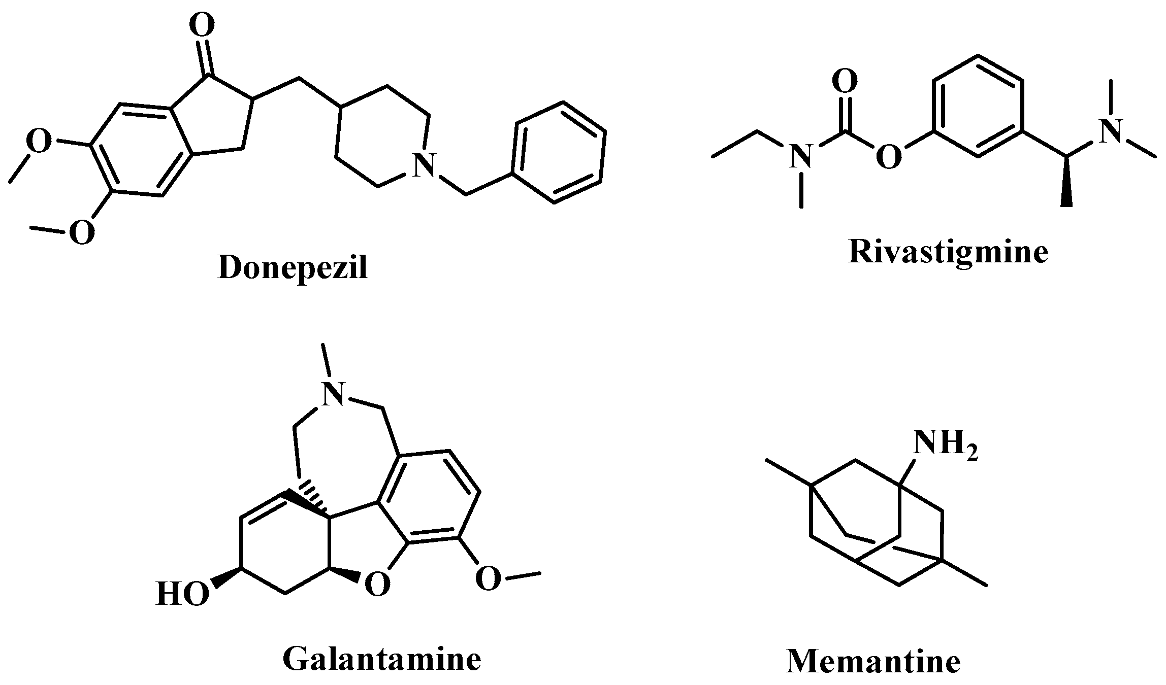
Figure 1. Anti-AD FDA-approved drugs.
Pharmaceuticals 18 01214 g001
Figure 2. Reasoned analysis of the recently created synthetic materials.
Pharmaceuticals 18 01214 g002
Scheme 1. Synthesis of cyclohepta[b]thienophen derivatives 2 and 4.
Pharmaceuticals 18 01214 sch001
Scheme 2. Formation of hybrids of cyclohepta[b]thieno-oxazepine 1116.
Pharmaceuticals 18 01214 sch002
Figure 3. Impact on (A) escape latency/time spent in target quadrant and (B) Y-maze SAP%. Data is offered as means ± SE (n = 6).
Pharmaceuticals 18 01214 g003aPharmaceuticals 18 01214 g003b
Figure 4. Effects on BACE1 and amyloid-β biomarker. Data is offered as means ± SE (n = 6). (A) BACE1 (ng/mL); (B) Amyloid-β (ng/g, Tissue).
Pharmaceuticals 18 01214 g004
Figure 5. Impact on brain neurotransmitters: (A) 5-HT, DA, and AChE; (B) BDNF; (C) NE. Data is offered as means ± SE (n = 6).
Pharmaceuticals 18 01214 g005aPharmaceuticals 18 01214 g005b
Figure 6. Impact on (A) TNF-α and IL-1β; (B) mRNA expression of TLR4, NFκβ, and NLRP3. Data is recorded as means ± SE (n = 6).
Pharmaceuticals 18 01214 g006aPharmaceuticals 18 01214 g006b
Figure 7. Effects on apoptosis of biomarkers Caspase-1, BAX, and Bcl2. The data are offered as means ± SE (n = 6).
Pharmaceuticals 18 01214 g007
Figure 8. Effects on Wnt3/β-catenin/GSK3β signaling pathway. Data is recorded as means ± SE (n = 6). (A) Wnt3; (B) GSK3β signaling; (C) β-catenin.
Pharmaceuticals 18 01214 g008
Figure 9. Effect on: (A) SOD, (B) TAC, (C) MDA, and (D) Nrf-2 and HO-1 expression. Data is recorded as means ± SE (n = 6).
Pharmaceuticals 18 01214 g009aPharmaceuticals 18 01214 g009b
Figure 10. A micrograph of a section in the CC, ST, FD, and SN. (AD) Negative control. (EH) AlCl3-induced AD group, showing a high number of shrunken degenerated neurons with severe NP in the CC, NP, FD, and necrotic neurons in ST. (IL) Donepezil group, showing moderate NP in CC, ST, FD, and SN. (MP) Compound 2 group, showing moderate NP in CC, FD, and SN, in addition to normal histology in ST. (QT) Compound 4 group, showing moderate NP in CC and SN, a moderate number of necrotic neurons in FD, as well as normal histology of ST neurons.
Pharmaceuticals 18 01214 g010
Figure 11. A micrograph from section in CC, ST, FD, and SN. (A′–D′) Compound 11, showing mild NP in neurons in CC and moderate NP in ST, FD, and SN. (E′–H′) Compound 12, showing normal histology in CC and FD, as well as mild NP in ST and SN. (I′–L′) Compound 13, showing normal histology of CC and FD, and moderate NP in ST and SN. (M′–P′) Compound 14, showing moderate NP in CC, FD, and SN, normal histology of neurons in ST. (Q′–T′) Compound 15, showing normal histology of CC and FD, and moderate NP in ST and SN. (U′–X′) Compound 16, showing mild NP in neurons in CC, as well as moderate NP in neurons of ST, FD, and SN.
Pharmaceuticals 18 01214 g011
Figure 12. IC50 values of hybrids investigated against AChE/BChE.
Pharmaceuticals 18 01214 g012
Figure 13. SAR of cyclohepta[b]thieno[1,4]oxazepine hybrids.
Pharmaceuticals 18 01214 g013
Figure 14. Bioavailability radar comparing the studied drugs with donepezil and tacrine.
Pharmaceuticals 18 01214 g014aPharmaceuticals 18 01214 g014b
Figure 15. (A) Bound ligand (green) and re-docked pose (orange) of donepezil (RMSD = 0.117 Å). (B) 2D complex overlay in 4EY7. (C) Bound ligand (green) and re-docked pose (orange) overlay (3D) of tacrine (RMSD = 0.589 Å). (D) Two-dimensional complex overlay in 4BDS.
Pharmaceuticals 18 01214 g015
Figure 16. Two-dimensional and three-dimensional binding modes of the most promising hybrids—(A) 2, (B) 14, and (C) 16—with AChE (PDB ID: 4EY7).
Pharmaceuticals 18 01214 g016aPharmaceuticals 18 01214 g016b
Figure 17. Two-dimensional and three-dimensional binding modes of the most promising hybrids—(A) 2, (B) 14, and (C) 16—with BChE (PDB ID: 4BDS).
Pharmaceuticals 18 01214 g017aPharmaceuticals 18 01214 g017b
Table 1. The impact of 2 and 4 on memory, learning and SAP%.
Pharmaceuticals 18 01214 i001
Cpd. No.RMemory (s)Learning (s)SAP%
Control-53.67 ± 1.418 ± 0.5890.83 ± 1.01
AlCl3-induced AD-9.83 ± 1.49 a49.17 ± 2.63 a47.67 ± 1.48 a
Donepezil-42.67 ± 0.61 ab14.78 ± 0.83 ab85 ± 0.58 ab
2H24.17 ± 1.22 abc33.33 ± 0.8 abc62.33 ± 1.05 abc
4-CO-CH2-Cl26.67 ± 1.54 abc31.5 ± 1.48 abc72.67 ± 1.02 abc
Note: Significance (p < 0.05) relative to (a) control group, (b) AlCl3-induced AD group, and (c) donepezil (data ± SE; n = 6).
Table 2. The effect of (1116) on memory (s), learning (s), and SAP%.
Pharmaceuticals 18 01214 i002
Cpd. No.R1Memory (s)Learning (s)SAP%
Control-53.67 ± 1.418 ± 0.5890.83 ± 1.01
AlCl3-induced AD-9.83 ± 1.49 a49.17 ± 2.63 a47.67 ± 1.48 a
Donepezil-42.67 ± 0.61 ab14.78 ± 0.83 ab85 ± 0.58 ab
11H35.17 ± 0.95 abc25.17 ± 1.25 abc69.67 ± 1.69 abc
12Cl37.17 ± 1.52 abc23.5 ± 1.23 abc76.5 ± 1.52 abc
13CN40.50 ± 2.62 abc14.17 ± 0.95 abc82.67 ± 1.28 abc
14-OCH2CH329.50 ± 2 abc21 ± 1.13 abc77.5 ± 2.11 abc
15OH42.50 ± 1.06 abc19.17 ± 1.01 abc82.5 ± 0.89 abc
16-OCH336.00 ± 1.27 abc26.83 ± 1.2 abc71.67 ± 1.15 abc
Note: Significance (p < 0.05) relative to (a) control group, (b) AlCl3-induced AD group, and (c) donepezil (data ± SE; n = 6).
Table 3. Effects of 2 and 4 on BACE1 (ng/mL) and amyloid-β(ng/g tissue).
Pharmaceuticals 18 01214 i003
Cpd. No.RBACE1 (ng/mL)Amyloid-β (ng/g Tissue)
Control-1.01 ± 0.011.59 ± 0.03
AlCl3-induced AD-29.68 ± 0.2 a27.92 ± 0.32 a
Donepezil-4.93 ± 0.04 ab8.10 ± 1.71 ab
2H23.60 ± 0.2 abc19.52 ± 0.15 abc
4-CO-CH2-Cl21.89 ± 0.27 abc17.05 ± 0.2 abc
Note: Significance (p < 0.05) relative to (a) control group, (b) AlCl3-induced AD group, and (c): donepezil (data ± SE; n = 6).
Table 4. Effects of (1116) on BACE1(ng/mL) and amyloid-β(ng/g tissue).
Pharmaceuticals 18 01214 i004
Cpd. No.R1BACE1 (ng/mL)Amyloid-β (ng/g Tissue)
Control-1.01 ± 0.011.59 ± 0.03
AlCl3-induced AD-29.68 ± 0.2 a27.92 ± 0.32 a
Donepezil-4.93 ± 0.04 ab8.10 ± 1.71 ab
11H18.83 ± 0.13 abc14.25 ± 0.16 abc
12Cl13.68 ± 0.2 abc10.51 ± 0.1 abc
13CN11.35 ± 0.31 abc9.04 ± 0.9 abc
14-OCH2CH316.66 ± 0.38 abc11.66 ± 0.17 abc
15OH10.50 ± 0.2 abc7.46 ± 0.9 abc
16-OCH317.44 ± 0.23 abc12.44 ± 0.14 abc
Note: Significance (p < 0.05) relative to (a) control group, (b) AlCl3-induced AD group, and (c) donepezil (data ± SE; n = 6).
Table 5. Effect of 2 and 4 on 5-HT (ng/g tissue), DA (ng/g tissue), and NE (nmol/g tissue). BDNF (ng/mL) and AChE (ng/g tissue) levels in brain tissues compared to donepezil and negative control.
Pharmaceuticals 18 01214 i005
Cpd. No.R5-HT (ng/g Tissue)DA (ng/g Tissue)NE (nmol/g Tissue)BDNF (ng/mL)AChE (ng/g Tissue)
Control-13.59 ± 0.2264.73 ± 0.36730.6 ± 0.36165.5 ± 0.1411.93 ± 0.26
AlCl3-induced AD-1.887 ± 0.49 a11.84 ± 1.42 a185.7 ± 1.42 a42.56 ± 0.04 a67.34 ± 0.22 a
Donepezil-11.23 ± 0.41 ab58.12 ± 14.01 ab635.4 ± 14.01 ab122.4 ± 0.48 ab13.91 ± 0.42 b
2H3.16 ± 0.63 abc24.71 ± 1.62 abc256.9 ± 1.62 abc64.95 ± 0.01 abc51.41 ± 0.13 abc
4-CO-CH2-Cl4.29 ± 0.76 abc30.69 ± 1.2 abc282.6 ± 1.2 abc75.75 ± 0.01 abc43.89 ± 0.19 abc
Note: Significance (p < 0.05) relative to (a) control group, (b) AlCl3-induced AD group, and (c) donepezil (data ± SE; n = 6).
Table 6. Effect of (1116) on 5-HT (ng/g tissue), DA (ng/g tissue), and NE (nmol/g tissue). BDNF (ng/mL) and AChE (ng/g tissue) levels in brain tissues compared to donepezil and negative control.
Pharmaceuticals 18 01214 i006
Cpd. No.R15-H (ng/g Tissue)DA (ng/g Tissue)NE (nmol/g Tissue)BDNF (ng/mL)AChE (ng/g Tissue)
Control-13.59 ± 0.2264.73 ± 0.36730.6 ± 0.36165.5 ± 0.1411.93 ± 0.26
AlCl3-induced AD-1.887 ± 0.49 a11.84 ± 1.42 a185.7 ± 1.42 a42.56 ± 0.04 a67.34 ± 0.22 a
Donepezil-11.23 ± 0.41 ab58.12 ± 0.14 ab635.4 ± 14.01 ab122.4 ± 0.48 ab13.91 ± 0.42 ab
11H9.13 ± 0.36 abc54.63 ± 6.11 abc321.2 ± 6.11 abc127.3 ± 0.01 abc20.09 ± 0.45 abc
12Cl5.07 ± 0.18 abc38.37 ± 2.86 abc420.5 ± 2.86 abc83.62 ± 0.06 abc35.66 ± 0.19 abc
13CN7.39 ± 0.1 abc48.65 ± 2.3 abc449.6 ± 2.3 abc104.9 ± 0.07 abc24.35 ± 0.8 abc
14-OCH2CH38.22 ± 0.19 abc51.88 ± 2.66 abc386.7 ± 2.66 abc115.8 ± 0.07 abc19.4 ± 0.39 abc
15OH6.77 ± 0.22 abc44.92 ± 2.85 abc499.8 ± 2.85 abc94.48 ± 0.05 abc25.73 ± 0.37 abc
16-OCH310.65 ± 0.71 abc52.96 ± 1.52 abc366.4 ± 1.52 abc134.8 ± 0.01 abc16.83 ± 0.29 abc
Note: Significance (p < 0.05) relative to (a) control group, (b) AlCl3-induced AD group, and (c) donepezil (data ± SE; n = 6).
Table 7. Effect of 2 and 4 on TNF-α (pg/g tissue) and IL-β (pg/g tissue), along with mRNA expression levels of TLR4, NFκβ, and NLRP3.
Pharmaceuticals 18 01214 i007
Cpd. No.RTNF-α (pg/g Tissue)IL-β (pg/g Tissue)TLR4NFκβNLRP3
Control-26.75 ± 0.1631.3 ± 0.281.02 ± 0.011.07 ± 0.011.04 ± 0.004
AlCl3-induced AD-176 ± 1.05 a118.4 ± 0.5 a9.63 ± 0.08 a8.48 ± 0.15 a10.68 ± 0.09 a
Donepezil-61.7 ± 1.12 ab58.75 ± 0.99 ab2.72 ± 0.02 ab3.0 ± 0.05 ab2.90 ± 0.04 ab
2H121.4 ± 0.97 abc93.93 ± 1.26 abc7.66 ± 0.12 abc6.53 ± 0.08 abc8.29 ± 0.09 abc
4-CO-CH2-Cl113.6 ± 0.83 abc82.82 ± 1.1 abc6.39 ± 0.12 abc5.77 ± 0.19 abc6.30 ± 0.09 abc
Note: Significance (p < 0.05) relative to (a) control group, (b) AlCl3-induced AD group, and (c) donepezil (data ± SE; n = 6).
Table 8. Effect of (1116) on TNF-α (pg/g tissue) and IL-β (pg/g tissue), in addition to mRNA expression levels of TLR4, NFκβ, and NLRP3.
Pharmaceuticals 18 01214 i008
Cpd. No.R1TNF-α (pg/g Tissue)IL-β (pg/g Tissue)TLR4NFκβNLRP3
Control-26.75 ± 0.1631.3 ± 0.281.02 ± 0.011.07 ± 0.011.04 ± 0.004
AlCl3-induced AD-176 ± 1.05 a118.4 ± 0.5 a9.63 ± 0.08 a8.48 ± 0.15 a10.68 ± 0.09 a
Donepezil-61.7 ± 1.12 ab58.75 ± 0.99 ab2.72 ± 0.02 ab3.0 ± 0.05 ab2.90 ± 0.04 ab
11H99.23 ± 0.42 abc68.56 ± 0.54 abc5.56 ± 0.1 abc4.93 ± 0.08 abc5.39 ± 0.11 abc
12Cl65.98 ± 0.91 abc54.51 ± 1.3 abc2.75 ± 0.06 abc4.30 ± 0.14 abc3.21 ± 0.12 abc
13CN52.38 ± 0.59 abc45.79 ± 1.34 abc4.09 ± 0.27 abc3.49 ± 0.07 abc2.86 ± 0.11 abc
14-OCH2CH373.21 ± 0.58 abc59.92 ± 0.98 abc3.60 ± 0.12 abc3.73 ± 0.04 abc3.92 ± 0.04 abc
15OH44.6 ± 0.6 abc45.66 ± 1.03 abc4.57 ± 0.09 abc2.98 ± 0.10 abc3.36 ± 0.12 abc
16-OCH385.17 ± 0.46 abc53.04 ± 0.61 abc4.43 ± 0.08 abc4.70 ± 0.12 abc4.32 ± 0.10 abc
Note: Significance (p < 0.05) relative to (a) control group, (b) AlCl3-induced AD group, and (c) donepezil (data ± SE; n = 6).
Table 9. Effect of 2 and 4 on Caspase-1, BAX, and Bcl-2 gene expression.
Pharmaceuticals 18 01214 i009
Cpd. No.RCaspase-1BAXBcl-2BAX/Bcl-2 Ratio
Control-1.02 ± 0.0031.01 ± 0.0041.08 ± 0.0060.94
AlCl3-induced AD-9.52 ± 0.009 a9.68 ± 0.160 a0.12 ± 0.002 a80.67
Donepezil-2.92 ± 0.050 ab2.25 ± 0.058 ab0.98 ± 0.002 ab2.30
2H7.46 ± 0.120 abc8.13 ± 0.070 abc0.27 ± 0.007 abc30.11
4-CO-CH2-Cl6.46 ± 0.120 abc7.42 ± 0.030 abc0.38 ± 0.020 abc19.53
Note: Significance (p < 0.05) relative to (a) control group, (b) AlCl3-induced AD group, and (c) donepezil (data ± SE; n = 6).
Table 10. Effects of compounds 1116 on Caspase-1, BAX, and Bcl-2 gene expression.
Pharmaceuticals 18 01214 i010
Cpd. No.R1Caspase-1BAXBcl-2BAX/Bcl-2 Ratio
Control-1.02 ± 0.0031.01 ± 0.0041.08 ± 0.0060.94
AlCl3-induced AD-9.52 ± 0.009 a9.68 ± 0.160 a0.12 ± 0.002 a80.67
Donepezil-2.92 ± 0.050 ab2.25 ± 0.0580 ab0.98 ± 0.002 ab2.30
11H5.92 ± 0.040 abc6.84 ± 0.090 abc0.52 ± 0.020 abc13.15
12Cl4.54 ± 0.090 abc4.85 ± 0.110 abc0.82 ± 0.020 abc5.91
13CN3.85 ± 0.070 abc5.80 ± 0.120 abc0.79 ± 0.020 abc7.34
14-OCH2CH35.08 ± 0.050 abc5.49 ± 0.100 abc0.70 ± 0.004 abc7.84
15OH2.94 ± 0.110 abc4.47 ± 0.040 abc0.86 ± 0.020 abc5.20
16-OCH35.04 ± 0.130 abc6.14 ± 0.090 abc0.63 ± 0.020 abc9.75
Note: Significance (p < 0.05) relative to (a) control group, (b) AlCl3-induced AD group, and (c) donepezil (data ± SE; n = 6).
Table 11. Effects of 2 and 4 on Wnt3/β-catenin/GSK-3β signaling.
Pharmaceuticals 18 01214 i011
Cpd. No.RWnt3a (ng/g Tissue)GSK3ββ-Catenin (ng/L)
Control-1.01 ± 0.0031.01 ± 0.00416.94 ± 0.14
AlCl3-induced AD-0.12 ± 0.003 a10.20 ± 0.03 a1.578 ± 0.09 a
Donepezil-0.98 ± 0.01 ab2.05 ± 0.07 ab12.92 ± 0.48 ab
2H0.25 ± 0.01 abc8.80 ± 0.12 abc4.262 ± 0.12 abc
4-CO-CH2-Cl0.34 ± 0.01 abc6.86 ± 0.15 abc6.17 ± 0.07 abc
Note: Significance (p < 0.05) relative to (a) control group, (b) AlCl3-induced AD group, and (c) donepezil (data ± SE; n = 6).
Table 12. Effects of compounds (1116) on Wnt3/β-catenin/GSK-3β signaling.
Pharmaceuticals 18 01214 i012
Cpd. No.R1Wnt3a (ng/g Tissue)GSK3ββ-Catenin (ng/L)
Control-1.01 ± 0.0031.01 ± 0.00416.94 ± 0.14
AlCl3-induced AD-0.12 ± 0.003 a10.20 ± 0.03 a1.578 ± 0.09 a
Donepezil-0.98 ± 0.01 ab2.05 ± 0.07 ab12.92 ± 0.48 ab
11H0.47 ± 0.01 abc2.34 ± 0.06 abc7.25 ± 0.04 abc
12Cl0.76 ± 0.01 abc5.36 ± 0.07 abc9.91 ± 0.15 abc
13CN0.84 ± 0.01 abc3.63 ± 0.04 abc11.67 ± 0.13 abc
14-OCH2CH30.66 ± 0.02 abc3.13 ± 0.07 abc8.4 ± 0.09 abc
15OH0.88 ± 0.01 abc4.72 ± 0.05 abc13.86 ± 0.11 abc
16-OCH30.52 ± 0.01 abc2.18 ± 0.10 abc7.34 ± 0.12 abc
Note: Significance (p < 0.05) relative to (a) control group, (b) AlCl3-induced AD group, and (c) donepezil (data ± SE; n = 6).
Table 13. Effect of 2 and 4 on SOD, TAC, and MDA levels, as well as Nrf-2 and HO-1 mRNA expression in brain tissues compared to donepezil and negative control.
Pharmaceuticals 18 01214 i013
Cpd. No.RSOD(U/g)TAC (µmol/g Tissue)MDA
(nmol/g Tissue)
Nrf-2HO-1
Control-3.052 ± 0.446.68 ± 0.057.105 ± 0.031.07 ± 0.0061.02 ± 0.003
AlCl3-induced AD-0.38 ± 0.38 a9.55 ± 0.250 a116.3 ± 0.07 a0.16 ± 0.020 a0.12 ± 0.007 a
Donepezil-5.78 ± 0.97 ab34.88 ± 1.510 ab25.25 ± 0.35 ab0.82 ± 0.001 ab0.81 ± 0.002 ab
2H0.68 ± 0.26 abc18.26 ± 0.395 abc92.07 ± 0.04 abc0.25 ± 0.008 abc0.34 ± 0.032 abc
4-CO-CH2-Cl1.75 ± 0.31 abc25.87 ± 0.965 abc77.14 ± 0.06 abc0.31 ± 0.004 abc0.39 ± 0.007 abc
Note: Significance (p < 0.05) relative to (a) control group, (b) AlCl3-induced AD group, and (c) donepezil (data ± SE; n = 6).
Table 14. Effect of (1116) on SOD, TAC, and MDA levels, as well as Nrf-2 and HO-1 mRNA expression in brain tissues compared to donepezil and negative control.
Pharmaceuticals 18 01214 i014
Cpd. No.R1SOD(U/g)TAC (µmol/g Tissue)MDA (nmol/g Tissue)Nrf-2HO-1
Control-3.05 ± 0.446.68 ± 0.057.10 ± 0.031.07 ± 0.0061.02 ± 0.003
AlCl3-induced AD-0.38 ± 0.38 a9.55 ± 0.25 a116.3 ± 0.07 a0.16 ± 0.02 a0.12 ± 0.007 a
Donepezil-5.78 ± 0.97 ab34.88 ± 1.51 ab25.25 ± 0.35 ab0.82 ± 0.01 ab0.81 ± 0.002 ab
11H2.46 ± 0.32 abc38.23 ± 0.50 abc36.75 ± 0.09 abc0.35 ± 0.006 abc0.53 ± 0.004 abc
12Cl2.15 ± 0.39 abc29.33 ± 0.57 abc72.02 ± 0.05 abc0.64 ± 0.020 abc0.69 ± 0.020 abc
13CN2.53 ± 0.25 abc32.9 ± 1.22 abc52.58 ± 0.08 abc0.78 ± 0.010 abc0.75 ± 0.010 abc
14OCH2CH32.38 ± 0.78 abc36.96 ± 0.51 abc47.6 ± 0.12 abc0.50 ± 0.020 abc0.64 ± 0.009 abc
15OH2.56 ± 0.54 abc31.67 ± 0.42 abc63.68 ± 0.12 abc0.82 ± 0.020 abc0.82 ± 0.005 abc
16OCH32.92 ± 0.20 abc42.42 ± 0.84 abc31.63 ± 0.05 abc0.47 ± 0.005 abc0.50 ± 0.020 abc
Note: Significance (p < 0.05) relative to (a) control group, (b) AlCl3-induced AD group, and (c) donepezil (data ± SE; n = 6).
Table 15. IC50 values and Selectivity Index (SI) of 2 and 4 against AChE/BChE vs. donepezil and tacrine.
Pharmaceuticals 18 01214 i015
Cpd. No.RIC50(µM)Selectivity Index
BChE/AChE
AChEBChE
2H1.10 ± 0.045.72 ± 0.205.21
4-CO-CH2-Cl41.22 ± 1.4256.19 ± 1.931.36
Donepezil-0.22 ± 0.01--
Tacrine--0.18 ± 0.01-
Table 16. IC50 values and Selectivity Index (SI) of compounds (1116) against AChE/BChE vs. donepezil and tacrine.
Pharmaceuticals 18 01214 i016
Cpd. No.R1IC50 (µM)Selectivity Index
BChE/AChE
AChEBChE
11H2.67 ± 0.0913.85 ± 0.485.18
12Cl10.90 ± 0.3865.86 ± 2.276.04
13CN5.04 ± 0.174.22 ± 0.140.84
14-OCH2CH30.39 ± 0.012.93 ± 0.107.50
15OH1.59 ± 0.060.70 ± 0.020.44
16-OCH30.76 ± 0.035.23 ± 0.186.89
Donepezil-0.22 ± 0.01--
Tacrine--0.18 ± 0.01-
Table 17. Physicochemical attributes of synthetic hybrids, donepezil, and tacrine.
Molecule#H-Bond Donors#H-Bond AcceptorsMLogPMWPAINS AlertsSynthetic Accessibility
2132.17240.3203.14
4042.83316.8003.48
11033.18327.4003.34
12033.68361.8403.33
13042.51352.4103.38
14043.08371.4503.58
15142.62343.4003.37
16042.85357.4203.47
Donepezil043.06379.4903.36
Tacrine112.33198.2602.08
Table 18. TPSA and determined percentage of ABS for generated compounds compared to donepezil and tacrine.
Cpd. No.TPSA (Å2)%ABS
280.84081.110
480.84081.110
1157.78089.060
1257.78089.060
1367.01085.880
1467.01085.880
1557.78089.060
1657.78089.060
Donepezil38.77095.620
Tacrine38.91095.580
Table 19. ADME/pharmacokinetic attributes of the hybrids investigated.
Cpd. NoBBB Perm-eantGI
Abs
P-gp SubstrateCYP1A2 InhibitorCYP2C19 InhibitorCYP2C9 InhibitorCYP2D6 InhibitorCYP3A4 InhibitorLog Kp
20.199High+++−4.84
40.112High+++−5.18
110.382High++++−5.26
120.824High+++−5.02
130.017High++++−5.61
140.002High+++++−5.29
150.027High+++++−5.60
160.027High+++++−5.46
Donepezil0.353High+++−5.58
Tacrine0.977High+++−5.59
Table 20. Drug-likeness of the investigated hybrids.
Cpd. No.LipinskiVeberGhoseMueggeEganBioavailability
Score
2YesYesYesYesYes0.55
4YesYesYesYesYes0.55
11YesYesYesYesYes0.55
12YesYesYesYesYes0.55
13YesYesYesYesYes0.55
14YesYesYesYesYes0.55
15YesYesYesYesYes0.55
16YesYesYesYesYes0.55
DonepezilYesYesYesYesYes0.55
TacrineYesYesYesNoYes0.55
Table 21. Toxicity of hits 2, 14, and 16 versus donepezil and tacrine.
pkCSM
Test2 14 16 DonepezilTacrine
AMES toxicityNoNoYesNoYes
Max. tolerated dose (human)0.668−0.116−0.18−0.2170.55
hERG I inhibitionNoNoNoNoNo
hERG II inhibitionNoNoNoYesNo
LD502.2012.6662.7622.7532.33
LOAEL1.8842.0482.1130.9911.204
HepatotoxicityNoNoNoYesYes
Skin SensitizationYesNoNoNoNo
T. Pyriformis toxicity0.981.0520.9810.8040.642
Minnow toxicity1.138−0.0410.05−2.0110.206
ProTox-II prediction
LD50 (mg/kg)15002000200050540
Toxicity ClassIVIVIVIVII
Immunotoxicity(−) (0.99)(−) (0.91)(−) (0.89)(+) (0.95)(−) (0.98)
Mutagenicity(−) (0.74)(−) (0.67)(−) (0.66)(−) (0.53)(+) (0.91)
Cytotoxicity(−) (0.76)(+) (0.50)(−) (0.50)(+) (0.63)(−) (0.72)
(Tumor Suppressor) p53(−) (0.85)(−) (0.80)(−) (0.79)(−) (0.94)(−) (0.95)
Table 22. (S) values (Kcal·mol−1), interactions, and distances (Å) of donepezil (PDB ID: 4EY7) and tacrine (PDB ID: 4BDS).
Drug(S) (kCal/mol)Receptor Amino AcidInteractionDistance (Å)
Donepezil−17.710Phe295H-acceptor2.98
Tyr341H–arene3.67
Tyr337H–arene3.71
Trp286H–arene3.87
Trp286arene-arene3.88
Tacrine−10.080His438H-donor3.32
His438H–arene4.54
Trp82arene-arene3.89
Table 23. (S) values (Kcal·mol−1), interactions, and distances (Å) of 2, 14, and 16 against AChE and BChE.
Cpd.
No.
(S) (kCal/mol)Receptor Amino AcidInteractionDistance (Å)(S) (kCal/mol)Receptor Amino AcidInteractionDistance (Å)
AChEBChE
2−14.45Ser293
Trp286
Trp286
Phe338
H-donor
H-pi
H-pi
H-pi
3.8
4.07
4.1
4.53
−14.97Asp70
His438
Trp82
H-donor
H-pi
pi-pi
4.01
4.68
3.85
14−14.74Phe295
Trp286
H-acceptor
pi-pi
3.1
3.6
−18.10Asp70
Gly78
Trp82
H-donor
H-donor
pi-pi
4.22
3.68
3.89
16−15.09Phe295
Trp286
Tyr341
H-acceptor
H-pi
pi-H
2.77
4.01
4.21
−16.41Asp70
Gly439
Gly116
Trp82
H-donor
H-acceptor
H-acceptor
pi-pi
4.4
3.34
3.08
3.72
Disclaimer/Publisher’s Note: The statements, opinions and data contained in all publications are solely those of the individual author(s) and contributor(s) and not of MDPI and/or the editor(s). MDPI and/or the editor(s) disclaim responsibility for any injury to people or property resulting from any ideas, methods, instructions or products referred to in the content.