Abstract
Chromodomain helicase DNA-binding protein 8 (CHD8), a frequently mutated gene in autism spectrum disorder (ASD), is an ATP-dependent chromatin remodeler with emerging roles in hematopoiesis. While CHD8 is known to maintain hematopoietic stem and progenitor cells (HSPCs) in the bone marrow, its function during developmental hematopoiesis remains undefined. Here, using a zebrafish model, we demonstrate that chd8 loss severely depletes the HSPC pool in the caudal hematopoietic tissue through a p53-dependent apoptotic mechanism. Furthermore, chd8−/− embryos exhibit a p53-independent expansion of myelopoiesis. chd8 deficiency upregulates brd4, which promotes systemic inflammatory cytokine expression. Inhibiting brd4 alleviates cytokine expression, suppresses excessive myelopoiesis, and restores HSPC development. Our findings reveal a dual regulatory mechanism in which chd8 governs HSPC development by repressing p53-mediated apoptosis and constraining brd4-driven immune cell differentiation.
1. Introduction
Hematopoietic stem and progenitor cells (HSPCs) are the foundation of the vertebrate hematopoietic system, responsible for the lifelong production and maintenance of all blood cell lineages [1]. HSPCs first arise during embryogenesis from the dorsal aorta in the aorta–gonad–mesonephros (AGM) region. Following their emergence, HSPCs migrate to the fetal liver in mammals or the caudal hematopoietic tissue (CHT) in zebrafish before ultimately colonizing the mammalian bone marrow or zebrafish kidney, where they sustain adult hematopoiesis [2,3,4].
In the fetal liver or CHT, HSPCs undergo rapid expansion and differentiation. The extensive proliferation induces replicative stress, which can trigger apoptosis to eliminate compromised cells and preserve the integrity of the HSPC pool [5,6,7,8]. Conversely, concurrent survival signals are essential to maintain a balance between cell death and propagation, ensuring proper HSPC expansion and preventing excessive depletion [9]. This critical equilibrium between apoptosis and survival, and between expansion and differentiation, is tightly regulated by elaborate signaling pathways, including transcriptional and epigenetic mechanisms [5].
Chromatin remodeling enzymes are essential regulators of chromatin structure and function, playing critical roles in stem cell biology. Among these, the chromodomain helicase DNA-binding (CHD) family is a major subtype defined by the presence of two chromodomains, a central helicase/ATPase domain, and a DNA-binding domain [10]. These proteins utilize energy from ATP hydrolysis to modulate DNA accessibility for transcription factors and other epigenetic modifiers, making them indispensable for proper developmental processes [11]. Notably, several CHD proteins, including CHD2, CHD4, and CHD7, have been established as key regulators of stem cell maintenance and differentiation [12,13,14].
CHD8 was originally identified as one of the most frequently mutated genes in autism spectrum disorders (ASDs) [15,16]. Prior research has largely focused on its critical role in brain development, including regulating pluripotency and early neurodifferentiation [17,18]. Recent studies have revealed its functions in hematopoiesis. CHD8 governs HSPC survival and stemness by restricting p53 activity in the adult bone marrow [19,20], promotes erythroid differentiation [21] and regulates Treg cells [22]. Although CHD8 is expressed throughout development, its role in HSPC development during embryogenesis remains unknown because of the early embryonic lethality of Chd8-null mice [23].
We previously demonstrated that a chd8 mutation in zebrafish disrupted the intestinal epithelial integrity, leading to a microbiota imbalance that impairs HSPC development [24]. But whether chd8 intrinsically affects HSPC survival remains unknown. In this study, we found that chd8 is dispensable for the initial emergence and migration of HSPCs, but it is essential for their subsequent survival within the CHT by suppressing p53-dependent apoptosis. We also found that chd8 loss leads to p53-independent myeloid differentiation, and that Brd4 inhibition rebalances HSPC development and myeloid differentiation.
2. Results
2.1. Chd8 Is Expressed in Developing HSPCs and chd8−/− Zebrafish Exhibit Significant Lethality
To study the intrinsic role of chd8 in hematopoiesis, we first analyzed its expression at different developmental stages and within hematopoietic cells using real-time quantitative PCR (RT-qPCR). Chd8 exhibited a dynamic expression profile, with transcript levels peaking during early developmental stages and gradually declining as embryogenesis proceeded (Figure 1A). Furthermore, chd8 expression is highly enriched in fluorescence-activated cell sorting (FACS)-purified drl-GFP+ hematopoietic cells [25] and CD41-GFP+ HSPCs [8] compared to the whole embryos. This enrichment was also observed in embryonic kidneys, the site of definitive hematopoiesis in zebrafish (Figure 1B). The specific enrichment of chd8 in HSPC-containing tissues and populations supports its potential for a cell-intrinsic function in these cells.
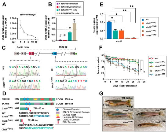
Figure 1.
Characterization of chd8 mRNA expression and two chd8 mutant lines. (A) Developmental expression profile of chd8 in whole embryos. Transcript levels were quantified by RT-qPCR and normalized to gapdh. (B) chd8 expression in whole embryos, embryonic kidneys, sorted drl-GFP+-hematopoietic cells, and CD41-GFP+-HSPCs. (C) Upper panel: genomic structure of zebrafish chd8, with targeted exons 3 and 12 highlighted. Lower panel: Sanger sequencing chromatograms confirming the deletion in chd8−/− mutants. The deleted nucleotides are shown in squares. (D) Domain architecture of human CHD8, zebrafish Chd8, and the truncated mutants (M1, M2) predicted from the genomic deletions. The asterisk indicates the premature stop codon. (E) chd8 transcript levels in WT, chd8+/−, and chd8−/− embryos from two mutant lines. (F) Survival analysis of WT, chd8+/−, and chd8−/− zebrafish from 1 dpf to 1 mpf (n = 100–200 per group). (G) Representative images of adult WT and chd8+/−, and chd8−/− (6 mpf). Data are presented as mean ± SD from three independent experiments (two-tailed Student’s t-test, * p < 0.05, ** p < 0.01, # p < 0.001). WT, wild type; dpf/mpf, days/months post-fertilization; bp, base pairs; aa, amino acids.
To study the hematopoietic development, we used two zebrafish chd8−/− mutant lines previously established in our laboratory, chd8M1−/− and chd8M2−/− [24]. The zebrafish Chd8 gene and the mammalian CHD8 protein are highly conserved. The amino acid homology between zebrafish Chd8 and human CHD8 is approximately 70%. The full-length zebrafish Chd8 protein comprises 2555 amino acids, and the two lines produce truncated proteins of 800 aa and 50 aa, respectively, due to premature termination codons (Figure 1C,D). RT-qPCR analysis showed a ~50% reduction in chd8 mRNA in heterozygotes (chd8+/−) and nearly undetectable levels in homozygous mutants (chd8−/−) (Figure 1E). Both mutant lines exhibited significant mortality rates, reaching 35–45% by 5 days post-fertilization (dpf), with a continued increase thereafter (Figure 1F). A small fraction of mutants survived to adulthood but were notably leaner than their wild-type (WT) counterparts at 6 months post-fertilization (mpf) (Figure 1G). chd8 heterozygotes showed no significant mortality or morphological abnormalities (Figure 1E,G). The viable chd8 mutant embryos give us a useful tool to study embryonic hematopoiesis. The two mutant lines exhibited similar hematological phenotypes. Unless specifically mentioned, the subsequent results are primarily from the chd8M1−/−, referred to hereafter as chd8−/− for simplicity.
2.2. Loss of chd8 Does Not Affect Primitive Hematopoiesis
Similar to mammals, zebrafish hematopoiesis occurs in two consecutive waves: primitive and definitive [4]. We first investigated whether chd8 mutants exhibited defects in primitive hematopoiesis. Using whole-mount in situ hybridization (WISH), we found that the expression of myeloid markers Pu.1 (at 22 hpf), mpx and mfap4 (at 24 hpf) showed no difference between chd8−/− mutants and their WT siblings. Similarly, the expression of erythroid progenitor marker gata 1 (at 24 hpf), and the erythrocyte markers hbbe3 and hbbe1 (at 22–24 hpf) were unaltered in chd8−/− embryos (Figure 2). These results suggest that primitive myeloid and erythroid development remains intact in the absence of chd8, indicating that chd8 is not required for primitive hematopoiesis.
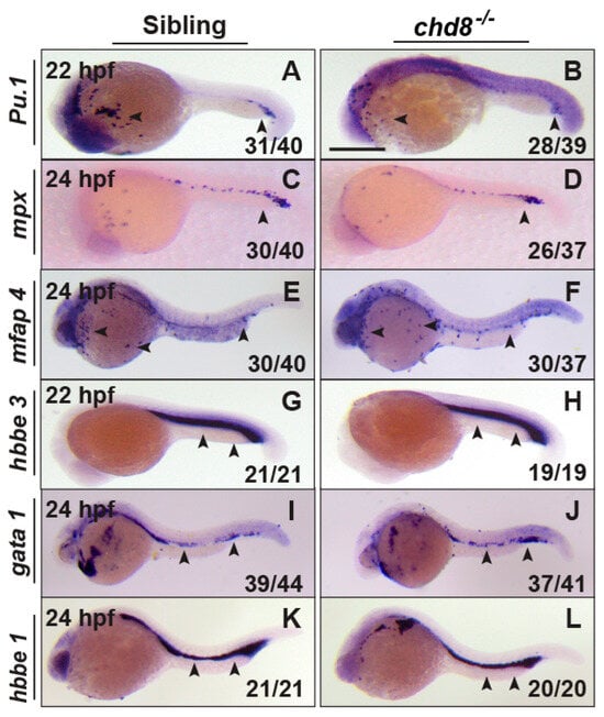
Figure 2.
Chd8 loss does not affect primitive hematopoiesis. WISH analysis of PU.1 (A,B), mpx (C,D), mfap4 (E,F), hbbe3 (G,H), gata 1 (I,J), and hbbe1 (K,L) in chd8−/− and their corresponding WT sibling embryos at 22–24 hpf. The fraction (n/n) in the bottom right corner indicates the number of embryos displaying the representative phenotype over the total number examined. WISH, whole-mount in situ hybridization; Scale bar, 200 µm.
2.3. Loss of chd8 Impairs HSPC Development in the CHT
To characterize the role of chd8 in definitive hematopoiesis, we analyzed the expression of HSPC markers cmyb and runx1. At 30 and 36 hpf, the expression of c-myb and runx1 in the AGM was properly specified and slightly elevated in chd8−/− mutants (Figure 3A,B), indicating HSPC emergence is normal or potentially enhanced. At 2 dpf, cmyb and runx1 expression remained intact, suggesting successful HSPC migration to the CHT. However, by 3 dpf, cmyb expression in the CHT was decreased, and by 5 dpf, it was strongly reduced in chd8−/− mutants (Figure 3C,D). This reduction persisted in the embryonic kidneys at 7 dpf (Figure 3E). Taken together, these results revealed that while HSPC initiation and migration were unaffected in chd8−/− mutants, their transient expansion within the CHT is severely impaired. We also examined heterozygous mutants and observed no hematopoietic defects, indicating that a single allele loss of chd8 does not impair hematopoiesis in zebrafish (Figure S1).
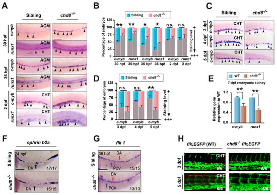
Figure 3.
chd8 is required for maintaining HSPCs in the CHT. (A,C) Representative WISH images tracking definitive HSPC markers c-myb and runx1 in WT sibling and chd8−/− embryos from 30 hpf to 5 dpf. (B,D) Quantification of the WISH signal for c-myb and runx1 in (A,C), respectively. (E) RT-qPCR analysis of c-myb and runx1 expression in the embryonic kidney at 7 dpf. (F,G) WISH of the arterial marker ephrinb2a at 24 hpf and the vascular marker flk1 at 36 hpf. (H) Live imaging of vascular plexus in the CHT region of Tg (flk: GFP) WT and chd8−/− embryos at 3 and 5 dpf. Data in (B,D,E) are presented as mean ± SD from three independent experiments (two-tailed Student’s t-test, n = 20–30 embryos per group, each experiment, * p < 0.05, ** p < 0.01, n.s., not significant). AGM, aorta–gonad–mesonephros; CHT, caudal hematopoietic tissue. Scale bars, 100 µm (A,F,G); 50 µm (H).
Since HSPCs originate from arterial vessels and their development is influenced by vasculogenesis and blood flow [26], we assessed vascular integrity. The expression of arterial (ephrinb2) and endothelial (flk1) markers was normal at 24 and 36 hpf (Figure 3F,G). Live imaging of the vascular plexus in Tg(flk1:EGFP) [5] embryos at 3 and 5 dpf further confirmed the normal vascular morphology in chd8−/− mutants (Figure 3H). We also did not find any circulation defects in the mutants. These findings together support a specific effect of chd8 in definitive hematopoiesis.
2.4. Loss of chd8 Leads to Enhanced Immune Cell Differentiation
To explore the impact of chd8 on HSPC function, we analyzed a series of lineage markers in chd8−/− embryos. WISH revealed a significant upregulation of the neutrophil markers mpx and L-plastin at 3, 4 and 5 dpf in chd8−/− embryos (Figure 4A–C). Similarly, expression of the macrophage markers mfap4, apoE, and neutral red-stained macrophages was increased in the chd8−/− CHT. We also observed elevated expression of the lymphoid marker rag1. These findings indicate that chd8 deficiency enhances both myeloid and lymphoid differentiation. As with HSPC, chd8 haploinsufficiency did not produce detectable abnormal phenotypes in differentiation (Figure S1).
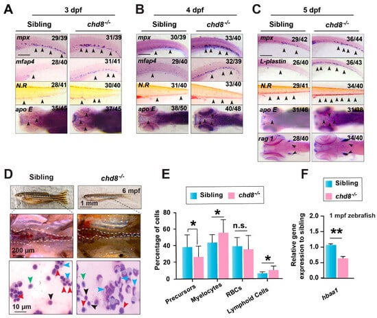
Figure 4.
chd8 loss leads to excessive myeloid and lymphoid differentiation. (A–C) WISH for hematopoietic lineage markers in WT sibling and chd8−/− embryos from 3 to 5 dpf. Neutrophils were labeled with mpx and l-plastin, and macrophages were labeled with mfap4, apoE, and neutral red (N.R.) staining; lymphoid cells were labeled with rag1. (D) Kidney morphology and May-Grünwald-Giemsa-stained WKM cells from 6-month-old WT and chd8−/− zebrafish. Cell types are indicated by arrows: blue, myelocytes; black, lymphoid cells; red, precursors; green, red blood cells. (E) Quantification of different cell populations in the WKM, based on morphological analysis in (D). (F) RT-qPCR analysis of hemoglobin gene hbaa1 expression in 1-month-old WT and chd8−/− zebrafish. Data are presented as mean ± SD from three independent experiments (two-tailed Student’s t-test, n = 5–6 fish per experiment, * p < 0.05, ** p < 0.01, n.s., not significant). WKM, whole kidney marrow. Scale bars, 100 µm (A–C).
We next asked whether the increased differentiation potential persists into adulthood. May-Grünwald-Giemsa staining of whole kidney marrow (WKM) from 6-month-old fish showed that, consistent with the embryonic phenotype, precursor cells were reduced while mature lymphoid and myeloid populations were expanded in chd8−/− (Figure 4D,E). Tu et al. reported that Chd8 loss causes anemia in mice [21], and we observed an increase in the erythroid marker hbbe1 and benzidine-stained globin in chd8−/− (Figure S2). But we found a significant reduction in hbaa1 expression starting from 1-month-old mutants (Figure 4F), suggesting a potential late-onset erythroid defect. These hematopoietic phenotypes were recapitulated in a second mutant allele, chd8M2−/− (Figure S3). Thus, our results demonstrate that chd8 loss disrupts HSPCs development and lineage output.
2.5. Loss of chd8 Induces p53-Mediated Apoptosis in HSPCs
Previous studies have shown that chd8 safeguards HSPCs by suppressing p53-dependent apoptosis in the bone marrow [22]. To determine if a similar mechanism operates in zebrafish, we assessed apoptosis in chd8-deficient embryos. Quantitative analysis revealed a significant upregulation of key apoptosis-related genes, including p53, caspase-3, and baxa, in whole embryos at 1, 3, and 5 dpf (Figure 5A). This elevation was even more pronounced within sorted drl-GFP+ hematopoietic cells at 5 dpf (Figure 5B), suggesting hematopoietic cell-intrinsic activation of the apoptotic program. Consistent with this, TUNEL assays demonstrated a substantial increase in apoptotic CD41-GFP+ HSPCs in the CHT of chd8−/− embryos (Figure 5C,D). These findings imply that the definitive hematopoiesis defect in chd8−/− originates from increased HSPC apoptosis. To directly test whether p53 upregulation mediates this phenotype, we knocked down p53 in chd8−/− embryos using a morpholino (MO) [27]. WISH showed a complete rescue of cmyb+ HSPCs in both the CHT and the embryonic kidney of chd8−/− embryos (Figure 5E,F), confirming that the HSPC loss is p53-dependent. Interestingly, p53 knockdown failed to suppress the enhanced myelopoiesis in chd8−/− embryos (Figure 5G,H), and did not inhibit increased erythroid differentiation (Figure S2D,E). In contrast, it partially rescued rag1 expression (Figure 5I,J). These results suggest the enhanced myeloid and erythroid differentiation in chd8−/− embryos occurs largely via a p53-independent mechanism. This finding aligns with the work of Tu et al., indicating that Chd8 possesses p53-independent functions in murine erythroid development. Collectively, our data demonstrate that chd8 deficiency depletes HSPCs by triggering p53-dependent apoptosis, but inhibiting p53 restores survival without fully rescuing their lineage differentiation defects.
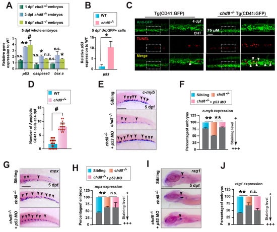
Figure 5.
HSPCs undergo p53-dependent apoptosis in chd8−/− embryos. (A) RT-qPCR analysis of p53, caspase3, and baxa expression in WT and chd8−/− embryos at 1, 3, and 5 dpf. (B) RT-qPCR analysis of p53 in drl-GFP+ cells sorted from WT and chd8−/− embryos. (C) Representative images of CD41-GFP+ and TUNEL double staining in WT and chd8−/− embryos at 4 dpf. The white arrowheads indicate apoptotic HSPCs in the CHT. (D) Quantification of TUNEL-positive HSPCs from the images in (C) (n = 6 embryos per group). (E,G,I) WISH analysis for c-myb, mpx, rag1 in WT and chd8−/− embryos following injection with p53 MO. (F,H,J) Quantitative analysis of the WISH results in (E,G,I), respectively. Data are presented as mean ± SD from three independent experiments (two-tailed Student’s t-test, n ≈ 10–20 embryos per group, each experiment, * p < 0.05, ** p < 0.01, # p < 0.001, n.s., not significant). MO, morpholino. Scale bars: 100 µm (E,G,I).
2.6. BET Inhibitor PFI-1 Restores HSPC Production and Differentiation
To identify additional regulators in chd8-mediated hematopoiesis, we performed a rescue screen with the Cayman Chemical Epigenetics Screening Library, based on the known role of chd8 in epigenetic regulation. Embryos from chd8+/− intercrosses were treated with library compounds (n = 96) from 3 to 5 dpf and assessed for c-myb expression at 5 dpf (Figure 6A). Several compounds restored c-myb expression in chd8−/−, with the BET (Bromodomain and Extra-Terminal) proteins inhibitor PFI-1 [28] exhibiting the strongest rescue effect (Figure 6B,C). BET proteins are transcriptional regulators that mainly bind to acetylated histones to control gene expression in fundamental processes such as cell proliferation, differentiation, and apoptosis [29]. To further validate their role, we employed additional BET inhibitors, JQ-1 and ABBV-075 [29]. Treatment with these compounds also significantly restored c-myb+-HSPC populations in chd8−/− embryos (Figure 6D–G). We next investigated whether BET inhibition influences HSPC survival. In chd8−/− embryos, PFI-1 treatment significantly downregulated the expression of p53, caspase-3, and baxa (Figure 6H). Consistently, the TUNEL assay demonstrated a marked reduction in apoptotic CD41-GFP+-HSPCs in the CHT following PFI-1 treatment (Figure 6I,J), indicating that PFI-I antagonizes p53-mediated apoptosis to rescue HSPC survival. Impotently, PFI-1 treatment also significantly reduced the enhanced expression of mpx and rag1 in chd8−/− mutant embryos at 5 dpf, suggesting it also counteracts the aberrant lineage differentiation driven by chd8 loss. Notably, these drug doses did not affect hematopoiesis in WT siblings (Figure 6B–G,K–N), indicating a specific corrective effect in the mutant context. Thus, BET inhibition restores HSPC development by suppressing p53-mediated apoptosis and normalizing excessive immune cell differentiation in the chd8−/− embryos.
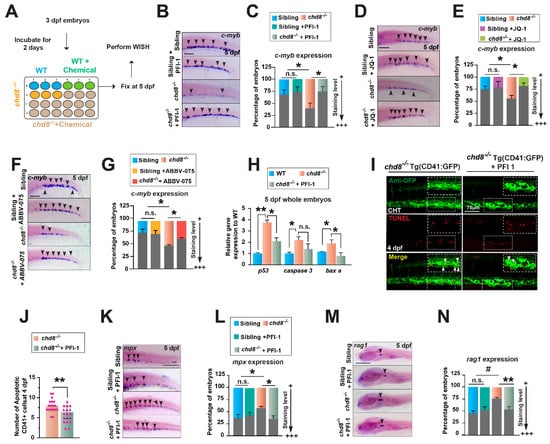
Figure 6.
The compound PFI-1 restores HSPC survival and differentiation. (A) Schematic of the chemical treatment regimen. Embryos were exposed to compounds from 3 to 5 dpf and subsequently analyzed by WISH. (B,D,F) WISH analysis of c-myb at 5 dpf in WT and chd8−/− embryos treated with DMSO or the BET inhibitors PFI-1 (25 µM), JQ-1 (0.5 µM), and ABBV-075 (0.05 µM). (C,E,G) Quantification of WISH results in (B,D,F). (H) RT-qPCR analysis of p53, caspase3, and baxa in WT and chd8−/− embryos treated with DMSO or PFI-1. (I) Representative images of CD41-GFP and TUNEL double staining in chd8−/− embryos at 4 dpf treated with DMSO or PFI-1. Apoptotic HSPCs are indicated by white arrowheads in the CHT. (J) Quantification of the apoptotic HSPCs in (I). (K, M) WISH for mpx and rag1 at 5 dpf in WT siblings and chd8−/− embryos treated with DMSO or PFI-1. (L,N) Quantification of WISH results in (K,M). Data are presented as mean ± SD from three independent experiments (two-tailed Student’s t-test, n ≈ 20 embryos per group in each experiment, * p < 0.05, ** p < 0.01, # p < 0.001, n.s., not significant). Scale bars, 100 µm (B,D,F,K,M).
2.7. PFI-1 Targets Brd4 in chd8−/− to Restore HSPC Production and Differentiation
We next sought to identify the potential protein target through which PFI-1 exerts its rescue effect in chd8−/−. PFI-1 is a potent BET inhibitor, particularly blocking the activity of BRD2 and BRD4 [28]. We first characterized the expression of their zebrafish homologs, brd2a, brd2b, and brd4, and confirmed their presence in hematopoietic cells (Figure S4A,B). To determine which of these is functionally required for the phenotype, we performed MO-mediated knockdown of each gene. While knockdown of brd2a, brd2b or a combination of both failed to restore HSPCs in chd8−/− mutants (Figure S5A–F), knockdown of brd4 completely rescued HSPC production and suppressed the enhanced myeloid differentiation (Figure 7A–D). Furthermore, brd4 inhibition also partially restored lymphoid differentiation (Figure 7E,F). Moreover, simultaneous knockdown of brd2a and brd2b in the brd4 morphant background did not augment this rescue (Figure S5G–L), indicating that brd4 is the primary target of PFI-1 in chd8−/−.
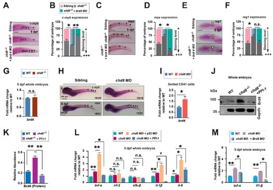
Figure 7.
brd4 knockdown rescues HSPCs in chd8−/− embryos. (A,C,E) WISH analysis for c-myb, mpx, and rag1 at 5 dpf in WT siblings, and chd8−/− embryos with and without brd4 morpholino injection. (B,D,F) Quantification of WISH analysis in (A,C,E). (G) RT-qPCR analysis of brd4 expression in WT and chd8−/− whole embryos. (H) WISH analysis for c-myb, mpx in WT siblings and embryos injected with chd8 MO. n/n in the lower right corner shows the number of embryos with similar staining patterns/total number of embryos examined. (I) RT-qPCR analysis of brd4 expression in sorted CD41-GFP+ cells from WT and chd8 morphant embryos (5 dpf). (J) Western blot analysis for Brd4 protein in WT and chd8−/− embryos treated with DMSO or PFI-1. GAPDH is shown as a loading control. (K) Quantification of Brd4 protein in (J). (L) RT-qPCR analysis of cytokine expression in WT and chd8 morphant embryos treated with DMSO or PFI-1 or injected with p53 MO. (M) RT-qPCR analysis of cytokine expression in WT and chd8 morphant embryos injected with brd4 MO. Data are expressed as mean ± SD from three independent experiments (two-tailed Student’s t-test, n ≈ 20 embryos per group, each experiment, * p < 0.05, ** p < 0.01, # p < 0.001, n.s., not significant). ng, nanogram. Scale bars, 100 µm (A,C,E).
Next, we examined the relationship between Chd8 and Brd4. brd4 mRNA levels was unchanged in the whole chd8−/− embryos (Figure 7G). To test if chd8 regulates brd4 in HSPCs, we knocked down chd8 in Tg(CD41:GFP) embryos using a MO [30], which recapitulated the chd8−/− mutant phenotypes (Figure 7H). This knockdown significantly increased brd4 expression in CD41-GFP+ HSPCs (Figure 7I). Furthermore, Brd4 protein was strongly upregulated in the whole chd8−/− embryos (Figure 7J,K), suggesting potential post-transcriptional dysregulation. The fact that chd8 loss causes brd4 upregulation, while brd4 knockdown rescues the hematopoietic defects in chd8−/− mutants, positions brd4 as a critical downstream effector of chd8. Therefore, we conclude that Chd8 mediates definitive hematopoiesis through at least two parallel pathways: by repressing p53-dependent apoptosis and by constraining Brd4 activity.
Finally, we previously demonstrated that the microbiota dysbiosis in chd8−/− drives elevated cytokine expression, which contributes to the host hematopoietic defects [24]. As Brd4 is known to regulate inflammatory gene expression, we asked whether Brd4 participates in the inflammatory response. Both PFI-1 treatment and brd4 knockdown successfully attenuated the systemic cytokine expression in chd8−/− mutants, whereas p53 knockdown did not produce this effect (Figure 7L,M). Therefore, Brd4 inhibition restores normal hematopoiesis partially through mitigating the pro-inflammatory cytokine expression.
3. Discussion
Recent studies have shown that CHD8 functions as a key regulator of HSPC survival and stemness in the bone marrow via suppressing p53 activity [19,20]. However, its role in HSPC development during embryogenesis remains unknown. Our study reveals that the zebrafish chd8−/− embryos retained primitive hematopoiesis but showed severe defects in definitive hematopoiesis in the CHT. chd8 is essential for HSPC survival by suppressing p53-dependent apoptosis. Furthermore, chd8 loss leads to enhanced myeloid differentiation through a p53-independent mechanism. Inhibiting brd4 attenuated myeloid differentiation and promoted HSPC development in chd8−/− partially via decreasing elevated inflammatory cytokine expression. Our results demonstrate that chd8 plays critical and multidimensional regulatory roles in HSPC development.
Definitive HSPCs transiently emerge from the AGM before migrating to the CHT. Chd8 does not regulate early HSPC emergence but is critical for their survival and development in the CHT from 3 dpf. Nascent HSPCs seldom proliferate in the AGM region but enter an active in cell cycle and undergo rapid division in the CHT [7,31]. Such extensive cell division in a short timeframe poses a risk of replicative stress, potentially activating the DNA replication checkpoint and inducing p53-dependent apoptosis [7,32]. We hypothesize that chd8 functions to suppress unwanted apoptosis in proliferating HSPCs, providing a critical window for DNA damage repair and ensuring the successful expansion of the HSPC pool. Previous studies have shown that dysregulation of alternative splicing activates p53-dependent apoptosis in HSPCs [33,34]. Meanwhile, Wdr5-mediated H3K4 methylation is crucial for HSPC survival in the CHT by maintaining genomic stability [7]. Our study identified another essential regulator involved in this process. In chd8−/−, both primitive hematopoiesis and vascular systems develop normally. And chd8 mRNA is highly enriched in CD41-GFP+ HSPCs. These data support that chd8 likely maintains HSPC survival through intrinsic mechanisms. Future studies, including the use of p53 mutants, are needed to formally prove that this function is cell autonomous. Furthermore, in the mouse bone marrow, CHD8 deficiency activates ATM kinase to stabilize P53 protein and CHD8 also directly binds to P53 to regulate its transactivation [20]. How Chd8 suppresses p53 activation during zebrafish development remains unknown. Future omics studies, such as those utilizing activator-targeted chromatin accessibility sequencing (ATAC-seq), will reveal the chromatin accessibility regulated by Chd8, thereby helping elucidate the mechanism by which it suppresses p53 activation.
In addition to HSPC defects, chd8−/− zebrafish embryos displayed enhanced immune cell differentiation that persisted into adulthood. Chd8−/− mice die between embryonic days 5.5 and E7.5 (E5.5-E7.5) [23]. Using the Mx-Cre inducible system, deletion of Chd8 in murine hematopoietic cells causes a significant reduction in HSPCs and lineage differentiation [19,20]. This discrepancy may be largely attributed to the distinction between a constitutive systemic knockout in zebrafish and a hematopoietic-specific deletion in mice. The phenotype in chd8−/− zebrafish likely involves both cell-autonomous and non-autonomous mechanisms. Chd8 may function cell-intrinsically to maintain HSPC survival. We previously showed that chd8 mutants harbor an imbalanced microbiota that drives elevated inflammatory cytokines (e.g., tnfa and il1b) in the host, which in turn non-cell-autonomously impairs HSPC differentiation and production [24]. In this study, we found that BET inhibitors and Brd4 knockdown reduced systemic cytokine production and restored balanced HSPC development in chd8−/−. In contrast, p53 knockdown did not affect cytokine production nor restore differentiation, but specifically rescued HSPC survival. This supports that enhanced myelopoiesis is related to elevated cytokines. Moreover, enhanced differentiation persisted in germ-free (GF) chd8−/− embryos [24], indicating that additional, microbiota-independent, genetic mechanisms promote differentiation. Previous studies showed that Chd8 directly regulates pluripotency and early neural differentiation through chromatin structure in mouse embryonic stem cells (ESCs) and neural progenitor cells (NPCs) [17,18]. We downloaded the RNA-seq data from mouse HSPC (WT vs Chd8−/−) [22], and found that despite no apparent enhancement in differentiation, myeloid and lymphoid differentiation and inflammatory pathways were significantly upregulated in mouse chd8−/− HSCs (Figure S6). This suggests that Chd8 may intrinsically negatively regulate these pathways in HSPCs, possibly through chromatin compaction, thereby contributing to precise differentiation control. The differentiation defects in chd8−/− zebrafish are likely the collective effects of HSPC-intrinsic regulation and extrinsic signaling. Future studies using tissue-specific knockdown and rescue experiments will help elucidate the cell-autonomous and non-autonomous functions of Chd8 in HSPC development. However, given that CHD8 is a high-risk gene for ASD—a condition often associated with immune dysregulation [35,36]—and its mutations are constitutive, the viable chd8−/− zebrafish larvae provide a powerful tool for analyzing hematopoietic defects caused by CHD8 deficiency at the organismal level.
PFI-1 is a highly selective BET inhibitor that particularly targets BRD2 and BRD4 by blocking their ability to bind to acetylated histone tails—a binding crucial for gene transcription [28]. Our study revealed that in chd8−/− embryos, PFI-1 primarily targets Brd4 to alleviate hematopoietic defects. As a key epigenetic regulator, BRD4 cooperates with various master transcription factors to control gene expression [29] and is known to positively regulate inflammatory genes such as IL1B [37,38]. Knocking down Brd4 may directly repress these inflammatory cytosines, thereby restoring the developmental balance of HSPCs. We also found that Brd4 inhibition partially rescues HSPC apoptosis in chd8−/−, likely by mitigating the pro-apoptotic effects induced by high levels of inflammatory cytokines such as IL1B and TNFα [39,40]. Indeed, we found that MO-mediated inhibition of il1b in chd8−/− partially reduced the expression of apoptosis-related gene (Figure S6). In addition, brd4 is known to directly promote granulopoiesis in zebrafish [41]. Thus, upregulation of Brd4 may drive myeloid differentiation directly. The exact function of Brd4 during HSPC development requires further study. Although brd4 expression remained unchanged in whole embryos, it was significantly upregulated in CD41+-HSPCs. Thus, chd8 may mediate brd4 expression within HSPCs. Notably, the increase in Brd4 protein levels in chd8 mutants far exceeded that of its mRNA levels, implying that Chd8 may regulate Brd4 protein synthesis or stability through post-transcriptional mechanisms. The cell type-specific regulation of Brd4 by Chd8 and the underlying regulatory mechanisms require further investigation. This study reveals that Chd8 restricts Brd4 activity during embryogenesis.
In conclusion, our findings demonstrate that Chd8 maintains HSPC development by suppressing p53-dependent activity. Furthermore, Chd8 loss drives p53-independent enhanced myeloid differentiation, and Brd4 inhibition restores normal myeloid differentiation and supports HSPC development. Our studies elucidate the critical roles of Chd8 in regulating developmental hematopoiesis.
4. Materials and Methods
4.1. Zebrafish Maintenance and Embryo Handling
Zebrafish (Danio rerio) strains, including wild-type AB and transgenic lines Tg(CD41: GFP), Tg(drl: GFP), and Tg(flk: GFP), were raised and maintained with a 14 h light/10 h dark cycle in a circulating water system at 28.5 °C, conductance 450–550 μS, and pH 7.0 ± 0.5 [42]. WT Siblings and mutant embryos were collected at the desired stage and grown in an E3 medium (5 mM NaCl, 0.17 mM KCl, 0.33 mM CaCl2, 0.33 mM MgSO4) with a density of 80–120 embryos per 10 cm Petri dish. Embryos were staged by hours post-fertilization (hpf) and days post-fertilization (dpf). Zebrafish were maintained, handled, and bred according to standard protocols from the Institutional Animal Care and Use Committee of Shanghai Jiao Tong University, China.
4.2. Genotyping of Mutant Lines
The chd8−/−(M1) and chd8−/−(M2) mutant lines were generate by CRISPR/Cas9 technology as previously described [24]. For genotyping, DNA fragments of 388 bp and 420 bp (flanking the respective chd8 target sites) were amplified by polymerase chain reaction (PCR) from genomic DNA. Mutants were identified using the T7 endonuclease I assay (NEB, M0302S) and confirmed by Sanger sequencing. The primers used to amplify the chd8 target sequences are listed in Table 1.

Table 1.
PCR primers used for genotyping.
4.3. Whole Mount In Situ Hybridization
Whole-mount in situ hybridization (WISH) was performed on 4% paraformaldehyde (PFA) -fixed zebrafish embryos. The anti-sense probes (c-my, pu.1, runx1, hbbe1 and hbbe3, mpx, mfap4, and rag1) were synthesized by in vitro transcription using SP6 or T7 polymerase (Ambion) with Digoxigenin RNA Labelling Mix (Roche, Mannheim, Germany). The WISH procedure was carried out as previously described [43]. Stained embryos were mounted in 100% glycerol and imaged using an Olympus SZX16 stereomicroscope or a BX53 microscope (Olympus, Tokyo, Japan).
4.4. Neutral Red, Benzidine, and Sudan Black Staining
To minimize pigmentation and enhance staining signal, embryos were treated with 1-phenyl 2-thiourea (PTU) until the desired stages. For macrophage visualization, live 4–6 dpf embryos were incubated in the dark for 6–8 h at 28.5 °C with 2.5 µg/mL Neutral Red (A600652, Sangon Biotech, Shanghai, China) in E3 medium [44]. For hemoglobin detection, live embryos were incubated in the dark for 30 min in a benzidine solution, prepared by combining 2 mL of 5 mg/mL benzidine (B108444, Aladdin, CA, USA) in methanol, 16.7 µL of 3 M sodium acetate, 100 µL of H2O2, and 2.483 mL of H2O [45]. Following incubation, embryos were washed with PBS containing 0.1% Tween 20 (PBT) and fixed overnight in 4% paraformaldehyde (PFA).
For Sudan Black staining, embryos at the desired stages were fixed overnight in 4% methanol-free formaldehyde (Polysciences), rinsed three times with PBS, and incubated in Sudan black B (SB; Sigma-Aldrich, Saint-Quentin Fallavier, France) solution at room temperature for 1 h [46]. Embryos were thoroughly washed with 70% ethanol and rehydrated with PBS and 0.1% Tween 20 (PBT). To better visualize the staining, embryos were incubated in a bleach solution (10% KOH, 30% H2O2, 10% Tween 20) for 5 min and then washed with PBT. Images of the stained embryos were taken through an SZX16 stereomicroscope or a BX53 microscope (Olympus, Tokyo, Japan).
4.5. Morpholinos
The antisense morpholinos (MOs) were purchased from Gene Tools and resuspended in nuclease-free water to a 1 mM stock solution. Embryos were injected at the 1–4 cell stage with 1–7 ng/nL of MO into the yolk. Injected embryos were raised to 5 dpf for subsequent staining. The MO sequence is provided in Table 2.

Table 2.
List of Morpholino (MO) used.
4.6. Chemical Screen
A screen of epigenetic small molecules (Cayman Chemicals, 11076) was performed at concentrations of 6, 12.5, and 25 µM, using DMSO as a vehicle control. WT and chd8−/− mutant embryos at 3 dpf were arrayed into 24-well plates (15 embryos per well in 600 µL of E3 medium). Plates were wrapped in aluminum foil to protect light-sensitive compounds and incubated at 28.5 °C until 5 dpf. The chemicals were then washed out 3 times with E3 medium. The toxicity of the chemicals was determined, and healthy embryos at 5 dpf were then fixed in 4% PFA at 4 °C overnight (O/N) for WISH. The additional BET inhibitors were obtained commercially: PFI-1 (Lot# QLDMMEO), (+)-JQ1 (Med Chem Express, Cat# HY-13030/CS-0581), and ABBV-075 (Mivebresib).
4.7. May–Grünwald–Giemsa Staining of Adult Whole Kidney Marrow Cells
Adult zebrafish of the desired genotype were euthanized in 0.4% tricaine. The kidney was then dissected via a midline incision and placed in 0.9× PBS containing 5% FBS. Whole kidney marrow (WKM) cells were isolated by mechanical dissociation through strenuous pipetting in 0.4 mL of the same buffer, followed by filtration through a 40-µm cell strainer (Falcon, New York, NY, USA). The resulting cell suspension was centrifuged onto glass slides using a cytocentrifuge (Sigma-Aldrich) at 400 rpm for 4 min. Slides were stained with May-Grünwald and Giemsa (Sigma-Aldrich) for morphological analysis and differential cell counting.
4.8. Gene Expression by Real-Time qPCR
Total RNA was isolated from whole embryos, kidneys, and sorted cells using Trizol reagent (Thermo Fisher Scientific, 10296028, Carlsbad, CA, USA) according to the manufacturer’s instructions. RNA was reverse-transcribed into cDNA using either the PrimeScript RT reagent Kit with gDNA Eraser (Takara, RR047A, Kyoto, Japan) or the Hifair® II 1st Strand cDNA Synthesis SuperMix for qPCR (Yeasen Biotech, 11123ES10, Shanghai, China). Quantitative real-time PCR (qPCR) was performed on a StepOnePlus system (Applied Biosystems, San Francisco, CA, USA) with TB Green Premix Ex Taq II (Takara, RR820A, Japan) or Hieff® qPCR SYBR Green Master Mix (Yeasen Biotech, 11203ES08, Shanghai, China). Gene expression was quantified using the 2–ΔΔCT method. The RT-qPCR primers are listed in Table 3.

Table 3.
Primers used for RT-qPCR.
4.9. Flow Cytometry and Cell Sorting
The CD41 GFP+ and drl GFP+ cells from WT and chd8−/− embryos were sorted out using FACS AriaIII (Becton, Dickinson and Company, Franklin Lakes, NJ, USA). Briefly, embryos of the desired stage were anesthetized in 0.4% tricaine, and single cells were collected by chopping the embryos with a blade. Following that, the chopped embryos were incubated for 20 min at 37 °C in 38 µg/mL Liberase (05401119001, Roche, Basel, Switzerland). The liberase reaction was stopped by adding 10% FBS, followed by filtration through a 40 µm filter and centrifugation (1500 rpm, at 4 °C for 10 min). The supernatant was discarded, and cells were resuspended in PBS containing 1% FBS for subsequent studies.
4.10. TUNEL Immunostaining
WT sibling and chd8−/−/ Tg (CD41: GFP) embryos were fixed at 4 dpf in 4% PFA overnight at 4 °C and dehydrated in methanol at −20 °C for 1 h. After serial rehydration, embryos were permeabilized with proteinase K (20 mg/mL) at room temperature (RT) for 40 min. The apoptotic cells were stained with Bright Red Apoptosis Detection Kit (Vazyme Biotech Co., Ltd., Shanghai, China) for 90 min at 37 °C. After washing with 1XPBS, embryos were blocked for 2 h at RT in blocking buffer (PBS + 1% DMSO + 0.1% Triton X-100+ 1% bovine serum albumin). To amplify the GFP signal, embryos were incubated with an anti-GFP antibody (Dia-an, Cat# 2057), followed by Alexa Fluor 488-conjugated goat anti-mouse secondary antibody (Invitrogen, Waltham, MA, USA) incubation. Images were taken through a confocal microscope (Leica, Wetzlar, Germany).
4.11. Statistical Analysis
GraphPad Prism 8.0.2 (GraphPad Software, San Diego, CA, USA, 2019) was used to analyze the data. The values of all triplicate experiments are presented as mean ± standard deviation (SD), and significance was determined by a 2-tailed Student t-test. For WISH and other staining results, embryos were divided into strong and weak staining groups. For statistical analysis, only the strongly stained groups of wild-type and mutant/morphant were compared.
Supplementary Materials
The following are available online at https://www.mdpi.com/article/10.3390/ijms262110805/s1.
Author Contributions
Conceptualization, A.A., W.L. and L.J.; methodology, A.A., X.W., D.Z., R.U. and L.J.; writing—original draft preparation, A.A. and L.J.; writing—review and editing, A.A., W.L. and L.J.; supervision, W.L. and L.J.; funding acquisition, W.L. and L.J. All authors have read and agreed to the published version of the manuscript.
Funding
This research was funded by the National Natural Science Foundation of China, grant number 32370875; The Open Platform on Target Identification of Innovative Drugs of Shanghai Jiao Tong University, grant number WH101117001; and the Natural Science Foundation of Shanghai, grant number 23ZR1430500.
Institutional Review Board Statement
The animal study protocol was approved by the Institutional Review Board of Shanghai Jiao Tong University (protocol code 202201259 and 14 March 2022 of approval).
Informed Consent Statement
Not applicable.
Data Availability Statement
The original contributions presented in this study are included in the article/Supplementary Material. Further inquiries can be directed to the corresponding authors.
Acknowledgments
We would like to thank Haixia Jiang from the Core Facility and Technical Service Center for SLSB, School of Life Sciences & Biotechnology, Shanghai Jiao Tong University, for technical support.
Conflicts of Interest
The authors declare no conflicts of interest.
References
- Laurenti, E.; Göttgens, B. From haematopoietic stem cells to complex differentiation landscapes. Nature 2018, 553, 418–426. [Google Scholar] [CrossRef] [PubMed]
- Lewis, K.; Yoshimoto, M.; Takebe, T. Fetal liver hematopoiesis: From development to delivery. Stem Cell Res. Ther. 2021, 12, 139. [Google Scholar] [CrossRef]
- Gao, X.; Xu, C.; Asada, N.; Frenette, P.S. The hematopoietic stem cell niche: From embryo to adult. Development 2018, 145, 139691. [Google Scholar] [CrossRef]
- Jing, L.; Zon, L.I. Zebrafish as a model for normal and malignant hematopoiesis. Dis. Model. Mech. 2011, 4, 433–438. [Google Scholar] [CrossRef]
- Xia, J.; Kang, Z.; Xue, Y.; Ding, Y.; Gao, S.; Zhang, Y.; Lv, P.; Wang, X.; Ma, D.; Wang, L.; et al. A single-cell resolution developmental atlas of hematopoietic stem and progenitor cell expansion in zebrafish. Proc. Natl. Acad. Sci. USA 2021, 118, e2015748118. [Google Scholar] [CrossRef]
- Mochizuki-Kashio, M.; Otsuki, N.; Fujiki, K.; Abdelhamd, S.; Kurre, P.; Grompe, M.; Iwama, A.; Saito, K.; Nakamura-Ishizu, A. Replication stress increases mitochondrial metabolism and mitophagy in FANCD2 deficient fetal liver hematopoietic stem cells. Front. Oncol. 2023, 13, 1108430. [Google Scholar] [CrossRef]
- Wang, X.; Liu, M.; Zhang, Y.; Ma, D.; Wang, L.; Liu, F. Wdr5-mediated H3K4 methylation facilitates HSPC development via maintenance of genomic stability in zebrafish. Proc. Natl. Acad. Sci. USA 2025, 122, e2420534122. [Google Scholar] [CrossRef] [PubMed]
- Wu, J.; Li, J.; Chen, K.; Liu, G.; Zhou, Y.; Chen, W.; Zhu, X.; Ni, T.T.; Zhang, B.; Jin, D.; et al. Atf7ip and Setdb1 interaction orchestrates the hematopoietic stem and progenitor cell state with diverse lineage differentiation. Proc. Natl. Acad. Sci. USA 2023, 120, e2209062120. [Google Scholar] [CrossRef] [PubMed]
- Chen, J.; Li, G.; Lian, J.; Ma, N.; Huang, Z.; Li, J.; Wen, Z.; Zhang, W.; Zhang, Y. Slc20a1b is essential for hematopoietic stem/progenitor cell expansion in zebrafish. Sci. China Life Sci. 2021, 64, 2186–2201. [Google Scholar] [CrossRef]
- Micucci, J.A.; Sperry, E.D.; Martin, D.M. Chromodomain helicase DNA-binding proteins in stem cells and human developmental diseases. Stem Cells Dev. 2015, 24, 917–926. [Google Scholar] [CrossRef]
- Ho, L.; Crabtree, G.R. Chromatin remodelling during development. Nature 2010, 463, 474–484. [Google Scholar] [CrossRef]
- Nagarajan, P.; Onami, T.M.; Rajagopalan, S.; Kania, S.; Donnell, R.; Venkatachalam, S. Role of chromodomain helicase DNA-binding protein 2 in DNA damage response signaling and tumorigenesis. Oncogene 2009, 28, 1053–1062. [Google Scholar] [CrossRef] [PubMed][Green Version]
- Zhen, T.; Kwon, E.M.; Zhao, L.; Hsu, J.; Hyde, R.K.; Lu, Y.; Alemu, L.; Speck, N.A.; Liu, P.P. Chd7 deficiency delays leukemogenesis in mice induced by Cbfb-MYH11. Blood 2017, 130, 2431–2442. [Google Scholar] [CrossRef]
- Yoshida, T.; Hazan, I.; Zhang, J.; Ng, S.Y.; Naito, T.; Snippert, H.J.; Heller, E.J.; Qi, X.; Lawton, L.N.; Williams, C.J.; et al. The role of the chromatin remodeler Mi-2beta in hematopoietic stem cell self-renewal and multilineage differentiation. Genes. Dev. 2008, 22, 1174–1189. [Google Scholar] [CrossRef]
- Katayama, Y.; Nishiyama, M.; Shoji, H.; Ohkawa, Y.; Kawamura, A.; Sato, T.; Suyama, M.; Takumi, T.; Miyakawa, T.; Nakayama, K.I. CHD8 haploinsufficiency results in autistic-like phenotypes in mice. Nature 2016, 537, 675–679. [Google Scholar] [CrossRef] [PubMed]
- Bernier, R.; Golzio, C.; Xiong, B.; Stessman, H.A.; Coe, B.P.; Penn, O.; Witherspoon, K.; Gerdts, J.; Baker, C.; Vulto-van Silfhout, A.T.; et al. Disruptive CHD8 mutations define a subtype of autism early in development. Cell 2014, 158, 263–276. [Google Scholar] [CrossRef]
- Ding, S.; Lan, X.; Meng, Y.; Yan, C.; Li, M.; Li, X.; Chen, J.; Jiang, W. CHD8 safeguards early neuroectoderm differentiation in human ESCs and protects from apoptosis during neurogenesis. Cell Death Dis. 2021, 12, 981. [Google Scholar] [CrossRef]
- Sood, S.; Weber, C.M.; Hodges, H.C.; Krokhotin, A.; Shalizi, A.; Crabtree, G.R. CHD8 dosage regulates transcription in pluripotency and early murine neural differentiation. Proc. Natl. Acad. Sci. USA 2020, 117, 22331–22340. [Google Scholar] [CrossRef]
- Nita, A.; Muto, Y.; Katayama, Y.; Matsumoto, A.; Nishiyama, M.; Nakayama, K.I. The autism-related protein CHD8 contributes to the stemness and differentiation of mouse hematopoietic stem cells. Cell Rep. 2021, 34, 108688. [Google Scholar] [CrossRef] [PubMed]
- Tu, Z.; Wang, C.; Davis, A.K.; Hu, M.; Zhao, C.; Xin, M.; Lu, Q.R.; Zheng, Y. The chromatin remodeler CHD8 governs hematopoietic stem/progenitor survival by regulating ATM-mediated P53 protein stability. Blood 2021, 138, 221–233. [Google Scholar] [CrossRef]
- Tu, Z.; Fan, C.; Davis, A.K.; Hu, M.; Wang, C.; Dandamudi, A.; Seu, K.G.; Kalfa, T.A.; Lu, Q.R.; Zheng, Y. Autism-associated chromatin remodeler CHD8 regulates erythroblast cytokinesis and fine-tunes the balance of Rho GTPase signaling. Cell Rep. 2022, 40, 111072. [Google Scholar] [CrossRef] [PubMed]
- Yang, J.Q.; Wang, C.; Nayak, R.C.; Kolla, M.; Cai, M.; Pujato, M.; Zheng, Y.; Lu, Q.R.; Guo, F. Genetic and epigenetic regulation of Treg cell fitness by autism-related chromatin remodeler CHD8. Cell Mol. Biol. Lett. 2025, 30, 36. [Google Scholar] [CrossRef] [PubMed]
- Nishiyama, M.; Oshikawa, K.; Tsukada, Y.; Nakagawa, T.; Iemura, S.; Natsume, T.; Fan, Y.; Kikuchi, A.; Skoultchi, A.I.; Nakayama, K.I. CHD8 suppresses p53-mediated apoptosis through histone H1 recruitment during early embryogenesis. Nat. Cell Biol. 2009, 11, 172–182. [Google Scholar] [CrossRef] [PubMed]
- Zhong, D.; Jiang, H.; Zhou, C.; Ahmed, A.; Li, H.; Wei, X.; Lian, Q.; Tastemel, M.; Xin, H.; Ge, M.; et al. The microbiota regulates hematopoietic stem and progenitor cell development by mediating inflammatory signals in the niche. Cell Rep. 2023, 42, 112116. [Google Scholar] [CrossRef]
- Henninger, J.; Santoso, B.; Hans, S.; Durand, E.; Moore, J.; Mosimann, C.; Brand, M.; Traver, D.; Zon, L. Clonal fate mapping quantifies the number of haematopoietic stem cells that arise during development. Nat. Cell Biol. 2017, 19, 17–27, Erratum in Nat. Cell Biol. 2017, 19, 142. [Google Scholar] [CrossRef]
- North, T.E.; Goessling, W.; Peeters, M.; Li, P.; Ceol, C.; Lord, A.M.; Weber, G.J.; Harris, J.; Cutting, C.C.; Huang, P.; et al. Hematopoietic stem cell development is dependent on blood flow. Cell 2009, 137, 736–748. [Google Scholar] [CrossRef]
- Plaster, N.; Sonntag, C.; Busse, C.E.; Hammerschmidt, M. p53 deficiency rescues apoptosis and differentiation of multiple cell types in zebrafish flathead mutants deficient for zygotic DNA polymerase delta1. Cell Death Differ. 2006, 13, 223–235. [Google Scholar] [CrossRef]
- Picaud, S.; Da Costa, D.; Thanasopoulou, A.; Filippakopoulos, P.; Fish, P.V.; Philpott, M.; Fedorov, O.; Brennan, P.; Bunnage, M.E.; Owen, D.R.; et al. PFI-1, a highly selective protein interaction inhibitor, targeting BET Bromodomains. Cancer Res. 2013, 73, 3336–3346. [Google Scholar] [CrossRef]
- Doroshow, D.B.; Eder, J.P.; LoRusso, P.M. BET inhibitors: A novel epigenetic approach. Ann. Oncol. 2017, 28, 1776–1787. [Google Scholar] [CrossRef]
- Sugathan, A.; Biagioli, M.; Golzio, C.; Erdin, S.; Blumenthal, I.; Manavalan, P.; Ragavendran, A.; Brand, H.; Lucente, D.; Miles, J.; et al. CHD8 regulates neurodevelopmental pathways associated with autism spectrum disorder in neural progenitors. Proc. Natl. Acad. Sci. USA 2014, 111, E4468–E4477. [Google Scholar] [CrossRef]
- Orkin, S.H.; Zon, L.I. Hematopoiesis: An evolving paradigm for stem cell biology. Cell 2008, 132, 631–644. [Google Scholar] [CrossRef]
- Alvarez, S.; Díaz, M.; Flach, J.; Rodriguez-Acebes, S.; López-Contreras, A.J.; Martínez, D.; Cañamero, M.; Fernández-Capetillo, O.; Isern, J.; Passegué, E.; et al. Replication stress caused by low MCM expression limits fetal erythropoiesis and hematopoietic stem cell functionality. Nat. Commun. 2015, 6, 8548. [Google Scholar] [CrossRef]
- Yu, S.; Jiang, T.; Jia, D.; Han, Y.; Liu, F.; Huang, Y.; Qu, Z.; Zhao, Y.; Tu, J.; Lv, Y.; et al. BCAS2 is essential for hematopoietic stem and progenitor cell maintenance during zebrafish embryogenesis. Blood 2019, 133, 805–815. [Google Scholar] [CrossRef] [PubMed]
- Zhao, Y.; Wu, M.; Li, J.; Meng, P.; Chen, J.; Huang, Z.; Xu, J.; Wen, Z.; Zhang, W.; Zhang, Y. The spliceosome factor sart3 regulates hematopoietic stem/progenitor cell development in zebrafish through the p53 pathway. Cell Death Dis. 2021, 12, 906. [Google Scholar] [CrossRef] [PubMed]
- Ahmad, S.F.; Nadeem, A.; Ansari, M.A.; Bakheet, S.A.; Attia, S.M.; Zoheir, K.M.; Al-Ayadhi, L.Y.; Alzahrani, M.Z.; Alsaad, A.M.; Alotaibi, M.R.; et al. Imbalance between the anti- and pro-inflammatory milieu in blood leukocytes of autistic children. Mol. Immunol. 2017, 82, 57–65. [Google Scholar] [CrossRef]
- Nadeem, A.; Ahmad, S.F.; Al-Harbi, N.O.; Al-Ayadhi, L.Y.; Sarawi, W.; Attia, S.M.; Bakheet, S.A.; Alqarni, S.A.; Ali, N.; AsSobeai, H.M. Imbalance in pro-inflammatory and anti-inflammatory cytokines milieu in B cells of children with autism. Mol. Immunol. 2022, 141, 297–304. [Google Scholar] [CrossRef]
- Mann, M.W.; Fu, Y.; Gearhart, R.L.; Xu, X.; Roberts, D.S.; Li, Y.; Zhou, J.; Ge, Y.; Brasier, A.R. Bromodomain-containing Protein 4 regulates innate inflammation via modulation of alternative splicing. Front. Immunol. 2023, 14, 1212770. [Google Scholar] [CrossRef] [PubMed]
- Franzè, E.; Laudisi, F.; Maresca, C.; Di Grazia, A.; Iannucci, A.; Pacifico, T.; Ortenzi, A.; Sica, G.; Lolli, E.; Stolfi, C.; et al. Bromodomain-containing 4 is a positive regulator of the inflammatory cytokine response in the gut. J. Crohns Colitis 2024, 18, 1995–2009. [Google Scholar] [CrossRef]
- Xiao, Y.; Li, H.; Zhang, J.; Volk, A.; Zhang, S.; Wei, W.; Zhang, S.; Breslin, P.; Zhang, J. TNF-α/Fas-RIP-1-induced cell death signaling separates murine hematopoietic stem cells/progenitors into 2 distinct populations. Blood 2011, 118, 6057–6067. [Google Scholar] [CrossRef][Green Version]
- Yamashita, M.; Passegué, E. TNF-α Coordinates Hematopoietic Stem Cell Survival and Myeloid Regeneration. Cell Stem Cell 2019, 25, 357–372.e357. [Google Scholar] [CrossRef]
- Yan, L.; Tan, S.; Wang, H.; Yuan, H.; Liu, X.; Chen, Y.; de Thé, H.; Zhu, J.; Zhou, J. Znf687 recruits Brd4-Smrt complex to regulate gfi1aa during neutrophil development. Leukemia 2024, 38, 851–864. [Google Scholar] [CrossRef]
- Westerfield, M. The Zebrafish Book: A Guide for the Laboratory Use of Zebrafish (Danio rerio); University of Oregon Press: Eugene, OR, USA, 2007. [Google Scholar]
- Thisse, C.; Thisse, B. High-resolution in situ hybridization to whole-mount zebrafish embryos. Nat. Protoc. 2008, 3, 59–69. [Google Scholar] [CrossRef]
- Herbomel, P.; Thisse, B.; Thisse, C. Zebrafish early macrophages colonize cephalic mesenchyme and developing brain, retina, and epidermis through a M-CSF receptor-dependent invasive process. Dev. Biol. 2001, 238, 274–288. [Google Scholar] [CrossRef] [PubMed]
- Kaplow, L.S. Simplified myeloperoxidase stain using benzidine dihydrochloride. Blood 1965, 26, 215–219. [Google Scholar] [CrossRef] [PubMed]
- Le Guyader, D.; Redd, M.J.; Colucci-Guyon, E.; Murayama, E.; Kissa, K.; Briolat, V.; Mordelet, E.; Zapata, A.; Shinomiya, H.; Herbomel, P. Origins and unconventional behavior of neutrophils in developing zebrafish. Blood 2008, 111, 132–141. [Google Scholar] [CrossRef] [PubMed]
- Bielczyk-Maczyńska, E.; Serbanovic-Canic, J.; Ferreira, L.; Soranzo, N.; Stemple, D.L.; Ouwehand, W.H.; Cvejic, A. A loss of function screen of identified genome-wide association study Loci reveals new genes controlling hematopoiesis. PLoS Genet. 2014, 10, e1004450. [Google Scholar] [CrossRef]
Disclaimer/Publisher’s Note: The statements, opinions and data contained in all publications are solely those of the individual author(s) and contributor(s) and not of MDPI and/or the editor(s). MDPI and/or the editor(s) disclaim responsibility for any injury to people or property resulting from any ideas, methods, instructions or products referred to in the content. |
© 2025 by the authors. Licensee MDPI, Basel, Switzerland. This article is an open access article distributed under the terms and conditions of the Creative Commons Attribution (CC BY) license (https://creativecommons.org/licenses/by/4.0/).