The authors wish to make the following corrections to this paper [1].
In the original Figure 1A, the same image was mistakenly selected for two panels illustrating the lysosome number in HaCaT cells grown in medium containing a low concentration of calcium, non-activated or “cytokine mix”-activated.
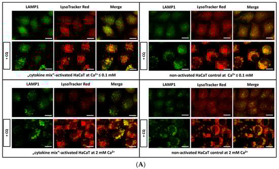
Figure 1.
Fluorescence microscopy analysis of lysosomal compartments and other acidic vesicles in keratinocytes. Shown are representative microscopy images of HaCaT cells: (i) Activated with “cytokine mix”: interleukin 1 alpha (IL-1α), IL-17A, IL-22, oncostatin M (OSM), and tumor necrosis factor alpha (TNF-α) at a concentration of 2 ng/mL each constituent and cultured in medium containing Ca2+ ≤ 0.1 mM; (ii) non-activated HaCaT cell control cultured in medium containing Ca2+ ≤ 0.1 mM; (iii) “cytokine mix”-activated HaCaT cells cultured in medium containing 2 mM Ca2+; and (iv) non-activated HaCaT cell control cultured in medium containing 2 mM Ca2+. Control with chloroquine (CQ) was used to increase the total amount of lysosomes. All images were captured from randomly selected ten microscopic fields containing 50–100 cells, each from three independent experiments (n = 3), using a fluorescence microscope. Scale bar represents 0.25 µm. (A) LysoTracker Red DND-99 staining showing the distribution of acidic organelles, including mature lysosomes and lysosomal-associated membrane protein 1 (LAMP1) staining of the lysosomal membrane.
The authors would like to apologize for any inconvenience caused to the readers by these changes. This change has no impact on the conclusions.
Conflicts of Interest
The authors declare that they have no conflict of interest.
Reference
- Bocheńska, K.; Moskot, M.; Malinowska, M.; Jakóbkiewicz-Banecka, J.; Szczerkowska-Dobosz, A.; Purzycka-Bohdan, D.; Pleńkowska, J.; Słomiński, B.; Gabig-Cimińska, M. Lysosome Alterations in the Human Epithelial Cell Line HaCaT and Skin Specimens: Relevance to Psoriasis. Int. J. Mol. Sci. 2019, 20, 2255. [Google Scholar] [CrossRef] [PubMed]

© 2020 by the authors. Licensee MDPI, Basel, Switzerland. This article is an open access article distributed under the terms and conditions of the Creative Commons Attribution (CC BY) license (http://creativecommons.org/licenses/by/4.0/).