You are currently viewing a new version of our website. To view the old version click .
Molecules
  • Article
  • Open Access

4 November 2025

Electrodeposition of BiVO4 Nanoparticles on TiO2 Nanotubes: Characterization and Synergetic Photocatalytic Degradation Activity of Amido Black Dye

,
,
,
,
,
,
,
and
1
Desalination and Natural Water Valorization Laboratory (LaDVEN), Water Research and Technologies Center (CERTE), P.O. Box 273, Soliman 8020, Tunisia
2
Photovoltaic Laboratory, Research and Technology Centre of Energy (CRTEn), P.O. Box 95, Hammam-Lif 2050, Tunisia
3
School of Design Engineering, Departamento de Fisica Aplicada, Universitat Politecnica de Valencia, Cami de Vera, 46022 Valencia, Spain
4
Chemistry Department, College of Science, Imam Mohammad Ibn Saud Islamic University (IMSIU), Riyadh 11623, Saudi Arabia
This article belongs to the Section Physical Chemistry

Abstract

To enhance the photocatalytic performance of TiO2 nanotubes (NTs) for the degradation of Amido Black as an organic pollutant, electrodeposition of bismuth vanadate (BiVO4) nanostructures was successfully applied. The effect of electrodeposited BiVO4 (25 s, 50 s, 150 s, 250 s), followed by a thermal treatment on TiO2-NTs, was studied. The structures of the as-prepared samples were characterized by X-ray diffraction (XRD). Morphological behavior was investigated using Scanning Electron Microscopy (SEM) and Transmission Electron Microscopy (TEM), both coupled with EDX. Optical characterizations were performed using photoluminescence and diffuse reflectance spectroscopy. The BiVO4/TiO2 NTs sample with 50 s deposition time gave the highest photocatalytic performance for Amido Black degradation, 99.4% after 150 min under UV light. This result has been achieved due to the structure and the optical properties of the sample. The heterojunction of both catalysts showed the synergetic effect on the photocatalytic performance where they remained stable after five cycling runs. Furthermore, quenching tests were conducted and proved that superoxide radicals (O2) are the main active species during photodegradation process.

1. Introduction

Dyes have been in widespread use in a wide range of industries for decades. Uncontrolled discharge of untreated hazardous dyes into freshwater causes harmful impacts on human health and ecosystems. Therefore, the mineralization of dyes in aquatic systems has become a top priority due to its negative effects. One of the most widely used dyes in clothing, beauty, food, printing, and even the pharmaceutical industry is Amido Black [,,]. In order to achieve the complete degradation of such a pollutant in water, several technologies have been applied [,,,,]. The most reputable and efficient techniques for organic materials removal are Advanced Oxidation Processes (AOPs). The AOP’s methods are designed for the removal of organic materials by way of reaction with reactive oxygen species (ROS), such as superoxide free radicals (O2) and hydroxyl radicals (HO), which are highly oxidative to the point of utter degradation or mineralization of the mentioned pollutants [,]. As these methods continue to evolve throughout the years and diversify in terms of energy sources for the generation of free ROS in aqueous solution, including sonocatalysis, electrocatalysis, photocatalysis, and piezocatalysis [,,,], the catalysts also evolve in terms of type, state, morphology, and size. One of the most exploited photocatalysts is TiO2, with its excellent properties and its band edge positioned for adequate potential for radical generation [,]. The variation in morphology of TiO2 expressed different results for the photodegradation of organic pollutants. The techniques for the TiO2 nanotubes synthesization varied as many emerged, such as the hydrothermal method [], the template method [] and the commonly used the anodization method [], which is a simple zero-cost method that consists of immersing the electrodes in electro-catalytic solution for 2 h at 60 V; according to the literature, the obtained morphology is highly ordered and vertically oriented. Due to these characteristics, the nanotubular morphology possesses a high specific surface area, a fast electron transfers and great photocatalytic activity [,,]. Even though the TiO2 nanotubes exhibit potential properties, they possess some limitations towards their effectiveness and yield, as this material presents a high recombination rate and limited spectrum performance [,]. To overcome these obstacles, a modification of the band alignment is proposed by coupling TiO2 with other photocatalysts, such as Cu2O, PbS, ZnO, NiO, BiVO4 and Fe2O4 [,,,,,,].
For instance, for the degradation of Methylene Blue, Shaban et al. [] synthesized TiO2 nanoribbons and obtained a photocatalytic degradation of 97.5% for 180 min under UV light, while Alias et al. [] prepared TiO2 nanoflowers and achieved a photodegradation of 98.95% for 180 min under solar light. Dong et al. [] also reached a 100% degradation of Methylene Blue in only 40 min under UV light with hierarchical mesoporous TiO2 nanoshell. However, TiO2 nanotubes are considered one of the best morphologies and are highly adequate for photocatalytic application [,,], as Lin et al. [] reported the difference between TiO2 nanoparticles and TiO2 nanotubes for the degradation of Methylene Blue, where the nanotubular morphology excelled with a removal efficiency of 99% in 25 min under UV light. Recently, Hajjaji M.A. et al. [] demonstrated that deposition of Pt on to the surface of TiO2 nanotubes contributes to the improvement in photocatalytic activity of Black Amido degradation compared to the pure sample. The authors reached the highest photocatalytic performance (97% degradation) after 90 min under UV irradiation.
The following Table 1 is a comparison between some results related to the use of catalysts/TiO2 for the degradation of Amido Black as an organic dye. BiVO4 has proven itself to be an excellent photocatalyst due to its narrow band gap and potential properties; Missaoui et al. [] prepared nanoflower-like BiVO4 nanostructured films for peroxymonosulfate (PMS) activation for the degradation of Rhodamine B (RhB) in aqueous solution. Under optimized conditions, they achieved total degradation in 17 min. Also, Nhat et al. [] decorated BiVO4 pine-like morphology for the photodegradation of Sulfamethoxazole; they reached a degradation of 98.8% under 210 min irradiation. The adequate band edge position of BiVO4 and its excellent photocatalytic properties makes it a potential candidate for a heterojunction with TiO2 nanotubes; this will be of interest to reducing the recombination by separating the pairs’ electron/holes, thus enhancing the photocatalytic performances. For instance, Saidurga et al. [] demonstrate the remarkable efficiency of hydrothermally synthesized BiVO4–TiO2 nanocomposites for the degradation of Rhodamine B. With a dye concentration of 5 × 10⁻⁵ M and a catalyst amount of 2 g/L, the 1:3 BiVO4–TiO2 composite achieved 97.1% degradation in 60 min under sunlight, with stability maintained over five cycles. Also, the mechanism analysis reveals that hydroxyl radicals are the main active species.
Table 1. Comparison of catalysts/TiO2 for the photodegradation of AB.
In this work, an effective approach to fabricating Z-scheme photocatalysts and a heterojunction of BiVO4/TiO2 nanotubes will be synthesized with the electrodeposition method at different conditions to study the effect of the deposition on the properties of TiO2 nanotubes, and photocatalytic activity will be investigated for the degradation of Black Amido.

2. Results

2.1. Structural Analysis

2.1.1. XRD Analysis

To investigate the structural properties and crystallinity of bare TiO2 nanotubes and BiVO4/TiO2 nanocomposites at different electrodeposition times of 25, 50, 150 and 250 s, X-ray diffraction was performed. Figure 1 shows the XRD patterns of each sample, where they all show a preferred orientation with high crystallinity corresponding to TiO2 anatase (101) at 2θ = 25.7°; these samples also show other orientations for the anatase phase at different angles (112), (004), (200), (105), (211), (204), (116), (202) and (107), corresponding to JCPDS card no. 21-1272. After the deposition of BiVO4 on the surface of the TiO2 nanotubes, one may notice the appearance of small, sharp peaks at 2 θ = 29.6° and 32° related to the (112) and (004) monoclinic scheelite BiVO4 phase (JCPDS card No. 75–1866). One can observe the presence of narrow XRD peaks corresponding to (111) and (120) Bi2O3 according to the reference JCPDS card No. 76-1730. These BiVO4 and Bi2O3 peaks are only visible for deposition times at 150 s and 250 s; it seems that for the lower deposition times, the peaks are unlikely to be detected, which could be due to their small size compared to the higher deposition time.
Figure 1. XRD patterns of BiVO4/TiO2 at different electrodeposition times (25, 50, 150, 250 s).
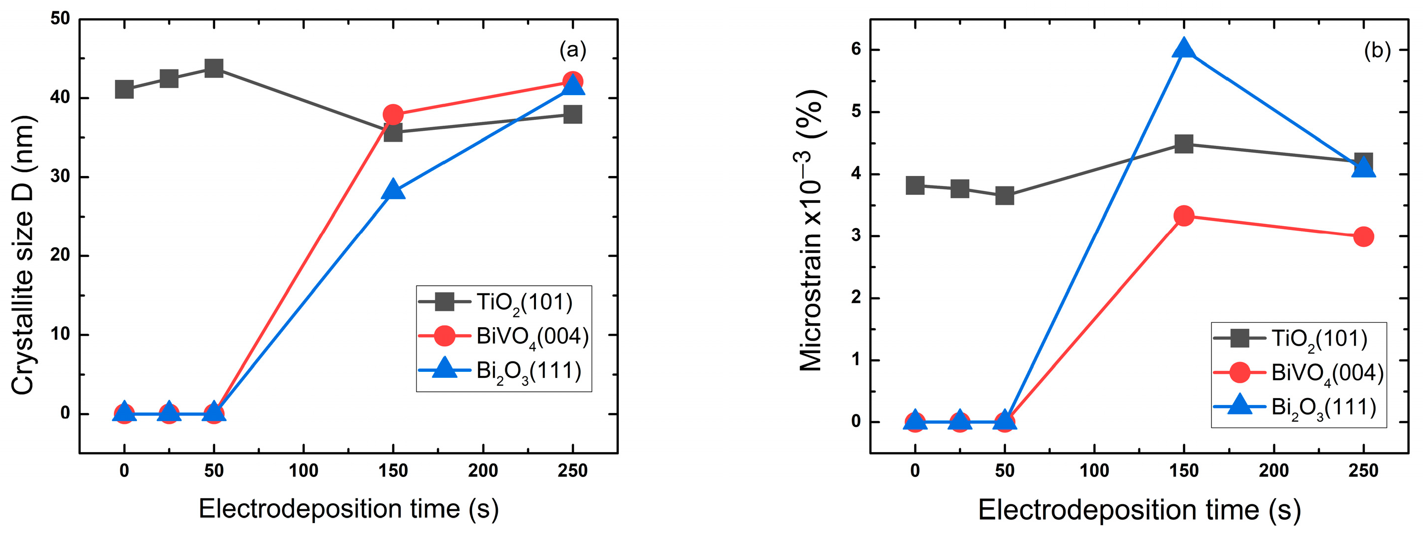
Figure 2 shows the variation in crystallite size and microstrain with the variation in Bi deposition time of the preferred orientation for each composite: TiO2 (101), BiVO4 (004) and Bi2O3 (111). These variations were estimated through the following equations [].
Figure 2. Crystallite size (a) and microstrain (b) of BiVO4/TiO2 at different electrodeposition times: 25 s, 50 s, 150 s, 250 s.
Debye–Scherer formula:
D = K λ β cos θ
Wilson equations:
ε = β 4 t a n ( θ )
where K is the shape factor, λ the X-ray wavelength, β the full width at half maximum of the peak, and θ the Bragg angle.
The exhibited values of the crystallite size corresponding to TiO2 (101) in Table 2 increase as the Bi deposition time increases from 0 to 50 s to reach 43.7 nm, then unexpectedly decreases for 150 and 250 s. This could be in correlation with the formation mechanism of the Bi layers. However, the crystallite size of BiVO4 and Bi2O3, as shown in Table 2, continues to increase with the deposition time to a maximum of 42 and 41.3 nm, respectively. The correlation between crystallite size and microstrain [] is distinguishable for the BiVO4, where the microstrain decreases with increasing crystallite size and electrodeposition time, which means the structure is stabilizing with less defects and dislocations. For Bi2O3, it appears that this structure has the highest microstrain, which could be due to the high defects present in the structure and low crystallinity. However, the 50 s sample TiO2 (101) has the largest crystallite size and the lowest microstrain; this could improve charge transport efficiency with fewer grain boundaries for charge carriers to overcome [].
Table 2. Crystallite size and microstrain of BiVO4/TiO2 for the preferred orientation TiO2 (101), BiVO4 (004) and Bi2O3 (111) at different electrodeposition times.

2.1.2. XPS Analysis

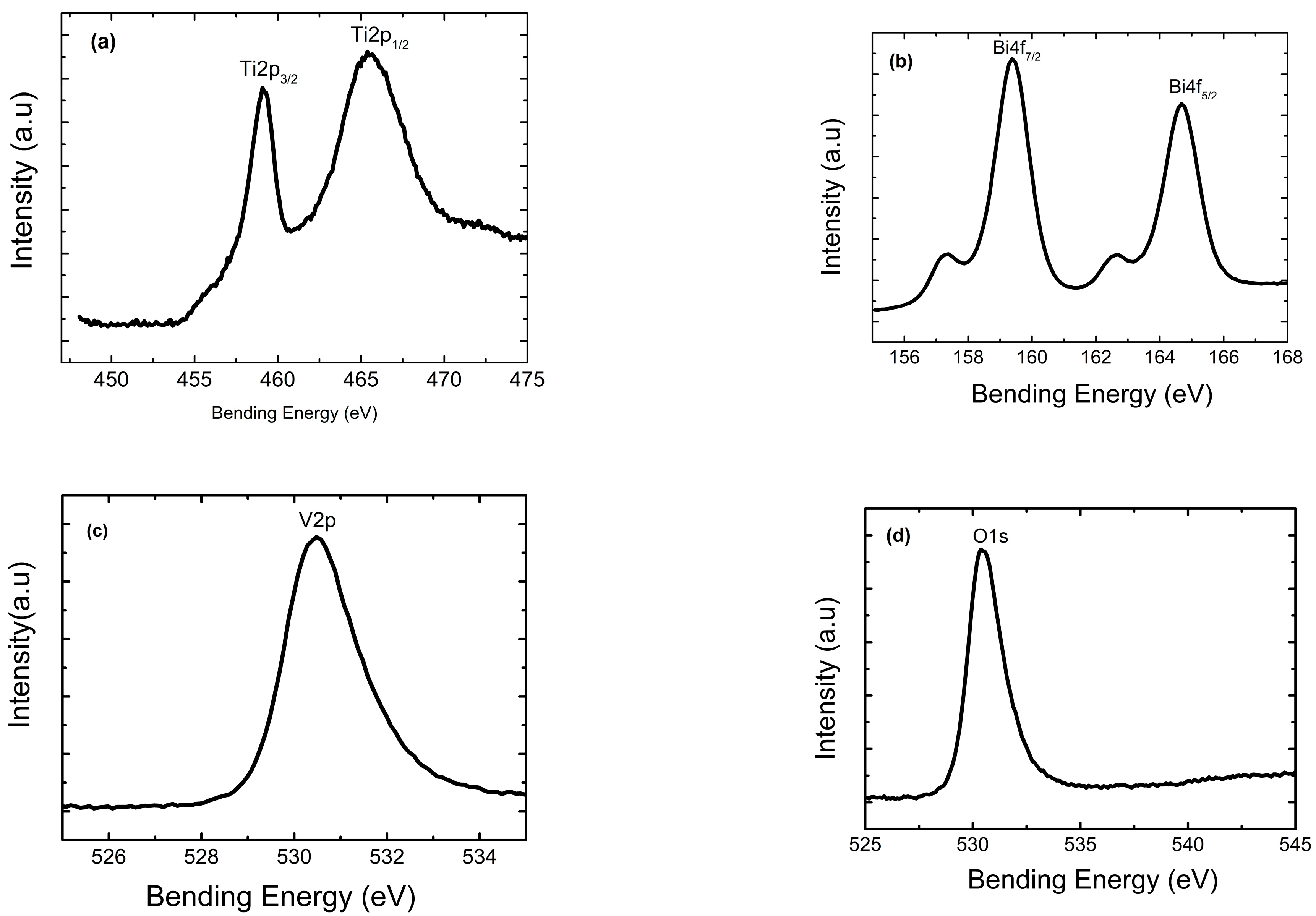
The high-resolution XPS spectra of Ti 2p, Bi 4f, V 2p and O 1s for the BiVO4/NTs-TiO2 nanocomposite are shown in Figure 3, implying the existence of the elements Ti, Bi, V and O in the nanocomposite film. The strong peaks at 459.1 eV and 465.4 eV were assigned to Ti 2p3/2 and Ti 2p1/2 (Figure 3a), respectively, and the energy difference between them was 6.3 eV, matching well with the data of Ti4+ in TiO2 [,]. The peaks appearing at 159.4 eV and 164.7 eV were attributed to Bi 4f7/2 and Bi 4f5/2 (Figure 3b), respectively, and the peaks at 530.5 eV originated from V 2p (Figure 3c). The above XPS results confirmed that the valence states of Bi and V were Bi3+ and V5+ in the composite film, respectively [,]. The characteristic peak of O 1s in Figure 3d at 530.0 eV is attributed to oxygen. Combining the results from XRD and XPS analyses, we concluded that the monoclinic BiVO4-modified TiO2 nanotube nanocomposite film has been successfully prepared through the anodic oxidation and different electrodeposition times.
Figure 3. High-resolution XPS spectra of the BiVO4/NTs-TiO2 50 s composite film: (a) Ti 2p, (b) Bi 4f, (c) V 2p, and (d) O 1s.

2.2. Morphological Analysis

2.2.1. SEM Analysis

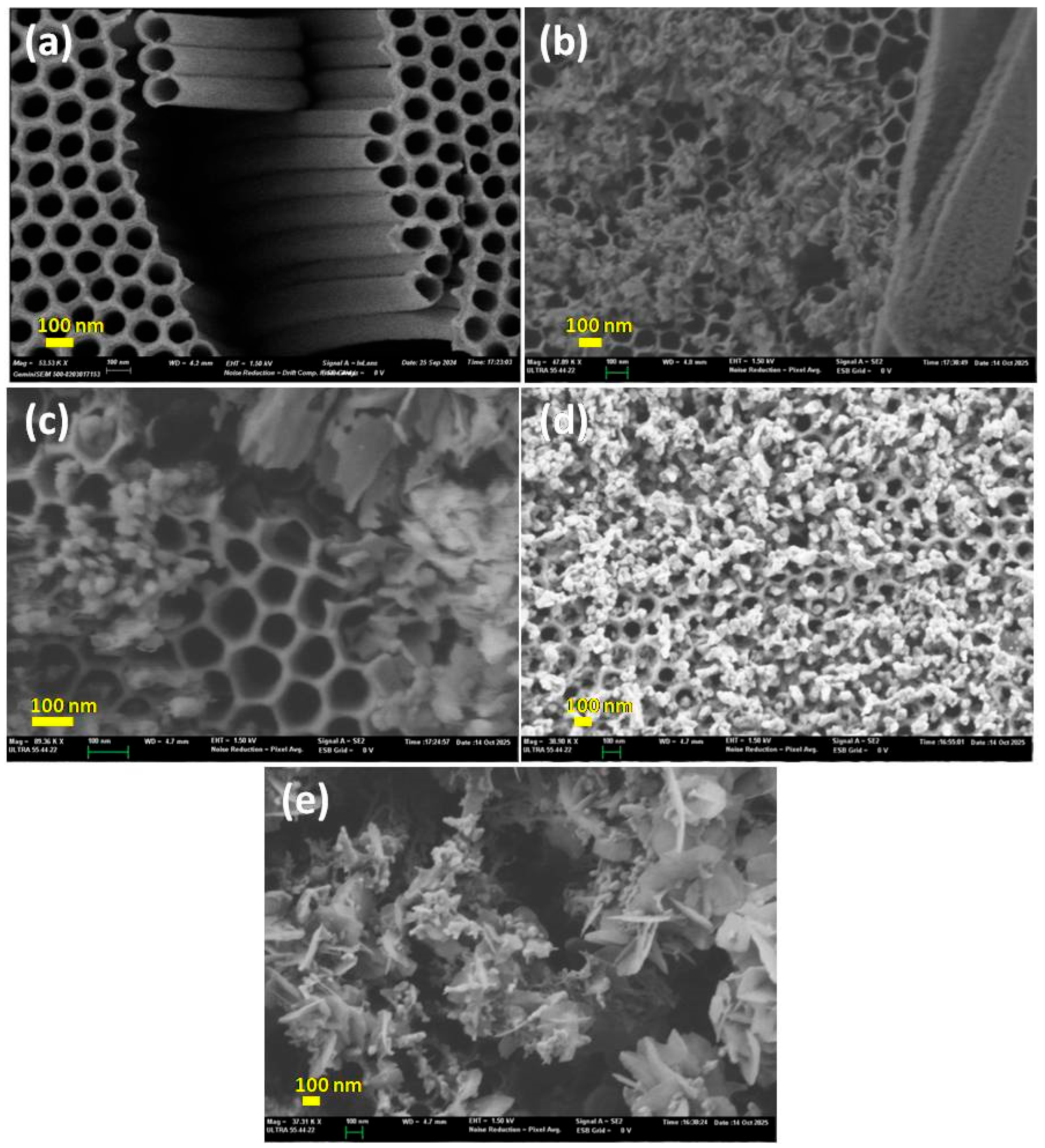
To investigate the effect of the deposition time on morphology of the samples, Scanning Electron Microscopy was conducted for pure TiO2 nanotubes and BiVO4/TiO2 nanotubes composites at different Bi deposition time 25 s, 50 s, 150 s and 250 s. The SEM images were taken at different magnifications to emphasize complementary structural features of the BiVO4/TiO2 samples. While it was not possible to acquire all images under identical magnification, the scale bars are provided to allow accurate comparison. This approach ensures that both the general morphology and the nanoscale features are clearly visible. Figure 4a shows the formation of TiO2 in regular and vertically ordered tubes on the surface of Ti substrate; after the deposition of BiVO4, irregularly shaped particles started to appear at 25 s deposition time (Figure 4b) and the nanosheets of BiVO4 were dispersed unevenly and agglomerated at some points; with the increase in deposition time to 50 s (Figure 4c), the size and number of the nanosheets increased. These nanosheets grew in size and entangled each other to almost form porous, nanoflower-like structures covering the TiO2 nanotubes at higher Bi deposition time (Figure 4d,e), where the nucleation kinetics change, while Bi nanoparticles nucleate and various BiVO4 morphologies emerge [].
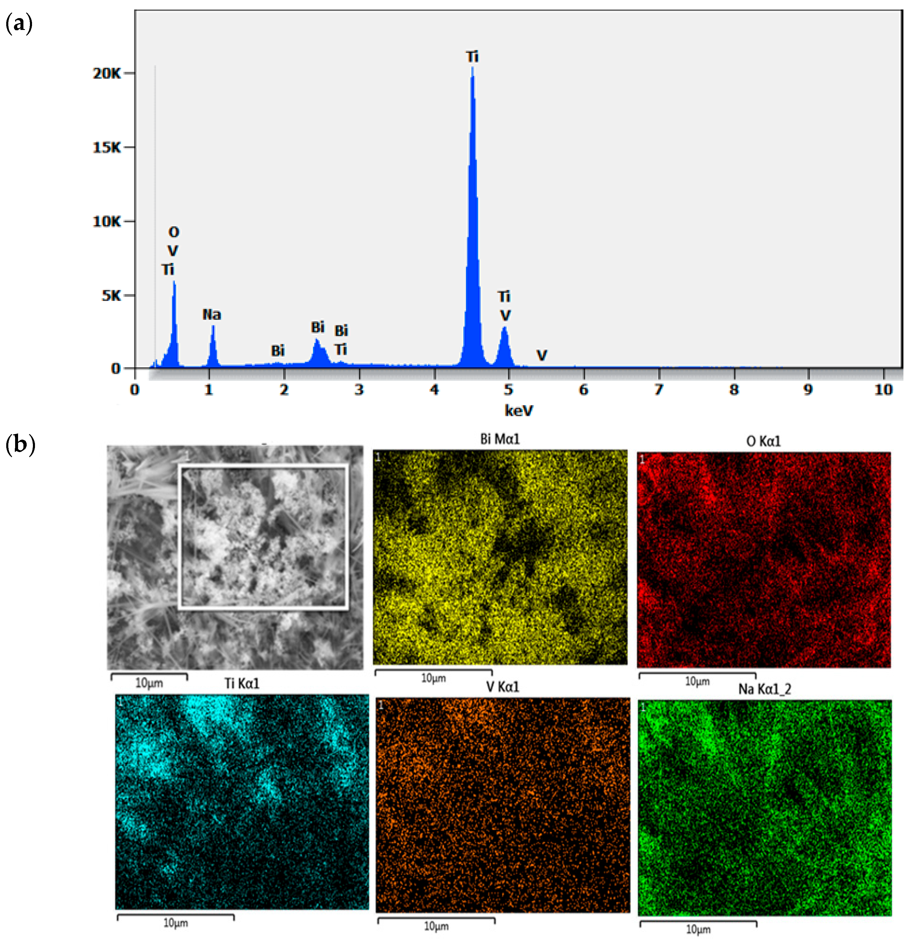
For a more in-depth study, coupled SEM characterization and EDX were conducted to identify the elements and the purity of the samples. Figure 5a shows the EDX spectra where peaks of Ti, O, V, and Bi are extremely visible for the 250 s BiVO4/TiO2 sample; Figure 5b shows the mapping images for the element where the vanadium and bismuth are largely distributed on the surface, which confirms the successful deposition of BiVO4 nanoparticles on the surface of TiO2 nanotubes. Table 3 exhibits the weight percentage of each element. It is distinguishable that Na has about 10% of total wt%; this could be due to the final 30 min wash of the samples with 1 M NaOH to eliminate the yellow-green residual V2O5 excess layer.
Table 3. Weight % of the chemical composition of BiVO4/TiO2 at 250 s electrodeposition time.

2.2.2. TEM Analysis

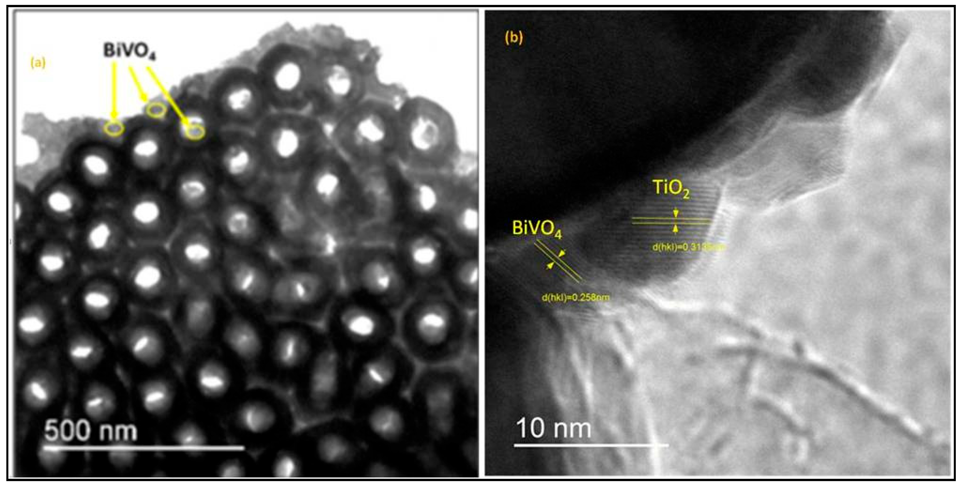
The Transmission Electron Microscopy investigation shows the TEM Images of BiVO4 nanoparticles deposited on TiO2 nanotubes at 50 s electrodeposition time; Figure 6a is bottom view image of the nanotubes with a diameter of 150 nm and an internal diameter of 68 nm. It is clearly visible that the BiVO4 nanoparticles are attached to the external and internal membrane of the nanotubes. Figure 6b is a high-resolution TEM image of the BiVO4/TiO2 interface. The HRTEM micrograph in Figure 6b, with its sharply resolved lattice fringes, clearly confirms the formation of this crucial BiVO4/TiO2 heterojunction. Careful analysis of the fringes shows two key interplanar spacings: the d(hkl) = 0.258 nm corresponding to the (200) plane of monoclinic BiVO4 (JCPDS No. 75-1866), while the d(hkl) = 0.313 nm is indexed to a specific lattice plane of TiO2 anatase (JCPDS No. 21-1272). This intimate physical contact is essential for our composite’s functional performance, as it ensures efficient charge transfer and the successful separation of photogenerated electron–hole pairs, which is a key mechanism for enhancing our photocatalytic system.
Figure 4. SEM imaging of (a) pure TiO2 nanotubes and BiVO4/TiO2 at different electrodeposition times (b) 25 s, (c) 50 s, (d) 150 s, (e) 250 s.
Figure 5. (a) EDX spectra of BiVO4/TiO2 at 250 s electrodeposition time, (b) mapping image of the elements.
Figure 6. (a) Bottom view TEM image of TiO2 nanotubes decorated with BiVO4 nanoparticles at 50 s electrodeposition time, (b) HRTEM imaging of the BiVO4/TiO2 interface.
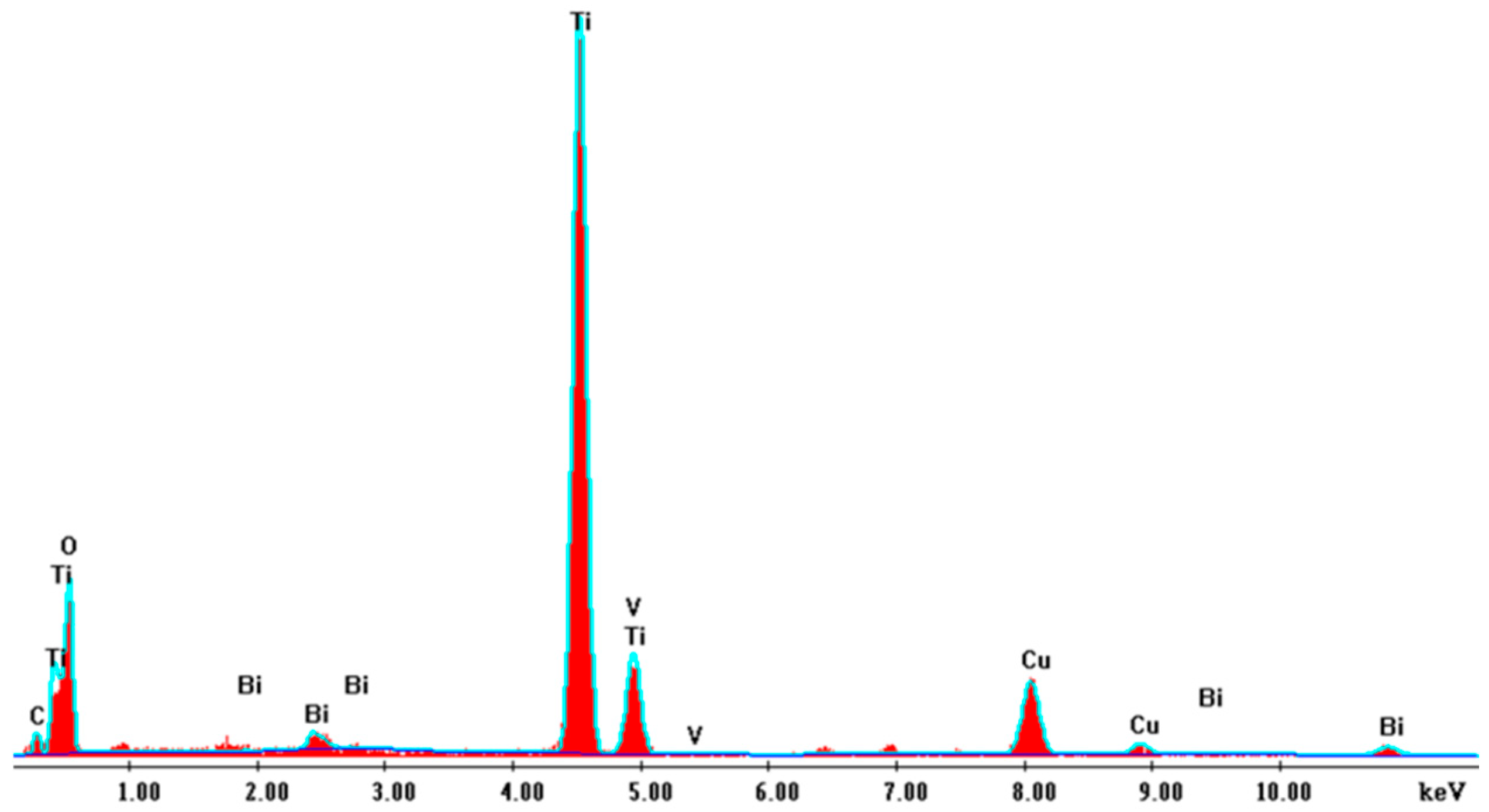
The performed EDX in Figure 7 confirms the high purity of the sample and the evidence of existing elements Ti, O, Bi, V.
Figure 7. EDX spectra of BiVO4/TiO2 at 50 s electrodeposition time.

2.3. Optical Analysis

2.3.1. Photoluminescence

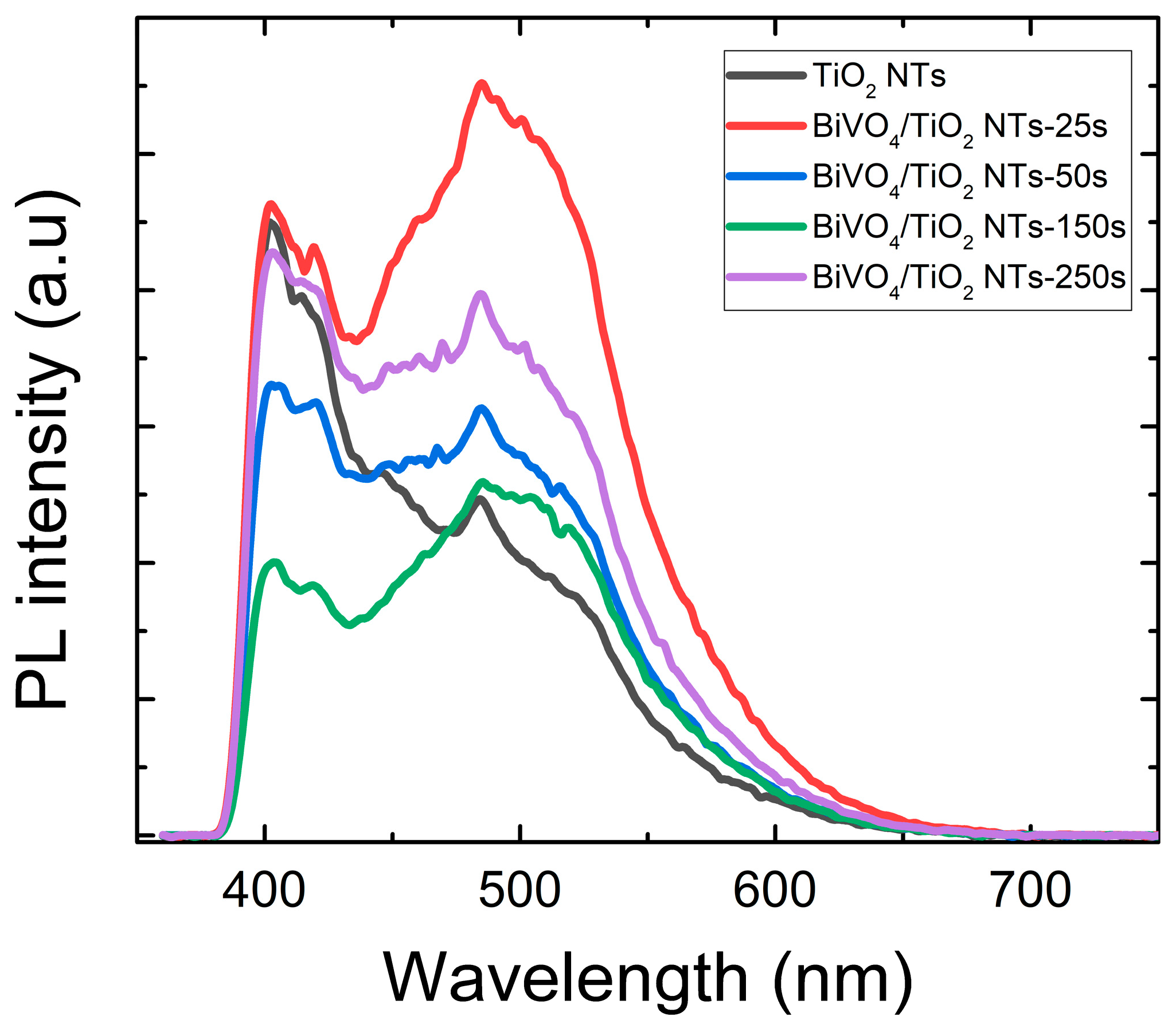
The migration, transfer and trapping of the photogenerated charge carrier’s electron–hole pairs in TiO2 were investigated with photoluminescence assessment, which also apprises the defects present in the band gap []. Figure 8 depicts the photoluminescence spectra of the BiVO4/TiO2 samples at different electrodeposition time 25 s, 50 s, 150 s and 250 s to better understand the impact of BiVO4 depositions on the optical properties of TiO2. Compared to the bare TiO2 nanotubes, it seems that all the samples present the same peaks located between 390 nm and 420 nm corresponding to 3~3.2 eV, which is compatible with the electronic transition between conduction and valence bands. The collection of peaks detected between 450 nm and 550 nm is related to the oxygen vacancies in TiO2 structure. Since the recombination of electron/hole is derived from the PL emission, the weaker the PL intensity, the lower the recombination rate.
Figure 8. Photoluminescence spectra of bare TiO2 and BiVO4/TiO2 nanotubes with different electrodeposition times: 25 s, 50 s, 150 s, 250 s.
The detected data reveal a non-monotonic trend in charge carrier recombination. Compared to the bare TiO2 nanotubes, the 50 s and 150 s samples exhibit a significant decrease in PL intensity. This indicates a lower electron–hole recombination rate, which we attribute to the successful formation of a BiVO4/TiO2 heterojunction at these deposition times. The heterojunction promotes the spatial separation of photogenerated charges, thereby suppressing their recombination. In contrast, the 25 s and 250 s samples show a higher PL intensity than pure TiO2, signaling enhanced recombination. For the 25 s sample, the low BiVO4 coverage is likely insufficient for optimal heterojunction formation; instead, the deposited material may introduce defective states that act as recombination centers. For the 250 s sample, the excessively thick and aggregated BiVO4 layer (as seen in SEM) introduces a high density of grain boundaries and defects, hindering charge transfer and promoting non-radiative recombination within the overlayer itself. This trend confirms that an optimal BiVO4 deposition time is critical to maximizing charge separation, with the 50 s sample achieving the most effective balance [].

2.3.2. Diffuse Reflectivity

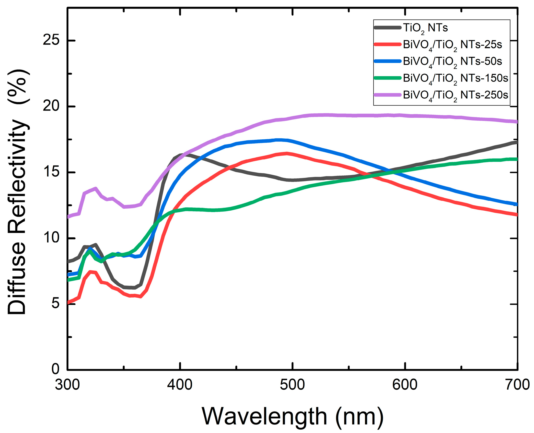
The diffuse reflectivity spectra of as-fabricated samples have been investigated with UV–Vis spectroscopy as shown in Figure 9; the spectra depict a minimum for all samples up to 380 nm, which corresponds to the gap energy of anatase TiO2 3.2 eV; then, the diffuse reflectivity increases for higher wavelengths. It seems that 250 s electrodeposition time of BiVO4 nanoparticles presents the highest reflectivity, which could be due to the surface characteristics; however, on the contrary, the 25 s sample showed a lower reflectivity in comparison to pure TiO2 and other samples. To determine the band gap of each sample, the Kubelka–Munk formula was used to create the Tauc plots (F(R))1/2 vs. hʋ [].
F R = ( 1 R ) 2 2 R
Figure 9. Diffuse reflectivity spectra of bare TiO2 and BiVO4/TiO2 nanotubes with different electrodeposition times: 25 s, 50 s, 150 s, 250 s.
After the exploitation of the Tauc plots, the collected optical band gap energy of each sample is presented in Table 4. The bare TiO2 nanotube presented band gap energy of 3.3 eV, which is almost consistent with the literature, in that the deposition time has an intense effect on the optical band gap energy. The band gap decreased to a minimum of 2.6 eV for the 50 s sample, which possesses the largest TiO2 crystallite size and lowest microstrain, indicating high crystallinity. However, for longer deposition times (150 s, 250 s), it increased to 2.66 eV and 2.7 eV, respectively. This reversal coincides with a significant reduction in the apparent TiO2 crystallite size (from 43.74 nm to 35.64 nm) and a probable increase in microstrain (Figure 2b). We attribute this not to a contradiction with the XRD results, but to the effects of thermal treatment on the composite. At high Bi loadings, the vigorous interfacial reaction and stress during the formation of a thick BiVO4 layer introduce lattice defects and strain into the TiO2 substrate. This disrupts the long-range order, broadening the XRD peaks and reducing the calculated crystallite size. The associated increase in band gap is consistent with the quantum confinement effect induced by this effective reduction in crystalline domain size and the rise in defect density within the TiO2 structure [,].
Table 4. Band gap energy and crystallite size of bare TiO2 and BiVO4/TiO2 nanotubes with different electrodeposition times: 25 s, 50 s, 150 s, 250 s.
The size of the crystal plays an important role in determining the band gap of the semiconductor, with smaller sizes leading to larger band gaps due to quantum confinement effects, where small particle size leads to discrete energy levels and band splitting []. Furthermore, the effect of crystal size on the band gap shift is found to be parallel to that of intrinsic strain [].

2.4. Photodegradation of Black Amido with BiVO4/TiO2

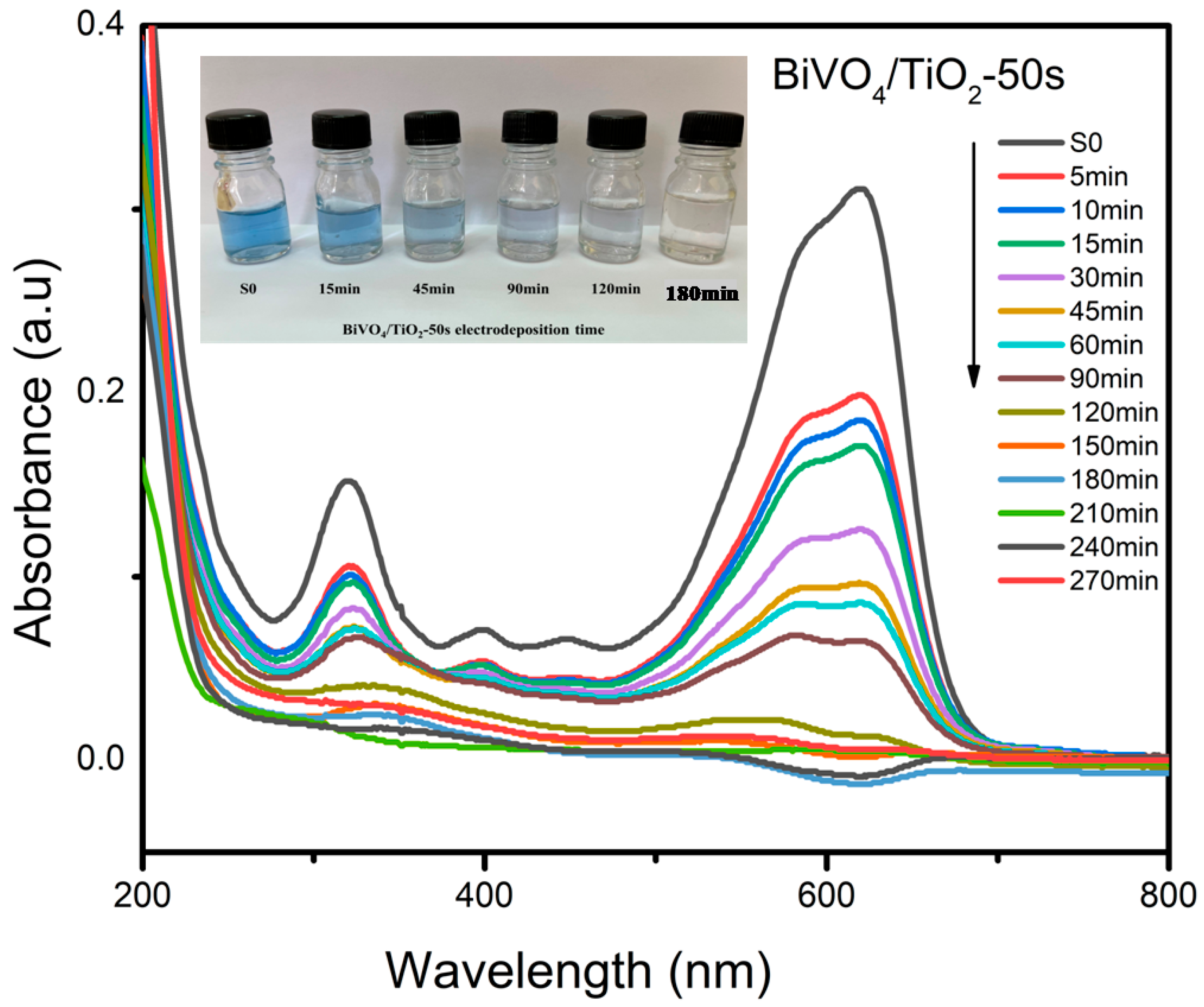
The photodegradation of Black Amido was conducted under UV light at different times (5, 10, 15, 30, 45, 60, 90, 120, 150, 180, 210, 240 and 270 min) and with bare TiO2 nanotubes and BiVO4-NPs/TiO2-NTs as photocatalysts with different electrodeposition times 25 s, 50 s, 150 s, and 250 s. Black Amido was chosen as an organic pollutant due to its complex chemical structure; the evaluation of the concentration during the photocatalysis process was assessed with UV–Vis absorption studies using the Beer–Lambert relation at a maximum wavelength of λ= 618 nm. Figure 10 depicts the absorbance spectra after the photocatalytic activity of Amido Black AB. The inset image shows how the blue color of AB is gradually decreasing in intensity as the irradiation time increases, and is completely gone at 180 min.
Figure 10. The absorbance of Amido Black after photodegradation for bare TiO2 and BiVO4/TiO2 nanocomposites at 50 s electrodeposition time.
Following Equation (4), the removal % of AB with BiVO4/TiO2 photocatalyst was determined [].
Removal   of   dye   ( % ) = ( 1 C C 0 ) × 100
It seems that the sample BiVO4/TiO2-50 s electrodeposition time reached a total degradation of Black Amido 99.4% at 150 min under UV light as illustrated in Figure 11, which is considered the highest compared to the other samples; this result agrees with the characterization results and the addition of BiVO4 to favor the production of reactive oxygen species. The active sites responsible for the degradation of the pollutant increase with the presence of BiVO4.
Figure 11. Photodegradation efficiency of bare TiO2 and BiVO4/TiO2 nanocomposites with different electrodeposition times: 25 s, 50 s, 150 s, 250 s for Amido Black removal.
The rate of the photodegradation kinetic was investigated with the given equation below []:
ln C 0 C t = k t
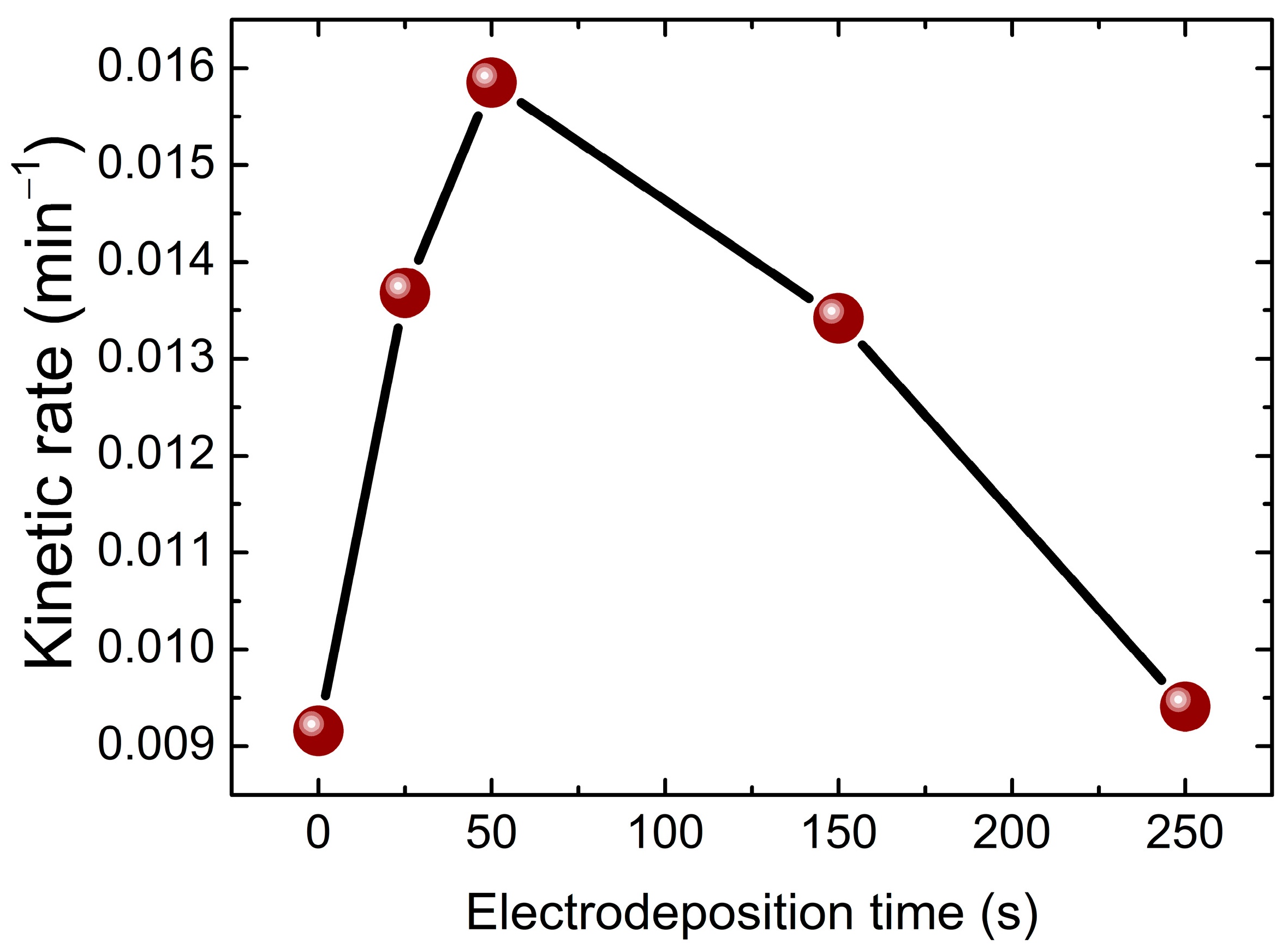
where C0 and Ct are the concentrations of the dye at time 0 and t, respectively, and k is the rate constant. The slope of ln C 0 C t plots provide the kinetic of photodegradation reaction rate ‘k’, which is presented in Figure 12. Compared to the pure TiO2 nanotubes, all the samples deposited with BiVO4 nanoparticles exhibited higher kinetic rate constant (k), especially the 50 s sample with k = 0.0159 min−1, which corresponds to the maximum removal percentage 99% and is in correlation with the optical and structural results where the 50 s sample had the largest crystallite size and lowest microstrain as well as the lowest band gap energy.
Figure 12. Kinetic constant of bare TiO2 and BiVO4/TiO2 nanocomposites with different electrodeposition time 25 s, 50 s, 150 s, 250 s for Amido Black degradation reaction.
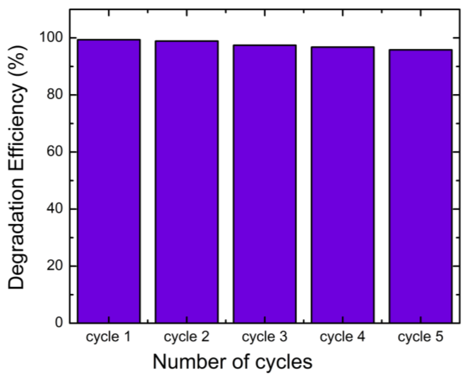
The stability and the reusability of the sample was investigated as shown in Figure 13. The photocatalytic activity of BiVO4/TiO2 composites remains at a high capacity even after five consecutive photocatalytic performances for AB degradation. It only displays less than 4% deactivation after five cycles.
Figure 13. Recycle test for BiVO4/TiO2-50 s electrodeposition time for five consecutive cycles of photodegradation of AB.

2.5. Quenching Test

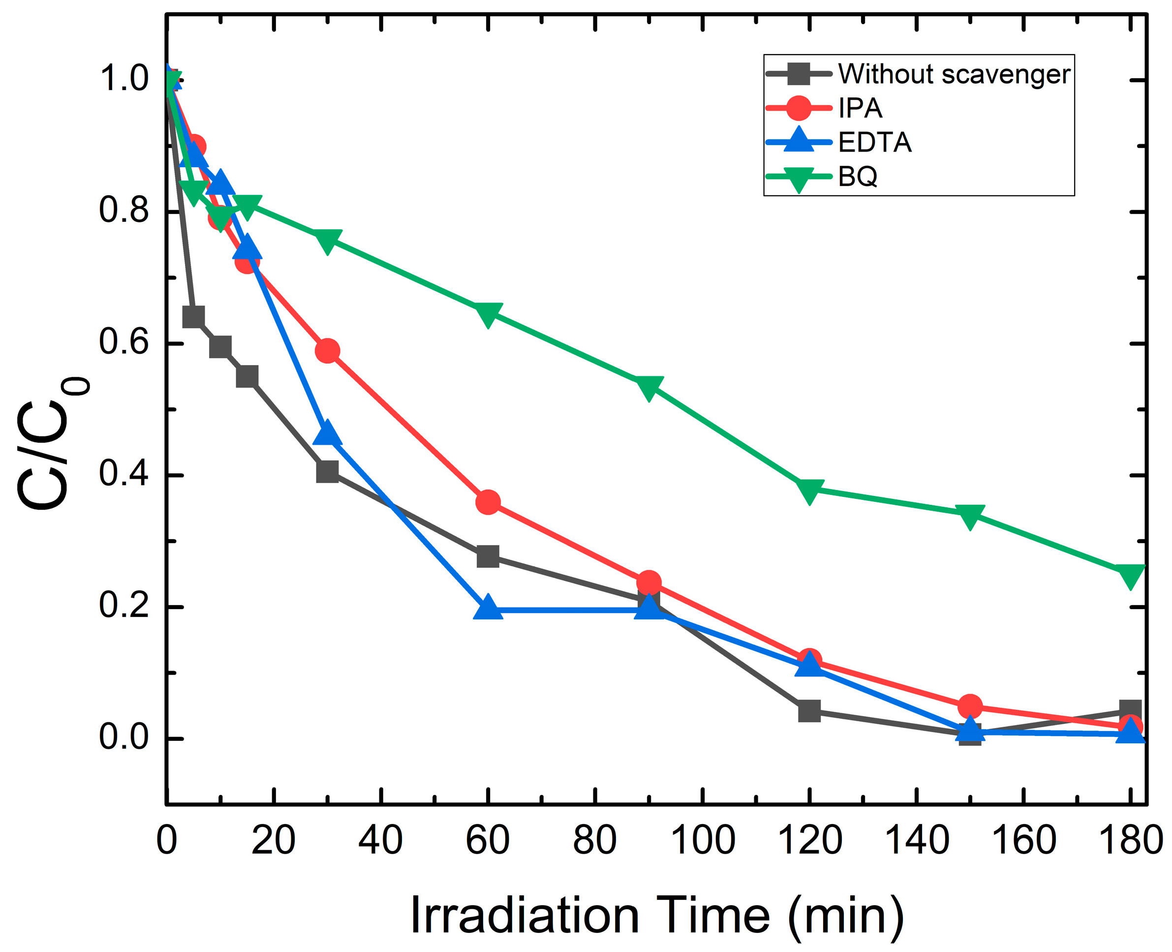
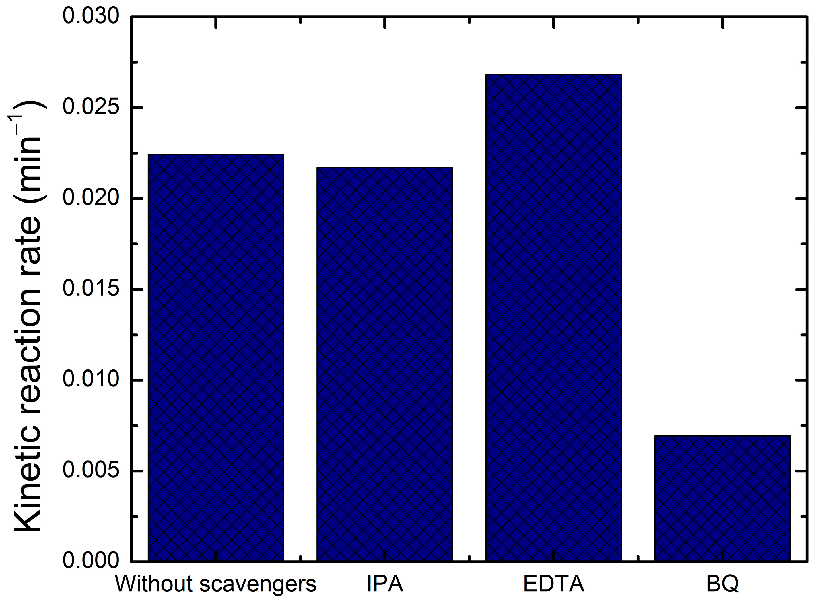
There are a variety of scavengers to use for the quenching of active oxygen species, including IPA, EDTA and BQ. These scavengers can trap holes (h+), hydroxyl radicals (OH) and superoxide radicals (O2•−), respectively []. Figure 14 depicts the effect of scavengers on the degradation of AB under UV light for 180 min. The scavenger trapping test was conducted to better understand the photocatalytic mechanism and assess the effect of the responsible reactive molecules in the photocatalytic approach.
Figure 14. Photocatalytic evaluation of AB degradation for BiVO4/TiO2-50 s electrodeposition time with the presence of active species scavengers.
Figure 15 is a comparison of kinetic rate constant k for the BiVO4/TiO2-50 s electrodeposition time with and without scavengers; it seems that the kinetic rate intensely decreases with the addition of BQ, which means that the superoxide radicals (O2•−)are the ones responsible for the degradation of Black Amido or precisely, the ones that are mostly generated in these reactions. The significant increase in k with the addition of EDTA could be explained by the capturing of holes, which increases the separation of electron/hole pairs, thus, decreasing the recombination rate and the excessive existence of electrons favor the generation of superoxide radicals (O2•−), which were previously proven to be responsible for the degradation of AB. However, with the addition of IPA, the constant rate vaguely decreases, indicating that hydroxyl radicals (OH) are not as important in the photodegradation of AB. These results are in accordance with the published literature [], where the only vague decrease in the rate constant upon the addition of IPA (a scavenger of OH radicals) provides crucial evidence that a classical OH mediated oxidation pathway is minor. This is a key signature of the proposed Z-scheme mechanism. In this system, the photogenerated holes in the TiO2 valence band, which are the typical source of OH radicals, are predominantly consumed by internal recombination with electrons from the BiVO4 conduction band. Consequently, the generation of OH is suppressed. The primary oxidative power is instead provided by the holes accumulated in the BiVO4 valence band, which can directly oxidize the pollutant, alongside the dominant action of superoxide radicals (O2•−).
Figure 15. Photocatalytic reaction rate k of AB degradation for BiVO4/TiO2-50 s electrodeposition time with and without the presence of active species scavengers.

2.6. Mechanism of Photocatalysis

The synthesization of p-n heterojunction BiVO4/TiO2 and the position of band edges after the alignment is of great interest for the amelioration of photocatalytic performances. Under irradiation, both photocatalysts photogenerate electron/hole pairs that migrate to the surface of the material p-n heterojunction, where the electrons will react with oxygen molecules for the development of superoxide radicals (O2•−)and the holes will react with water molecules for the development of hydroxyl radicals OH, as demonstrated in the following equations []
TiO2 + hν → e (CB) + h+ (VB)
O2 + e → (O2•−)
h+ + H2O → OH + H+
h+ + OH → OH
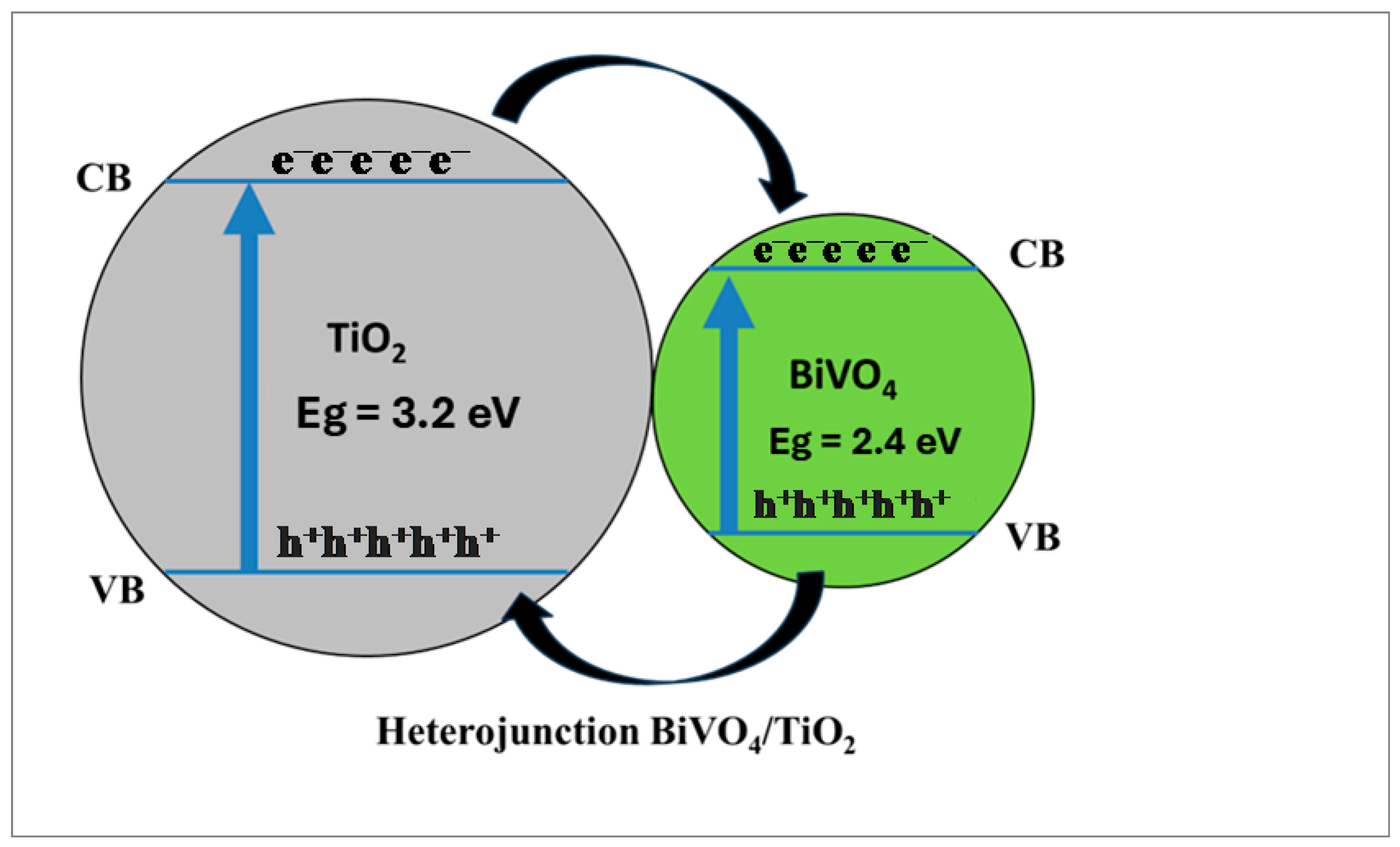
The same mechanism is performed on the surface of both photocatalysts, and after the production of these reactive species, ROS, they engage in the degradation of organic pollutants present in wastewater. The separation phenomenon of the photogenerated charge carries effectively reduces the recombination rate and enhances the photocatalytic efficiency of BiVO4/TiO2 nanocomposites; according to the literature, BiVO4 nanoparticles exhibit a work function of χ = 6.04 eV, while TiO2 possesses a χ = 5.8 eV [] after contact and the band alignment, as shown in Figure 16, is where the electrons transfer from the conduction band of TiO2 to BiVO4 and the holes migrate from the valence band of BiVO4 to TiO2.
Figure 16. Charge carrier transfer and separation mechanism for BiVO4/TiO2 nanocomposite.

3. Discussion

The method used demonstrates that electrodeposition of BiVO4 on TiO2 nanotubes is an effective and simple method for enhancing photocatalytic activity. The fabrication of BiVO4 nanostructures was successful via a two-step process: electrodeposition of Bi nanoparticles, followed by heat treatment. The Bi deposition time had a significant impact on the composite properties. SEM analysis revealed that with increasing deposition time (from 25 s to 250 s), the morphology of the deposited BiVO4 changed from irregular and dispersed nanosheets (25 s, 50 s) to larger and aggregated structures, forming a porous film partially covering the TiO2 nanotubes at longer times (150 s, 250 s).
The photocatalytic performance for the degradation of Amido Black was highly dependent on the electrodeposition time. The BiVO4/TiO2 sample with a deposition time of 50 s exhibited the best degradation efficiency (99.4% in 150 min) and the fastest reaction kinetics. This optimal performance is attributed to a synergistic combination of structural and optical properties. XRD analysis indicated that the TiO2 (101) crystallites in the 50 s sample possessed the largest size (43.74 nm) and the lowest microstrain, suggesting that high crystallinity is favorable for charge transport. Although the size of the BiVO4 crystallites themselves is smaller at short deposition times, their presence and optimal distribution, confirmed by TEM and EDX, are crucial. The BiVO4 nanoparticles act as a co-catalyst, forming a heterojunction with TiO2 that promotes the separation of photogenerated electron–hole pairs. This is supported by the photoluminescence (PL) spectra, where the 50 s sample exhibited lower PL intensity, indicating reduced charge carrier recombination.
The optical properties were also optimized for the 50 s deposition time, as this sample exhibited the lowest optical band gap (2.6 eV). The heterojunction between BiVO4 and TiO2 facilitates a Z-type charge transfer mechanism, where electrons from the TiO2 conduction band combine with holes from the BiVO4 valence band. This process effectively separates the strongest reducing and oxidizing agents, increasing the generation of reactive oxygen species (ROS). Trapping tests confirmed that superoxide radicals (O2•−) are the main active species in the degradation process. Furthermore, the BiVO4/TiO2-50 s composite demonstrated excellent stability, maintaining high photocatalytic activity over five consecutive cycles with minimal deactivation (<4%). In conclusion, the BiVO4/TiO2 heterojunction, particularly with an optimized electrodeposition time of 50 s, represents a promising and effective catalyst for water remediation and pollutant degradation applications. Moreover, the performance of BiVO4/TiO2 in AB degradation was found to be better or comparable to those of the similar tabular forms of catalysts reported in the recent literature, as summarized in Table 5.
Table 5. Comparative work of BiVO4/TiO2 under different photocatalytic conditions.

4. Materials and Methods

4.1. Chemicals

The reagents used in this work are bismuth (III) nitrate pentahydrate (Bi(NO3)3.5H2O, 99.99%), lithium perchlorate (LiClO4, 99%), dimethyl sulfoxide (DMSO, extra pure grade), vanadyl acetylacetonate (VO (acac)2, 99%), and sodium hydroxide (NaOH, extra pure grade). All these chemicals were purchased from Sigma-Aldrich (St. Louis, MO, USA).

4.2. Synthesis of TiO2 Nanotubes (NTs)

A second-grade Ti foil with a dimension of 2.5 cm × 2.5 cm × 1 mm (99.7%) was used. The surface of the foil was first polished with sanding papers of grades ranging from 320 to 2200. Then, it was ultrasonically cleaned for 10 min, with ethanol, acetone, and double-distilled water, in that order. The elaboration of TiO2 NTs from Ti substrates involved a two-step anodization: 45 min for the first anodization and 120 min for the second one under a fixed voltage of 60 V at room temperature. The process was performed in an electrolytic cell containing 100 mL of ethylene glycol + 1 wt% NH4F + 2 v% H2O. To ensure the anatase crystal phase of TiO2 was obtained, the samples were annealed in an oven at 450 °C for 3 h.

4.3. Synthesis of BiVO4 Nanoparticles on TiO2-NTs

Two steps are required for the electrodeposition of BiVO4 on titanium dioxide NTs. Firstly, the Bi thin films were prepared by the electrodeposition method for four periods of time: 25, 50, 150 and 250 s. We used an electrolyte bath containing 50 mL of dimethyl sulfoxide (DMSO), 20 mM of Bi(NO3)3.5H2O, and 0.1 M LiClO4. A three-electrode Autolab PGSTAT30 potentiostat/galvanostat PGSTAT30 was used for the electrodeposition process: titanium NTs substrate as working electrode, Ag/AgCl (3 M NaCl) as reference electrode, and Pt wire as counter electrode. The temperature of the plating solution was fixed at 60 °C and the optimized cathodic potential was −1.6 V (vs. Ag/AgCl). After each electrodeposition, the dark-colored Bi films were carefully immersed in DMSO solution for 3 min and dried in air at 70 °C for 1 h. Then, in order to convert Bi to BiVO4, a DMSO solution containing 50 μL of 50 mM VO (acac)2 was spread over the entire surface of the Bi film by dip coating and completely immersing the electrode in the solution. The film was then heated at 450 °C for 3 h in air (ramping rate = 2 °C min−1). During the heating process, Bi-metal and VO2+ species were oxidized to Bi2O3 and V2O5, respectively. Then, they reacted with each other to form BiVO4. The VO2+ ions were in excess to ensure the complete conversion of Bi to BiVO4. This excess led to the formation of yellow-green residual V2O5 layer that could be easily removed by soaking the samples in 1 M NaOH solution for 30 min while stirring. BiVO4 samples were obtained for four Bi deposition times (25, 50, 150, and 250 s).

4.4. Samples Characterization

In order to examine the morphology of the prepared samples, we used a 15 kV Scanning Electron Microscope (SEM, FEI XL30 ESEM, Hatfield, PA, USA) and Transmission Electron Microscopy (TEM, FEI Tecnai G2) equipped with X-ray energy dispersive analysis (EDX) operating at 200 kV. For structural study, X-ray diffraction was performed using Philips X’ PERTMPD diffractometer (Amsterdam, The Netherlands), which uses CuKα radiation (λ = 1.5406 Å). The diffraction patterns ranged in 2θ = 10–80°. The spectra were collected with a Thermo Scientific K-Alpha X-Ray Photoelectron Spectrometer (XPS, Waltham, MA, USA) System using monochromatized Al Kα (1486.6 eV). Optical properties of pure TiO2 NTs and BiVO4-TiO2NTs were investigated by UV–Vis-NIR photoluminescence (PL) using a Perkin Elmer Lambda 950 spectrophotometer (Waltham, MA, USA) between 200 and 1200 nm and Perkin Elmer LS55 coupled to xenon lamp at an excitation wavelength of λ = 340 nm.

4.5. Photocatalytic Activity Measurements

4.5.1. Photodegradation of Black Amido

The photocatalytic activity of the prepared samples (bare TiO2 NTs and BiVO4/TiO2 nanocomposites) was evaluated by monitoring the degradation of Amido Black 10B (AB) dye under UV light irradiation. The process was conducted at ambient temperature (~25 °C).
Aqueous solutions of AB dye with a concentration of 5 mg·L−1 were prepared. For each test, approximately 10 mL of the dye solution was placed in a Petri dish, and the photocatalyst sample (a thin film on a Ti foil substrate with dimensions of 2.5 cm × 1 cm) was immersed into the solution.
Prior to irradiation, the system was kept in complete darkness under constant magnetic stirring for 15 min to establish an adsorption–desorption equilibrium between the dye molecules and the catalyst surface. This step ensures that the subsequent degradation was due to photocatalytic reactions and not merely physical adsorption.
After the dark adsorption period, the solution was irradiated using a 15 W OSRAM germicidal UV lamp with a primary emission wavelength of λ = 256 nm. The lamp, with a length of 45 cm, was positioned at a fixed distance above the Petri dishes to ensure uniform illumination. Appropriate safety measures (UV-protective glasses and gloves) were strictly followed during irradiation.
At predetermined time intervals (5, 10, 15, 30, 45, 60, 90, 120, 150, 180, 210, 240, and 270 min), small aliquots (~1 mL) of the solution were extracted from the Petri dish. The concentration of AB dye in the collected samples was monitored by measuring the absorbance at its characteristic maximum wavelength (λmax = 618 nm) using a UV–Vis spectrophotometer. The degradation efficiency was calculated based on the decrease in absorbance relative to the initial concentration after the dark adsorption step.

4.5.2. Trapping Test

To better understand which of the generated ROS are responsible for the massive degradation of Black Amido with the photocatalysts BiVO4/TiO2, a collective group of scavengers have been used, such as EDTA, IPA and BQ; each is assigned to capture a species from the ROS hole h+, radical’s hydroxyl OH and radical’s superoxide (O2•−). The equations below explain the quenching processes [,]:
  • Quenching of Hydroxyl Radicals (OH) by Isopropanol (IPA)
    Isopropanol reacts with hydroxyl radicals via hydrogen abstraction, leading to the formation of acetone and water.
Chemical Equation:
(CH3)2CH-OH + OH → (CH3)2C-OH + H2O
2 (CH3)2C-OH → (CH3)2C(OH)-C(OH)(CH2)2 (Dimerization)
(CH3)2C-OH + O2 → (CH3)2C=O + HO2 (Formation of Acetone and a hydroperoxyl radical)
Quenching of Superoxide Radical Anions (O2•−) by Benzoquinone (BQ)
Benzoquinone (BQ) readily accepts an electron from the superoxide radical, forming a semiquinone radical and then hydroquinone after protonation.
Chemical Equation:
O2•− + BQ → O2 + BQ•− (Semiquinone Radical Anion)
BQ•− + H+ → HBQ (Semiquinone Radical)
HBQ + O2•− + H+ → H2BQ (Hydroquinone) + O2
Quenching of Photogenerated Holes (h+) by Ethylenediaminetetraacetic Acid (EDTA)
EDTA acts as a sacrificial electron donor, capturing the photogenerated holes before they can generate OH radicals or directly oxidize the dye.
Chemical Equation (Simplified):
EDTA + h+ → EDTA•+ (Oxidized EDTA)

5. Conclusions

In summary, this study demonstrates that electrodeposition of BiVO4 on TiO2 nanotubes is an effective and simple method for enhancing photocatalytic activity. The fabrication of BiVO4 nanostructures was successful via a two-step process: electrodeposition of Bi nanoparticles followed by heat treatment. The Bi deposition time had a significant impact on the composite properties. SEM analysis revealed that with increasing deposition time (from 25 s to 250 s), the morphology of the deposited BiVO4 changed from irregular and dispersed nanosheets (25 s, 50 s) to larger and aggregated structures, forming porous nanoflower-like structures partially covering the TiO2 nanotubes at longer times (150 s, 250 s).
The photocatalytic activity of the BiVO4/TiO2 composites was found to be strongly influenced by the electrodeposition time, with the sample deposited for 50 s exhibiting the highest degradation efficiency toward Amido Black (99.4% within 150 min). This outstanding performance results from a well-balanced combination of structural and optical characteristics. XRD analysis revealed that the TiO2 (101) crystallites in the 50 s sample possessed the largest size and lowest microstrain, reflecting enhanced crystallinity and improved charge carrier transport. Meanwhile, TEM and EDX analyses confirmed the uniform dispersion of BiVO4 nanoparticles, which promotes the formation of a well-defined BiVO4/TiO2 heterojunction. Such intimate interfacial contact effectively facilitates charge separation, as further evidenced by the reduced PL emission intensity.
In addition, the narrowed optical band gap (2.6 eV) of the BiVO4/TiO2-50 s composite enhances visible-light absorption and contributes to superior photocatalytic efficiency. The Z-scheme charge transfer mechanism established between BiVO4 and TiO2 maintains strong redox potentials, favoring the formation of reactive oxygen species—primarily superoxide radicals (O2•−) as confirmed by radical-trapping experiments. Furthermore, the BiVO4/TiO2-50s composite displayed remarkable cycling stability, preserving over 96% of its initial performance after five consecutive runs. Overall, the optimized BiVO4/TiO2 heterojunction with a 50 s deposition time demonstrates high efficiency, robustness, and durability, highlighting its great promise as a sustainable photocatalyst for wastewater treatment and environmental remediation.

Author Contributions

Conceptualization, K.B.M. and A.H.; methodology, K.B.M. and A.H.; software, S.S., R.B.Z. and I.K.; validation, K.B.M., A.B., B.M.S. and A.H.; formal analysis, K.B.M., S.S., R.B.Z., H.E. and A.H.; investigation, K.B.M., S.S., I.K., R.B.Z., L.K., H.E., A.B., B.M.S. and A.H.; data curation, A.H. and L.K.; writing—original draft preparation, K.B.M., S.S., A.B. and A.H.; writing—review and editing, K.B.M., S.S., A.B., A.H. and B.M.S.; visualization, L.K.; supervision, A.H. All authors have read and agreed to the published version of the manuscript.

Funding

This work has been funded by the Conselleriad’Innovació, Universitats, Ciènciai Societat Digital (Generalitat Valenciana) under the Prometeus 2023 program, grant CIPROM2022/03).

Institutional Review Board Statement

Not applicable.

Data Availability Statement

The original contributions presented in this study are included in the article. Further inquiries can be directed to the corresponding authors.

Conflicts of Interest

The authors declare no conflicts of interest. The funders had no role in the design of the study; in the collection, analyses, or interpretation of data; in the writing of the manuscript; or in the decision to publish the results.

References

  1. Jaishree, G.; Divya, G.; Rao, T.S.; Chippada, M.L.V.P.; Raju, I.M. Biogenic surfactant mediated facile synthesis of visible light sensitized Zn/Mg co-doped TiO2 nanomaterials—A green approach: Evaluation of photocatalytic activity by degradation of Amido Black 10B. Sustain. Environ. Res. 2022, 32, 38. [Google Scholar] [CrossRef]
  2. Forgacs, E.; Cserháti, T.; Oros, G. Removal of synthetic dyes from wastewaters: A review. Environ. Int. 2004, 30, 953–971. [Google Scholar] [CrossRef]
  3. Khan, S.; Noor, T.; Iqbal, N.; Yaqoob, L. Photocatalytic Dye Degradation from Textile Wastewater: A Review. ACS Omega 2024, 9, 21751–21767. [Google Scholar] [CrossRef]
  4. Lee, K.M.; Lai, C.W.; Ngai, K.S.; Juan, J.C. Recent developments of zinc oxide based photocatalyst in water treatment technology: A review. Water Res. 2016, 88, 428–448. [Google Scholar] [CrossRef]
  5. Burakov, A.E.; Galunin, E.V.; Burakova, I.V.; Kucherova, A.E.; Agarwal, S.; Tkachev, A.G.; Gupta, V.K. Adsorption of heavy metals on conventional and nanostructured materials for wastewater treatment purposes: A review. Ecotoxicol. Environ. Saf. 2018, 148, 702–712. [Google Scholar] [CrossRef]
  6. Assadi, A.A.; Bouzaza, A.; Lemasle, M.; Wolbert, D. Removal of trimethylamine and isovaleric acid from gas streams in a continuous flow surface discharge plasma reactor. Chem. Eng. Res. Des. 2015, 93, 640–651. [Google Scholar] [CrossRef]
  7. Martínez-Huitle, C.A.; Panizza, M. Electrochemical oxidation of organic pollutants for wastewater treatment. Curr. Opin. Electrochem. 2018, 11, 62–71. [Google Scholar] [CrossRef]
  8. Muddemann, T.; Haupt, D.; Sievers, M.; Kunz, U. Electrochemical reactors for wastewater treatment. ChemBioEng Rev. 2019, 6, 142–156. [Google Scholar] [CrossRef]
  9. Su, R.; Zhu, Y.; Gao, B.; Li, Q. Progress on mechanism and efficacy of heterogeneous photocatalysis coupled oxidant activation as an advanced oxidation process for water decontamination. Water Res. 2024, 251, 121119. [Google Scholar] [CrossRef]
  10. Feijoo, S.; Yu, X.; Kamali, M.; Appels, L.; Dewil, R. Generation of oxidative radicals by advanced oxidation processes (AOPs) in wastewater treatment: A mechanistic, environmental and economic review. Rev. Environ. Sci. Bio/Technol. 2023, 22, 205–248. [Google Scholar] [CrossRef]
  11. Terki, M.; Triaa, S.; Ali, F.K.; Youcef, R.; Brahim, I.O.; Trari, M. Sono-assisted degradation of rhodamine B using the Fe modified MgO nanostructures: Characterization and catalytic activity. React. Kinet. Catal. Lett. 2023, 136, 1143–1155. [Google Scholar] [CrossRef]
  12. Huang, L.; Li, D.; Liu, J.; Yang, L.; Dai, C.; Ren, N.; Feng, Y. Construction of TiO2 nanotube clusters on Ti mesh for immobilizing Sb-SnO2 to boost electrocatalytic phenol degradation. J. Hazard. Mater. 2020, 393, 122329. [Google Scholar] [CrossRef]
  13. Waghchaure, R.H.; Adole, V.A.; Jagdale, B.S. Photocatalytic degradation of methylene blue, rhodamine B, methyl orange and Eriochrome black T dyes by modified ZnO nanocatalysts: A concise review. Inorg. Chem. Commun. 2022, 143, 109764. [Google Scholar] [CrossRef]
  14. Zhou, C.; Liu, W.; Li, H.; Yang, M.; Yang, Z. Separable magnetic Fe3O4@ MoS2 composite for adsorption and piezo-catalytic degradation of dye. Catalysts 2021, 11, 1403. [Google Scholar] [CrossRef]
  15. Chen, D.; Cheng, Y.; Zhou, N.; Chen, P.; Wang, Y.; Li, K.; Huo, S.; Cheng, P.; Peng, P.; Zhang, R.; et al. Photocatalytic degradation of organic pollutants using TiO2-based photocatalysts: A review. J. Clean. Prod. 2020, 268, 121725. [Google Scholar] [CrossRef]
  16. Peiris, S.; de Silva, H.B.; Ranasinghe, K.N.; Bandara, S.V.; Perera, I. Recent development and futurs prospects of TiO2 photocatalysis. J. Chin. Chem. Soc. 2021, 68, 738–769. [Google Scholar] [CrossRef]
  17. Alkanad, K.; Hezam, A.; Al-Zaqri, N.; Bajiri, M.A.; Alnaggar, G.; Drmosh, Q.A.; Almukhlifi, H.A.; Krishnappagowda, L.N. One-step hydrothermal synthesis of anatase TiO2 nanotubes for efficient photocatalytic CO2 reduction. ACS Omega 2022, 7, 38686–38699. [Google Scholar] [CrossRef] [PubMed]
  18. Ikreedeegh, R.R.; Hossen, A.; Tahir, M.; Aziz, A.A. A comprehensive review on anodic TiO2 nanotube arrays (TNTAs) and their composite photocatalysts for environmental and energy applications: Fundamentals, recent advances and applications. Co-ord. Chem. Rev. 2024, 499, 215495. [Google Scholar] [CrossRef]
  19. Zakir, O.; Idouhli, R.; Elyaagoubi, M.; Khadiri, M.; Aityoub, A.; Koumya, Y.; Rafqah, S.; Abouelfida, A.; Outzourhit, A. Fabrication of TiO2 nanotube by electrochemical anodization: Toward photocatalytic application. J. Nanomater. 2020, 2020, 4745726. [Google Scholar] [CrossRef]
  20. David, T.M.; Dev, P.R.; Wilson, P.; Sagayaraj, P.; Mathews, T. A critical review on the variations in anodization parameters toward microstructural formation of TiO2 nanotubes. Electrochem. Sci. Adv. 2022, 2, e202100083. [Google Scholar] [CrossRef]
  21. Puga, M.; Venturini, J.; Caten, C.T.; Bergmann, C. Influencing parameters in the electrochemical anodization of TiO2 nanotubes: Systematic review and meta-analysis. Ceram. Int. 2022, 48, 19513–19526. [Google Scholar] [CrossRef]
  22. Hossen, A.; Aziz, A.A.; Ikreedeegh, R.R.; Muhammad, A.D.; Yaacof, N.; Leong, K.H.; Wu, L. Optimization of anodizing parameters for the morphological properties of TiO2 nanotubes based on response surface methodology. Next Mater. 2024, 2, 100061. [Google Scholar] [CrossRef]
  23. Beranek, R.; Tsuchiya, H.; Sugishima, T.; Macak, J.M.; Taveira, L.; Fujimoto, S.; Kisch, H.; Schmuki, P. Enhancement and limits of the photoelectrochemical response from anodic TiO2 nanotubes. Appl. Phys. Lett. 2005, 87, 243114. [Google Scholar] [CrossRef]
  24. Dong, H.; Zeng, G.; Tang, L.; Fan, C.; Zhang, C.; He, X.; He, Y. An overview on limitations of TiO2-based particles for photocatalytic degradation of organic pollutants and the corresponding countermeasures. Water Res. 2015, 79, 128–146. [Google Scholar] [CrossRef]
  25. Abidi, M.; Assadi, A.; Bouzaza, A.; Hajjaji, A.; Bessais, B.; Rtimi, S. Photocatalytic indoor/outdoor air treatment and bacterial inactivation on CuxO/TiO2 prepared by HiPIMS on polyester cloth under low intensity visible light. Appl. Catal. B Environ. 2019, 259, 118074. [Google Scholar] [CrossRef]
  26. Wang, J.; Chen, S.; Liu, D.; Chen, C.; Li, R.; Peng, T. Fabrication of PbS nanocrystal-sensitized ultrafine TiO2 nanotubes for efficient and unusual broadband-light-driven hydrogen production. Mater. Today Chem. 2020, 17, 100310. [Google Scholar] [CrossRef]
  27. Rezaei, M.; Nezamzadeh-Ejhieha, A. The ZnO-NiO nano-composite: A brief characterization, kinetic and thermodynamic study and study the Arrhenius model on the sulfasalazine photodegradation. Int. J. Hydrogen Energy 2020, 45, 24749–24764. [Google Scholar] [CrossRef]
  28. Lettieri, S.; Pavone, M.; Fioravanti, A.; Amato, L.S.; Maddalena, P. Charge carrier processes and optical properties in TiO2 and TiO2-based heterojunction photocatalysts: A Review. Materials 2021, 14, 1645. [Google Scholar] [CrossRef] [PubMed]
  29. Hajjaji, A.; Jemai, S.; Rebhi, A.; Trabelsi, K.; Gaidi, M.; Alhazaa, A.; Al-Gawati, M.; El Khakani, M.; Bessais, B. Enhancement of photocatalytic and photoelectrochemical properties of TiO2 nanotubes sensitized by SILAR—Deposited PbS nanoparticles. J. Mater. 2020, 6, 62–69. [Google Scholar] [CrossRef]
  30. Hamdi, D.; Mansouri, L.; Srivastava, V.; Sillanpaa, M.; Bousselmi, L. Enhancement of Eu and Ce doped TiO2 thin films photoactivity: Application on amido black photodegradation. Inorg. Chem. Commun. 2021, 133, 108912. [Google Scholar] [CrossRef]
  31. Missaoui, K.; Ouertani, R.; Jbira, E.; Boukherroub, R.; Bessaïs, B. Morphological influence of BiVO4 nanostructures on peroxymonosulfate activation for highly efficient catalytic degradation of rhodamine B. Environ. Sci. Pollut. Res. 2021, 28, 52236–52246. [Google Scholar] [CrossRef]
  32. Shaban, M.; Ashraf, A.M.; Abukhadra, M.R. TiO2 nanoribbons/carbon nanotubes composite with enhanced photocatalytic activity; fabrication, characterization, and application. Sci. Rep. 2018, 8, 781. [Google Scholar] [CrossRef]
  33. Alias, S.S.; Harun, Z.; Azhar, F.H.; Ibrahim, S.A.; Johar, B. Comparison between commercial and synthesised nano flower-like rutile TiO2 immobilised on green super adsorbent towards dye wastewater treatment. J. Clean. Prod. 2020, 251, 119448. [Google Scholar] [CrossRef]
  34. Dong, G.; Wang, Y.; Lei, H.; Tian, G.; Qi, S.; Wu, D. Hierarchical mesoporous titania nanoshell encapsulated on polyimide nanofiber as flexible, highly reactive, energy saving and recyclable photocatalyst for water purification. J. Clean. Prod. 2020, 253, 120021. [Google Scholar] [CrossRef]
  35. Sahrin, N.T.; Nawaz, R.; Chong, F.K.; Lee, S.L.; Wirzal, M.D. Current perspectives of anodized TiO2 nanotubes towards photodegradation of formaldehyde: A short review. Environ. Technol. Innov. 2021, 22, 101418. [Google Scholar] [CrossRef]
  36. Feng, Y.; Rijnaarts, H.H.; Yntema, D.; Gong, Z.; Dionysiou, D.D.; Cao, Z.; Miao, S.; Chen, Y.; Ye, Y.; Wang, Y. Applications of anodized TiO2 nanotube arrays on the removal of aqueous contaminants of emerging concern: A review. Water Res. 2020, 186, 116327. [Google Scholar] [CrossRef] [PubMed]
  37. Palmas, S.; Mais, L.; Mascia, M.; Vacca, A. Trend in using TiO2 nanotubes as photoelectrodes in PEC processes for wastewater treatment. Curr. Opin. Electrochem. 2021, 28, 100699. [Google Scholar] [CrossRef]
  38. Niu, L.; Zhao, X.; Tang, Z.; Lv, H.; Wu, F.; Wang, X.; Zhao, T.; Wang, J.; Wu, A.; Giesy, J. Difference in performance and mechanism for methylene blue when TiO2 nanoparticles are converted to nanotubes. J. Clean. Prod. 2021, 297, 126498. [Google Scholar] [CrossRef]
  39. Hajjaji, M.A.; Missaoui, K.; Trabelsi, K.; Bouzaza, A.; Bessais, B.; Hajjaji, A.; Assadi, A.A. Electrodeposited Platinum Nanoparticles on Highly Ordered Titanium Dioxide Nanotubes for Photocatalytic Application: Enhancement of Photocatalytic Degradation of Amido Black Dye. Catal. Lett. 2024, 154, 1242–1254. [Google Scholar] [CrossRef]
  40. Le-Duy, N.; Hoang, L.-A.T.; Nguyen, T.D.; Lee, T. Pd nanoparticles decorated BiVO4 pine architectures for photocatalytic degradation of sulfamethoxazole. Chemosphere 2023, 321, 138118. [Google Scholar] [CrossRef]
  41. Rajkumar, S.; Perumalsamy, S.V.; Chidambaram, S.G.T.; Kulandaivel, J.; Paramasivam, T.; Ponnamma, D. Exploration of photocatalytic dye degradation and non-linear optical absorption of BiVO4–TiO2 nanocomposites. J. Mater. Sci. Mater. Electron. 2024, 35, 2209. [Google Scholar] [CrossRef]
  42. Kashyap, J.; Gautam, S.; Ashraf, S.M.; Riaz, U. Synergistic performance of sonolytically synthesized Poly (1-naphthylamine)/TiO2 nanohybrids: Degradation studies of amido black-10B dye. ChemistrySelect 2018, 3, 11926–11934. [Google Scholar] [CrossRef]
  43. Ali, I.; Alharbi, O.M.L.; Alothman, Z.A.; Badjah, A.Y. Kinetics, Thermodynamics, and modeling of amido black dye photodegradation in water using Co/TiO2 nanoparticles. Photochem. Photobiol. 2018, 94, 935–941. [Google Scholar] [CrossRef] [PubMed]
  44. Sassi, S.; Bouich, A.; Hajjaji, A.; Khezami, L.; Bessais, B.; Soucase, B.M. Cu-Doped TiO2 Thin Films by Spin Coating: Investigation of Structural and Optical Properties. Inorganics 2024, 12, 188. [Google Scholar] [CrossRef]
  45. Goktas, A.; Modanlı, S.; Tumbul, A.; Kilic, A. Facile synthesis and characterization of ZnO, ZnO:Co, and ZnO/ZnO:Co nano rod-like homojunction thin films: Role of crystallite/grain size and microstrain in photocatalytic performance. J. Alloys Compd. 2022, 893, 162334. [Google Scholar] [CrossRef]
  46. Deb, A.K.; Chatterjee, P. Microstrain and lattice disorder in nanocrystalline titanium dioxide prepared by chemical route and its relation with phase transformation. J. Theor. Appl. Phys. 2020, 14, 285–293. [Google Scholar] [CrossRef]
  47. Sun, J.; Li, X.; Zhao, Q.; Tadé, M.O.; Liu, S. Quantum-sized BiVO4 modified TiO2 microflower composite heterostructures: Efficient production of hydroxyl radicals towards visible light-driven degradation of gaseous toluene. J. Mater. Chem. A 2015, 3, 21655–21663. [Google Scholar] [CrossRef]
  48. Guan, Z.-C.; Wang, H.-P.; Wang, X.; Hu, J.; Du, R.-G. Fabrication of heterostructured β-Bi2O3-TiO2 nanotube array composite film for photoelectrochemical cathodic protection applications. Corros. Sci. 2018, 136, 60–69. [Google Scholar] [CrossRef]
  49. Park, Y.; McDonald, K.J.; Choi, K.-S. Progress in bismuth vanadate photoanodes for use in solar water oxidation. Chem. Soc. Rev. 2013, 42, 2321–2337. [Google Scholar] [CrossRef]
  50. Komaraiah, D.; Radha, E.; Sivakumar, J.; Reddy, M.R.; Sayanna, R. Photoluminescence and photocatalytic activity of spin coated Ag+ doped anatase TiO2 thin films. Opt. Mater. 2020, 108, 110401. [Google Scholar] [CrossRef]
  51. Sadeghzadeh-Attar, A. Boosting the photocatalytic ability of hybrid BiVO4-TiO2 heterostructure nanocomposites for H2 production by reduced graphene oxide (rGO). J. Taiwan Inst. Chem. Eng. 2020, 111, 325–336. [Google Scholar] [CrossRef]
  52. Khemissi, I.; Khezami, L.; Trabelsi, K.; Guesmi, A.; Kouki, A.; Kiwi, J.; Bessais, B.; Rtimi, S.; Hajjaji, A. Stable Ta2O5 nanotubes decorated by PbS by the SILAR method for photocatalytic dye degradation. J. Photochem. Photobiol. A Chem. 2023, 444, 114937. [Google Scholar] [CrossRef]
  53. Diwan, B.D.; Dubey, V.K. Influence of size on effective band gap of silicon nano-wire. Adv. Mater. Res. 2014, 938, 322–326. [Google Scholar] [CrossRef]
  54. Khammar, M.; Ynineb, F.; Guitouni, S.; Bouznit, Y.; Attaf, N. Crystallite size and intrinsic strain contribution in band gap energy redshift of ultrasonic-sprayed kesterite CZTS nanostructured thin films. Appl. Phys. A 2020, 126, 398. [Google Scholar] [CrossRef]
  55. Abidi, M.; Hajjaji, A.; Bouzaza, A.; Trablesi, K.; Makhlouf, H.; Rtimi, S.; Assadi, A.; Bessais, B. Simultaneous removal of bacteria and volatile organic compounds on Cu2O-NPs decorated TiO2 nanotubes: Competition effect and kinetic studies. J. Photochem. Photobiol. A Chem. 2020, 400, 112722. [Google Scholar] [CrossRef]
  56. Jemai, S.; Khezami, L.; Gueddana, K.; Trabelsi, K.; Hajjaji, A.; Amlouk, M.; Soucase, B.M.; Bessais, B.; Rtimi, S. Impact of annealing on ZrO2 nanotubes for photocatalytic application. Catalysts 2023, 13, 558. [Google Scholar] [CrossRef]
  57. Rebhi, A.; Amri, C.; Khemissi, I.; Zaghouani, R.B.; Gaidi, M.; Hajjaji, A.; Choubani, K.; Almeshaal, M.A.; Papathi, M.P.; Ben Rabha, M. Porous silicon layer decorated with PbS nanoparticles by SILAR method for enhanced photocatalytic degradation of amido black dye. J. Nanopart. Res. 2024, 26, 47. [Google Scholar] [CrossRef]
  58. Liaqat, M.; Kausar, S.; Iqbal, T.; Afsheen, S.; Younas, A.; Zubair, M.; Syed, A.; Elgorban, A.M.; Wong, L.S. Synergistic photocatalytic activity of TiO2/BiVO4 nanocomposites: Optimization, characterization, and recyclability for dye and antibiotic degradation. J. Inorg. Organomet. Polym. Mater. 2024, 34, 3246–3257. [Google Scholar] [CrossRef]
  59. Sassi, S.; Trabelsi, K.; El Jery, A.; Abidi, M.; Hajjaji, A.; Khezami, L.; Karrech, A.; Gaidi, M.; Soucase, B.; Bessais, B. Synergistic effect of CuxOy-NPs/TiO2-NTs heterostructure on the photodegradation of amido black staining. Optik 2023, 272, 170234. [Google Scholar] [CrossRef]
  60. Zalfani, M.; Van Der Schueren, B.; Hu, Z.Y.; Rooke, J.C.; Bourguiga, R.; Wu, M.; Li, Y.; Van Tendeloo, G.; Su, B.L. Novel 3DOM BiVO4/TiO2 nanocomposites for highly enhanced photocatalytic activity. J. Mater. Chem. A 2015, 3, 21244–21256. [Google Scholar] [CrossRef]
  61. Drisya, K.T.; Solís-López, M.; Ríos-Ramírez, J.J.; Durán-Álvarez, J.C.; Rousseau, A.; Velumani, S.; Asomoza, R.; Kassiba, A.; Jantrania, A.; Castaneda, H. Electronic and optical competence of TiO2/BiVO4 nanocomposites in the photocatalytic processes. Sci. Rep. 2020, 10, 13507. [Google Scholar] [CrossRef] [PubMed]
  62. Wetchakun, N.; Chainet, S.; Phanichphant, S.; Wetchakun, K. Efficient photocatalytic degradation of methylene blue over BiVO4/TiO2 nanocomposites. Ceram. Int. 2015, 41, 5999–6004. [Google Scholar] [CrossRef]
  63. Zhu, M.; Lu, J.; Hu, Y.; Liu, Y.; Hu, S.; Zhu, C. Photochemical reactions between 1,4-benzoquinone and O2•−. Environ. Sci. Pollut. Res. 2020, 27, 31289–31299. [Google Scholar] [CrossRef]
  64. Mitra, S.P. Pharmacology and biochemistry behind the use of natural herbs to control arthritis—A review. Indian J. Nat. Prod. Resour. (IJNPR) [Former. Nat. Prod. Radiance (NPR)] 2018, 8, 204–223. [Google Scholar]
Disclaimer/Publisher’s Note: The statements, opinions and data contained in all publications are solely those of the individual author(s) and contributor(s) and not of MDPI and/or the editor(s). MDPI and/or the editor(s) disclaim responsibility for any injury to people or property resulting from any ideas, methods, instructions or products referred to in the content.

Article Metrics

Citations

Article Access Statistics

Multiple requests from the same IP address are counted as one view.