Abstract
The liver is a crucial organ among body organs due to its wide functions, in particular, detoxification and metabolism. Exposure to detrimental chemicals or viral infections may provoke liver dysfunction and ultimately induce liver tissue damage. Finding natural substances for liver disease treatment to overcome the conventional treatments’ side effects has attracted the attention of researchers worldwide. Our current work was conducted to investigate the hepato-therapeutic activities of essential oil (EO) isolated from Tagetes patula flowers. EO was extracted using the hydro-distillation (HD) technique and its chemical composition was identified by GC/MS. Then, the hepatic treatment potential of extracted EO was evaluated in vivo against CCL4 in rats. HD of T. patula flowers yielded highly chemical constituents of EO along with significant antioxidant potential. A coherent molecular network was fashioned via the Global Natural Products Social Molecular Networking (GNPS) to visualize the essential components and revealed that the sesquiterpene (E)-β-caryophyllene was the most predominant volatile constituent which accounted for 24.1%. The treatment of CCL4 led to significant induced oxidative stress markers malonaldehyde, total protein, and non-protein sulfhydryl, as well as elevated serum aminotransferase, gamma-glutamyl transferase, alkaline phosphatase, and bilirubin. In addition, it disrupted the level of lipid profile. The post-treatment using T. patula EO succeeded in relieving all toxic effects of CCl4 and recuperating the histopathological signs induced by CCL4. Silymarin was used as a standard hepatoprotective agent. The obtained results demonstrated that the extracted EO exerted high protective activities against the toxicity of CCL4. Moreover, the T. patula flowers EO can be used as a natural remedy to relieve many contemporary liver diseases related to oxidative stress.
1. Introduction
The liver is a main organ in the body that plays a vital role in the metabolism, detoxification, and removal of different toxic chemicals from the body. It regulates different physiochemical functions, such as oxidation, reduction, hydroxylation, hydrolysis, conjugation, sulfation, and acetylation [1]. Currently, liver diseases pose serious health issues worldwide. Environmental pollutants, food contaminants, and some chemical drugs are associated with liver damage, via producing free radicals agents when metabolized in the liver [2,3,4]. Carbon tetrachloride (CCl4) is a compound that is most commonly used to induce liver injuries in experimental animals model [5,6]. It is a well-known hepatotoxic agent, which may provoke liver injury through multiple mechanisms, including oxidative stress, inflammatory response, and apoptosis [7,8]. In hepatocytes, CCL4 is converted to trichloromethyl free radicals that can inaugurate cell damage by reacting with cell macromolecules [9] causing oxidative stress, inflammation, and cellular necrosis, which leads to hepatocellular damages, such as fibrosis, cirrhosis, and atrophy [10].
Due to the potential adverse effects of chemical hepatic drugs, contemporary research focuses on finding alternative natural hepatoprotective and hepato-therapeutic products from natural resources, such as plants, with high efficacy for the treatment and protection from liver disorders [11,12]. Essential oil (EO) extracted from different parts of plants or their exudates have major therapeutic effects in aromatherapy [13].
Tagetes patula (French marigold), which belongs to the family composite, is vastly recognized for its phytochemical and therapeutic properties. Traditionally, the plant is used to treat many symptoms and diseases, such as cough, colic, constipation, diarrhea, rheumatism, and eye problems. T. patula is known for its antimicrobial, antioxidants, antiseptic, blood purifying, and diuretic activities. In addition, its flowers are edible and used in refreshing drinks [10]. Different parts of the plant contain many phytochemical components, such as carotenes, trepans, steroids, flavonoids, and thiophenes [14].
Few studies were performed on the chemical composition and biological action of T. patula essential oil (EO). Therefore, this current study particularly aimed at the extraction of EO from the flowers of the T. patula that exist in the Kingdom of Saudi Arabia. Then, an evaluation of its phytochemical constituent content is carried out as well as an appraisal of its efficacy against hepatotoxicity provoked by CCl4. To the best of our knowledge, our work is the first time to investigate the hepato-therapeutic properties of the essential oil obtained from the flowers of T. patula growing in the Kingdom of Saudi Arabia.
2. Materials and Methods
2.1. Plant Material, Essential Oil Extraction, and GC/MS Analysis
The fresh flowers (100 g) of T. patula were collected during the flowering stage from the FAYFA Mountains, Jizan province, Kingdom of Saudi Arabia, in April 2021. The plant samples were identified by taxonomist Dr. Rajakrishnan Rajagopal, KSU, Riyadh, Saudi Arabia, and the specimen was deposited in the Herbarium, KSU (Voucher #KSU24550). The flower of T. patula was extracted by hydro-distillation (HD) as described in [15]. Moreover, the GC/MS analysis and identification were performed using the methods proposed in [15].
2.2. Molecular Networking GC Workflow
A molecular network was created using the Library Search/Molecular Networking GC workflow at GNPS [16]. The precursor ion mass tolerance was set to 20,000 Da and the MS/MS fragment ion tolerance to 0.5 Da. Then, a molecular network was created, where edges were filtered to have a cosine score above 0.7 and more than six-matched peaks. Furthermore, edges between two nodes remained in the network only if each of the nodes appeared in each other’s respective top 10 most similar nodes. Finally, the maximum size of a molecular family was set to 100, and the lowest-scoring edges were removed from molecular families until the molecular family size was below this threshold. All of the matches that remained between network spectra and library spectra were required to have a score above 0.85 and at least six-matched peaks. The molecular networks were visualized using Cytoscape software (3.9.0, The Cytoscape Consortium, San Diego, CA, USA) [17].
2.3. Hepatoprotection Study of Essential Oil Extracted from T. Patula
2.3.1. Antioxidant Activity
The in vitro antioxidant activity of EO extracted from T. patula flower was evaluated based on a 2,2-diphenyl-1-picrylhydrazyl (DPPH) scavenging assay, nitric oxide (NO) radical scavenging assay, and ferric reducing antioxidant power assay (FRAP), according to Koksal et al. [18], Rao [19], and Benzie and Strain [20], respectively.
2.3.2. Biological Evaluation of the Hepatotherapy for EO of T. Patula (In Vivo)
Acute Toxicity of Extracted EO of T. Patula
Acute toxicity of extracted EO of T. patula was ascertained orally, in which (OECD, 401) thirty adult Wistar albino rats were treated separately with six nominal experimental concentrations of extracted EO (10, 20, 50, 100, and 200 mg/kg BW). The tests were performed semi-statically for 96 h. Mortality was recorded after 96 h of exposure period. The symptoms of tremors, convulsions, salivation, micturition, defecation, lethargy, sleep and coma, respiration, sedation piloerection, and writhing were recorded during the first 4 h. The recorded mortality data were used to calculate the 96-h lethal dose (LD50) value according to Mamza et al. [21].
Animals and Experimental Protocol
A total of 40 weaned Wistar albino rats weighing between 180 and 200 g were provided by the Experimental Animal Care Center of the College of Pharmacy, King Saud University, Riyadh. The animals were maintained on a standard chow diet and housed in polycarbonate cages in a room free from any source of chemical contamination, artificially illuminated (12 h dark/light cycle), and thermally controlled (25 ± 2 °C) at the animal facility. All of the animals received humane care in compliance with the guidelines of the Ethics Committee of the Experimental Animal Care Society, College of Pharmacy, King Saud University, Riyadh, Saudi Arabia. Moreover, all of the rats were given 1 week of acclimatization, prior to being randomly allocated to form five groups (eight animals in each group) and treated as follows (illustration in Figure 1).
- ○
- Control group: Untreated group.
- ○
- CCl4 group (CCL4): Rats were treated with intraperitoneal CCL4 at a dose of 1.25 mL kg−1 BW for 15 days.
- ○
- CCL4/Silymarin group (CCL4/SL): Rats received CCL4 at the abovementioned dose for 15 days, and then treated orally with silymarin at a dose of 10 mg kg−1 BW for the next 15 days.
- ○
- CCL4/Essential oil: (CCL4/EO-5): Rats were exposed to CCL4 at a dose of 1.25 mg kg−1 BW for 15 days, and then the extracted EO was administered orally by gavage at a dose of 5 mg kg−1 BW for the next 15 days.
- ○
- CCL4/Essential oil: (CCL4/EO-10): Rats received CCL4 at a dose of 1.25 mg kg−1 BW for 15 days, and then the total phenolic content (TP) of EO was administered orally by gavage at a dose of 10 mg kg−1 BW for the next 15 days.

Figure 1.
Scheme of the experimental protocol CCL4: Carbon tetrachloride; SL: Silymarin; iP: Intraperitoneal.
Figure 1.
Scheme of the experimental protocol CCL4: Carbon tetrachloride; SL: Silymarin; iP: Intraperitoneal.

The dose of EO was selected on the basis of the acute toxicity test, while the intoxicant dose of CCL4 was selected according to the previously published studies [22]. The blood specimens were collected from the Orbital sinus of the experimental rats under short anesthesia using isoflurane on the 16th day for Group 2. Moreover, on the 32nd day for groups 1, 3, 4, and 5, serum samples were obtained by centrifugation of the blood samples at 3000 rpm for 15 min. Then, the animals were sacrificed using ether anesthesia and the liver tissue was dissected and used for biochemical and histological inspection.
Histopathological Inspection
Tissues excised from formalin-fixed liver were used for paraffin block preparation, then sliced into 4-μm sections. The sections were mounted onto slides, blotted by H&E, and examined using a microscope (Olympus BX51, Tokyo, Japan) with an Olympus E-330 camera [23].
Oxidative Stress Biomarkers
Malondialdehyde (MDA) was assayed spectrophotometrically by the reaction with thiobarbituric acid as an indicator of lipid peroxidation according to the procedure reported by Ohkawa et al. [24]. Then, the content of MDA (nmol/g) was calculated by reference to a standard curve of MDA solution. The hepatic non-protein sulfhydryl (NP-SH) was measured according to the methods reported by [25] in tissue homogenates. The absorbance was carried out within 5 min of adding the DTNB solution at 412 nm against a reagent blank. Meanwhile, the total protein (TP) of hepatic was estimated using the method of Lowery [26].
Serum Liver Functions Marker
Aminotransferase enzymes GOT and GPT were assessed as stated by Reitman and Frankel [27]. Alkaline phosphatase (ALP), gamma-glutamyl transferase (GGT), and bilirubin were determined according to the report by Otto et al. [28], Whitfield [29], and Doumas et al. [30], respectively. All of the kits were purchased from Roche Diagnostics GmbH, Mannheim, Germany.
Estimation of Lipid Profiles
Serum total cholesterol, triglycerides, and high-density lipoproteins (HDL) were estimated in serum samples according to kit instructions. The low-density lipoprotein (LDL) was computed using a standard formula suggested by Davidson and Rosenson [31]:
LDL = Total cholesterol − [HDL + triglycerides/5] × 100
2.4. Statistical Analysis
The data obtained in this study were expressed as mean ± SD. For assessments of the results, one-way analysis of variance (ANOVA) was used, followed by a post hoc Tukey test. The value of p < 0.05 was considered as statistically significant between the empirical groups. All the statistical analyses were performed using SPSS software (v.11.5, IBM, Armonk, NY, USA) and the figures were created using the GraphPad Prism 8 (GraphPad Software, San Diego, CA, USA).
3. Results
3.1. T. Patula EO Chemical Constitutions
The HD technique was used to extract the essential oil of T. patula flowers prior to injection into the GC/MS to detect the different volatile skeletons, as shown in Figure 2. The nominated metabolites of the T. patula flowers were provided in Table 1. The HD of the T. patula flowers produced a yellow oil with a yield of 0.84% v/w. According to GC/MS results, T. patula EO was found to contain 79 constituents, with 89.8% identified constituents (Table 1).
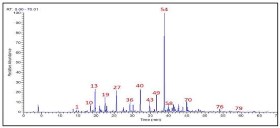
Figure 2.
Total ion chromatogram (TIC) for GC/MS of T. patula flowers volatile constituents.

Table 1.
Phytochemical compositions of T. patula flowers volatile constituents.
Although (E)-β-caryophyllene represents the most predominant volatile sesquiterpene constituent (24.1%), the monoterpenoids exhibited the major existing structures (31 structures, 39.24%), as shown in Figure 3. Moreover, phenylpropanoids increased to the third level with 10.13%. On the other hand, the volatile ketones, fatty acid alkyl ester, volatile alcohols, benzene derivatives, and the mixed structures of monoterpenes esters or ketones represented 7.59%, 3.80%, 3.80%, 2.53%, and 2.53%, respectively. Furthermore, the volatiles diterpene, ester, and ether showed the lowest structures with 1.27% for each one of them. Finally, the only acyclic diterpene identified was neophytadiene with 0.66% (Table 1).
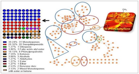
Figure 3.
Molecular network of the GC/MS analysis of the identified volatile constituents from T. patula, and the percent of metabolites classes. All nodes are labeled with the retention time from the GNPS-GC/MS spectral libraries.
As shown in Table 1, the identified volatile compounds can be arranged in descending order as follows: (E)-β-caryophyllene (24.1%) > 2-undecanone + bornyl acetate (12.2%) > 2-nonanone (9.7%) > camphor (4.4%) > limonene (4.3%) > biphenyl (I.S.) (3.2%) > 2-nonyl acetate + cis-ocimenone (2.3%) > piperitenone (2.0%) > cis-davanone (1.8%) > germacrene D (1.3%) > δ-cadinene (1.3%) > linalool (1.2%) > carvone (1.2%) > (Z)-ethyl cinnamate (1.2%), respectively, depending on their percent areas.
Next, the molecular network (MN) was built from the GC/MS data using the GNPS online plate form to visualize the volatiles and analyze the results. The created MN (Figure 3) demonstrated that the (E)-β-caryophyllene is the most abundant metabolite.
3.2. Antioxidants Activity of EO Extracted from T. Patula
Data summarized in Table 2 revealed that the T. patula EO showed high antioxidant activities against DPPH, NO, and FRAP with EC50 29.85 ± 4.53, 33.19 ± 3.8, and 30.22 ± 2.12 µg/mL, respectively compared with the activities of ascorbic acid.
Table 2.
Antioxidant activity as EC50 (μg/mL) for DPPH and NO assay or FE (μg/mL) for FRAP assay of T. patula EO extracts (mean ± SD).
3.3. Acute Oral Toxicity of Essential Oil of T. Patula
The effects of extracted TP EO on groups, doses of 100 and 200 mg/kg BW demonstrated symptoms of micturition, defecation sedation, and minor agitation. In Table 3, the mortality was recorded and the values were used to estimate the lethal dose (LD50) using the Karbar method. The calculated LD50 was estimated as 126 mg/kg BW.
Table 3.
LD50 determination according to the Karbar method.
3.4. Histopathology Findings
The curative effects of EO extracted from T. patula against CCL4 toxicities in rats were asserted by the histopathological inspection. The normal architecture of the hepatocytes and clear sinusoids were observed in the hepatic section of the untreated group (Figure 4A). On the other hand, the liver section of CCL4-treated rats manifests multiple histological alterations, including altered hepatocyte morphology, loss of cell membrane, increased nuclear size, and connective tissue infiltration with prominent necrosis and vacuoles (Figure 4B). Furthermore, the liver of rats treated with vehicle-treated control exhibits normal architecture hepatocytes, intact cell membrane, and central vein (Figure 4C). Meanwhile, the rats treated with EO after CCL4 at 5 and 10 mg/kg BW, showed notable recovery of the liver histopathological signs when compared with the CCL4 group (Figure 5E,F). The rats treated with silymarin after CCL4 did not manifest any pathological alterations compared with the control slide.
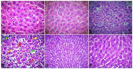
Figure 4.
A representative photomicrograph of a section of the liver tissues of rats exposed to CCL4 (1.25 mg/kg BW) for 15 days, followed by silymarin (10 mg/kg BW) and extracted EO at two doses (5 and 10 mg/kg BW) for 15 days (E&F). Control rats ((A), 100 μm) show a normal architecture of hepatocytes and intact cell membrane. ((B), 100 μm) section of liver of rats treated by CCL4 shows altered hepatocyte morphology, with loss of cell membrane, increased nuclear size, and connective tissue infiltration with prominent necrosis and vacuoles. ((C), 100 μm) section of liver of rat’s vehicle-treated control exhibits normal architecture hepatocytes, intact cell membrane, and central vein. ((D), 100 μm) section of liver of rats treated by CCL4 (1.25 mg/kg BW) for 15 days and followed by silymarin for the next 15 days exhibits potent hepatoprotective activity with radially arranged hepatocytes similar to the control with intact cell membrane, as well as infiltration of cells formation of normal hepatic cards and absence of necrosis and vacuoles. ((E), 100 μm) section of liver of rats treated by CCL4 (1.25 mg/kg BW) and followed by extracted EO (5 mg/kg BW) exhibits moderate hepato-therapeutic activity with radially arranged hepatocytes similar to the control with intact cell membrane and infiltration of cells. ((F), 100 μm) section of liver of rats treated by CCL4 (1.25 mg/kg BW) and followed by extracted EO (10 mg/kg BW) exhibits highly hepato-therapeutic effect with radially arranged hepatocytes similar to the control and silymarin with intact cell membrane and infiltration of cells formation of normal hepatic cards, as well as absence of necrosis and vacuoles.
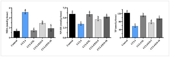
Figure 5.
Effect of EO extracted from T. patula on MDA, TP, and NP-SH in liver of rats treated with CCL4. Data represented as mean ± SD (n = 6). CCL4: Carbon tetrachloride; SL: Silymarin; EO-5: Essential oil (5 mg/kg BW); EO-10: Essential oil (10 mg/kg BW); MDA: Malondialdehyde; TP: Total protein; NP-SH: Non-protein sulfhydryls. Different letters indicate statistically significant differences between groups at (p < 0.05).
3.5. EO Effect on MDA, TP, and NP-SH in CCL4-Treated Rats
The malondialdehyde (MDA), non-protein sulfhydryl (NP-SH), and total protein (TP) were assessed in liver tissue as characteristics of oxidative stress. Figure 5 shows that CCL4-administration provoked a notable reduction (p < 0.05) in TP and NP-SH in the CCL4-treated group in comparison to the control group. In contrast, CCL4 administration increases lipid peroxidation events, measured as MDA products. Meanwhile, rats treated with EO after CCL4 showed significant enhancement in TP and NP-SH levels at the two tested doses. Moreover, significant recovery in lipid peroxidation was similarly noted in the silymarin effect.
3.6. EO of T. Patula Effect on Serum Liver Functions in CCL4-Treated Rats
CCL4 induced pathologic changes in the liver, including hepatomegaly and serologic changes, along with increased activities of GOT, GPT, bilirubin, and GGT. Figure 6 shows that CCL4 treatment significantly (p < 0.05) increased the activities of GOT, GPT, GGT, and bilirubin, while the post-administration of EO (5 and 10 mg/kg BW) after CCL4 significantly (p < 0.05) mitigated these functional markers toward near-normal levels. T. patula EO at a dose of 10 mg/kg BW was more effective than the lowest dose of 5 mg/kg BW. A similar biological effect was noticed in rats treated with silymarin.
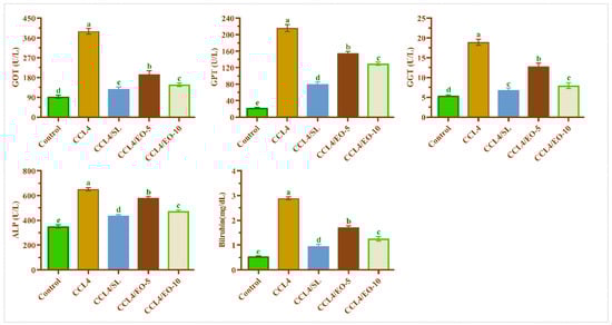
Figure 6.
Effect of EO extracted from T. patula on liver function markers of rats treated with CCL4. Data represented as mean ± SD (n = 6). CCL4: Carbon tetrachloride; SL: Silymarin; EO-5: Essential oil (5 mg/kg BW); EO-10: Essential oil (10 mg/kg BW); GOT: Glutamic oxaloacetic transaminase; GPT: Glutamic pyruvic transaminase; GGT: Gamma-glutamyl transpeptidase; ALP: Alkaline phosphatase. Different letters indicate statistically significant differences between groups at (p < 0.05).
3.7. EO Effect on Lipid Profile in CCL4-Treated Rats
To evaluate the palliation impacts of T. patula EO on lipid profile markers in CCL4-treated rats, the serum cholesterol, TriG, LDL, and HDL were estimated. The deduced results in Figure 7 illustrated that the CCL4 treatment resulted in a marked increase in total cholesterol, TriG, and LDL with a significant decrease in HDL levels compared with the untreated group. In contrast, post-treatment of extracted EO after CCL4 showed significant (p < 0.05) restoration in tested lipid profile markers when compared with the control group, indicating improvement of the lipid metabolism in EO post-treated rats at two doses. The TriG levels were significantly lower in rats treated with 10 mg of EO than those treated with silymarin following CCL4 treatment.
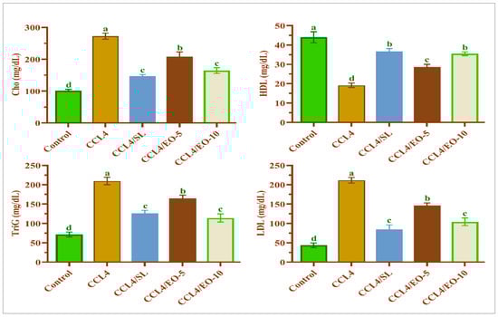
Figure 7.
Effect of EO extracted from T. patula on lipid profile of rats treated with CCL4. Data represented as mean ± SD (n = 6). CCL4: Carbon tetrachloride; SL: Silymarin; EO-5: Essential oil (5 mg/kg BW); EO-10: Essential oil (10 mg/kg BW); Cho: Cholesterol; TriG: Triglycerides; HDL: High-density lipoprotein; LDL: Low-density lipoprotein. Different letters indicate statistically significant differences between groups at (p < 0.05).
4. Discussion
The liver recreates crucial functions in the metabolism and biotransformation of xenobiotics. On account of its position amidst the intestinal tract and circulatory system, it receives extensive quantities of xenobiotics and nutrients absorbed via the digestive tract and the portal vein, becoming the target organ of sundry categories of natural or synthetic toxicants [1].
This study aimed to extract the essential oil from the flower of T. patula and assess its protective utility against CCl4-induced liver damage in rats. T. patula EO was extracted using the HD, common, reliable, sensitive, and accurate technique [32]. The phytochemical composition results showed that the T. patula EO contains 79 compounds that represent 89.8% total volume of the extracted EO. The primary groups of detected constitutes are monoterpenes and sesquiterpenoids, which represented 39.2% and 25.32%, respectively. These were documented to exert many biological properties, including antioxidant and anti-inflammatory properties [33]. Furthermore, the prime compound in the extracted EO is E-β-caryophyllene, which represents 24.1% of the total extracted EO. This component is documented to have good antioxidant and anti-inflammatory activities [34].
According to the present findings, CCl4 caused liver disorders in rats, which were portrayed by oxidative stress, pathological tissue damage, elevated liver enzymes, and disruption of lipid metabolism. These results are in agreement with the results of [35,36,37,38]. The toxic mechanism of CCl4 occurs during liver metabolism, wherein the cytochrome P450 (CYP) enzyme converts CCl4 to the trichloromethyl radical (CCl3) [39]. This process impairs many vital cellular processes, induces vast cell damage, and causes the release of the aminotransferase enzyme into blood circulation. In hepatic apoptosis, the synthesis of cellular phospholipids refers to the amalgamation of phospholipids into lipoproteins, conducting the assembly of triglycerides [40].
Moreover, CCl4 metabolism in the liver caused oxidative stress and suppressed the antioxidant defense system [41,42]. The current results showed that the CCl4 treatment resulted in significant alleviation of MDA, NP-SH, and TP. Lipid peroxidation is the principal mechanism of hepatic injury [43]. In the liver, CCl4 transforms to trichloromethyl (CCl3) under the catalytic activity of cytochrome P450. CCl3 is a free radical, that mainly can react with oxygen to produce toxic trichloromethyl peroxyl (CCl3O2) radicals [39]. The outcome CCl3O2 has the prospect to bind to myriad proteins or lipids and induce the lipids’ peroxidation [44,45]. In the present investigation, treatment with CCL4 led to the initiation of the degradation of lipids in the cellular membrane. This caused the generation of MDA products that result in a loss of cell membrane integrity and liver injury [46]. The treatment by the extracted EO from T. patula markedly lessened the production of the (MDA) in CCl4-treated rats. This shows that the administration of T. patula essential oils at two tested doses efficaciously minimized lipid peroxidation which is influenced by CCl4. These results are harmonious with results published by Riaz et al. [47] as well as Singh and Thakur [48].
Furthermore, the decreased NP-SH content is ideal evidence of hepatotoxicity. The current results showed a marked reduction of the liver NP-SH content in CCL4 rats, which could induce further damage and dysfunction of the liver. The treatment by EO of T. patula or silymarin significantly re-increased the level of NP-SH in liver tissues indicating a therapeutic potential of the extracted EO.
The shortage of TP is also an indicator of hepatotoxicity. This decrease in total protein could trigger hydration, which is hurtful to cellular homeostasis. This will negatively impact the metabolic activities of the liver, and thus body health [49]. Enhancement of the levels of TP by EO at two tested doses and silymarin denotes a lessening of the oxidative stress, and thus mitigation of hepatic toxicity. In addition to the mitigation of liver toxicity, the extracted EO showed good in vitro antioxidants assays (DPPH, FRAP, and NO). The antioxidant activity of the T. Patula EO may be attributed to its constituents and their antioxidant potency.
As stated in the current findings, a significant increase in the liver damage markers was noted in the CCL4-treated rats. The excess serum GOT, ALP, GGT, GPT, and bilirubin levels were due to hepatocytes damage [50]. Particularly, the excessive release of bilirubin into the serum of CCL4-treated rats has demonstrated the decreased ability of the liver for bile extraction [51].
Therefore, the elevation of the levels of these biochemical markers is a straightforward repercussion of alterations in the hepatic tissue’s structural integrity, which also has been confirmed by histological findings. The post-administration of the extracted oil at two tested doses significantly succeeded in protecting the liver and diminished the hepatic damage markers. These results are congruous with the previously published results regarding the protection of the liver from oxidative stress caused by CCl4 using different agents, such as gallic acid and docosahexaenoic acid [22,52,53]. The lipid profile markers are the reliable biomarkers for investigating liver health. The activity of serum lipid profiles, such as triglycerides, total cholesterol, HDL, and LDL was significantly elevated in CCL4-treated rats, indicating deterioration in hepatic functions due to the damage caused by CCl4 metabolites.
According to our results, liver protection against CCl4 has been achieved by administration of EO of T. patula at two tested doses through stimulating the regeneration of liver cells or via the enhancement of the antioxidants system, thus scavenging the formed free radicals and preventing their reaction. The hepatoprotective action of the essential oil isolated from T. patula can be attributed to its content of monoterpenes and sesquiterpene compounds. Monoterpenes and sesquiterpenes were reported to have manifold pharmacological influences, such as antioxidant and anti-inflammatory activities [54]. According to the chemical composition results, (E)-β caryophyllene (24.1%) is the major compound in the T. patula EO. This sesquiterpene component was documented as a hepatoprotective component against CCL4 via exerting its antioxidant and anti-inflammatory effects [34,55]. The hepato-therapeutic effects of T. patula EO were associated with mitigation of the oxidative stress in CCL4 rats treated by T. patula EO compared with the rats treated with CCl4 only.
5. Conclusions
CCl4 is a well-known liver toxic, and commonly used in hepatotoxic models. With increasing cases of liver diseases, the identification, evaluation, and preparation of hepatoprotective drugs from plant sources has become an impressive approach. The extracted essential oil from the flower of T. patula yielded 0.43% w/v. Seventy-nine components were identified by GC/MS analysis and these components represent 89.8% of the oil components. Monoterpenes were the major components of the oil, representing 39.24% followed by sesquiterpenes at 25.32%. The T. patula EO showed high antioxidant activities toward DPPH scavenging assay, NO, and FRAP. The sesquiterpene of (E)-β-caryophyllene as the most predominant volatile constituent among the flower accounted for 24.1%. The impact of the EO was noted as reducing the MDA level toward normal levels. Moreover, restoring TP and NP-SH groups was superior to the effect parallel with the silymarin treatment group. On other hand, the histopathological study showed complete recovery of hepatic tissues in the group treated with CCl4 and 10 mg/kg BW T. patula EO. Additionally, T. patula EO administration restores liver functions and maintains lipid profile at two tested doses. These results suggest that T. patula EO can be used to protect and enhance the recovery of the liver to overcome the adverse side effects of some drugs as well as food toxic contaminants.
Author Contributions
H.Y.A., M.E. and M.M.S. conceived and designed the experiments; H.Y.A., S.A., J.A.-Q., A.A. and J.W. performed the experiments; H.Y.A., M.E., J.A.-Q. and M.M.S. analyzed the data and wrote the primary draft of the manuscript; S.A., M.M.S., M.E. and A.A. participated in the sample collections and manuscript writing and editing. All authors have read and agreed to the published version of the manuscript.
Funding
The authors would like to thank Researchers Supporting Project number (RSP2022R504), King Saud University, Riyadh, Saudi Arabia.
Institutional Review Board Statement
Not applicable.
Informed Consent Statement
Not applicable.
Data Availability Statement
Not applicable.
Conflicts of Interest
The authors declare no conflict of interest.
References
- Gu, X.; Manautou, J.E. Molecular mechanisms underlying chemical liver injury. Expert Rev. Mol. Med. 2012, 14, 1–25. [Google Scholar] [CrossRef] [PubMed]
- Feijóo, M.; Túnez, I.; Ruiz, A.; Tasset, I.; Muñoz, E.; Collantes, E. Oxidative stress biomarkers as indicator of chronic inflammatory joint diseases stage. Reumatol. Clin. 2010, 6, 91–94. [Google Scholar] [CrossRef] [PubMed]
- Chandan, B.; Saxena, A.; Shukla, S.; Sharma, N.; Gupta, D.; Singh, K.; Suri, J.; Bhadauria, M.; Qazi, G. Hepatoprotective activity of Woodfordia fruticosa Kurz flowers against carbon tetrachloride induced hepatotoxicity. J. Ethnopharmacol. 2008, 119, 218–224. [Google Scholar] [CrossRef] [PubMed]
- Farzaei, M.H.; Zobeiri, M.; Parvizi, F.; El-Senduny, F.F.; Marmouzi, I.; Coy-Barrera, E.; Naseri, R.; Nabavi, S.M.; Rahimi, R.; Abdollahi, M. Curcumin in liver diseases: A systematic review of the cellular mechanisms of oxidative stress and clinical perspective. Nutrients 2018, 10, 855. [Google Scholar] [CrossRef] [PubMed]
- Kuo, D.-H.; Kang, W.-H.; Shieh, P.-C.; Chen, F.-A.; Chang, C.-D.; Tsai, M.-L.; Cheng, A.-C.; Ho, C.-T.; Pan, M.-H. Protective effect of Pracparatum mungo extract on carbon tetrachloride-induced hepatotoxicity in rats. Food Chem. 2010, 123, 1007–1012. [Google Scholar] [CrossRef]
- Karimi, J.; Mohammadalipour, A.; Sheikh, N.; Khodadadi, I.; Hashemnia, M.; Goudarzi, F.; Khanjarsim, V.; Solgi, G.; Hajilooi, M.; Bahabadi, M. Protective effects of combined Losartan and Nilotinib on carbon tetrachloride (CCl4)-induced liver fibrosis in rats. Drug Chem. Toxicol. 2020, 43, 468–478. [Google Scholar] [CrossRef]
- Huang, H.-L.; Wang, Y.-J.; Zhang, Q.-Y.; Liu, B.; Wang, F.-Y.; Li, J.-J.; Zhu, R.-Z. Hepatoprotective effects of baicalein against CCl4-induced acute liver injury in mice. World J. Gastroenterol. 2012, 18, 6605–6613. [Google Scholar] [CrossRef]
- Chen, X.; Meng, Q.; Wang, C.; Liu, Q.; Sun, H.; Huo, X.; Sun, P.; Yang, X.; Peng, J.; Liu, K. Protective effects of calycosin against CCl 4-induced liver injury with activation of FXR and STAT3 in mice. Pharm. Res. 2015, 32, 538–548. [Google Scholar] [CrossRef]
- Hashem, M.M.; Salama, M.M.; Mohammed, F.F.; Tohamy, A.F.; El Deeb, K.S. Metabolic profile and hepatoprotective effect of Aeschynomene elaphroxylon (Guill. & Perr.). PLoS ONE 2019, 14, 1–24. [Google Scholar]
- Unsal, V.; Cicek, M.; Sabancilar, İ. Toxicity of carbon tetrachloride, free radicals and role of antioxidants. Rev. Environ. Health 2020, 36, 279–295. [Google Scholar] [CrossRef]
- Liu, J.; Wang, X.; Liu, R.; Liu, Y.; Zhang, T.; Fu, H.; Hai, C. Oleanolic acid co-administration alleviates ethanol-induced hepatic injury via Nrf-2 and ethanol-metabolizing modulating in rats. Chem.-Biol. Interact. 2014, 221, 88–98. [Google Scholar] [CrossRef] [PubMed]
- Seif, M.; Deabes, M.; El-Askary, A.; El-Kott, A.F.; Albadrani, G.M.; Seif, A.; Wang, Z. Ephedra sinica mitigates hepatic oxidative stress and inflammation via suppressing the TLR4/MyD88/NF-κB pathway in fipronil-treated rats. Environ. Sci. Pollut. Res. 2021, 28, 62943–62958. [Google Scholar] [CrossRef] [PubMed]
- Dunning, T. Aromatherapy: Overview, safety and quality issues. OA Altern. Med. 2013, 1, 1–6. [Google Scholar] [CrossRef]
- Faizi, S.; Fayyaz, S.; Bano, S.; Yawar Iqbal, E.; Siddiqi, H.; Naz, A. Isolation of nematicidal compounds from Tagetes patula L. yellow flowers: Structure–activity relationship studies against cyst nematode Heterodera zeae infective stage larvae. J. Agric. Food Chem. 2011, 59, 9080–9093. [Google Scholar] [CrossRef] [PubMed]
- Aati, H.Y.; Perveen, S.; Orfali, R.; Al-Taweel, A.M.; Aati, S.; Wanner, J.; Khan, A.; Mehmood, R. Chemical composition and antimicrobial activity of the essential oils of Artemisia absinthium, Artemisia scoparia, and Artemisia sieberi grown in Saudi Arabia. Arab. J. Chem. 2020, 13, 8209–8217. [Google Scholar] [CrossRef]
- Wang, M.; Carver, J.J.; Phelan, V.V.; Sanchez, L.M.; Garg, N.; Peng, Y.; Nguyen, D.D.; Watrous, J.; Kapono, C.A.; Luzzatto-Knaan, T. Sharing and community curation of mass spectrometry data with Global Natural Products Social Molecular Networking. Nat. Biotechnol. 2016, 34, 828–837. [Google Scholar] [CrossRef]
- Shannon, P.; Markiel, A.; Ozier, O.; Baliga, N.S.; Wang, J.T.; Ramage, D.; Amin, N.; Schwikowski, B.; Ideker, T. Cytoscape: A software environment for integrated models of biomolecular interaction networks. Genome Res. 2003, 13, 2498–2504. [Google Scholar] [CrossRef]
- Koksal, E.; Bursal, E.; Dikici, E.; Tozoglu, F.; Gulcin, I. Antioxidant activity of Melissa officinalis leaves. J. Med. Plants Res. 2011, 5, 217–222. [Google Scholar]
- Rao, M. Nitric oxide scavenging by curcuminoids. J. Pharm. Pharmacol. 1997, 49, 105–107. [Google Scholar]
- Benzie, I.F.; Strain, J.J. The ferric reducing ability of plasma (FRAP) as a measure of “antioxidant power”: The FRAP assay. Anal. Biochem. 1996, 239, 70–76. [Google Scholar] [CrossRef]
- Mamza, U.; Sodipo, O.; Abdulrahman, F.; Khan, I. Evaluation of phytochemical, antimicrobial and toxicity studies of ethanolic stem extract of Phyllanthus amarus Thonn and Schum (Euphorbiacea). Int. J. Sci. Technoledge 2015, 3, 262–268. [Google Scholar]
- Ojeaburu, S.; Oriakhi, K. Hepatoprotective, antioxidant and, anti-inflammatory potentials of gallic acid in carbon tetrachloride-induced hepatic damage in Wistar rats. Toxicol. Rep. 2021, 8, 177–185. [Google Scholar] [CrossRef] [PubMed]
- Toor, S.S.; Reddy, H.; Deng, S.; Hoffmann, J.; Spangsmark, D.; Madsen, L.B.; Holm-Nielsen, J.B.; Rosendahl, L.A. Hydrothermal liquefaction of Spirulina and Nannochloropsis salina under subcritical and supercritical water conditions. Bioresour. Technol. 2013, 131, 413–419. [Google Scholar] [PubMed]
- Ohkawa, H.; Ohishi, N.; Yagi, K. Assay for lipid peroxides in animal tissues by thiobarbituric acid reaction. Anal. Biochem. 1979, 95, 351–358. [Google Scholar] [CrossRef]
- Sedlak, J.; Lindsay, R.H. Estimation of total, protein-bound, and nonprotein sulfhydryl groups in tissue with Ellman’s reagent. Anal. Biochem. 1968, 25, 192–205. [Google Scholar] [CrossRef]
- Lowery, G.H. A Quantitative Study of The Nocturnal Migration of Birds; University of Kansas: Lawrence, KS, USA, 1951. [Google Scholar]
- Reitman, S.; Frankel, S. A colorimetric method for the determination of serum glutamic oxalacetic and glutamic pyruvic transaminases. Am. J. Clin. Pathol. 1957, 28, 56–63. [Google Scholar] [CrossRef]
- Otto, A.; Oliver, H.; Jane, M. A method for the rapid determination of alkaline phosphatase with five cubic millimeters of serum. J. Biol. Chem. 1946, 164, 321–329. [Google Scholar]
- Whitfield, J. Gamma glutamyl transferase. Crit. Rev. Clin. Lab. Sci. 2001, 38, 263–355. [Google Scholar] [CrossRef]
- Doumas, B.T.; Kwok-Cheung, P.P.; Perry, B.W.; Jendrzejczak, B.; McComb, R.B.; Schaffer, R.; Hause, L.L. Candidate reference method for determination of total bilirubin in serum: Development and validation. Clin. Chem. 1985, 31, 1779–1789. [Google Scholar] [CrossRef]
- Davidson, M.H.; Rosenson, R.S. Novel targets that affect high-density lipoprotein metabolism: The next frontier. Am. J. Cardiol. 2009, 104, 52E–57E. [Google Scholar] [CrossRef]
- Abdel-Hameed, E.-S.S.; Salman, M.S.; Fadl, M.A.; Elkhateeb, A.; Hassan, M.M. Chemical composition and biological activity of Mentha longifolia L. essential oil growing in Taif, KSA extracted by hydrodistillation, solvent free microwave and microwave hydrodistillation. J. Essent. Oil Bear. Plants 2018, 21, 1–14. [Google Scholar] [CrossRef]
- Rondon, M.; Velasco, J.; Hernandez, J.; Pecheneda, M.; Rojas, J.; Morales, A.; Carmona, J.; Diaz, T. Chemical composition and antibacterial activity of the essential oil of Tagetes patula L. (Asteraceae) collected from the Venezuela Andes. Rev. Latinoam. Quím. 2006, 34, 32–36. [Google Scholar]
- Francomano, F.; Caruso, A.; Barbarossa, A.; Fazio, A.; La Torre, C.; Ceramella, J.; Mallamaci, R.; Saturnino, C.; Iacopetta, D.; Sinicropi, M.S. β-Caryophyllene: A sesquiterpene with countless biological properties. Appl. Sci. 2019, 9, 5420. [Google Scholar] [CrossRef]
- Mughal, T.A.; Saleem, M.Z.; Ali, S.; Anwar, K.K.; Bashir, M.M.; Babar, M.; Khan, M.A. Evaluation of Hepatotoxicity of Carbon Tetrachloride and Pharmacological Intervention by Vitamin E in Balb C Mice. Pak. J. Zool. 2019, 51, 755–761. [Google Scholar] [CrossRef]
- El-Hadary, A.E.; Elsanhoty, R.M.; Ramadan, M.F. In vivo protective effect of Rosmarinus officinalis oil against carbon tetrachloride (CCl4)-induced hepatotoxicity in rats. PharmaNutrition 2019, 9, 1–7. [Google Scholar] [CrossRef]
- Al-Rasheed, N.M.; Fadda, L.M.; Ali, H.M.; Abdel Baky, N.A.; El-Orabi, N.F.; Al-Rasheed, N.M.; Yacoub, H.I. New mechanism in the modulation of carbon tetrachloride hepatotoxicity in rats using different natural antioxidants. Toxicol. Mech. Methods 2016, 26, 243–250. [Google Scholar] [CrossRef]
- Moreira, P.R.; Maioli, M.A.; Medeiros, H.C.; Guelfi, M.; Pereira, F.T.; Mingatto, F.E. Protective effect of bixin on carbon tetrachloride-induced hepatotoxicity in rats. Biol. Res. 2014, 47, 1–7. [Google Scholar] [CrossRef]
- Weber, L.W.; Boll, M.; Stampfl, A. Hepatotoxicity and mechanism of action of haloalkanes: Carbon tetrachloride as a toxicological model. Crit. Rev. Toxicol. 2003, 33, 105–136. [Google Scholar] [CrossRef]
- Lombardi, B.; Ugazio, G.; Raick, A. Choline-deficiency fatty liver: Relation of plasma phospholipids to liver triglycerides. Am. J. Physiol.-Leg. Content 1966, 210, 31–36. [Google Scholar] [CrossRef]
- Ullah, H.; Khan, A.; Baig, M.W.; Ullah, N.; Ahmed, N.; Tipu, M.K.; Ali, H.; Khan, S. Poncirin attenuates CCL4-induced liver injury through inhibition of oxidative stress and inflammatory cytokines in mice. BMC Complement. Med. Ther. 2020, 20, 1–14. [Google Scholar] [CrossRef]
- Ma, J.-Q.; Ding, J.; Zhang, L.; Liu, C.-M. Ursolic acid protects mouse liver against CCl4-induced oxidative stress and inflammation by the MAPK/NF-κB pathway. Environ. Toxicol. Pharmacol. 2014, 37, 975–983. [Google Scholar] [CrossRef] [PubMed]
- Recknagel, R.O.; Glende, E.A.; Britton, R.S. Free radical damage and lipid peroxidation. In Hepatotoxicology; CRC Press: Boca Raton, FL, USA, 2020; pp. 401–436. [Google Scholar]
- Li, L.; Li, W.; Kim, Y.-h.; Lee, Y.W. Chlorella vulgaris extract ameliorates carbon tetrachloride-induced acute hepatic injury in mice. Exp. Toxicol. Pathol. 2013, 65, 73–80. [Google Scholar] [CrossRef] [PubMed]
- Lee, K.J.; Jeong, H.G. Protective effect of Platycodi radix on carbon tetrachloride-induced hepatotoxicity. Food Chem. Toxicol. 2002, 40, 517–525. [Google Scholar] [CrossRef]
- Ayala, A.; Muñoz, M.F.; Argüelles, S. Lipid peroxidation: Production, metabolism, and signaling mechanisms of malondialdehyde and 4-hydroxy-2-nonenal. Oxidative Med. Cell. Longev. 2014, 2014, 1–31. [Google Scholar] [CrossRef] [PubMed]
- Riaz, M.; Ahmad, R.; Rahman, N.U.; Khan, Z.; Dou, D.; Sechel, G.; Manea, R. Traditional uses, Phyto-chemistry and pharmacological activities of Tagetes patula L. J. Ethnopharmacol. 2020, 255, 1–16. [Google Scholar] [CrossRef]
- Singh, N.; Thakur, R. A Review on Pharmacological aspects of Tagetes erecta Linn. PharmaTutor 2019, 7, 16–24. [Google Scholar]
- Adeyemi, O.S.; Fambegbe, M.; Daniyan, O.R.; Nwajei, I. Yoyo Bitters, a polyherbal formulation influenced some biochemical parameters in Wistar rats. J. Basic Clin. Physiol. Pharmacol. 2012, 23, 135–138. [Google Scholar] [CrossRef]
- Zafar, R.; Ali, S.M. Anti-hepatotoxic effects of root and root callus extracts of Cichorium intybus L. J. Ethnopharmacol. 1998, 63, 227–231. [Google Scholar] [CrossRef]
- Sadeghi, H.; Jahanbazi, F.; Sadeghi, H.; Omidifar, N.; Alipoor, B.; Kokhdan, E.P.; Mousavipoor, S.M.; Mousavi-Fard, S.H.; Doustimotlagh, A.H. Metformin attenuates oxidative stress and liver damage after bile duct ligation in rats. Res. Pharm. Sci. 2019, 14, 122–129. [Google Scholar] [CrossRef]
- Li, R.; Yang, W.; Yin, Y.; Ma, X.; Zhang, P.; Tao, K. 4-OI Attenuates Carbon Tetrachloride-Induced Hepatic Injury via Regulating Oxidative Stress and the Inflammatory Response. Front. Pharmacol. 2021, 12, 1–13. [Google Scholar] [CrossRef]
- He, J.; Bai, K.; Hong, B.; Zhang, F.; Zheng, S. Docosahexaenoic acid attenuates carbon tetrachloride-induced hepatic fibrosis in rats. Int. Immunopharmacol. 2017, 53, 56–62. [Google Scholar] [CrossRef] [PubMed]
- Bartikova, H.; Hanusova, V.; Skalova, L.; Ambroz, M.; Bousova, I. Antioxidant, pro-oxidant and other biological activities of sesquiterpenes. Curr. Top. Med. Chem. 2014, 14, 2478–2494. [Google Scholar] [CrossRef] [PubMed]
- Calleja, M.A.; Vieites, J.M.; Montero-Meterdez, T.; Torres, M.I.; Faus, M.J.; Gil, A.; Suárez, A. The antioxidant effect of β-caryophyllene protects rat liver from carbon tetrachloride-induced fibrosis by inhibiting hepatic stellate cell activation. Br. J. Nutr. 2013, 109, 394–401. [Google Scholar] [CrossRef] [PubMed]


Publisher’s Note: MDPI stays neutral with regard to jurisdictional claims in published maps and institutional affiliations. |
© 2022 by the authors. Licensee MDPI, Basel, Switzerland. This article is an open access article distributed under the terms and conditions of the Creative Commons Attribution (CC BY) license (https://creativecommons.org/licenses/by/4.0/).