Abstract
Here, we describe the case of a 74-year-old male patient with a high-risk prostate carcinoma who underwent positron-emission tomography/computed tomography (PET/CT) with [68Ga]Ga-prostate-specific membrane antigen ([68Ga]Ga-PSMA-11) for staging. [68Ga]Ga-PSMA-11 PET/CT detected an extensive area of increased tracer uptake at the prostatic level, involving both lobes. Additionally, a rounded lesion approximately 4 cm in diameter was identified in the celiac region adjacent to the stomach, exhibiting moderate tracer uptake. Based on these imaging findings, the patient underwent radiation therapy applied to the prostate and pelvis and a biopsy of the suspected lesion adjacent to the stomach, which was positive for Siewert type III gastroesophageal junction adenocarcinoma (HER2-negative, PDL-1 60%). This case demonstrates the importance of not overlooking incidental tracer uptakes in PSMA PET/CT imaging in the stomach, as they could represent neoplastic lesions.
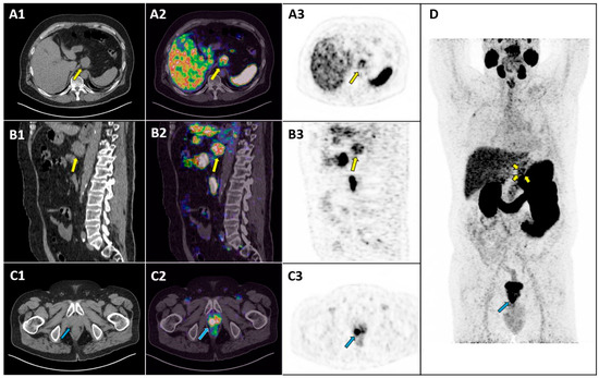
Figure 1.
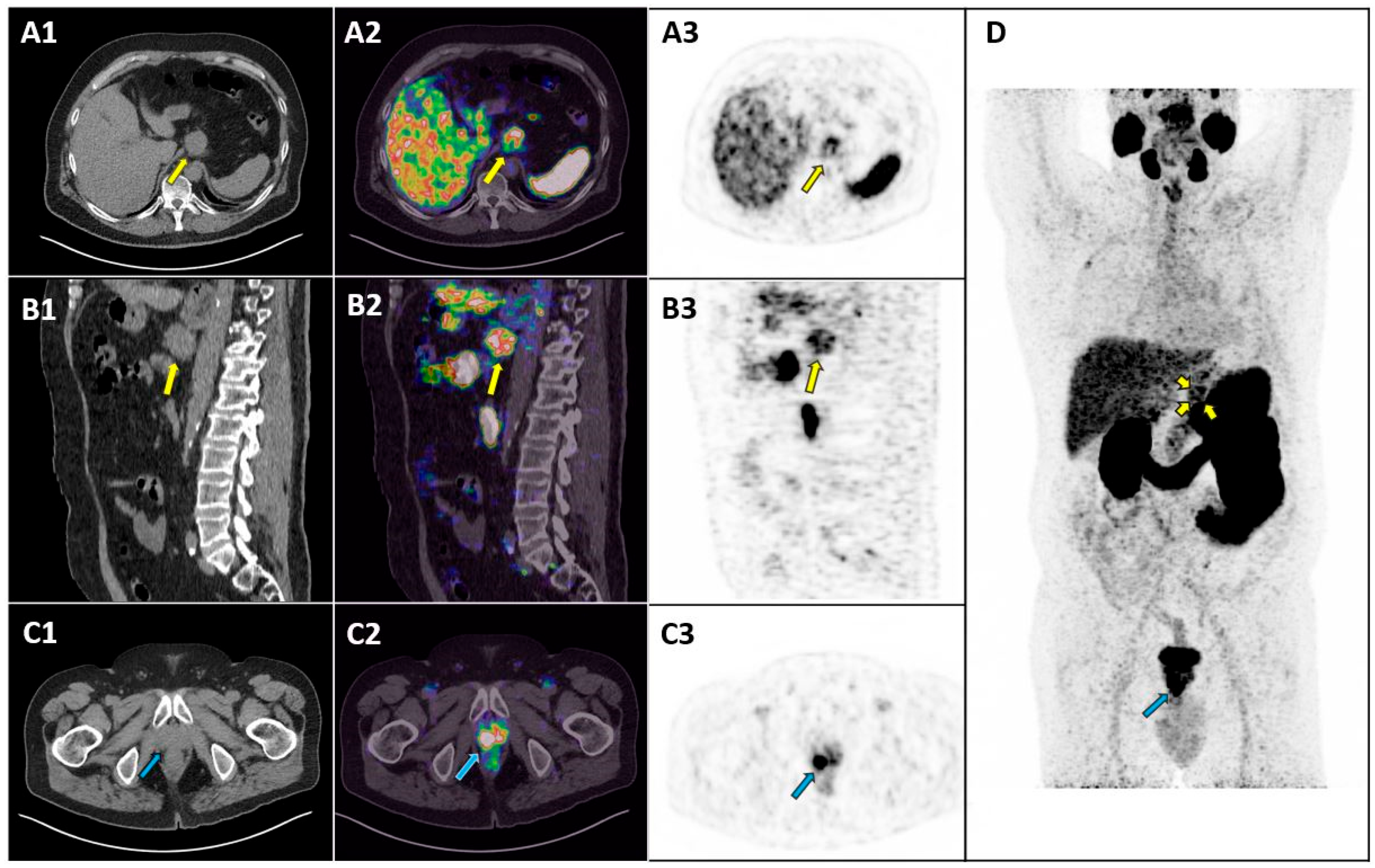
A 74-year-old male patient with a high-risk prostate carcinoma underwent positron-emission tomography/computed tomography (PET/CT) with [68Ga]Ga-prostate-specific membrane antigen ([68Ga]Ga-PSMA-11) for staging. PET/CT was performed 60 min after an intravenous injection of 250 MBq of [68Ga]Ga-PSMA-11. PET image analysis was performed using qualitative criteria: areas of increased tracer uptake compared to the background, excluding sites of physiological uptake, were considered abnormal. Axial CT image (A1), axial fused PET/CT image (A2), axial PET image (A3), sagittal CT image (B1), sagittal fused PET/CT image (B2) and sagittal PET image (B3) at gastric level revealed a rounded lesion (approximately 4 cm in diameter) adjacent to the stomach in the celiac region, exhibiting moderate tracer uptake (yellow arrows in (A1–A3,B1–B3,D); SUVmax: 9.7). This lesion appeared inseparable from the stomach wall. Axial CT image (C1), axial fused PET/CT image (C2) and axial PET image (C3) at the pelvic level revealed an extensive area of increased tracer uptake at the prostatic level involving both lobes (blue arrows in (C1–C3,D)). The maximum-intensity-projection (MIP) PET image (D) highlighted both the prostatic lesion (blue arrow) and the gastric lesion (yellow arrow), indicating abnormal tracer uptake at both sites. Upon physical examination after the PET/CT scan, the patient did not show any intestinal symptoms such as decreased appetite, indigestion, or abdominal distension. Based on these imaging findings, the patient underwent radiation therapy applied to the prostate and pelvis and a biopsy of the suspected lesion adjacent to the stomach. Endoscopic examination revealed a large vegetative lesion extending along the small curvature of the stomach from the cardia to the level of the gastric antrum. Histopathological examination demonstrated a Siewert type III gastroesophageal junction adenocarcinoma (HER2-negative, PDL-1 60%). PSMA PET/CT has an established role in prostate cancer imaging [1], but non-prostatic tumor lesions may exhibit PSMA overexpression and increased PSMA ligand uptake in PET/CT [2,3]. This case demonstrates the importance of not overlooking gastric incidental tracer uptakes in PET/CT with PSMA ligands, as they may represent a malignant tumor.
Figure 1.
A 74-year-old male patient with a high-risk prostate carcinoma underwent positron-emission tomography/computed tomography (PET/CT) with [68Ga]Ga-prostate-specific membrane antigen ([68Ga]Ga-PSMA-11) for staging. PET/CT was performed 60 min after an intravenous injection of 250 MBq of [68Ga]Ga-PSMA-11. PET image analysis was performed using qualitative criteria: areas of increased tracer uptake compared to the background, excluding sites of physiological uptake, were considered abnormal. Axial CT image (A1), axial fused PET/CT image (A2), axial PET image (A3), sagittal CT image (B1), sagittal fused PET/CT image (B2) and sagittal PET image (B3) at gastric level revealed a rounded lesion (approximately 4 cm in diameter) adjacent to the stomach in the celiac region, exhibiting moderate tracer uptake (yellow arrows in (A1–A3,B1–B3,D); SUVmax: 9.7). This lesion appeared inseparable from the stomach wall. Axial CT image (C1), axial fused PET/CT image (C2) and axial PET image (C3) at the pelvic level revealed an extensive area of increased tracer uptake at the prostatic level involving both lobes (blue arrows in (C1–C3,D)). The maximum-intensity-projection (MIP) PET image (D) highlighted both the prostatic lesion (blue arrow) and the gastric lesion (yellow arrow), indicating abnormal tracer uptake at both sites. Upon physical examination after the PET/CT scan, the patient did not show any intestinal symptoms such as decreased appetite, indigestion, or abdominal distension. Based on these imaging findings, the patient underwent radiation therapy applied to the prostate and pelvis and a biopsy of the suspected lesion adjacent to the stomach. Endoscopic examination revealed a large vegetative lesion extending along the small curvature of the stomach from the cardia to the level of the gastric antrum. Histopathological examination demonstrated a Siewert type III gastroesophageal junction adenocarcinoma (HER2-negative, PDL-1 60%). PSMA PET/CT has an established role in prostate cancer imaging [1], but non-prostatic tumor lesions may exhibit PSMA overexpression and increased PSMA ligand uptake in PET/CT [2,3]. This case demonstrates the importance of not overlooking gastric incidental tracer uptakes in PET/CT with PSMA ligands, as they may represent a malignant tumor.
Author Contributions
Conceptualization, G.T. and B.M.; formal analysis, C.M.I.; G.P. and M.C.; data curation, C.M.I.; writing—original draft preparation, C.M.I.; writing—review and editing, G.T.; supervision, G.P. and B.M. All authors have read and agreed to the published version of the manuscript.
Funding
This research received no external funding.
Institutional Review Board Statement
Not applicable.
Informed Consent Statement
Written informed consent has been obtained from the patient to publish this paper.
Data Availability Statement
The data presented in this article are available upon request from the corresponding author.
Conflicts of Interest
The authors declare no conflicts of interest.
References
- Oprea-Lager, D.E.; MacLennan, S.; Bjartell, A.; Briganti, A.; Burger, I.A.; de Jong, I.; De Santis, M.; Eberlein, U.; Emmett, L.; Fizazi, K.; et al. European Association of Nuclear Medicine Focus 5: Consensus on Molecular Imaging and Theranostics in Prostate Cancer. Eur. Urol. 2024, 85, 49–60. [Google Scholar] [CrossRef] [PubMed]
- Rizzo, A.; Dall’Armellina, S.; Pizzuto, D.A.; Perotti, G.; Zagaria, L.; Lanni, V.; Treglia, G.; Racca, M.; Annunziata, S. PSMA Radioligand Uptake as a Biomarker of Neoangiogenesis in Solid Tumours: Diagnostic or Theragnostic Factor? Cancers 2022, 14, 4039. [Google Scholar] [CrossRef] [PubMed]
- de Galiza Barbosa, F.; Queiroz, M.A.; Nunes, R.F.; Costa, L.B.; Zaniboni, E.C.; Marin, J.F.G.; Cerri, G.G.; Buchpiguel, C.A. Nonprostatic diseases on PSMA PET imaging: A spectrum of benign and malignant findings. Cancer Imaging 2020, 20, 23. [Google Scholar] [CrossRef] [PubMed]
Disclaimer/Publisher’s Note: The statements, opinions and data contained in all publications are solely those of the individual author(s) and contributor(s) and not of MDPI and/or the editor(s). MDPI and/or the editor(s) disclaim responsibility for any injury to people or property resulting from any ideas, methods, instructions or products referred to in the content. |
© 2025 by the authors. Licensee MDPI, Basel, Switzerland. This article is an open access article distributed under the terms and conditions of the Creative Commons Attribution (CC BY) license (https://creativecommons.org/licenses/by/4.0/).