Interventional Oncology

A topical collection in Medicina (ISSN 1648-9144). This collection belongs to the section "Oncology".

Viewed by 44338

Editors


E-Mail Website
Guest Editor
Radiologie & Oncologie Interventionnelles, Sorbonne Université, Hôpital Tenon, Paris, France
Interests: interventional oncology; percutaneous ablations (cryoablation, electroporation, microwave ablation, radiofrequency ablation); osteoplasty and fixation; soft tissue; bone; spine; pain management
Special Issues, Collections and Topics in MDPI journals

E-Mail Website
Co-Guest Editor
Radiologie & Oncologie Interventionnelles, Sorbonne Université, Hôpital Tenon, Paris, France
Interests: interventional radiology; interventional oncology; percutaneous ablation techniques; targeted tumor drug delivery; patient-radiologist relationship

E-Mail Website
Co-Guest Editor
Department of Neuroradiology, Université Grenoble Alpes, Grenoble, France
Interests: interventional radiology; pain management; interventional neuroradiology; vertebroplasty; neurolysis; infiltration; CT guidance

E-Mail Website
Co-Guest Editor
2nd Department of Radiology, University General Hospital "ATTIKON" Medical School, National and Kapodistrian University of Athens, Athens, Greece
Interests: interventional oncology; ablation; vertebral augmentation; MSK interventions
Special Issues, Collections and Topics in MDPI journals

Topical Collection Information

Dear Colleagues,

Interventional oncology (IO) is a medical specialty offering innovative diagnostic and therapeutic minimally invasive procedures in the oncologic field, but also allowing the management of all issues related to cancer. One critical aspect of IO is that the concept unifying these procedures remains imaging guidance.

IO has made considerable improvements over the last few years, driven by breakthrough innovations and technological developments. Percutaneous biopsies are widely proposed for the diagnosis of cancer but also now iteratively to offer critical insights into a disease, allowing the provision of the best care in the era of personalized medicine. Furthermore, IO treatments now include a wide array of techniques that are able to provide local control or symptomatic relief. Ablations (cryoablation, radio frequency ablation, microwave ablation, electroporation) are routinely used in the liver, kidney, lung, bone, or soft tissues. A better understanding of biological effects has been observed thanks to preclinical evaluation. Embolization is also commonly performed. Osteoplasty and fixation are gaining importance in pain palliation. They interest a large community of physicians, including oncologists, radiation therapists, surgeons, anesthesiologists, and interventional radiologists.

The purpose of this topical collection is to present these image-guided treatments as alternatives to conservative therapies or surgery and to promote basic and clinical research on this topic as these techniques can serve as attractive alternatives to other options, with a clear effect on quality of life.

We encourage authors to submit both preclinical and clinical studies in the field. Clinical studies may include systematic reviews, retrospective studies, case reports, and prospective studies emphasizing the role and need for IO.

We remain at your disposal for additional questions.

Prof. Dr. François Cornelis
Dr. Matthias Barral
Dr. Adrian Kastler
Dr. Dimitrios Filippiadis
Guest Editors

Manuscript Submission Information

Manuscripts should be submitted online at www.mdpi.com by registering and logging in to this website. Once you are registered, click here to go to the submission form. Manuscripts can be submitted until the deadline. All submissions that pass pre-check are peer-reviewed. Accepted papers will be published continuously in the journal (as soon as accepted) and will be listed together on the collection website. Research articles, review articles as well as short communications are invited. For planned papers, a title and short abstract (about 100 words) can be sent to the Editorial Office for announcement on this website.

Submitted manuscripts should not have been published previously, nor be under consideration for publication elsewhere (except conference proceedings papers). All manuscripts are thoroughly refereed through a single-blind peer-review process. A guide for authors and other relevant information for submission of manuscripts is available on the Instructions for Authors page. Medicina is an international peer-reviewed open access monthly journal published by MDPI.

Please visit the Instructions for Authors page before submitting a manuscript. The Article Processing Charge (APC) for publication in this open access journal is 2200 CHF (Swiss Francs). Submitted papers should be well formatted and use good English. Authors may use MDPI's English editing service prior to publication or during author revisions.

Keywords

  • interventional oncology
  • cancer
  • image guidance
  • biopsy
  • percutaneous thermal ablation
  • cryoablation
  • radiofrequency ablation
  • electroporation
  • microwave ablation
  • embolization
  • local control
  • pain palliation
  • vertebroplasty
  • osteoplasty
  • fixation
  • osteosynthesis

Published Papers (14 papers)

2024

Jump to: 2023, 2022, 2021

6 pages, 7819 KiB  
Case Report
Successful Endovascular Management of Pseudoaneurysm following Transarterial Chemoembolization: A Case Report
by Joo Yeon Jang, Tae Un Kim, Hwaseong Ryu, Ki Tae Yoon, Young Mi Hong and Ung Bae Jeon
Medicina 2024, 60(5), 701; https://doi.org/10.3390/medicina60050701 - 25 Apr 2024
Viewed by 842
Abstract
Background and Objectives: Transarterial chemoembolization (TACE) is a widely accepted treatment for hepatocellular carcinoma (HCC). Regarding TACE, arterial injuries, such as hepatic artery spasm or dissection, can also occur, although pseudoaneurysms are rare. We report a case of pseudoaneurysm following TACE. Materials [...] Read more.
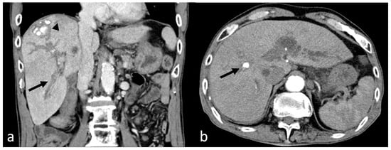
Background and Objectives: Transarterial chemoembolization (TACE) is a widely accepted treatment for hepatocellular carcinoma (HCC). Regarding TACE, arterial injuries, such as hepatic artery spasm or dissection, can also occur, although pseudoaneurysms are rare. We report a case of pseudoaneurysm following TACE. Materials and Methods: A 78-year-old man had been undergoing TACE for HCC in segment 8 of the liver for the past 5 years, with the most recent TACE procedure performed approximately 1 month prior. He presented to the emergency department with melena that persisted for 5 days. Computed tomography revealed a pseudoaneurysm in the S8 hepatic artery with hemobilia. Results: the pseudoaneurysm was successfully treated by N-Butyl-cyanoacrylate glue embolization. Conclusions: In patients that have undergone TACE presenting with melena and hemobilia identified on CT, consideration of hepatic artery pseudoaneurysm is crucial. Such cases can be safely and effectively treated with endovascular managements. Full article
Show Figures

Figure 1

12 pages, 1012 KiB  
Review
Biodegradable Microspheres for Transarterial Chemoembolization in Malignant Liver Disease
by Ornella Moschovaki-Zeiger, Nikolaos-Achilleas Arkoudis, Athanasios Giannakis, Stavros Grigoriadis, Fotis Anagnostopoulos and Stavros Spiliopoulos
Medicina 2024, 60(4), 678; https://doi.org/10.3390/medicina60040678 - 22 Apr 2024
Viewed by 1522
Abstract
Transarterial chemoembolization (TACE) has revolutionized the treatment landscape for malignant liver disease, offering localized therapy with reduced systemic toxicity. This manuscript delves into the use of degradable microspheres (DMS) in TACE, exploring its potential advantages and clinical applications. DMS-TACE emerges as a promising [...] Read more.
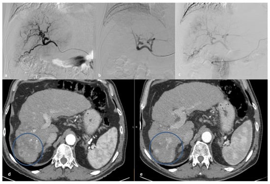
Transarterial chemoembolization (TACE) has revolutionized the treatment landscape for malignant liver disease, offering localized therapy with reduced systemic toxicity. This manuscript delves into the use of degradable microspheres (DMS) in TACE, exploring its potential advantages and clinical applications. DMS-TACE emerges as a promising strategy, offering temporary vessel occlusion and optimized drug delivery. The manuscript reviews the existing literature on DMS-TACE, emphasizing its tolerability, toxicity, and efficacy. Notably, DMS-TACE demonstrates versatility in patient selection, being suitable for both intermediate and advanced stages. The unique properties of DMS provide advantages over traditional embolic agents. The manuscript discusses the DMS-TACE procedure, adverse events, and tumor response rates in HCC, ICC, and metastases. Full article
Show Figures

Figure 1

2023

Jump to: 2024, 2022, 2021

11 pages, 4201 KiB  
Review
Ablative Techniques for Sarcoma Metastatic Disease: Current Role and Clinical Applications
by Evgenia Efthymiou, Georgios Charalampopoulos, Georgios Velonakis, Stauros Grigoriadis, Alexis Kelekis, Nikolaos Kelekis and Dimitrios Filippiadis
Medicina 2023, 59(3), 485; https://doi.org/10.3390/medicina59030485 - 1 Mar 2023
Cited by 3 | Viewed by 2084
Abstract
Sarcomas are heterogenous mesenchymal neoplasms with more than 80 different histologic subtypes. Lung followed by liver and bone are the most common sites of sarcoma metastatic disease. Ablative techniques have been recently added as an additional alternative curative or palliative therapeutic tool in [...] Read more.
Sarcomas are heterogenous mesenchymal neoplasms with more than 80 different histologic subtypes. Lung followed by liver and bone are the most common sites of sarcoma metastatic disease. Ablative techniques have been recently added as an additional alternative curative or palliative therapeutic tool in sarcoma metastatic disease. When compared to surgery, ablative techniques are less invasive therapies which can be performed even in non-surgical candidates and are related to decreased recovery time as well as preservation of the treated organ’s long-term function. Literature data upon ablative techniques for sarcoma metastatic disease are quite heterogeneous and variable regarding the size and the number of the treated lesions and the different histologic subtypes of the original soft tissue or bone sarcoma. The present study focuses upon the current role of minimal invasive thermal ablative techniques for the management of metastatic sarcoma disease. The purpose of this review is to present the current minimally invasive ablative techniques in the treatment of metastatic soft tissue and bone sarcoma, including local control and survival rates. Full article
Show Figures

Figure 1

2022

Jump to: 2024, 2023, 2021

6 pages, 1692 KiB  
Case Report
Intrapericardial Thymoma Presented as Pericardial Tamponade with Post-Operative Myasthenia Gravis
by Yueh-Hsun Tsai, Kai-Hsiung Ko, Hao Yen and Tsai-Wang Huang
Medicina 2022, 58(5), 609; https://doi.org/10.3390/medicina58050609 - 27 Apr 2022
Cited by 1 | Viewed by 2529
Abstract
Background: Thymoma is an epithelial mass arising from the thymus. Most thymomas are located in the anterior mediastinum. Ectopic intrapericardial thymoma is very unusual; to date, only eight cases of pericardial thymoma have been reported. Among thymoma patients, 20% to 25% are associated [...] Read more.
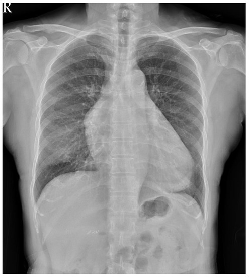
Background: Thymoma is an epithelial mass arising from the thymus. Most thymomas are located in the anterior mediastinum. Ectopic intrapericardial thymoma is very unusual; to date, only eight cases of pericardial thymoma have been reported. Among thymoma patients, 20% to 25% are associated with myasthenia gravis. However, postoperative myasthenia gravis occurs in less than 1% of cases. Here, we share a rare case of ectopic intrapericardial thymoma that developed postoperative myasthenia gravis six months after surgery. Case presentation: A 66-year-old woman visited the outpatient department due to productive cough and chest pain. Chest radiography showed increased soft tissue opacity over the mediastinum. A soft tissue mass in the pericardium and a ground glass nodule in right upper lung were noted using chest computed tomography. The diagnosis of thymoma, type B2, pT3N0M0, and stage IIIA and synchronous adenocarcinoma in situ of the right upper lung was confirmed after surgical removal. Six months later, the patient developed postoperative myasthenia gravis. Conclusions: Thymoma is rarely considered a differential diagnosis in pericardial tumors. Surgical removal with adjuvant radiation therapy should be performed considering the malignancy potential of thymomas and cardiac complications. In patients without myasthenia gravis, a small chance of postoperative myasthenia gravis remains. Patients should be carefully monitored for myasthenia gravis after surgery. Full article
Show Figures

Figure 1

2021

Jump to: 2024, 2023, 2022

8 pages, 1230 KiB  
Article
Percutaneous Navigation under Local Anesthesia for Computed Tomography-Guided Microwave Ablation of Malignant Liver Lesions Located in the Hepatic Dome
by Dimitrios K. Filippiadis, Georgios Velonakis, Argyro Mazioti, Athanasios Tsochatzis, Thomas Vrachliotis, Alexis Kelekis and Nikolaos Kelekis
Medicina 2021, 57(10), 1056; https://doi.org/10.3390/medicina57101056 - 3 Oct 2021
Cited by 6 | Viewed by 2393
Abstract
Background and Objectives: The aim of the present study was to report the safety and efficacy of percutaneous navigation under local anesthesia for computed tomography-guided microwave ablation of malignant liver lesions located in the hepatic dome. Patients with primary and secondary malignant liver [...] Read more.
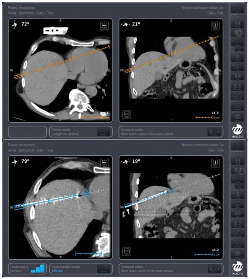
Background and Objectives: The aim of the present study was to report the safety and efficacy of percutaneous navigation under local anesthesia for computed tomography-guided microwave ablation of malignant liver lesions located in the hepatic dome. Patients with primary and secondary malignant liver lesions located in the hepatic dome who underwent percutaneous computed tomography-guided microwave ablation using a computer-assisted navigation system under local anesthesia were prospectively evaluated. The primary objective was technical success. Materials and Methods: The sample consisted of 10 participants (16 lesions) with a mean age of 60.60 years (SD = 9.25 years) and a mean size of 20.37 ± 7.29 cm, and the mean follow-up time was 3.4 months (SD = 1.41) months. Results: Primary technical success was 93.75%. Tumor remnant was noticed at one month follow-up in a single metastatic lesion, which was re-treated with an ablation session, and no tumor remnant was depicted in the subsequent imaging follow-up (secondary technical success 100%). Grade I self-limited complications (according to the CIRSE classification system) included small pleural effusion (n = 1) and minor bleeding post antenna removal (n = 1) requiring nothing but observation. Conclusions: the findings of the present study indicate that percutaneous navigation under local anesthesia is a safe and efficacious approach for computed tomography-guided microwave ablation of malignant liver lesions located in the hepatic dome. Large randomized controlled studies are warranted to observe treatment effectiveness and compare the results with those of other options. Full article
Show Figures

Figure 1

10 pages, 2800 KiB  
Article
Bipolar Radiofrequency Ablation of Painful Spinal Bone Metastases Performed under Local Anesthesia: Feasibility Regarding Patient’s Experience and Pain Outcome
by Adrian Kastler, Daniel-Ange Barbé, Guillaume Alemann, Georges Hadjidekov, Francois H. Cornelis and Bruno Kastler
Medicina 2021, 57(9), 966; https://doi.org/10.3390/medicina57090966 - 15 Sep 2021
Cited by 7 | Viewed by 2425
Abstract
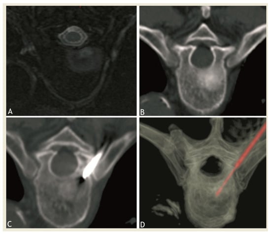
Background and objectives: To assess the pain relief of bipolar RFA combined or not with vertebroplasty in patients with painful vertebral metastases and to evaluate the feasibility and tolerance of the RFA procedure performed under local anesthesia. Materials and Methods: 25 patients [...] Read more.
Background and objectives: To assess the pain relief of bipolar RFA combined or not with vertebroplasty in patients with painful vertebral metastases and to evaluate the feasibility and tolerance of the RFA procedure performed under local anesthesia. Materials and Methods: 25 patients (18 men, 7 women, mean age: 60.X y.o) with refractory painful vertebral metastasis were consecutively included between 2012 and 2019. A total of 29 radiofrequency ablation (RFA) procedures were performed under CT guidance, local anesthesia and nitrous oxide inhalation, including 16 procedures combined with vertebroplasty for bone consolidation purposes. Pain efficacy was clinically evaluated using the visual analogue scale (VAS) at day 1, 1 month, 3 months, 6 months and 12 months, and the tolerance of the procedure was evaluated. Results: Procedure tolerance was graded as either not painful or tolerable in 97% of cases. Follow-up postprocedure mean VAS score decrease was 74% at day 1: 6.6 (p < 0.001), 79% at 1 month: 6.6 (p < 0.001), 79% at 3 months: 6.5 (p < 0.001), 77% at 6 months, and 79% at 12 months: 6.6 (p < 0.001). Conclusions: Bipolar RFA, with or without combined vertebroplasty, appears to be an effective and reliable technique for the treatment of refractory vertebral metastases in patients in the palliative care setting. It is a feasible procedure under local anesthesia which is well tolerated by patients therefore allowing to broaden the indications of such procedures. Field of study: interventional radiology. Full article
Show Figures

Figure 1

7 pages, 2093 KiB  
Article
Percutaneous Image-Guided Vertebral Fixation in Cancer-Related Vertebral Compression Fractures: A Case Series Study
by Francois H. Cornelis, Leo Razakamanantsoa, Mohamed Ben Ammar, Milan Najdawi, Francois Gardavaud, Sanaa El-Mouhadi and Matthias Barral
Medicina 2021, 57(9), 907; https://doi.org/10.3390/medicina57090907 - 30 Aug 2021
Cited by 3 | Viewed by 2072
Abstract
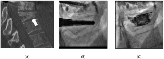
Background and objectives: Cancer-related vertebral compression fractures (VCF) may cause debilitating back pain and instability, affecting the quality of life of cancer patients. To further drive cement deposition during vertebroplasty, the aims of this restrospective case series study were to report the feasibility, [...] Read more.
Background and objectives: Cancer-related vertebral compression fractures (VCF) may cause debilitating back pain and instability, affecting the quality of life of cancer patients. To further drive cement deposition during vertebroplasty, the aims of this restrospective case series study were to report the feasibility, safety and short term efficacy (≤6 months) of percutaneous vertebral fixation in cancer-related vertebral compression fractures using various intravertebral implants. Methods: All consecutive cancer patients treated with percutaneous vertebral fixation for VCF were retrospectively included. Various devices were inserted percutaneously under image guidance and filled by cement. Descriptive statistics were used and a matched paired analysis of pain scores was performed to assess for changes following interventions. Results: A total of 18 consecutive patients (12 women (66.6%) and 6 men (33.3%); mean age 59.7 ± 15.5 years) were included. A total of 42 devices were inserted in 8 thoracic and 16 lumbar vertebrae. Visual analogue scale measurement significantly improved from 5.6 ± 1.8 preoperatively to 1.5 ± 1.7 at 1 week (p < 0.01) and to 1.5 ± 1.3 at 6 months (p < 0.01). No severe adverse events were observed, but three adjacent fractures occurred between 1 week and 5 months after implantation. Conclusions: Percutaneous vertebral fixation of cancer-related VCF is feasible and safe and allows pain relief. Full article
Show Figures

Figure 1

12 pages, 1712 KiB  
Article
Ablation Zone Involution of Liver Tumors Is Faster in Patients Treated with Irreversible Electroporation Than Microwave Ablation
by Fourat Ridouani, Mario Ghosn, Francois Cornelis, Elena N Petre, Meier Hsu, Chaya S Moskowitz, Peter T Kingham, Stephen B Solomon and Govindarajan Srimathveeravalli
Medicina 2021, 57(9), 877; https://doi.org/10.3390/medicina57090877 - 26 Aug 2021
Cited by 4 | Viewed by 2981
Abstract
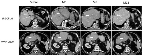
Background and Objectives: To compare ablation zone involution following microwave ablation (MWA) or irreversible electroporation (IRE) of liver tumors. Materials and Methods: MWA or IRE performed for colorectal cancer liver metastasis (CRLM) or hepatocellular carcinoma (HCC) during January 2011 to December [...] Read more.
Background and Objectives: To compare ablation zone involution following microwave ablation (MWA) or irreversible electroporation (IRE) of liver tumors. Materials and Methods: MWA or IRE performed for colorectal cancer liver metastasis (CRLM) or hepatocellular carcinoma (HCC) during January 2011 to December 2015 were analyzed. Patients with a tumoral response on 1-year follow-up computed tomography (CT) were included. Generalized estimating equations were used to evaluate the differences between the two modalities on ablation zone involution observed on CT at 6 (M6) and 12 months (M12), and on laboratory values (total bilirubin, alanine transaminase, aspartate transaminase, alkaline phosphatase, albumin, and platelets count). The likelihood ratio test was used to assess whether the association between ablation modalities and these outcomes differed over time. Results: Seventeen (17/44, 39%) women and 27 (27/44, 61%) men were included, with 25 HCC (25/44, 57%) and 19 CRLM (19/44, 43%) patients. IRE was used in 9 (9/19, 47%) CRLM and 5 (5/25, 20%) HCC patients, respectively. All other patients had MWA. Ablation zone size and involution between IRE and MWA differed significantly over time (interaction p < 0.01), with a mean of 241.04 vs. 771.08 mm2 (ratio 0.313; 95% CI, 0.165–0.592; p < 0.01) at M6 and 60.47 vs. 589.43 mm2 (ratio 0.103; 95% CI, 0.029–0.365; p < 0.01) at M12. Changes in liver enzymes did not differ significantly between IRE and MWA at both timepoints. Conclusions: Liver tumors treated with IRE underwent faster involution when compared to tumors treated with MWA, but liver enzymes levels were comparable. Full article
Show Figures

Figure 1

11 pages, 4912 KiB  
Article
Ultrasound-Guided Moving Shot Radiofrequency Ablation of Benign Soft Tissue Neoplasm
by Wei-Che Lin, Yi-Fan Tai, Meng-Hsiang Chen, Sheng-Dean Luo, Faye Huang, Wei-Chih Chen, Pi-Ling Chiang, Hsiu-Ling Chen, Mei-Hsiu Chen and Jung-Hwan Baek
Medicina 2021, 57(8), 830; https://doi.org/10.3390/medicina57080830 - 17 Aug 2021
Cited by 6 | Viewed by 2861
Abstract
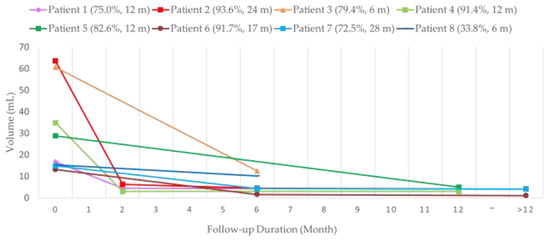
Background and Objective: To evaluate the effectiveness of radiofrequency ablation (RFA) using the moving-shot technique for benign soft tissue neoplasm. Materials and Methods: This retrospective study reviewed eight patients with benign soft tissue neoplasm presenting with cosmetic concerns and/or symptomatic issues [...] Read more.
Background and Objective: To evaluate the effectiveness of radiofrequency ablation (RFA) using the moving-shot technique for benign soft tissue neoplasm. Materials and Methods: This retrospective study reviewed eight patients with benign soft tissue neoplasm presenting with cosmetic concerns and/or symptomatic issues who refused surgery. Six patients had vascular malformation, including four with venous malformation and two with congenital hemangioma. The other two patients had neurofibroma. All patients underwent RFA using the moving-shot technique. Imaging and clinical follow-up were performed in all patients. Follow-up image modalities included ultrasound (US), computed tomography (CT), and magnetic resonance (MR) imaging. The volume reduction ratio (VRR), cosmetic scale (CS), and complications were evaluated. Results: Among the seven patients having received single-stage RFA, there were significant volume reductions between baseline (33.3 ± 21.2 cm3), midterm follow-up (5.1 ± 3.8 cm3, p = 0.020), and final follow-up (3.6 ± 1.4 cm3, p = 0.022) volumes. The VRR was 84.5 ± 9.2% at final follow-up. There were also significant improvements in the CS (from 3.71 to 1.57, p = 0.017). The remaining patient, in the process of a scheduled two-stage RFA, had a 33.8% VRR after the first RFA. The overall VRR among the eight patients was 77.5%. No complications or re-growth of the targeted lesions were noted during the follow-up period. Of the eight patients, two received RFA under local anesthesia, while the other six patients were under general anesthesia. Conclusions: RFA using the moving-shot technique is an effective, safe, and minimally invasive treatment for benign soft tissue neoplasms, achieving mass volume reduction within 6 months and significant esthetic improvement, either with local anesthesia or with general anesthesia under certain conditions. Full article
Show Figures

Figure 1

8 pages, 2522 KiB  
Article
Combined Microwave Ablation and Osteosynthesis for Long Bone Metastases
by Claudio Pusceddu, Giuseppe Dessì, Luca Melis, Alessandro Fancellu, Giuseppe Ruggiu, Paolo Sailis, Stefano Congia, Daniele Derudas, Riccardo Cau, Ignazio Senis, Nicola Ballicu and Luca Saba
Medicina 2021, 57(8), 825; https://doi.org/10.3390/medicina57080825 - 16 Aug 2021
Cited by 2 | Viewed by 2456
Abstract
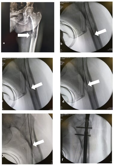
Background and Objectives: The purpose of this study was to evaluate the feasibility, safety and efficacy of microwave ablation (MWA) in combination with open surgery nail positioning for the treatment of fractures or impending fractures of long bone metastases. Material and Methods: Eleven [...] Read more.
Background and Objectives: The purpose of this study was to evaluate the feasibility, safety and efficacy of microwave ablation (MWA) in combination with open surgery nail positioning for the treatment of fractures or impending fractures of long bone metastases. Material and Methods: Eleven patients (four men, seven women) with painful bone metastases of the humerus, femur or tibia with non-displaced fractures (one case) or impending fractures (10 cases) underwent open MWA in combination with osteosynthesis by locked nail positioning. Pain intensity was measured using a VAS score before and after treatment. CT or MRI were acquired at one month before and 1, 3, 6, 12 and 18 months after treatment. Results: All procedures were successfully completed without major complications. The level of pain was significantly reduced one month after treatment. For the patients with humerus metastases, the complete recovery of arm use took 8 weeks, while for the patients with femoral metastases the complete recovery of walking capacity took 11 weeks. The VAS score ranged from 7 (4–9) before treatment to 1.5 (0–2.5) after treatment. During a mid-term follow-up of 18 months (range 4–29 months), none of the patients showed tumor relapse or new fractures in the treated site. Two patients died due to tumor disease progression. Conclusion: Results of this preliminary study suggest that combined MWA and surgical osteosynthesis with locked nails is a safe and effective treatment for pathological fractures or malignant impending fractures of long bone metastases of the humerus, femur and tibia. Further analyses with larger cohorts are warranted to confirm these findings. Full article
Show Figures

Figure 1

9 pages, 3384 KiB  
Case Report
Malignant Gastric Outlet Obstruction Caused by Duodenal Cervix Metastasis in a Young Woman: Rendezvous Technique
by Ester Marra, Pasquale Quassone, Pasquale Tammaro, Cinzia Cardalesi, Raffaele D’Avino, Fabio Cipolletta, Anna Del Prete, Angela Travaglino, Stefania Tamburrini, Giovanni Ferrandino, Giuseppe Sarti, Michele Iannuzzi, Pietro Maida and Gianpaolo Santini
Medicina 2021, 57(8), 765; https://doi.org/10.3390/medicina57080765 - 28 Jul 2021
Cited by 3 | Viewed by 2264
Abstract
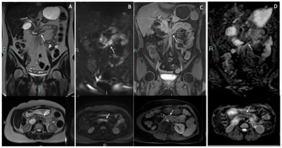
Background: Malignant gastric outlet obstruction (MGOD) is an extremely rare expression of advanced extra-gastrointestinal cancer, such as squamous cell carcinoma (SCC) of the cervix, and only sixcases are described in the literature.Because of the short life expectancyand the high surgical risk involving [...] Read more.
Background: Malignant gastric outlet obstruction (MGOD) is an extremely rare expression of advanced extra-gastrointestinal cancer, such as squamous cell carcinoma (SCC) of the cervix, and only sixcases are described in the literature.Because of the short life expectancyand the high surgical risk involving these patients, less invasive approaches have been developed over time, such asthe use of an enteral stent or less invasive surgical techniques (i.e., laparoscopic gastrojejunostomy). However, MGOD could make it difficult to perform an endoscopic retrograde cholangio-pancreatography (ERCP) for standard endoscopic drainage, so in this case a combined endoscopic-percutaneous technique may be performed. This article, therefore, aims to highlight the presence in the doctor’s armamentarium of the “rendezvous technique”, few case reports of whichare described in the literature, and, moreover, this article aims to underline the technique’sfeasibility. Case Presentation: The case is that of a 38-year-old woman who presented with MGOD three years after the diagnosis of SCC of the cervix, who successfully underwent the rendezvous technique with the resolution of duodenal obstruction. Endoscopic enteral stenting treatment with the placement of a metal stent (SEMSs) represents the mainstay of MGOD treatment compared withsurgery due to its lower morbidity, mortality, shorter hospitalization and earlier symptom relief. However, in patients with both duodenal and biliary obstruction, a combined endoscopic–percutaneous approach may be necessary because of the difficulty in passing the duodenal stricture or in accessing the papilla through the mesh of the duodenal SEMS. Conclusion: The rendezvous procedure is a technicallyfeasible and minimally invasive approach to the double stenting of biliary and duodenal strictures. It achieves the desired therapeutic result while avoiding the need to perform more invasive procedures that could have a negative impact on the patient’sprognosis. Full article
Show Figures

Figure 1

10 pages, 877 KiB  
Article
Clinical Usefulness of Ultrasound-Guided Fine Needle Aspiration and Core Needle Biopsy for Patients with Axillary Lymphadenopathy
by Tomoyuki Fujioka, Mio Mori, Kazunori Kubota, Emi Yamaga, Yuka Yashima, Goshi Oda, Tsuyoshi Nakagawa, Iichiroh Onishi, Toshiyuki Ishiba and Ukihide Tateishi
Medicina 2021, 57(7), 722; https://doi.org/10.3390/medicina57070722 - 16 Jul 2021
Cited by 4 | Viewed by 10014
Abstract
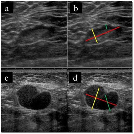
Background and Objectives: It is necessary to properly diagnose and manage axillary lymphadenopathy caused by a variety of diseases. This study aimed to evaluate the utility of ultrasound (US)-guided sampling in patients with axillary lymphadenopathy. Materials and Methods: Patients with axillary [...] Read more.
Background and Objectives: It is necessary to properly diagnose and manage axillary lymphadenopathy caused by a variety of diseases. This study aimed to evaluate the utility of ultrasound (US)-guided sampling in patients with axillary lymphadenopathy. Materials and Methods: Patients with axillary lymphadenopathy (excluding patients with newly diagnosed breast cancer) who underwent US-guided fine needle aspiration (FNA) or core needle biopsy (CNB) at a single center between February 2016 and September 2020 were retrospectively examined. The association between US imaging findings and malignancy was investigated and the diagnostic performance of US-guided sampling was assessed. Results: Fifty-five patients (including eight males) were included in the study; of these, 34 patients (61.8%) were finally diagnosed with a malignant lymph node lesion. Twenty-two patients (40.0%) had undergone FNA and 33 (60.0%) had undergone CNB. Larger short and long axis diameters, thicker lymph node cortex, and the absence of fatty hilum on the US were significantly associated with malignancy (p < 0.05). The diagnostic performance of FNA, CNB, and FNA + CNB was excellent (sensitivity, specificity, and accuracy of 0.909, 0.900, and 0.917 for FNA, 0.958, 1.000, and 0.970 for CNB, and 0.941, 0.952, and 0.945 for FNA + CNB, respectively). Conclusions: US-guided FNA and CNB play an important role in the diagnosis and management of patients with axillary lymphadenopathy. Full article
Show Figures

Figure 1

12 pages, 5087 KiB  
Article
Risk Analysis of Needle Injury to the Long Thoracic Nerve during Ultrasound-Guided C7 Selective Nerve Root Block
by Seok Kang, Ha-Mok Jeong, Beom-Suk Kim and Joon-Shik Yoon
Medicina 2021, 57(6), 635; https://doi.org/10.3390/medicina57060635 - 19 Jun 2021
Viewed by 3214
Abstract
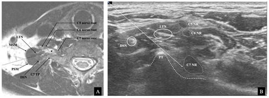
Background and Objectives: Ultrasound (US)-guided cervical selective nerve root block (SNRB) is a widely used treatment for upper limb radicular pain. The long thoracic nerve (LTN) passes through the middle scalene muscle (MSM) at the C7 level. The needle trajectory of US-guided [...] Read more.
Background and Objectives: Ultrasound (US)-guided cervical selective nerve root block (SNRB) is a widely used treatment for upper limb radicular pain. The long thoracic nerve (LTN) passes through the middle scalene muscle (MSM) at the C7 level. The needle trajectory of US-guided C7 SNRB pierces the MSM, therefore indicating a high probability of injury to the LTN. We aimed to identify the LTN and to investigate the risk of needle injury to the nerve during US-guided C7 SNRB. Materials and Methods: This retrospective observational study included 30 patients who underwent US-guided SNRB at the C7 level in a university hospital. We measured the maximal cross-sectional diameter (MCSD) of the LTN and cross-sectional area (CSA) of the C7 nerve root and assessed the injury risk of LTN during US-guided C7 SNRB by simulating the trajectory of the needle in the ultrasound image. Results: The LTN was detectable in all the cases, located inside and outside the MSM in 19 (63.3%) and 11 (36.7%) of cases, respectively. The LTN’s mean MCSD was 2.10 mm (SD 0.13), and the C7 root’s CSA was 10.78 mm2 (SD 1.05). The LTN location was within the simulated risk zone in 86.7% (26/30) of cases. Conclusion: Our findings suggest a high potential for LTN injury during US-guided C7 SNRB. The clear visualization of LTNs in the US images implies that US guidance may help avoid nerve damage and make the procedure safer. When performing US-guided C7 SNRB, physicians should take into consideration the location of the LTN. Full article
Show Figures

Figure 1

9 pages, 4134 KiB  
Review
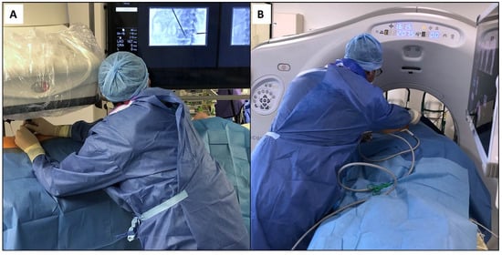
Ergonomics in Interventional Radiology: Awareness Is Mandatory
by Francois H. Cornelis, Leo Razakamanantsoa, Mohamed Ben Ammar, Raphael Lehrer, Idriss Haffaf, Sanaa El-Mouhadi, Francois Gardavaud, Milan Najdawi and Matthias Barral
Medicina 2021, 57(5), 500; https://doi.org/10.3390/medicina57050500 - 14 May 2021
Cited by 13 | Viewed by 4025
Abstract
Ergonomics in interventional radiology has not been thoroughly evaluated. Like any operators, interventional radiologists are exposed to the risk of work-related musculoskeletal disorders. The use of lead shielding to radiation exposure and the lack of ergonomic principles developed so far contribute to these [...] Read more.
Ergonomics in interventional radiology has not been thoroughly evaluated. Like any operators, interventional radiologists are exposed to the risk of work-related musculoskeletal disorders. The use of lead shielding to radiation exposure and the lack of ergonomic principles developed so far contribute to these disorders, which may potentially affect their livelihoods, quality of life, and productivity. The objectives of this review were to describe the different situations encountered in interventional radiology and to compile the strategies both available to date and in development to improve ergonomics. Full article
Show Figures

Figure 1

Back to TopTop