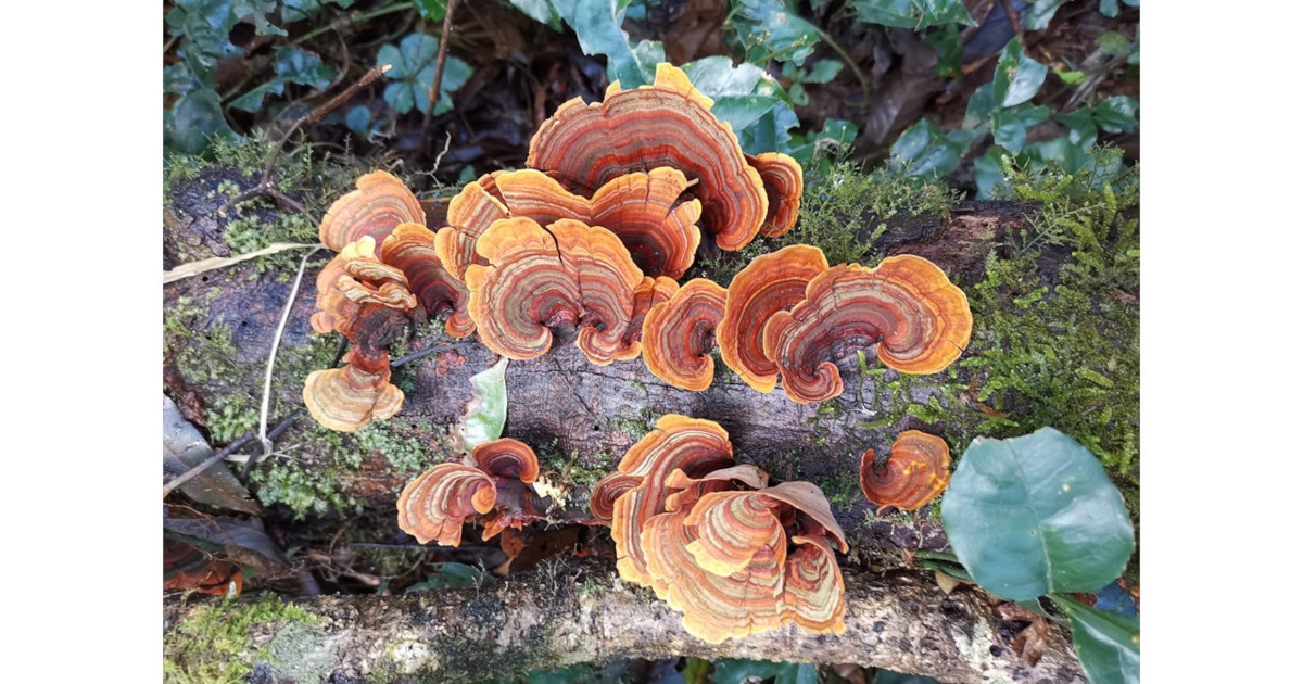
Diversity and Evolution of Fungi

A special issue of Diversity (ISSN 1424-2818). This special issue belongs to the section "Phylogeny and Evolution".

Deadline for manuscript submissions: closed (15 January 2023) | Viewed by 33778

Special Issue Editors


E-Mail Website
Guest Editor
1. School of Life Sciences, Tsinghua University, Beijing 100084, China
2. College of Biodiversity Conservation, Southwest Forestry University, Kunming 650224, China
Interests: molecular systematics; taxonomy; multigene phylogeny; medical fungi; wood-decaying fungi; fungal pathogens; biodiversity
Special Issues, Collections and Topics in MDPI journals
CAS Key Laboratory for Plant Diversity and Biogeography of East Asia, Kunming Institute of Botany, Chinese Academy of Sciences, Lanhei Road 132, Kunming 650201, China
Interests: fungal diversity; molecular systematics; taxonomy; multigene phylogeny; fungal ecology; ectomycorrhizal fungi

Special Issue Information

Dear Colleagues,

Fungi are a distinct, diverse, and ecologically important branch of the tree of life, in which these hardworking organisms play a vital role in ecosystems as diverse as soil, leaves, rocks, and pelagic zones of the ocean. Distinguished from plants by their heterotrophic nature, and distinct from animals by their external rather than internal digestion, fungi diverged from their sister kingdom the animals ∼1.3 billion years ago. The hidden and microscopic nature of many fungi also means that their diversity is undersampled, and perhaps less than 5% of the estimated two to four million species have been formally described. Fungal genomes are easy to obtain, and fungi have served as models for genome evolution and the reconstruction of phylogenetic relationships using genome-scale data, in which groundbreaking comparative genomic studies that take advantage of these features have already been published.  These pioneering studies are just the prelude to the period that is upon us now.

Despite the early embrace of molecular systematics by mycologists, both the discovery and classification of fungi are still in great flux, particularly among the more basal branches of the tree, whose true diversity is only now coming to light from genomic analyses and environmental DNA surveys. This Special Issue aims to bring together a collection of papers focusing on the biodiversity, molecular systematics, and taxonomy of fungi worldwide.

Dr. Chang-Lin Zhao
Dr. Zai-Wei Ge
Guest Editors

Manuscript Submission Information

Manuscripts should be submitted online at www.mdpi.com by registering and logging in to this website. Once you are registered, click here to go to the submission form. Manuscripts can be submitted until the deadline. All submissions that pass pre-check are peer-reviewed. Accepted papers will be published continuously in the journal (as soon as accepted) and will be listed together on the special issue website. Research articles, review articles as well as short communications are invited. For planned papers, a title and short abstract (about 100 words) can be sent to the Editorial Office for announcement on this website.

Submitted manuscripts should not have been published previously, nor be under consideration for publication elsewhere (except conference proceedings papers). All manuscripts are thoroughly refereed through a single-blind peer-review process. A guide for authors and other relevant information for submission of manuscripts is available on the Instructions for Authors page. Diversity is an international peer-reviewed open access monthly journal published by MDPI.

Please visit the Instructions for Authors page before submitting a manuscript. The Article Processing Charge (APC) for publication in this open access journal is 2100 CHF (Swiss Francs). Submitted papers should be well formatted and use good English. Authors may use MDPI's English editing service prior to publication or during author revisions.

Keywords

  • biodiversity
  • molecular systematics
  • taxonomy
  • multigene phylogeny
  • medical fungi
  • wood-decaying fungi
  • fungal pathogens
  • edible mushrooms
  • poisonous mushroom

Benefits of Publishing in a Special Issue

  • Ease of navigation: Grouping papers by topic helps scholars navigate broad scope journals more efficiently.
  • Greater discoverability: Special Issues support the reach and impact of scientific research. Articles in Special Issues are more discoverable and cited more frequently.
  • Expansion of research network: Special Issues facilitate connections among authors, fostering scientific collaborations.
  • External promotion: Articles in Special Issues are often promoted through the journal's social media, increasing their visibility.
  • e-Book format: Special Issues with more than 10 articles can be published as dedicated e-books, ensuring wide and rapid dissemination.

Further information on MDPI's Special Issue polices can be found here.

Published Papers (11 papers)

Order results
Result details
Select all
Export citation of selected articles as:

Research

12 pages, 2406 KiB  
Article
Analysis of Fungal Diversity before and after Discoloration of Rubberwood in Xishuangbanna
by Susu Yang, Xiaolong Wu, Lin Liu, Yan Yan, Jian Qiu and Lei Qin
Diversity 2023, 15(4), 471; https://doi.org/10.3390/d15040471 - 23 Mar 2023
Cited by 1 | Viewed by 1471
Abstract
The discoloration problem of rubber wood caused by the discoloration fungi has caused the degradation of rubber wood and greatly reduced its economic value, and the prevention and control of rubber wood discoloration have become the top priority of basic research on rubber [...] Read more.
The discoloration problem of rubber wood caused by the discoloration fungi has caused the degradation of rubber wood and greatly reduced its economic value, and the prevention and control of rubber wood discoloration have become the top priority of basic research on rubber wood protection and modification. To determine the fungal community diversity and dominant groups before and after discoloration of rubber wood, nine rubber wood samples were subjected to ITS sequencing using Illumina high-throughput sequencing technology. The results showed that the detected fungal communities comprised 5 phyla, 18 classes, 58 orders, 137 families, 218 genera, and 297 species. Discoloration of rubber wood is not caused by a single species, with the dominant genera for discolored rubber wood being Huntiella, Ceratocystis, and Acremonium and for undiscolored rubber wood, Phomopsis. Furthermore, the diversity, uniformity of species distribution, and richness of discolored rubber wood were found to be higher than those of undiscolored rubber wood. In conclusion, understanding the change trends in the structure of these fungal communities is essential for studying the biological control of rubberwood discoloration. Full article
(This article belongs to the Special Issue Diversity and Evolution of Fungi)
Show Figures

Figure 1

16 pages, 2743 KiB  
Article
Pseudocercospora rizhaoensis sp. nov. Causing Leaf Spot Disease of Ligustrum japonicum in China
by Yun Liu, Shumei Guo, Jin Liu and Xiangli Yang
Diversity 2022, 14(11), 990; https://doi.org/10.3390/d14110990 - 17 Nov 2022
Cited by 1 | Viewed by 2620
Abstract
Ligustrum japonicum is a common ornamental tree species in China. However, leaf spot disease has emerged in Rizhao City, Shandong Province of China in recent years. Members of Pseudocercospora are usually known as plant pathogens, mainly causing leaf spots and blights. Species of [...] Read more.
Ligustrum japonicum is a common ornamental tree species in China. However, leaf spot disease has emerged in Rizhao City, Shandong Province of China in recent years. Members of Pseudocercospora are usually known as plant pathogens, mainly causing leaf spots and blights. Species of this genus are distinguished mainly based on morphological differences on the host plants, as well as the molecular data. A new species named Pseudocercospora rizhaoensis on Ligustrum japonicum is introduced herein based on morphology and molecular data of combined ITS, LSU, act, tef1 and rpb2 sequences. Koch’s postulates were confirmed by a pathogenicity test, re-isolation and identification. Full article
(This article belongs to the Special Issue Diversity and Evolution of Fungi)
Show Figures

Figure 1

12 pages, 4529 KiB  
Article
Morphological and Phylogenetic Studies of Three New Species of Calocybe (Agaricales, Basidiomycota) from China
by Yue Qi, Aiguo Xu, Ye Zhou, Kexin Bi, Weiqiang Qin, Hongbo Guo and Xiaodan Yu
Diversity 2022, 14(8), 643; https://doi.org/10.3390/d14080643 - 11 Aug 2022
Cited by 2 | Viewed by 1688
Abstract
Three species, Calocybe lilacea, C. longisterigma, and C. subochraceus, were newly discovered in Inner Mongolia, Hunan, and Liaoning provinces of China. Calocybe lilacea is mainly characterized by its small-sized basidiocarp, brownish-orange pileus, lilac gray to dull violet stipe, and noncellular [...] Read more.
Three species, Calocybe lilacea, C. longisterigma, and C. subochraceus, were newly discovered in Inner Mongolia, Hunan, and Liaoning provinces of China. Calocybe lilacea is mainly characterized by its small-sized basidiocarp, brownish-orange pileus, lilac gray to dull violet stipe, and noncellular epicutis. The main characteristics of C. longisterigma are its light brown to brownish-orange pileus, long sterigmata, and noncellular epicutis. The main morphological features of C. subochraceus are its small-sized basidiocarp, pale-orange pileus, adnexed lamellae, tortuous stipe, and noncellular epicutis. These morphological features confirmed that the three species of Calocybe all belonged to Sect. Carneoviolaceae. Phylogenetic analysis based on a combined dataset (ITS–nrLSU–RPB2) determined that the three species belong to the genus Calocybe and form a distinct lineage. The morphological differences between the three new species and other related species of Calocybe are also discussed. Full article
(This article belongs to the Special Issue Diversity and Evolution of Fungi)
Show Figures

Figure 1

9 pages, 2070 KiB  
Article
Two New Species of Dacrymyces (Dacrymycetales, Basidiomycota) from Southwestern China
by Ya-Ping Lian, Ablat Tohtirjap and Fang Wu
Diversity 2022, 14(5), 379; https://doi.org/10.3390/d14050379 - 11 May 2022
Cited by 3 | Viewed by 2162
Abstract
Two new species of Dacrymyces, D. cerebriformis and D. sinostenosporus, are presented from southwestern China, based on morphological characteristics and phylogenetic analyses. Dacrymyces cerebriformis is characterized by obviously cerebriform basidiomata when mature, hyphae without clamp connections, and hyaline, thin-walled, allantoid, 0–7-septate [...] Read more.
Two new species of Dacrymyces, D. cerebriformis and D. sinostenosporus, are presented from southwestern China, based on morphological characteristics and phylogenetic analyses. Dacrymyces cerebriformis is characterized by obviously cerebriform basidiomata when mature, hyphae without clamp connections, and hyaline, thin-walled, allantoid, 0–7-septate basidiospores (18.4–23.1 × 5.5–7.7 μm). Dacrymyces sinostenosporus is characterized by discoid and applanate basidiomata when mature, hyphae without clamp connections, and hyaline, thin-walled, cylindrical to allantoid, 0–7-septate basidiospores (18.0–23.5 × 6.3–8.0 μm). Dacrymyces cerebriformis can be distinguished from D. sinostenosporus by the narrower thin- or thick-walled terminal cells in the marginal hyphae of the sterile surface (1.7–3.5 μm vs. 3.5–6.0 μm in diameter) and thinner basidiospores (Q = 3.14–3.28 vs. Q = 2.63–2.77). A phylogenetic analysis of Dacrymyces is performed by using a dataset composed of concatenated internal transcribed spacer regions (ITS) and a large subunit (nrLSU) of ribosomal DNA gene, and two new species nested in two distinct lineages with robust support. A full description and illustrations of the two new species are provided here. Full article
(This article belongs to the Special Issue Diversity and Evolution of Fungi)
Show Figures

Figure 1

17 pages, 3069 KiB  
Article
Six New Species of Leucoagaricus (Agaricaceae) from Northeastern China
by Yunrui Ma, Tiezhi Liu, Xiaodan Yu, Tiezheng Wei and Zai-Wei Ge
Diversity 2022, 14(5), 314; https://doi.org/10.3390/d14050314 - 20 Apr 2022
Cited by 15 | Viewed by 5584
Abstract
Six new species, Leucoagaricus albosquamosus, Leucoagaricus atroviridis, Leucoagaricus aurantioruber, Leucoagaricus candidus, Leucoagaricus centricastaneus and Leucoagaricus virens, collected from northeastern China are described based on morphological characters and molecular evidence. Illustrations of fresh basidiomata and line drawings of key anatomical characters are provided. [...] Read more.
Six new species, Leucoagaricus albosquamosus, Leucoagaricus atroviridis, Leucoagaricus aurantioruber, Leucoagaricus candidus, Leucoagaricus centricastaneus and Leucoagaricus virens, collected from northeastern China are described based on morphological characters and molecular evidence. Illustrations of fresh basidiomata and line drawings of key anatomical characters are provided. A phylogenetic tree inferred from internal transcribed spacer (ITS) region and large subunit ribosomal RNA gene (LSU) sequences shows that three of the new taxa are nested within the section Leucoagaricus and two of the new taxa are in the subgenus Sericeomyces, whereas the other new taxus is clustered with Leucoagaricus viriditinctus and Leucoagaricus irinellus, forming a clade that does not fit in any known section. Full article
(This article belongs to the Special Issue Diversity and Evolution of Fungi)
Show Figures

Figure 1

11 pages, 2379 KiB  
Article
Two New Species of Diatrype (Xylariales, Ascomycota) with Polysporous Asci from China
by Zhanen Yang, Bo Zhang, Zhi Qu, Zikun Song, Xiaoyan Pan, Changlin Zhao and Haixia Ma
Diversity 2022, 14(2), 149; https://doi.org/10.3390/d14020149 - 18 Feb 2022
Cited by 7 | Viewed by 2031
Abstract
Two new species of Diatrype collected in northeast China are described and illustrated based on morphological and molecular evidence. Diatrype larissae from Heilongjiang Province is characterised by having 3–6 perithecia in a stroma, asci polysporous, ascospores allantoid, aseptate, slightly or moderately curved, subhyaline. [...] Read more.
Two new species of Diatrype collected in northeast China are described and illustrated based on morphological and molecular evidence. Diatrype larissae from Heilongjiang Province is characterised by having 3–6 perithecia in a stroma, asci polysporous, ascospores allantoid, aseptate, slightly or moderately curved, subhyaline. Diatrype betulaceicola from Inner Mongolia has large stroma with 5–14 perithecia, perithecium immersed, asci polysporous, long-stalked, ascospores allantoid, aseptate, slightly curved, subhyaline. The phylogenies inferred from the data set of nrDNA ITS1-5.8S-ITS2 (ITS) and beta-tubulin (β-tubulin) supported the two new species both as members in the genus Diatrype and distinct species. The morphological similarities and dissimilarities of the new species with phylogenetically close relatives are discussed. A dichotomous identification key to the Diatrype spp. known from China is proposed. Full article
(This article belongs to the Special Issue Diversity and Evolution of Fungi)
Show Figures

Figure 1

12 pages, 1826 KiB  
Article
Three New Species of Absidia (Mucoromycota) from China Based on Phylogeny, Morphology and Physiology
by Heng Zhao, Yong Nie, Tongkai Zong, Yucheng Dai and Xiaoyong Liu
Diversity 2022, 14(2), 132; https://doi.org/10.3390/d14020132 - 13 Feb 2022
Cited by 11 | Viewed by 3915
Abstract
Species of Absidia are distributed widely in the environment, while their diversity is insufficiently studied. Three new species, A. frigida, A. gemella and A. longissima, are proposed herein from Xinjiang and Yunnan in China based on phylogenetic, morphological and physiological evidence. [...] Read more.
Species of Absidia are distributed widely in the environment, while their diversity is insufficiently studied. Three new species, A. frigida, A. gemella and A. longissima, are proposed herein from Xinjiang and Yunnan in China based on phylogenetic, morphological and physiological evidence. According to maximum likelihood (ML), maximum parsimony (MP) and Bayesian inference (BI) analyses, the phylogenetical results suggest that A. frigida, A. gemella and A. longissima are closely related to A. psychrophilia, A. turgida and A. zonata and A. koreana, respectively, based on ITS and LSU rDNA sequences. Absidia frigida is characterized by a lower growth temperature, which does not grow above 24 °C. It differs from A. psychrophilia by sporangiophores, sporangia, columellae, collars and projections. Absidia gemella is distinguished from A. turgida by hypha, sporangiospores, sporangia, projections and sporangiophores. Absidia longissima is discriminated from A. zonata and A. koreana by sporangiophores, columellae and collars. The three new species are described and illustrated in this article. Full article
(This article belongs to the Special Issue Diversity and Evolution of Fungi)
Show Figures

Figure 1

29 pages, 6201 KiB  
Article
Diversity of Wood-Decaying Fungi in Wuliangshan Area, Yunnan Province, P.R. China
by Xiao He and Chang-Lin Zhao
Diversity 2022, 14(2), 131; https://doi.org/10.3390/d14020131 - 11 Feb 2022
Cited by 9 | Viewed by 3409
Abstract
Five surveys were carried out in the Wuliangshan area, Yunnan Province, P.R. China, based on a combination of morphological features and molecular evidence. Around 2454 specimens of wood-decaying fungi were collected. The paper summarizes the obtained results on the wood-decaying fungi of this [...] Read more.
Five surveys were carried out in the Wuliangshan area, Yunnan Province, P.R. China, based on a combination of morphological features and molecular evidence. Around 2454 specimens of wood-decaying fungi were collected. The paper summarizes the obtained results on the wood-decaying fungi of this area, consisting in 95 species distributed in 59 genera, 23 families and 9 orders. Their hosts and substrates were also identified. A checklist of wood-decaying fungi is given. Sequences of the ITS nrRNA gene region of the studied specimens were generated and phylogenetic analysis was performed with maximum likelihood, maximum parsimony and Bayesian inference methods. The present list of wood-decaying fungi enriches the knowledge of fungal diversity worldwide and supplies the basic data for future applications. Full article
(This article belongs to the Special Issue Diversity and Evolution of Fungi)
Show Figures

Figure 1

14 pages, 3842 KiB  
Article
Morphological and Molecular Evidence for Two New Species within Russula Subgenus Brevipes from China
by Jie Song, Haijiao Li, Shijun Wu, Qianqian Chen, Guang Yang, Jinyun Zhang, Junfeng Liang and Bin Chen
Diversity 2022, 14(2), 112; https://doi.org/10.3390/d14020112 - 5 Feb 2022
Cited by 7 | Viewed by 2599
Abstract
Two new Russula species, R. subbrevipes and R. callainomarginis, from China are described based on morphological and molecular characteristics. Russula subbrevipes has thus far only been found in southwestern China at altitudes of higher than 3400 m and is characterized by [...] Read more.
Two new Russula species, R. subbrevipes and R. callainomarginis, from China are described based on morphological and molecular characteristics. Russula subbrevipes has thus far only been found in southwestern China at altitudes of higher than 3400 m and is characterized by a yellow ochre pileal surface, glabrous or tomentose stipe, fruity odor, subglobose to ellipsoid basidiospores, isolated or partially connected warts and pleurocystidia with a cap appendage. Russula callainomarginis is characterized by a cream to white pileus, light turquoise lamellae margin, spongy stipe, light turquoise zone on the top of the stipe, pungent odor, globose to ellipsoid basidiospores and dominant isolated warts. The phylogenetic tree of Russula was constructed with multi-gene sequences, including the internal transcribed spacer regions (ITS), the ribosomal large subunit (nrLSU), the small subunit of the mitochondrial rRNA gene (mtSSU) and the second largest subunit of RNA polymerase II (RPB2). The results show that both R. subbrevipes and R. callainomarginis represent new lineages in Russula subg. Brevipes. Description and illustration of the two new species are presented. Full article
(This article belongs to the Special Issue Diversity and Evolution of Fungi)
Show Figures

Figure 1

10 pages, 2418 KiB  
Article
The Morphological Characteristics and Phylogenetic Analyses Revealed an Additional Taxon in Heteroradulum (Auriculariales)
by Jia-Jin Li, Chang-Lin Zhao and Chao-Mao Liu
Diversity 2022, 14(1), 40; https://doi.org/10.3390/d14010040 - 9 Jan 2022
Cited by 9 | Viewed by 2206
Abstract
Auriculariales is diverse, embracing a number of corticioid, poroid, and hydnoid genera. The present study covers a new wood-inhabiting fungal species of Heteroradulum niveum sp. nov that is proposed on the basis of a combination of morphological features and molecular evidence. The [...] Read more.
Auriculariales is diverse, embracing a number of corticioid, poroid, and hydnoid genera. The present study covers a new wood-inhabiting fungal species of Heteroradulum niveum sp. nov that is proposed on the basis of a combination of morphological features and molecular evidence. The species is characterized by the resupinate basidiomata, a monomitic hyphal system with generative hyphae with clamp connections; tubular cystidia; two- to four-celled basidia; and allantoid, colorless, thin-walled, smooth, IKI–, CB–, basidiospores (6.5–13.5 × 2.7–5.5 µm). Sequences of ITS and nLSU rRNA gene regions of the specimens were generated, and phylogenetic analyses were carried out with methods of maximum parsimony, maximum likelihood, and Bayesian inference. These phylogenetic analyses inferred from ITS+nLSU indicated that H. niveum is nested in Heteroradulum within Auriculariales. Further study within Heteroradulum on the basis of ITS+nLSU dataset revealed that it formed a monophyletic lineage with a strong support (100% BS, 100% BP, 1.00 BPP) and then grouped with H. yunnanensis. Full article
(This article belongs to the Special Issue Diversity and Evolution of Fungi)
Show Figures

Figure 1

17 pages, 41286 KiB  
Article
Multi-Gene Phylogeny and Taxonomy of Hypoxylon (Hypoxylaceae, Ascomycota) from China
by Haixia Ma, Zikun Song, Xiaoyan Pan, Yu Li, Zhanen Yang and Zhi Qu
Diversity 2022, 14(1), 37; https://doi.org/10.3390/d14010037 - 7 Jan 2022
Cited by 14 | Viewed by 3428
Abstract
The Hypoxylon species play an important ecological role in tropical rainforest as wood-decomposers, and some might have benefical effects on their hosts as endophytes. The present work concerns a survey of the genus Hypoxylon from Hainan Tropical Rainforest National Park of China. Four [...] Read more.
The Hypoxylon species play an important ecological role in tropical rainforest as wood-decomposers, and some might have benefical effects on their hosts as endophytes. The present work concerns a survey of the genus Hypoxylon from Hainan Tropical Rainforest National Park of China. Four new species: H. wuzhishanense, H. hainanense, H.chrysalidosporum, and H.cyclobalanopsidis, were discovered based on a combination of morphological characteristics and molecular data. Hypoxylon wuzhishanense is characterized by Rust pulvinate stromata, amyloid apical apparatus and brown ascospores, with most of the perispore being indehiscent in 10% KOH. Hypoxylon hainanense has effused–pulvinate and Violet stromata, amyloid apical apparatus, light-brown to brown ascospores with straight germ slit and dehiscent perispore. Hypoxylonchrysalidosporum is distinguished by glomerate to pulvinate stromata, highly reduced or absent inamyloid apical apparatus, and light-brown to brown ascospores with very conspicuous coil-like ornamentation. Hypoxyloncyclobalanopsidis has Livid Purple pulvinate stromata, highly reduced amyloid apical apparatus, faint bluing, brown ascospores and dehiscent perispore, and it grows on dead branches of Cyclobalanopsis. Detailed descriptions, illustrations, and contrasts with morphologically similar species are provided. Phylogenetic analyses inferred from ITS, RPB2, LSU, and β-tubulin sequences confirmed that the four new species are distinct within the genus Hypoxylon. Full article
(This article belongs to the Special Issue Diversity and Evolution of Fungi)
Show Figures

Figure 1

Back to TopTop