Abstract
The discoloration problem of rubber wood caused by the discoloration fungi has caused the degradation of rubber wood and greatly reduced its economic value, and the prevention and control of rubber wood discoloration have become the top priority of basic research on rubber wood protection and modification. To determine the fungal community diversity and dominant groups before and after discoloration of rubber wood, nine rubber wood samples were subjected to ITS sequencing using Illumina high-throughput sequencing technology. The results showed that the detected fungal communities comprised 5 phyla, 18 classes, 58 orders, 137 families, 218 genera, and 297 species. Discoloration of rubber wood is not caused by a single species, with the dominant genera for discolored rubber wood being Huntiella, Ceratocystis, and Acremonium and for undiscolored rubber wood, Phomopsis. Furthermore, the diversity, uniformity of species distribution, and richness of discolored rubber wood were found to be higher than those of undiscolored rubber wood. In conclusion, understanding the change trends in the structure of these fungal communities is essential for studying the biological control of rubberwood discoloration.
1. Introduction
Hevea brasiliensis is native to the Amazon River basin in south America and is mainly grown in tropical areas between 10 degrees south and 15 degrees north latitude of the Earth. It is one of the most widely used woods for manufacturing furniture and other wood products because of its light white color, beautiful grain, low shrinkage, and good dimensional stability []. Since the second half of the 20th century, it has been widely cultivated in Hainan, Yunnan, Xishuangbanna, and other regions of China []. Rubber trees are a fast-growing multipurpose species with good economic benefits, and after 25–30 y of planting, they need to be renewed due to their own aging and decreasing rubber production capacity [].
After harvesting, rubber wood has become a valuable timber resource again, however, because its sugar, protein, ash, and other content are much higher than other species, providing rich nutrients for fungi, which proliferate rubber wood caneasily decay, mold, and discolor.The sapstainfungican survive on the surface of rubber wood, absorb the nutrients in the cells of rubber wood, grow and develop various colors of spores, through the spores in the rubber wood surface reproduction, propagation, infection, germination, and mycelial spread of discoloration, change the surface roughness and color of rubber wood, and affect the beauty of rubber wood surface. As a result, economic losses have been incurred in the timber industry worldwide [,]. Currently, research on the control of discoloration of rubber wood has shifted from physical and chemical control to more environmentally friendly biological control. Biological control of wood discoloration is a method to protect the wood from discoloration fungi by using microorganisms with antagonistic or space-occupying abilities. Antagonistic fungi reduce wood discoloration by reducing the chance of discoloration fungi invading the wood, in order to inhibit the growth of wood discoloration fungi on the wood, which is undoubtedly an effective way to prevent discoloration of rubber wood [,].
Research on the use of microorganisms to control discoloration began in the mid-1960s []. It was reported that Behrendt et al. found that the mycelium of Phlebiopsis gigantea could parasitize the sapstain fungi and cause them to decolorize or apoptosis []. In redpine woods, Ophiostomapiliferum achieved better results against discoloration fungi in laboratory tests []. Lasiodiplodiatheobromae(Pat.) is a weak wound fungal pathogen that causes discoloration of rubber wood, Trichoderma was found to inhibit the growth of L. theobromae under axenic conditions in the laboratory by Veenin et al. Sajitha and Florence found that Streptomyces SA18 could also inhibit the growth of L. theobromae under axenic conditions in the laboratory [,]. Benjamin (2003) et al. screened albino strains of Ophiostoma for biological control of discoloration fungi on New Zealand radiata pine (Pinus radiata) and obtained good results in field trials [].
Given the diversity of discoloration fungi, it is important to learn more about fungal-induced wood discoloration. There are many techniques available for the detection of fungi, the traditional method is morphological analysis according to fungal taxonomic guidelines, but this technique requires the isolation and identification of cultured microorganisms before analysis, which may seriously reduce the degree of fungal diversity because only about 17% of fungi can easily grow in the medium, i.e., most species are unculturable [,]. In comparison, high-throughput sequencing technology is more accurate, faster and less costly, and can directly sequence various microbial genes from wood DNA samples, which can reflect the wood microbial community structure more accurately and comprehensively; thus more and more studies are applying this technology to the characterization and study of wood biomes [].
In this context, this study used high-throughput sequencing technology to find the dominant fungal groups in rubber wood samples before and after discoloration, to assess the diversity of fungal community structure and its evolution trend in rubber wood before and after discoloration, to better understand the fungal community during discoloration of rubber wood, and to help study the prevention and control of rubber wood discoloration in-depth, as well as to provide a reference for later targeted screening of rubber wood endophytic fungi to control discoloration fungi. It also provides a reference for the later research on the screening of endophytic fungi to control discoloration and apply them on rubber tree living wood.
2. Materials and Methods
2.1. Sample Collection
The rubber trees were taken from the natural rubber production site (21°42′21″ N, 100°41′1″ E, 710 m above sea level) in Jinghong City, Xishuangbanna Dai Autonomous Prefecture, China, according to the GB/T 18261-2013 (test method for anti-mildew agents in controlling wood mold and stain fungi). Each tree intercepted a section of 1.2 m long specimen upward at the breast height area, and the specimen was required to be free of insects, discoloration, and mildew, and did not need to be dried. After the discoloration of fresh rubber wood in the natural environment, the discolored part of the sample was taken, the sample was chipped into wood chips, and liquid nitrogen was added to the mortar and pestle for rapid and repeated grinding so that the wood chips were fully crushed to powder form 2 g each of colorless rubber wood samples and discolored rubber wood samples were taken and 3 sets of replicates were performed. After disinfection of the sample surface, the samples were refrigerated at −20 °C before DNA extraction.
2.2. DNA Extraction and PCR Amplification of Samples
DNA extraction was performed according to the CTAB (Cetyl Trimethyl Ammonium Bromide) method as described by Kistler [], Place 100 mg of wood powder in a 2 mL centrifuge tube and add 700 μL of CTAB extraction buffer (Tris HCl (pH 8.0), NaCl 5 M, EDTA 0.5 M, CTAB 10%, Mercaptoethanol, PVP 1%, Aquadest Distilled Water). Mix the wood flour with the buffer solution, put it in a water bath at 65 °C for 90 min, and add chloroform-isoamyl alcohol (24:1) until the tube is full, and shake to mix the two well, place in a centrifuge at 10,000 rpm for 10 min, transfer the top supernatant to a new sterilized tube using a micropipette, add 600 µL of cold isopropanol, centrifuge at 10,000 rpm for 5 min and precipitate appears, drain the isolated liquid, add 800 µL of 75% ethanol, centrifuge for 2 min and drain the liquid, upright the centrifuge tube, dry the DNA precipitate, add 50 µL TE buffer, centrifuge at 10,000 rpm for 2 min, and store the isolated DNA in a refrigerator at −20 °C [,].
DNA was detected by 0.8% agarose gel electrophoresis, and DNA concentration was measured by UV spectrophotometer []. Based on the sequenced region, specific primers with index sequences were synthesized to amplify the samples, and the amplified primer sequences were ITS3 (5′-GATGAAGAACGYAGYRAA-3′) and ITS4 (5′-TCCTCCGCTTATTGATATGC-3′). The amplification conditions were pre-denaturation 94 °C for 1 min, 1 cycle; denaturation 94 °C for 20 s, annealing 54 °C for 30 s and extension 72 °C for 30 s, 25–30 cycles; 72 °C for 5 min, 1 cycle; and 4 °C for holding. Three PCR technical replicates were performed for each sample, and equal amounts of linear phase PCR products were taken and mixed for subsequent library construction. The PCR products were mixed with 6-fold buffer and subsequently detected by electrophoresis of the target fragments using a 2% agarose gel. After the PCR amplification results were detected by electrophoresis, the PCR products were directly sent to the Bionovogene Company (Suzhou, China) for sequencing. The ITS sequences of the endophytic fungi were compared with those available on NCBI by the blast, and the species names of the endophytic fungi were finally determined by morphological and molecular identification. High-throughput sequencing was performed using PE250 sequencing and the sequencing kit was Illmina’s Hiseq Rapid SBS Kit v2 (from the American company Illumina, San Diego, CA, USA). All samples were sequenced twice, with repeat testing from DNA extraction to high-throughput testing.
2.3. Data Analysis
Sequence data and quality control were performed using FLASH splicing of double-ended sequences, using QIIME (v1.9.0). The quality control criteria were filtering OTU sequences with average quality lower than 25, removing sequences with length less than 200 bp, and removing sequences with fuzzy base (N) number greater than 2 []. Based on the Usearch (10.0.240) software, OTU (Operational Taxonomic Units) clustering was performed using the UPARSE algorithm at a 97% consistency level, and the sequence with the highest frequency of occurrence in each OTU was selected as a representative sequence for OTU []. Homogenization was performed for each sample, and the least amount of data in the sample was used as the criterion for resampling. Various data transformations use R language (3.6.0) for community composition, alpha, and beta diversity analyses.
3. Results
3.1. Data Statistics of Rubber Wood Samples before and after Discoloration
After data preprocessing, the measured data are counted and the parameters such as the number of sequences, sequence length, and base content of each sample are processed (Table 1). The raw data were spliced, quality controlled, and filtered to produce a total of 323,642 high quality sequences. The proportion of effective sequences to the original sequences was above 87%, and the percentage of bases with base mass value greater than 30 in Effective Tags was above 93%, so the data were highly reliable.

Table 1.
Sequencing data of the samples.
After the OTU is obtained, the sparsity curve is plotted to determine whether the current sequencing depth of each sample was sufficient to reflect the microbial diversity contained in that community sample []. The dilution curves of the four discolored rubber woods and the five undiscolored rubber woods sequenced samples gradually flattened with the increase insequencing depth, indicating that the sequencing data had obtained most of the sample information and could already reflect the microbial community composition in the sequenced samples, and the continued increase indata volume would only produce a small number of low-abundance species (Figure 1).
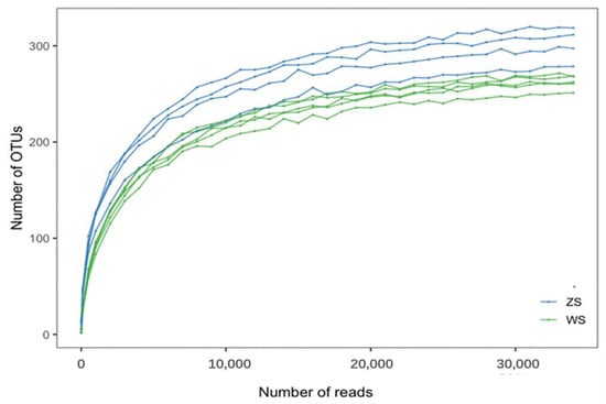
Figure 1.
Dilution curves of different samples before and after discoloration of rubber wood. Note: The blue line indicates the rubberwood sample after discoloration, the green line indicates the rubberwood sample before discoloration. zs: discolored rubber wood samples, ws: undiscolored rubber wood samples.
3.2. Changes in Fungal Diversity before and after Discoloration of Rubber Wood
The abundance rank curves of the samples were obtained by arranging the OTUs in the samples in order of their abundance along the horizontal coordinates, and the respective abundance values were used as the vertical coordinates to reflect the distribution pattern of OTU abundance in each sample. In the horizontal direction, species richness is reflected by the width of the curve, so, the higher the species abundance, the greater the range of the curve on the horizontal axis. The species evenness is measured by the slope, so, the flatter the slope, the higher the evenness []. It can be found that the span of discolored rubber wood samples was larger and the fold line was flat compared with that of un-discolored rubber wood samples, indicating that the uniformity and richness of surface microbial species distribution of discolored rubber wood were higher than that of un-discolored rubber wood (Figure 2).
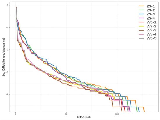
Figure 2.
Grade abundance curves of different samples before and after discoloration of rubber wood. Note: zs: discolored rubber wood samples, ws: undiscolored rubber wood samples.
Chao1 is a strain richness index, commonly used to estimate the total number of species; the Simpson index is used to estimate one of the microbial diversity indices, and the Shannon index indicates the structure of the community, originally proposed to understand community structure, and takes into account the relative abundance of different species []; these two diversity indices combine the evaluation of richness and evenness, taking into account both the richness of species and evenness. The Shannon index emphasizes the richness and rare cover types, while the Simpson index emphasizes more on evenness and dominant cover types [,]; the coverage index is used to reflect the cover of this species; PD (phylogenetic diversity) refers to phylogenetic diversity, which is the most commonly used indicator in the study of the evolutionary history of biodiversity and is the sum of branch lengths of all species in the phylogenetic tree in a region [].
The larger the Chao1 index, Simpson index, and Shannon index, the greater the abundance and diversity of the fungal flora; the coverage rate can assess the degree of sample sequence coverage in the library of the sample [].
The average species diversity index of the fungi in each sample was expressed by the alpha diversity index, observed species, Chao1, Shannon, Simpson, coverage, and PD as shown in Table 2, with a coverage of 0.99 or more for all samples, indicating a low probability of sequences not being detected in the samples. The Chao1 index, Shannon index, and Simpson index before discoloration were significantly lower than those after discoloration, indicating that the discoloration fungi were absolutely dominant in rubber wood after discoloration, and had a certain inhibitory effect on other fungi, and the species diversity and a total number of species were increased.

Table 2.
Statistics of data processing results of sample ITS sequencing.
The samples before and after discoloration contained a total of 538 fungal OTU, 193 fungal species in the discolored samples, and 101 fungal species in the undiscolored samples, indicating that the fungal species changed significantly before and after the discoloration of rubber wood, which is basically consistent with the results in Table 2 (Figure 3).
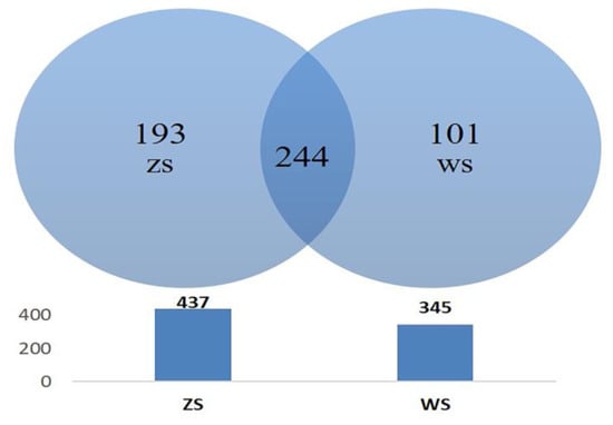
Figure 3.
Fungal OTU Venn diagram of rubberwood samples before and after discoloration.
3.3. Changes in Fungal Community Composition of Rubber Wood Samples before and after Discoloration
The top 10 species in terms of maximum abundance at each taxonomic level in the class, order, family, and genus were selected based on the OTU abundance in the samples, and cumulative histograms were generated for variance analysis in order to view and compare the species with higher abundance at different taxonomic levels in each sample and their proportions (Figure 4). As shown in Table 3, the dominant species and their abundance at each taxonomic level, the fungal communities of rubber wood before and after discoloration were dominant at the level of phylum Ascomycota, with an abundance of 97.06% and 93.82%, respectively. At the order level, the species communities of rubberwood before and after discoloration then differed significantly; the dominant fungal order before discoloration was Diaporthales (84.1%), while after discoloration it was Microascales (40.3%) and Hypocreales (26.23%). At the genus level, the dominant fungal genus of undiscolored rubberwood was Phomopsis (84.05%), and after discoloration was Huntiella (21.75%), Ceratocystis (18.54%) and Acremonium (13.88%).
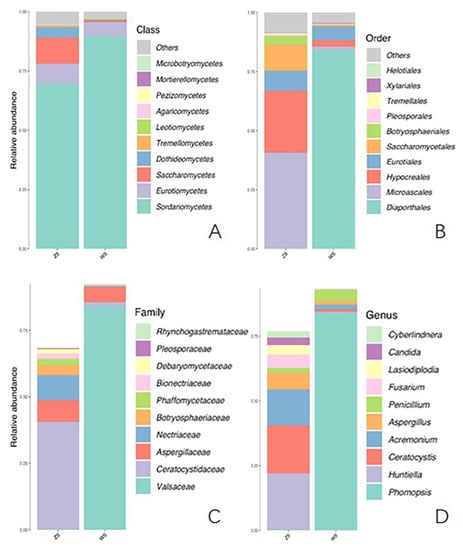
Figure 4.
Relative abundance of fungi in rubber wood at different taxonomic levels before and after discoloration histogram. ((A) is the relative abundance map at the class level, (B) is the relative abundance map at the order level, (C) is the relative abundance map at the family level, and (D) is the relative abundance map at the genus level.).

Table 3.
Dominant species at each level and their abundance in each sequenced sample.
Heat map cluster analysis is a development of general cluster analysis, which generally focuses on only one grouping and cannot explain the nature and basis of the grouping, while heat map cluster analysis can combine two groupings in one dimension []. The different OTUs were clustered in blocks to reflect the similarities, differences and clustering relationships of fungal communities in different samples before and after the discoloration of rubber wood based on the gradients of different colors in the heat map. Figure 5 shows the heat map of the top 50 OTUs based on relative abundance.
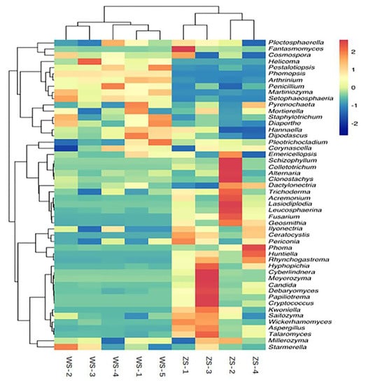
Figure 5.
Heat map of fungi in rubber wood at the genus level before and after discoloration. Note: The corresponding color of the heat map is mapped to the heat map matrix data; the depth of red inthe color of the rectangle in this figure indicates the overall high abundance of the genus in the sample, conversely, the depth of blue in the color of the rectangle indicates the overall low abundance of the genus in the sample.
3.4. Analysis of Fungal Differences between Rubber Wood Samples before and after Discoloration
PcoA (principal coordinates analysis), NMDS (non-metric multi-dimensional scaling) and cluster tree analysis based on OUTdata showed the relationship between the samples of rubber wood before and after discoloration, i.e., the closer the distance of fungal communities in rubber wood before and after discoloration, the higher the community similarity and the closer the clustering relationship. In the PCoA and NMDS plots (Figure 6), the positions of the unchanged rubberwood samples almost overlapped, while the positions of the changed rubberwood samples were relatively scattered. While the closer the samples were in the cluster dendrogram, the shorter the branch lengths were, indicating that the community structures of the two samples were more similar, and the branch lengths of the changed rubberwood samples were significantly longer compared to the unchanged rubberwood samples (Figure 7). This shows that the fungal community diversity of rubberwood increased after discoloration and the species diversity of rubberwood fungal communities varied greatly after discoloration.
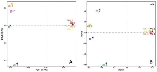
Figure 6.
PCoA plot (A) and NMDS plot (B) of fungi in rubber wood before and after discoloration.
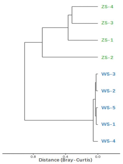
Figure 7.
Cluster analysis of fungi in each sample before and after discoloration of rubber wood.
4. Discussion
Given the diversity of sapstain fungi and the fact that some potential fungi used as sapstain fungal biocontrol agents do not work well in field trials, and the advantages of endophytic fungi that have evolved with trees over time and are adapted to certain resistant biomass inherent in trees and can interact directly with pathogens [,], we propose the theoretical hypothesis of using endophytic fungi of rubber wood to inhibit the growth of sapstain fungi. The vast majority of the fungi isolated and identified from undiscolored rubber wood are endophytic fungi, which are fungi that live inside healthy plant tissues and organs and have established a harmonious symbiotic relationship with the host plant in a long-term synergistic evolutionary process []. Some endophytic fungi can not only keep the host plant healthy by inhibiting and/or killing various pathogenic bacteria through competition or other effects but also produce secondary bioactive metabolic components with the same or similar activity as the host plant to synergize the host plant’s defense against pathogenic microorganisms [,]. Currently, flavonoids, terpenoids, alkaloids, steroids, peptides, cyclic peptides, and quinones have been isolated from the secondary metabolites of plant endophytic fungi, and these different types of compounds have various biological activities such as antibacterial, antitumor, and antifungal, which have wide application prospects in agriculture, medicine and other related fields with important ecological significance and have received wide attention from researchers worldwide []. Therefore, high-throughput sequencing technology was used to identify the endophytic fungi present in large numbers in undiscolored rubber wood and the dominant genera in discolored rubber wood to explore whether endophytic fungi that can be cultivated and can inhibit the growth of sapstain fungi can be isolated and screened for reference.
In this study, a total of 323,642 high-quality sequences and 538 OTUs were detected, including 193 color-changing rubberwoods and 101 uncolored rubberwoods, belonging to 297 species in 218 genera belonging to 5 phyla, 18 classes, 58 orders, 137 families. It can be found that the discoloration of rubberwood is closely related to the change of microbial community structure, the increase in the abundance of harmful microorganisms, and the decrease in the abundance of beneficial microorganisms after the felling of rubber trees. Compared with before color change, the composition and abundance of fungal communities of rubberwood changed after color change.
The results showed that the diversity of fungal communities in rubber wood samples changed in the process before and after discoloration. After the rubber tree felling, the fungi causing discoloration rapidly colonized and occupied absolute dominance on rubber wood, and had some inhibitory effect on other fungi, and the fungal diversity in rubber wood increased after discoloration. The differences in fungal alpha diversity indices in rubber wood samples before and after discoloration may be due to the different dominance of fungi with different nutritional and environmental preferences [].
The results of beta diversity analysis showed that the differences in fungal community diversity between the undiscolored rubber wood samples were small, while the differences in fungal communities between the discolored rubber wood samples were large, which also indicated that the fungi causing discoloration in rubber wood were more diverse. In addition, based on the community composition analysis, it was found that the dominant fungi in rubber wood changed relatively significantly before and after discoloration. At the genus level, the dominant fungal genus on undiscolored rubber wood was Phomopsis, while the dominant fungal genera on discolored rubber wood were Huntiella, Ceratocystis, and Acremonium, suggesting that fungi of these three genera may play a crucial role in rubber wood discoloration. These findings are consistent with previous studies, such as those showing that some species of Ceratocystis of the subphylum Cysticercus are the most common to cause discoloration of coniferous and broadleaf timber [], and in Canada, sapstain fungi of the genus Ceratocystis cause discoloration of sapwood in American poplar logs []. Fojutowski found the blue-stain fungus Ceratocystis imperfecta Mill. et Grenz not only caused severe surface discoloration of Pinus sylvesfris L. wood, but also caused different degrees of damage to physical properties such as wood strength, rigidity, and density []. Species of Huntiella produces spots on the stems of inoculated eucalyptus seedlings and is generally considered less pathogenic than Ceratocystis []. Huntiella spp. areusually considered to be related to wood saprobes or weak pathogens associated with relatively-minor lesions or sapstain of timber []. Acremonium is predominantly saprophytic and some species are capable of infesting plants under suitable conditions leading to disease []. Acremonium has become a common fungus in the inter-root soil of ginseng suffering from root rot, and it also shows as a dominant group when isolated from some medicinal plants such as species of Cordyceps [].
Author Contributions
Conceptualization, S.Y. and X.W.; methodology, S.Y. and L.L.; software, S.Y.; validation, S.Y. and Y.Y.; formal analysis, S.Y.; investigation, S.Y., X.W. and L.Q.; resources, S.Y. and J.Q.; data curation, S.Y. and X.W.; writing—original draftpreparation, S.Y.; writing—review and editing, S.Y. and L.Q.; visualization, S.Y. and L.L.; supervision, J.Q.; project administration, L.Q.; funding acquisition, L.Q. All authors have read and agreed to the published version of the manuscript.
Funding
The research was supported by the National Natural Science Foundation of China (Project No. 32160347), The Open Project of Key Laboratory of Southwest Mountain Forest Resources Conservation and Utilization, Ministry of Education, Southwest Forestry University (KLESWFU-202004), Yunnan Provincial Education Department Scientific Research Fund Project (2023Y0694).
Institutional Review Board Statement
Not applicable.
Data Availability Statement
The datasets presented in this study can be found in online repositories. The names of the repository/repositories and accession number(s) can be found below: SRA database and PRJNA940689.
Conflicts of Interest
The authors declare no conflict of interest.
References
- Killmann, W.; Hong, L.T. Rubberwood—The successof an agricultural by-product. Unasylva 2000, 51, 66–72. [Google Scholar]
- Ping, L.J.; Sun, B.L.; Shao, L.L.; Chai, Y.B.; Liu, J.L. Effect of Melamine-Urea-Glyoxal Resin on Physical and Mechanical Properties of Rubberwood. Wood Sci. Technol. 2021, 35, 38–43. (In Chinese) [Google Scholar] [CrossRef]
- Balsiger, J.; Bahdon, J.; Whiteman, A. The Utilization, Processing and Demand for Rubberwood as a Source of Wood Supply; Forestry Policy and Planning Division: Rome, Italy, 2000. [Google Scholar] [CrossRef]
- Bruce, A.; Stewart, D.; Verrall, S.; Wheatley, R.E. Effect of volatiles from bacteria and yeast on the growth and pigmentation of sapstain fungi. Int. Biodeterior. Biodegrad. 2003, 51, 101–108. [Google Scholar] [CrossRef]
- Florence, E.J.M.; Gnanaharan, R.; Singh, P.A.; Sharma, J.K. Weight loss and cell wall degradation in rubberwood caused by sapstain fungus Botryodiplodia theobromae. Holzforschung 2002, 56, 225–228. [Google Scholar] [CrossRef]
- Kidd, G.J.D. CCA-treated lumber poses danger fromarsenic and chromium. Pestic. You 2001, 21, 13–15. [Google Scholar]
- Suprapta, D.N. Potential of microbial antagonists as biocontrol agentsagainst plant fungal pathogens. J. ISSAAS 2012, 18, 1–8. [Google Scholar]
- Sajitha, K.L.; Dev, S.A.; Maria Florence, E.J. Biocontrol potential of Bacillus subtilis B1 against sapstain fungus in rubber wood. Eur. J. Plant Pathol. 2017, 150, 237–244. [Google Scholar] [CrossRef]
- Behrendt, C.J.; Blanchette, R.A. Biological processing of Pine Logs for Pulp and Paper Production with Phlebiopsis gigantea. Appl. Environ. Microbiol. 1997, 63, 1995–2000. [Google Scholar] [CrossRef]
- Behrendt, C.J.; Blanchette, R.A.; Farrell, R.L. Biological control of blue-stain fungi in wood. Phytopathology 1995, 85, 92–97. [Google Scholar] [CrossRef]
- Veenin, T.; Veenin, A.; Denrungruang, P. Biological control of stain fungus in rubberwood (Hevea brasiliensis Muell Arg) by Trichoderma sp. Thail. J. For. 1999, 18, 73–86. [Google Scholar]
- Sajitha, K.L.; Florence, E.J.M. Effects of Streptomyces sp. on growth of rubberwood sapstain fungus Lasiodiplodia theobromae. J. Trop. For. Sci. 2013, 25, 393–399. [Google Scholar]
- Held, B.W.; Thwaites, J.M.; Farrell, R.L.; Blanchette, R.A. Albino Strains of Ophiostoma Species for Biological Control of Sapstaining Fungi. Holzforschung 2003, 57, 237–242. [Google Scholar] [CrossRef]
- Hawksworth, D.L. The fungal dimension of biodiversity: Magnitude, significance, and conservation. Mycol. Res. 1991, 95, 641–655. [Google Scholar] [CrossRef]
- Kaeberlein, T.; Lewis, K.; Epstein, S.S. Isolating “Uncultivable” Microorganisms in Pure Culture in a Simulated Natural Environment. Science 2002, 296, 1127–1129. [Google Scholar] [CrossRef] [PubMed]
- Li, Y.; Xu, X.X. Research progress of high-throughput sequencing technology. China Med. Eng. 2019, 27, 32–37. [Google Scholar] [CrossRef]
- Kistler, L. Ancient DNA Extraction from Plants. In Ancient DNA: Methods and Protocols; Shapiro, B., Hofreiter, M., Eds.; Methods in Molecular Biology; Humana Press: Totowa, NJ, USA, 2012; Volume 840, pp. 71–79. [Google Scholar] [CrossRef]
- Siregar, I.Z.; Ramdhani, M.J.; Karlinasari, L.; Adzkia, U.; Arifin, M.Z.; Dwiyanti, F.G. DNA isolation success rates from dried and fresh wood samples of selected 20 tropical wood tree species for possible consideration in forensic forestry. Sci. Justice 2021, 61, 573–578. [Google Scholar] [CrossRef] [PubMed]
- Mundy, D.C.; Vanga, B.R.; Thompson, S.; Bulman, S. Assessment of Sampling and DNA Extraction Methods for Identification of Grapevine Trunk Microorganisms Using Metabarcoding. N. Z. Plant Prot. 2018, 71, 10–18. [Google Scholar] [CrossRef]
- Guo, Z.Q.; Wang, B.; Lu, J.Z.; Li, C.Y.; Kuang, L.D.; Tang, X.X.; Mei, X.L.; Xie, X.H. Analysis of the relationship between caecal flora difference and production performance of two rabbit species by high-throughput sequencing. Czech J. Anim. Sci. 2021, 66, 271–280. [Google Scholar] [CrossRef]
- Magoc, T.; Salzberg, S.L. FLASH: Fast length adjustment of short reads to improve genome assemblies. Bioinformatics 2011, 27, 2957–2963. [Google Scholar] [CrossRef] [PubMed]
- Baldrian, P.; Větrovský, T.; Lepinay, C.; Kohout, P. High-throughput sequencing view on the magnitude of global fungal diversity. Fungal Divers. 2021, 114, 539–547. [Google Scholar] [CrossRef]
- Shang, Z.D.; Tan, Z.K.; Kong, Q.H.; Shang, P.; Wang, H.H.; Zhaxi, W.J.; Zhaxi, C.; Liu, S.Z. Characterization of fungal microbial diversity in Tibetan sheep, Tibetan gazelle and Tibetan antelope in the Qiangtang region of Tibet. Mycoscience 2022, 63, 156–164. [Google Scholar] [CrossRef]
- Avolio, M.L.; Carroll, I.T.; Collins, S.L.; Houseman, G.R.; Hallett, L.M.; Isbell, F.; Koerner, S.E.; Komatsu, K.J.; Smith, M.D.; Wilcox, K.R. A comprehensive approach to analyzing community dynamics using rankabundance curves. Ecosphere 2019, 10, e02881. [Google Scholar] [CrossRef]
- Nguyen, T.V.; Cho, W.S.; Kim, H.; Jung, H.; Kim, Y.K.; Chon, T. Inferring community properties of benthic macroinvertebrates in streams using Shannon index and exergy. Front. Earth Sci. 2014, 8, 44–57. [Google Scholar] [CrossRef]
- Nagendra, H. Opposite trends in response for the Shannon and Simpson indices of landscape diversity. Appl. Geogr. 2022, 22, 175–186. [Google Scholar] [CrossRef]
- Zhang, M.; Yang, M.; Shi, Z.; Gao, J.; Wang, X. Biodiversityand Variations of ArbuscularMycorrhizal Fungi Associated withRoots along Elevations in Mt. Taibaiof China. Diversity 2022, 14, 626. [Google Scholar] [CrossRef]
- Faith, D.P. Conservation evaluation and phylogenetic diversity. Biol. Conserv. 1992, 61, 1–10. [Google Scholar] [CrossRef]
- Patrick, D.S.; Sarah, L.W.; Thomas, R.; Justine, R.H.; Martin, H.; Emily, B.H.; Ryan, A.L.; Brian, B.O.; Donovan, H.P.; Courtney, J.R.; et al. Introducing mothur: Open Source, Platform Independent, Community Supported Software for Describing and Comparing Microbial Communities. Appl. Environ. Microbiol. 2009, 75, 7537–7541. [Google Scholar] [CrossRef]
- Sakinah, A.I.; Musa, Y.; Farid, M.; Anshori, M.F.; Arifuddin, M.; Laraswati, A.A. Cluster heatmap for screening the drought tolerant rice through hydroponic culture. IOP Conf. Ser. Earth Environ. Sci. 2021, 807, 042045. [Google Scholar] [CrossRef]
- Seifert, K.A.; Breuil, C.; Rossignol, L.; Best, M.; Saddler, J.N. Screening for microorganisms with the potential forbiological control of sapstain on unseasoned lumber. Mater. Org. 1988, 23, 81–95. [Google Scholar]
- Lu, H.; Zou, W.X.; Meng, J.C. New bioactive metabolites produced by Colletorichum sp. an endophytic fungus in Artemisia annua. Plant Sci. 2000, 151, 67–73. [Google Scholar] [CrossRef]
- Arnold, A.E.; Mejia, L.C.; Kyllo, D. Fungal endophytes limit pathogen damage in a tropical tree. Proc. Nat. Acad. Sci. USA 2003, 100, 15649–15654. [Google Scholar] [CrossRef]
- Wiyakrutta, S.; Sriubolmas, N.; Panphut, W. Endophytic fungi with anti-microbial, anti-cancer andanti-malarial activities isolated from Thai medicinal plants. World J. Microbiol. Biotechnol. 2004, 20, 265–272. [Google Scholar] [CrossRef]
- Bamisile, B.S.; Dash, C.K.; Akutse, K.S.; Keppanan, R.; Afolabi, O.G.; Hussain, M.; Qasim, M.; Wang, L. Prospects of endophytic fungal entomopathogens as biocontrol and plant growth promoting agents: An insight on how artificial inoculation methods affect endophytic colonization of host plants. Microbiol. Res. 2018, 217, 34–50. [Google Scholar] [CrossRef] [PubMed]
- Wang, Z.C.; Wang, L.; Pan, Y.P.; Zheng, X.X.; Liang, X.N.; Sheng, L.L.; Zhang, D.; Sun, Q.; Wang, Q. Research advances on endophytic fungi and their bioactive metabolites. Bioprocess Biosyst. Eng. 2023, 46, 165–170. [Google Scholar] [CrossRef] [PubMed]
- Ren, C.; Liu, W.; Zhao, F.; Zhong, Z.; Deng, J.; Han, X.; Yang, G.; Feng, Y.; Ren, G. Soil bacterial and fungal diversity and compositions respond differently to forest development. Catena 2019, 181, 104071. [Google Scholar] [CrossRef]
- Payne, C.J.; Woodward, S.; Petty, J.A. Fungal spore of softwood timber drying kilns in Scotland. Mater. Org. 1998, 32, 109–125. [Google Scholar]
- Hiratsuka, Y.; Chakravarty, P. Role of Phialemoniumcurvatumas a potential biological control agent against a blue stain fungus on aspen. Eur. J. For. Pathol. 1999, 29, 305–310. [Google Scholar] [CrossRef]
- Fojutowski, A. The Selected Properties of Scots Pine Wood Blue-Stained by Fungus Cladosporium herbarum; Document No. IRG/WP/10484; International Research Group on Wood Preservation: Stockholm, Sweden, 2003. [Google Scholar]
- Liu, F.F.; Chen, S.F. A repertory of new species of Ceratocystis and Huntiella fungi from China:2014–2020. Eucalyptus Technol. 2021, 38, 62–72. [Google Scholar] [CrossRef]
- Liu, F.; Marincowitz, S.; Chen, S.; Mbenoun, M.; Tsopelas, P.; Soulioti, N.; Wingfield, M.J. Novel species of Huntiella from naturally-occurring forest trees in Greece and South Africa. MycoKeys 2020, 69, 33–52. [Google Scholar] [CrossRef]
- Mirtalebi, M.; Banihashemi, Z.; Sabahi, F.; Mafakheri, H. Dieback of rose caused by Acremonium sclerotigenum as a new causal agent of rose dieback in Iran. Span. J. Agric. Res. 2016, 14, e10SC03. [Google Scholar] [CrossRef]
- Mu, D.Y.; Lv, G.Z.; Sun, X.D. Two new Chinese record species isolated from the inter-root soil of medicinal plants. Mycol. Res. 2012, 10, 1–3. [Google Scholar] [CrossRef]
Disclaimer/Publisher’s Note: The statements, opinions and data contained in all publications are solely those of the individual author(s) and contributor(s) and not of MDPI and/or the editor(s). MDPI and/or the editor(s) disclaim responsibility for any injury to people or property resulting from any ideas, methods, instructions or products referred to in the content. |
© 2023 by the authors. Licensee MDPI, Basel, Switzerland. This article is an open access article distributed under the terms and conditions of the Creative Commons Attribution (CC BY) license (https://creativecommons.org/licenses/by/4.0/).