


Abstract
Three species, Calocybe lilacea, C. longisterigma, and C. subochraceus, were newly discovered in Inner Mongolia, Hunan, and Liaoning provinces of China. Calocybe lilacea is mainly characterized by its small-sized basidiocarp, brownish-orange pileus, lilac gray to dull violet stipe, and noncellular epicutis. The main characteristics of C. longisterigma are its light brown to brownish-orange pileus, long sterigmata, and noncellular epicutis. The main morphological features of C. subochraceus are its small-sized basidiocarp, pale-orange pileus, adnexed lamellae, tortuous stipe, and noncellular epicutis. These morphological features confirmed that the three species of Calocybe all belonged to Sect. Carneoviolaceae. Phylogenetic analysis based on a combined dataset (ITS–nrLSU–RPB2) determined that the three species belong to the genus Calocybe and form a distinct lineage. The morphological differences between the three new species and other related species of Calocybe are also discussed.
1. Introduction
Calocybe Kühner ex Donk (1962: 42), a well-known genus, was published as a valid name by Donk [1]. It was considered as a section of the genus Lyophyllum P. Karst at first [2,3]. On account of the brighter pileus of Calocybe species, Singer later proposed that Calocybe is an independent genus of Lyophyllaceae [4]. The species of Calocybe have characteristics of white to light-colored pileus, small spores, and pileipellis of a cutis or cellular type. On the basis of pileus color, spore surface, and hyphae of the epicutis, Singer divided Calocybe into four sections [4]. However, the classification opinion proposed by Singer has not been fully supported by molecular data. In recent years, many phylogenetic investigations on Lyophyllaceae indicated that Calocybe belongs to a monophyletic group [5,6,7,8,9,10,11,12,13,14]. However, the infrageneric classification system of Calocybe remains unclear. For instance, C. naucoria and C. chrysenteron are assigned to the same section, namely, Sect. Carneoviolaceae [4], but the two species belong to two distinct clades according to phylogenetic results [11,12,14].
The species of Calocybe have a widespread distribution all over the world. Currently, over 116 records have been listed in Index Fungorum (Available online: www.indexfungorum.org, accessed on 8 June 2022). In China, 16 species of Calocybe have been reported, most of which were discovered and described in the northern China [11,12,13,14,15,16,17,18,19,20]. In addition, several of these species are treated as edible by Dai et al. [21], including C. carnea (Bull.) Donk, C. gambosa (Fr.) Singer, and C. ionides (Bull.) Donk. To identify whether the three newly collected species of Calocybe are new to science, phylogenetic analysis and morphological feature determinations were conducted and discussed in the present study.
2. Material and Methods
2.1. Specimens and Morphological Study
All the collections used in this study were photographed in the field and deposited in the Fungal Herbarium of Shenyang Agricultural University (SYAU-FUNGI), with voucher collection numbers of SYAU-FUNGI-066 to SYAU-FUNGI-071 (Table 1). Color abbreviations followed Kornerup and Wanscher [22]. Methods for morphological observation followed Li et al. [11] and Pei et al. [23].
Table 1.
Species names, voucher numbers, and GenBank accession numbers of the Calocybe specimens analyzed in the study.
2.2. DNA Extraction, PCR Amplification, and Sequencing
Total DNA was extracted from fresh tissue blocks using the cetyltrimethylammonium bromide (CTAB) method [23]. The universal primer pairs ITS5/ITS4 [24], LR0R/LR5 [25], and b6F/b7.1R [26] were used for amplification of the ITS region, nrLSU region, and RPB2, respectively. The PCR protocol followed that in Li et al. [11]. The products of PCR amplification were examined on a 1% agarose gel and visualized by the Gel Imaging System (Bio-Rad, SYSTEM GelDoc XR+ IMAGELAB, CA). The fragments used for analysis in this study were provided by BGI Co, Ltd., Beijing, China.
2.3. Phylogenetic Analyses
BLAST (Available online: www.blast.ncbi.nlm.nih.gov, accessed on 13 May 2022) was used to search for the related species of the three new species. Then, high-quality and representative sequences of Calocybe and related genera in former phylogenetic studies [7,11,12,13,14,27] were downloaded from GenBank in order to reconstruct the phylogenetic relationships of the genus Calocybe. The newly generated sequences from this study were aligned with those retrieved from GenBank using MAFFT v7.313 [28]. A data partition homogeneity test [29] was implemented in PAUP 4.0b4a [30]. The result detected no conflicts among ITS, nrLSU, and RPB2 regions (p-value = 0.8), suggesting that the three regions can be combined. ModelFinder [31] and PartitionFinder 2 [32] were used for the selection of the best-fitting evolution mode. Maximum likelihood (ML) and Bayesian inference (BI) analyses were performed following the method of Pei et al. [23]. Both BI and ML was conducted under GTR mode, and the BI of the combined datasets was run for 8,000,000 generations. The best tree was viewed in FIGTREE v1.4.4 [33] and was compiled in Adobe Illustrator CC.
3. Results
Molecular Phylogenetic Inference
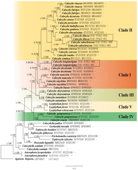
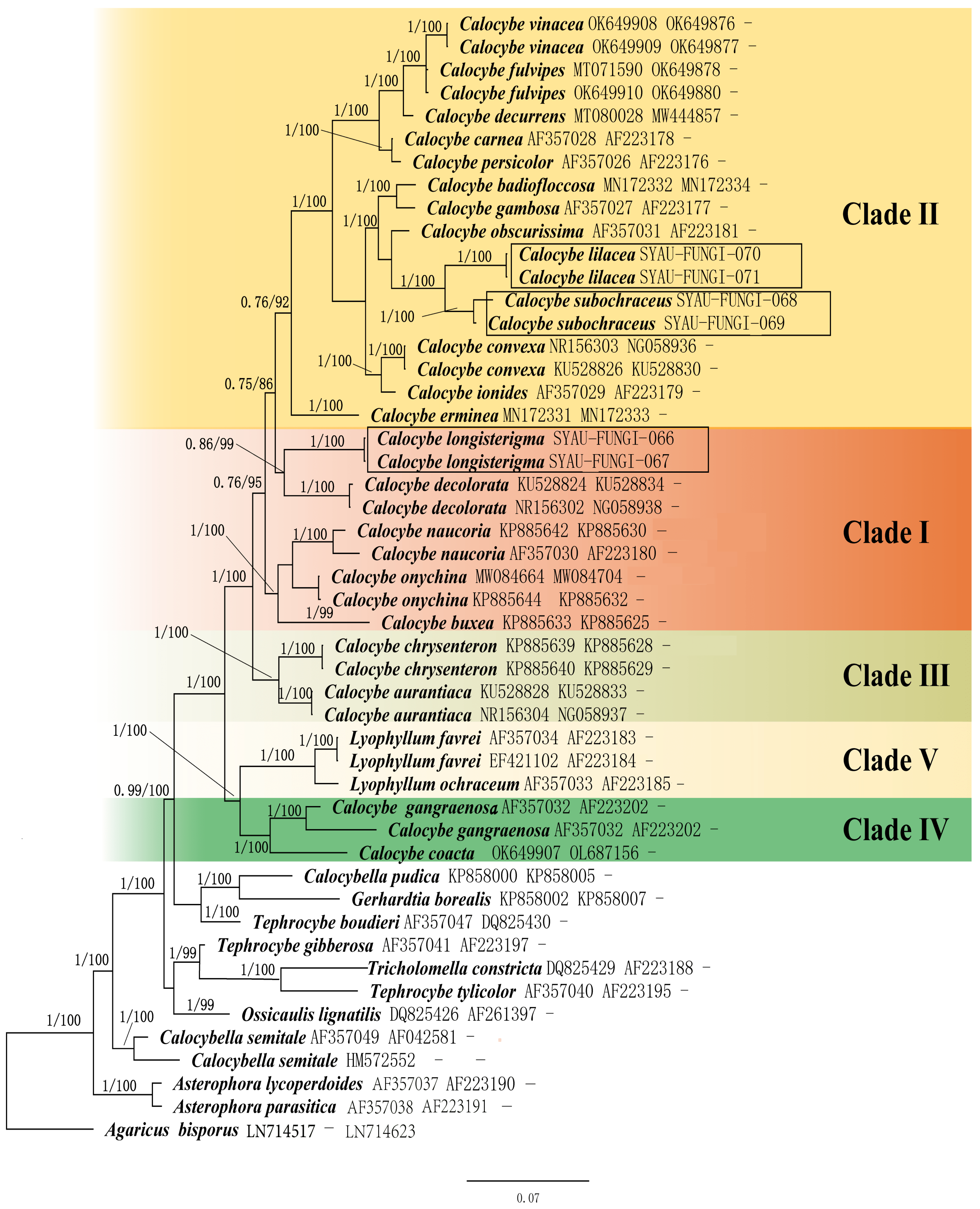
The dataset for phylogeny analysis consisted of three gene regions from 46 samples representing 33 species, and Agaricus bisporus (J.E. Lange) Imbach was selected as an outgroup in this study. All the sequences used in this study are listed in Table 1. ML and BI resulted in almost identical tree topologies, and the BI tree was selected for display (Figure 1). The results suggested that the species of Calocybe formed a distinct group supported by a strong value (1.00 PP and 100% BS), corroborated by former research [5,6,7,8,9,10,11,12,13,14]. A total of five clades can be viewed within species of Calocybe, which is in line with Li et al. [11]. Calocybe lilacea and C. subochraceus formed an independent lineage supported by a strong value (1.00 PP and 100% BS), located within clade II. Moreover, the two new species gathered in a separate lineage with high support (1.00 PP and 100% BS). In clade I, C. longisterigma formed a single clade with high support (1.00 PP and 100% BS), sister to the clade containing C. decolorata (0.86 PP and 99% BS).
Figure 1.
Bayesian phylogenetic analysis of Calocybe based on the combined ITS–nrLSU–RPB2 sequences. Node support (PP ≥ 0.90 and BS ≥ 75%) is annotated on the branches. The three new species and corresponding voucher numbers are shown in boxes.
4. Taxonomy
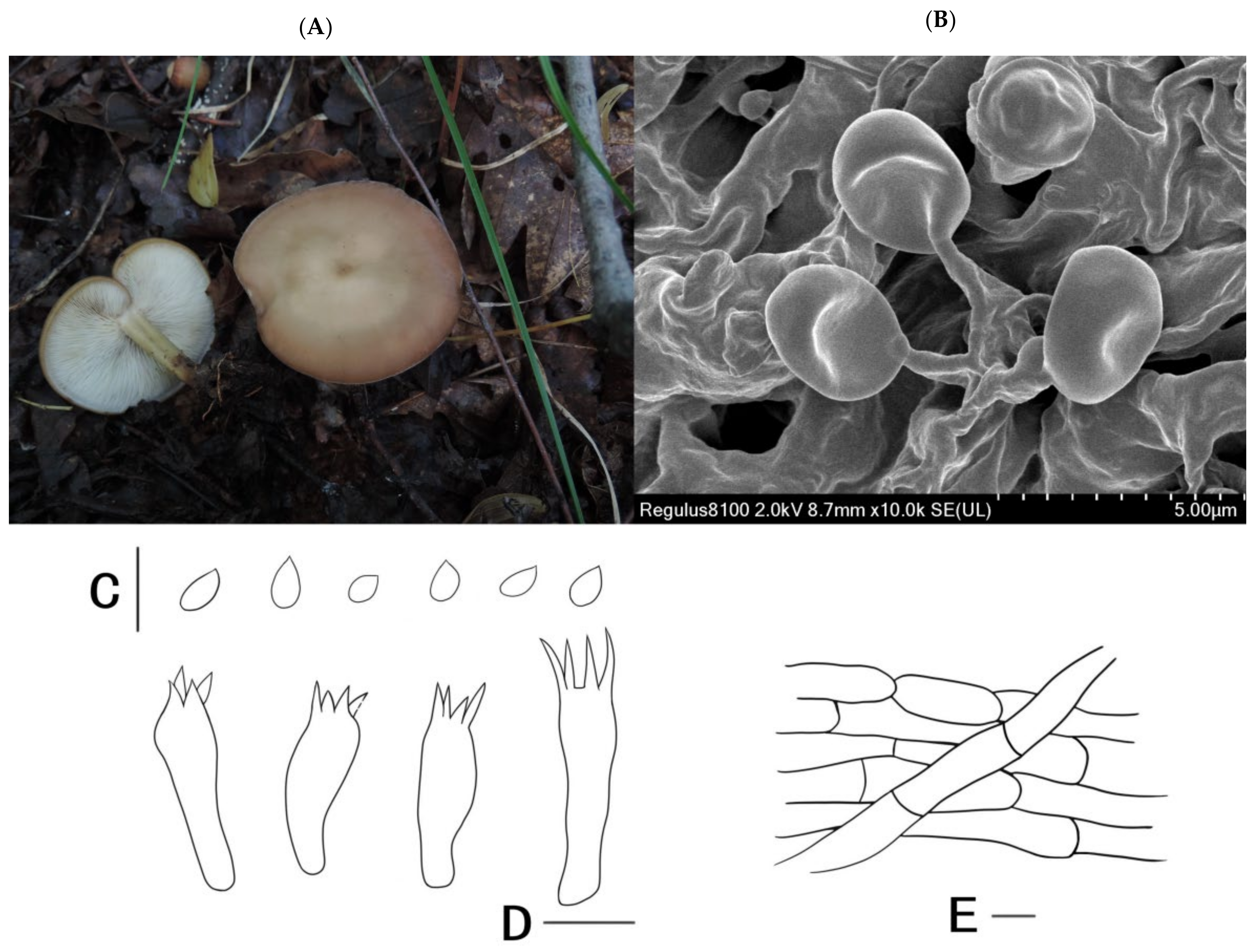
Calocybe lilacea X.D. Yu, Y. Zhou & W.Q. Qin, sp. nov. MycoBank No. MB 843876 (Figure 2A–E).
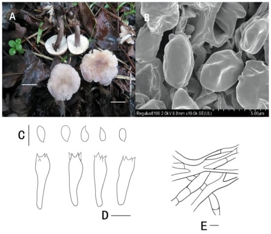
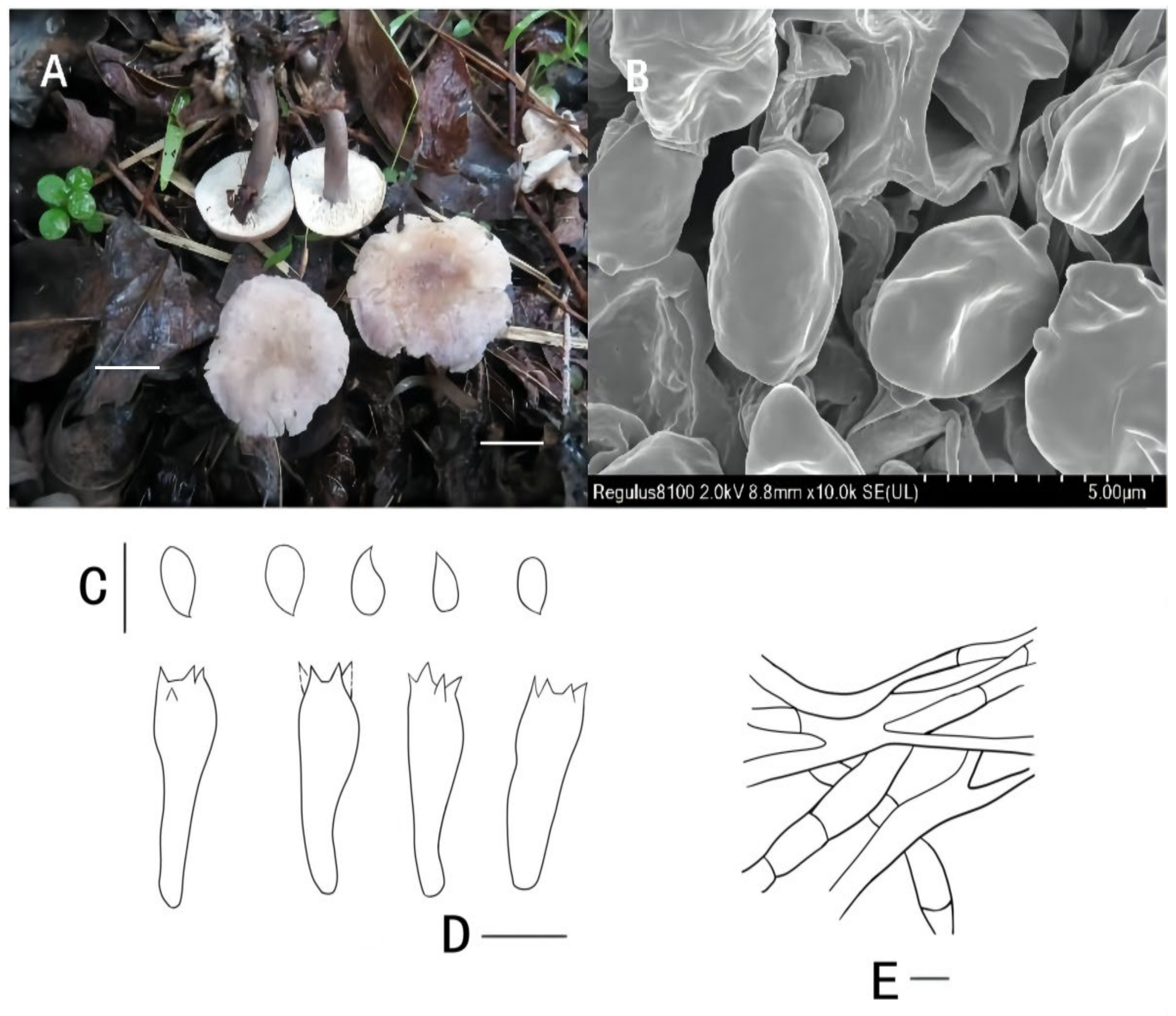
Figure 2.
Calocybe lilacea (holotype, SYAU-FUNGI-070): (A) macroscopic habitat and basidiomata; (B) surface of basidiospores; (C) basidiospores; (D) basidia; (E) pileipellis. Scale bars: (A) 1 cm; (B–E) 5 μm.
Etymology: The epithet “lilacea” refers to the lilac color of the stipe.
Holotype: China. Hunan Province: Zhangjiajie, Jishou University, on the soil, on 26 November 2019, W.Q. Qin (holotype: SYAU-FUNGI-070).
Description: Basidiomata slightly small-sized. Pileus 1.5–3.0 cm in diameter, convex at first, soon becoming plano-convex, surface hygrophanous, light brown (7D4 to 7D5) at center, becoming paler toward the margin, margin brownish orange (7C3) to reddish gray (7B2), slightly incurved at first, soon becoming extended and sometimes irregular. Lamellae 1.0–2.0 mm broad, moderately crowded, adnate, white, with lamellulae, edges concolorous and denticulate. Stipe 1.5–2.5 cm long × 0.3–0.5 cm diameter, central, stuffed, equal or slightly attenuated upward, surface lilac gray to dull violet (16C2 to 17E3). Pileus context 0.2–0.3 cm thick, white to creamy. Odor faint. Spore deposit white.
Basidiospores (3.1) 3.4–4.5 (4.9) × (2.7) 3.2–3.7 (4.1) μm, Q = (1.37) 1.40–1.51 (1.56) μm (n = 30), average Q = 1.44, subglobose, hyaline, inamyloid, smooth. Basidia (17.0) 18.0–20.5 (21.0) × (4.0) 4.5–7.0 (8.0) μm, clavate, four-spored, hyaline, siderophilous granules observed. Hymenial cystidia absent. Hymenophoral trama 90–130 μm thick, regular, consisting of 2.0–5.0 μm wide, thin-walled, hyaline hyphae. Subhymenium 18–26 μm thick. Pileipellis and cutis of dense, subparallel, repent branched hyphae, hyphae 2.4–4.8 μm wide, thin-walled, pigmented. Stipitipellis hyphae 3.4–8.6 μm wide, thin-walled, hyaline. Caulocystidia absent. Clamp connections absent.
Ecology and distribution: Saprophytic on soil by forests or roads. Known from Hunan province.
Additional specimen examined: CHINA. Hunan Province: Zhangjiajie, Jishou University, on the soil, on 26 November 2019, W.Q. Qin (SYAU-FUNGI-071).
Remarks:Calocybe lilacea is mainly characterized by its small basidiomata, brownish orange pileus, adnate and white lamellae, lilac gray to dull violet stipe, smooth basidiospores, and noncellular epicutis. On the basis of Singer’s taxonomical opinion and these morphological characteristics, Calocybe subochraceus is assigned to Sect. Carneoviolaceae [4]. The lilac gray to violet stipe also appears in other three Calocybe species of the section, i.e., C. africana, C. cyanella, and C. ionides. However, Calocybe lilacea features a brownish pileus, which is different from the three species with purple tones pileus [34,35,36]. Furthermore, C. decurrens is similar to C. lilacea in having a violet stipe when young [14]; however, C. decurrens is characterized by a much larger pileus up to 105 mm in diameter and much longer spores up to 9.3 μm in length [14].
Calocybe longisterigma X.D. Yu, Y. Zhou & H.B. Guo, sp. nov. MycoBank No. MB 843877 (Figure 3A–E).
Figure 3.
Calocybelongisterigma (holotype, SYAU-FUNGI-066): (A) macroscopic habitat and basidiomata; (B) surface of basidiospores; (C) basidiospores; (D) basidia; (E) pileipellis. Scale bars: (A) 1 cm; (B–E) 5 μm.
Etymology: The epithet “longisterigma” refers to the species characterized by long sterigmata.
Holotype: China. Inner Mongolia: Chifeng, Linxi County, Xinlin Town, Dauran Village, on the soil, on 22 July 2017, X.D. Yu (holotype: SYAU-FUNGI-066).
Description: Basidiomata medium-sized. Pileus 2.5–6.5 cm broad, convex to plano-convex at first, then gradually becoming flat, surface silky, hygrophanous, brownish orange (5C4 to 6C3) to grayish orange (6B3), often becoming light brown (7D4 to 7D5) at margin; edge entire. Lamellae 1.0–3.0 mm broad, adnate to decurrent, rather crowded, white, with lamellulae; edge entire, concolorous with the sides. Stipe 2.0–3.5 cm long × 0.3–0.5 cm diameter, central, stuffed, equal or and slightly attenuated downward; surface glabrous, non-striate or indistinctly striate, white at apex and light brown at base when young, brownish orange (5C4 to 5C5) at apex and brown at base (6D4 to 6D5) when mature, with whitish basal tomentum. Pileus context up to 0.3–0.6 cm thick, white to cream. Odor faint. Spore deposit white.
Basidiospores (2.1) 2.6–3.4 (3.9) × 1.7–2.6 (3.2) μm, Q = (1.39) 1.42–1.49 (1.52) μm (n = 30), average Q = 1.47, subglobose to ellipsoid, hyaline, inamyloid, smooth. Basidia (12) 13.0–16.0 (18.0) × (3.3) 3.6–5.4 (6.0) μm, clavate, four-spored, long sterigmata up to 3.8–8.2 μm, hyaline, siderophilous granules observed. Any type of cystidia absent. Hymenophoral trama 190–220 μm thick, regular, consisting of 2.0–4.0 μm wide, thin-walled, hyaline hyphae. Subhymenium 24–35 μm thick. Pileipellis and cutis of dense, subparallel, repent branched hyphae, hyphae 3.0–5.5 μm wide, thin-walled, sometimes pigmented. Stipitipellis hyphae 4.4–8.5 μm wide, thin-walled, hyaline. Caulocystidia absent. Clamp connections absent.
Ecology and distribution: Saprophytic on the grass. Known from Inner Mongolia.
Additional specimen examined: China. Inner Mongolia: Chifeng, Linxi County, Xinlin Town, Dawulan village, on the grass in woods, on 22 July 2017, H.B. Guo (SYAU-FUNGI-067).
Remarks: The main characteristics of Calocybe longisterigma are brownish orange to grayish orange pileus, adnate to decurrent lamellae, small basidiospores, long sterigmata, and non-cellular epicutis. According to Singer’s concept of infrageneric classification [4], it should be classified in Sect. Carneoviolaceae. In this section, Calocybe longisterigma resembles C. carnea, C. decurrens, and C. fulvipes because these members of this section all have carneous or browish pileus. The long sterigmata up to 8.2 μm and small basidiospores (2.6–3.4 × 1.7–2.6 μm) of C. longisterigma can distinguish it from these three species. Moreover, C. decurrens can be differentiated by its pastel violet stipe at first [14]. Calocybe carnea differs from C. longisterigma by its adnexed gills and white stipe [37]. Calocybe fulvipes has adnexed to emarginate lamellae, while C. longisterigma features adnate to decurrent lamellae, which can separate the two species. In addition, Calocybe fulvipes produces grayish-orange lamellae when bruised, which is absent in C. longisterigma [12].
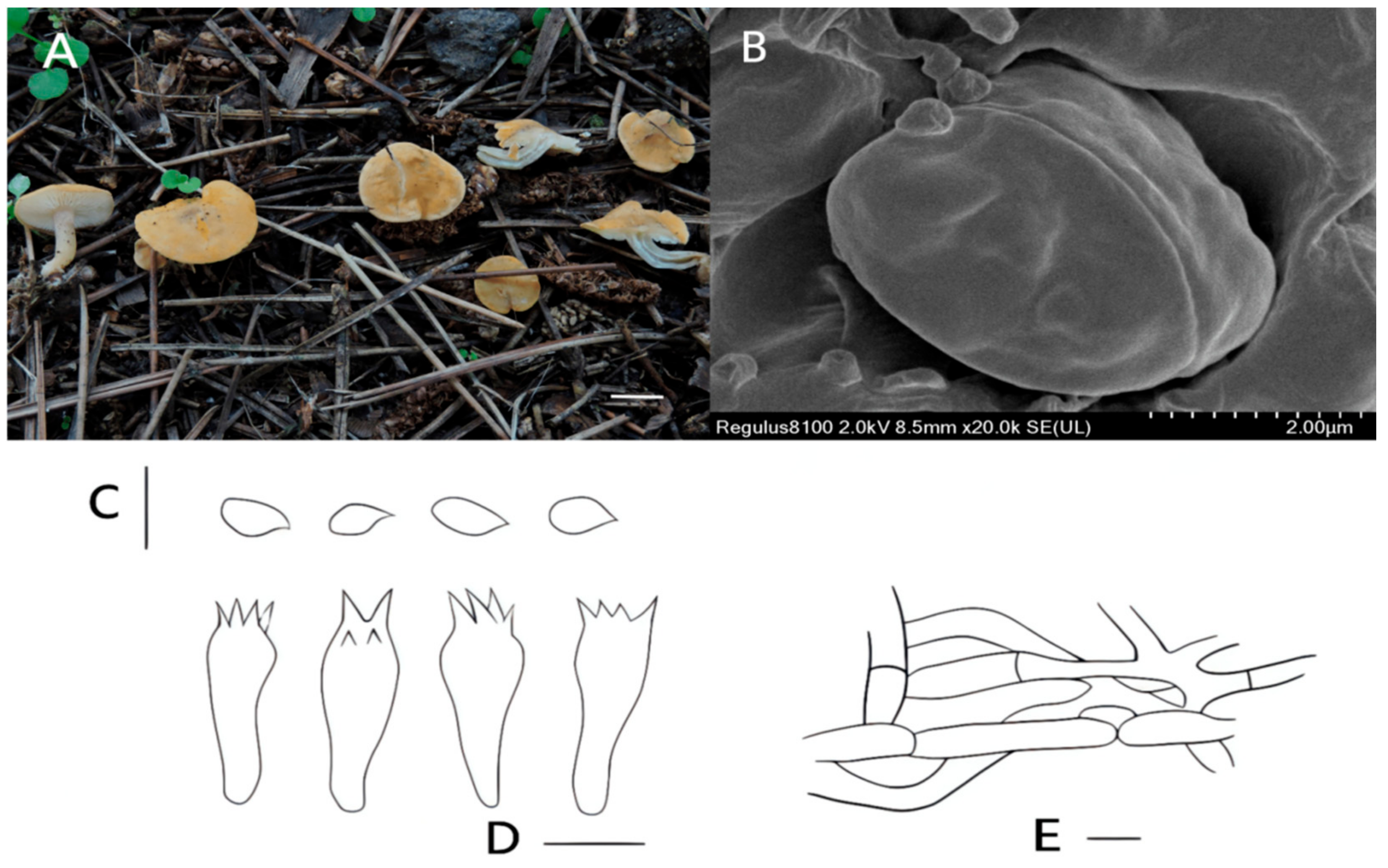
Figure 4.
Calocybe subochraceus (holotype, SYAU-FUNGI-068): (A) macroscopic habitat and basidiomata; (B) surface of basidiospores; (C) basidiospores; (D) basidia; (E) pileipellis. Scale bars: (A) 1 cm; (B) 2 μm; (C–E) 5 μm.
Holotype: China. Liaoning Province: Shenyang, Shenyang Agricultural University, on the grass in woods, on 21 July 2020, X.D. Yu (holotype: SYAU-FUNGI-068).
Etymology: The epithet “subochraceus” refers to the light-yellow color of the pileus, which is similar to the species Calocybe ochraceus.
Description: Basidiomata small-sized. Pileus 1.0–3.0 cm broad, convex to plano-convex at first, then becoming applanate, even slightly depressed at center with age; surface pale orange (5A3 to 5A5), often becoming paler orange/white (5A2) at margin; margin entire at first, then expanding to uplifted and flexuous with age. Lamellae 0.4–1.0 mm broad, adnexed, moderately close, white, with lamellulae; edge denticulate, concolorous with the sides. Stipe 1.6–2.3 cm long × 0.4–0.7 cm diameter, subcylindrical to flexuous, central, hollow, light brown to grayish brown (7D3 to 7D4) at first, then gradually becoming light orange to orange white to grayish orange (5A3, 5A2, 5B2 to 5B5), generally deeper in upper part, with whitish basal tomentum. Pileus context 0.3–0.5 cm thick, white. Odor faint. Spore deposit white.
Basidiospores (3.1) 3.3–4.5 (4.9) × (2.4) 2.9–3.6 (3.9) μm, Q = (1.49) 1.56–1.62 (1.64) μm (n = 30), average Q = 1.59, subglobose to elongate ellipsoid, hyaline, inamyloid, smooth. Basidia (15.5) 16.0–19.0 (20.0) × (5.0) 5.5–7.5 (8.0) μm, clavate, four-spored, subhyaline, siderophilous granules observed. Hymenial cystidia absent. Hymenophoral trama 100–160 μm thick, regular, consisting of 2.0–5.0 μm wide, thin-walled, hyaline hyphae. Subhymenium 17–24 μm thick. Pileipellis and cutis of dense, subparallel, repent branched hyphae, hyphae 2.6–4.9 μm wide, thin-walled, pigmented. Stipitipellis hyphae 2.6–7.8 μm wide, thin-walled, hyaline. Caulocystidia absent. Clamp connections absent.
Ecology and distribution: Saprophytic in small groups on soil in forests. Known from Liaoning province.
Additional specimen examined: China. Liaoning Province: Shenyang, Shenyang Agricultural University, on the grass, on 21 July 2020, X.D. Yu (SYAU-FUNGI-069).
Remarks: The main features of Calocybe subochraceus are its small basidiomata, pale orange pileus, white and adnexed lamellae, tortuous stipe, and noncellular epicutis. These key features suggest that C. subochraceus belongs to Sect. Carneoviolaceae [4]. Amongst this section, several members have similar orange pileus, including C. alpestris, C. carnea, and C. coniceps. However, C. alpestris (6.2–6.8 × 2.7–3.0 μm), C. coniceps (6.5–8.5 × 4.5–5.8 μm), and C. carnea (more than 5 μm long) are characterized by larger basidiospores [34,36], making them distinguishable from C. subochraceus. Furthermore, C. aurantiaca, C. convexa, and C. decolorata resemble C. subochraceus in having orange to yellow pileus. However, C. aurantiaca and C. decolorata are distinct from C. subochraceus in having decurrent gills. Calocybe convexa and C. decolorata have fusiform hymenial cystidia, which is absent in C. subochraceus.
5. Discussion
Calocybe lilacea, C. longisterigma, and C. subochraceus, newly discovered in Inner Mongolia, Hunan, and Liaoning provinces of China, respectively, were originally reported and described in detail in this study. Calocybe lilacea is mainly characterized by small-sized basidiomata and lilac gray to dull violet stipe. The main characteristics of C. longisterigma are brownish orange pileus, decurrent lamellae, small basidiospores, and long sterigmata. The key features of C. subochraceus are small basidiomata, pale orange pileus, white lamellae, and tortuous stipe. On account of the Singer’s infrageneric classification system, the three species of Calocybe are all assigned to the Sect. Carneoviolaceae [4]. Morphologically, unique characteristics of the three species of Calocybe can readily distinguish them from other related species, as elaborated above.
Phylogenetically, species of Calocybe appears to form a monophyletic group, which is consistent with previous studies [5,6,7,8,9,10,11,12,13,14]. Additionally, the three new species determined in this study occupy an independent position in the phylogenetic tree. Calocybe lilacea clusters in a single clade sister to the clade of C. longisterigma; however, the two species described in the study can be easily delimited using morphological methods. Calocybe lilacea has a smaller pileus and a lilac gray to dull violet stipe, but C. longisterigma has a slightly larger pileus and a light-brown stipe. Microscopically, the two Calocybe species can also be separated; Calocybe longisterigma is characterized by smaller basidiospores and much longer sterigmata, which is absent in C. lilacea. Calocybe subochraceus occupies an independent lineage sister to the clade containing C. decolorata, originally reported in northeastern China by Li et al. [11]. Calocybe decolorata has decurrent and blue lamellae when injured, which differs from C. subochraceus that has adnexed and unchanged lamellae when bruised. Additionally, C. decolorata has fusiform hymenial cystidia, which is absent in C. subochraceus.
In the phylogenetic analysis (Figure 1), five clades and 25 species were recognized within the genus Calocybe. The morphologically based infrageneric classification of Calocybe species by Singer [4] could not be fully supported by the results of our phylogenetic analyses. Except for C. onychina with an amaranthine pileus and C. longisterigma with a carneose pileus [38], the species of Clade I are characterized by a white to yellow pileus. The pileus color within Clade II varies, including white, yellow, brown, and violet shades [34,39]. In Clade III, C. chrysenteron and C. aurantiaca are yellow. Calocybe coacta and C. gangraenosa, located in Clade IV, have a white pileus. Clade V consists of C. ochracea and C. favrei, which have a gilded pileus [40]. Therefore, it is not appropriate to consider pileus color as the only criterion of the infrageneric classification of Calocybe, but it could provide taxonomic basis to some extent. The species of Calocybe within each phylogenetic clade lack uniform characteristics to work in identification. Further investigations and additional materials are still needed to reveal the infrageneric classification of Calocybe species.
Until now, a total of 19 species of Calocybe have been reported from China. On the basis of observations and the literature [11,12,13,14,18,39,40], a key for the Calocybe species from China is provided in Table 2.
Table 2.
A key to the Chinese species of Calocybe.
Author Contributions
Conceptualization, X.Y. and A.X.; methodology, Y.Z. and Y.Q.; software, Y.Z. and Y.Q; validation, X.Y. and Y.Q.; formal analysis, Y.Q; investigation, W.Q., K.B. and H.G.; resources, H.G. and W.Q.; data curation, Y.Z.; writing—original draft preparation, Y.Z. and Y.Q.; writing—review and editing, Y.Q. and X.Y.; visualization, Y.Z. and Y.Q.; supervision, X.Y.; project administration, X.Y.; funding acquisition, X.Y. and A.X. All authors have read and agreed to the published version of the manuscript.
Funding
This research was funded by Science and Technology Plan Project of Liaoning Province (2020-MZLH-33), the National Natural Science Foundation of China (No. 31770014), and research and development of wild macrofungal germplasm resources collection and preservation technology in Tibet Germplasm Resource Bank (ZDZ×2018000017).
Institutional Review Board Statement
Not applicable.
Informed Consent Statement
Not applicable.
Data Availability Statement
Data can be found within the manuscript.
Acknowledgments
The authors kindly thank Xin-Yu Ma and Jian-Xuan Hou for modifying the pictures in this study.
Conflicts of Interest
The authors declare no conflict of interest.
References
- Kühner, R. Utilisation du carmin acétique dans la classification des agarics leucosporés. Bull. Mens. Soc. Linn. Lyon 1938, 7, 204–212. [Google Scholar] [CrossRef] [Scilit]
- Kühner, R.; Romagnesi, H. Flore Analytique des Champignons Supérieurs (Agarics, Boelts, Chanterelles); Masson: Paris, France, 1953. [Google Scholar]
- Donk, M.A. The generic names proposed for Agaricaceae. Beih. Nova Hedwig. 1962, 5, 1–320. [Google Scholar]
- Singer, R. The Agaricales in Modern Taxonomy, 4th ed.; Koeltz Scientific Books: Koenigstein, Germany, 1986; pp. 1–981. [Google Scholar]
- Hofstetter, V.; Clémençon, H.; Vilgalys, R.; Moncalvo, J.M. Phylogenetic analyses of the Lyophylleae (Agaricales, Basidiomycota) based on nuclear and mitochondrial rDNA sequences. Mycol. Res. 2002, 106, 104–1059. [Google Scholar] [CrossRef] [Scilit]
- Hofstetter, V.; Redhead, S.A.; Kauff, F.; Moncalvo, J.M.; Matheny, P.B.; Vilgalys, R. Taxonomic revision and examination of ecological transitions of the Lyophyllaceae (Basidiomycota, Agaricales) based on a multigene phylogeny. Cryptogam. Mycol. 2014, 35, 399–425. [Google Scholar] [CrossRef] [Scilit]
- Moncalvo, J.M.; Vilgalys, R.; Redhead, S.A.; Johnson, J.E.; James, T.Y.; Aime, M.C.; Hofstetter, V.; Verduin, S.J.W.; Larsson, E.; Baroni, T.J.; et al. One hundred and seventeen clades of euagarics. Mol. Phylogenetics Evol. 2002, 23, 357–400. [Google Scholar] [CrossRef] [Scilit]
- Matheny, P.B.; Curtis, J.M.; Hofstetter, V.; Aime, M.C.; Moncalvo, J.M.; Ge, Z.W.; Yang, Z.L.; Slot, J.C.; Ammirati, J.F.; Baroni, T.J.; et al. Major clades of Agaricales: A multi-locus phylogenetic overview. Mycologia 2006, 98, 982–995. [Google Scholar] [CrossRef] [PubMed]
- Garnica, S.; Weiss, M.; Walther, G.; Oberwinkler, F. Reconstructing the evolution of agarics from nuclear gene sequences and basidiospore ultrastructure. Mycol. Res. 2007, 111, 1019–1029. [Google Scholar] [CrossRef] [Scilit]
- Bellanger, J.M.; Moreau, P.A.; Corriol, G.; Bidaud, A.; Chalange, R.; Dudova, Z.; Richard, F. Plunging hands into the mushroom jar: A phylogenetic framework for Lyophyllaceae (Agaricales, Basidiomycota). Genetica 2015, 143, 169–194. [Google Scholar] [CrossRef] [Scilit]
- Li, J.J.; Wu, S.Y.; Yu, X.D.; Zhang, S.B.; Cao, D.X. Three new species of Calocybe (Agaricales, Basidiomycota) from northeastern China are supported by morphological and molecular data. Mycologia 2017, 109, 55–63. [Google Scholar] [CrossRef] [Scilit] [PubMed]
- Xu, J.Z.; Yu, X.; Suwannarach, N.; Jiang, Y.; Zhao, W.; Li, Y. Additions to Lyophyllaceae s.l. from China. Fungi 2021, 7, 1101. [Google Scholar] [CrossRef] [Scilit] [PubMed]
- Xu, J.Z.; Yu, X.D.; Zhang, C.L.; Li, Y. Two new species of Calocybe (Lyophyllaceae) from northeast China. Phytotaxa 2019, 425, 219–232. [Google Scholar] [CrossRef] [Scilit]
- Xu, J.Z.; Yu, X.D.; Zhang, C.L.; Li, Y. Morphological characteristics and phylogenetic analyses revealed a new Calocybe (Lyophyllaceae, Basidiomycota) species from northeast China. Phytotaxa 2021, 490, 203–210. [Google Scholar] [CrossRef] [Scilit]
- Index Fungorum. 2022. Available online: http://www.indexfungorum.org (accessed on 29 March 2022).
- Pearson, A.A. New records and observations. III. Trans. Br. Mycol. Soc. 1946, 29, 191–210. [Google Scholar] [CrossRef] [Scilit]
- Zhang, J.W.; Ma, S.Y.; Qi, L.L.; Li, Y. Macrofungal flora diversity in Liangshui Nature Reserve, Heilongjiang Province. J. Fungal Res. 2017, 15, 170–176. [Google Scholar]
- Mao, X.L. The Macrofungi in China; Henan Science Technology Press: Zhengzhou, China, 2000. [Google Scholar]
- Li, Y.; Li, T.H.; Yang, Z.L.; Bau, T.; Dai, Y.C. Atlas of Chinese Macrofungal Resources; Central China Farmers Publishing House: Zhengzhou, China, 2015. [Google Scholar]
- Wang, Y. Study on the Diversity of Macrofungi in Tanggou Greenstone Valley National Forest Park, Liaoning Province; Jilin Agricultural University: Changchun, China, 2019; pp. 1–79. [Google Scholar]
- Dai, Y.C.; Zhou, L.W.; Yang, Z.L.; Wen, H.A.; Bau, T.; Li, T.H. A revised checklist of edible fungi in China. Mycosystema 2010, 29, 1–21. (In Chinese) [Google Scholar]
- Kornerup, A.; Wanscher, J.H. Methuen Handbook of Colour; Methuen and Co., Ltd.: London, UK, 1963; 242p. [Google Scholar]
- Pei, Y.; Guo, H.B.; Liu, T.Z.; Qin, W.Q.; Zhao, D.; Qi, X.J.; Yu, X.D. Three new Melanoleuca species (Agaricales, Basidiomycota) from north-eastern China, supported by morphological and molecular data. MycoKeys 2021, 80, 133–148. [Google Scholar] [PubMed]
- Doyle, J.J.; Doyle, J.L. A rapid DNA isolation procedure for small quantities of fresh leaf tissue. Phytochem. Bull. 1987, 19, 11–15. [Google Scholar]
- White, T.J.; Bruns, T.; Lee, S.; Taylor, J. Amplification and direct sequencing of fungal ribosomal RNA genes from phylogenetics. In PCR Protocols: Methods and Applications; Innes, M.A., Gelfand, D.H., Sninsky, J.S., White, T.J., Eds.; Academic Press: London, UK, 1990; pp. 315–322. [Google Scholar]
- Michot, B.; Hassouna, N.; Bachellerie, J. Secondary structure of mouse 28S rRNA and a general model for the folding of the large rRNA in eucaryotes. Nucleic Acids Res. 1984, 12, 4259–4279. [Google Scholar] [CrossRef] [Scilit]
- Matheny, P.B.; Wang, Z.; Binder, M.; Curtis, J.M.; Lim, Y.M.; Nilsson, R.H.; Hughes, K.W.; Hofstetter, V.; Ammirati, J.F.; Schoch, C.L.; et al. Contributions of rpb2 and tef-1α to the phylogeny of mushrooms and allies (Basidiomycota, Fungi). Mol. Phylogenetics Evol. 2007, 43, 430–451. [Google Scholar] [CrossRef] [Scilit] [PubMed]
- Katoh, K.; Standley, D.M. MAFFT multiple sequence alignment software version 7: Improvements in performance and usability. Mol. Biol. Evol. 2013, 30, 772–780. [Google Scholar] [CrossRef] [Scilit] [PubMed]
- Farris, J.S.; Källersjö, M.; Kluge, A.G.; Bult, C. Constructing a significance test for incongruence. Syst. Biol. 1995, 44, 570–572. [Google Scholar] [CrossRef]
- Swofford, D.L. PAUP. Phylogenetic Analysis Using Parsimony (*and other Methods); Version 4; Sinauer Associates: Sunderland, MA, USA, 2009. [Google Scholar]
- Kalyaanamoorthy, S.; Minh, B.Q.; Wong, T.K.F.; Haeseler, A.V.; Jermiin, L.S. ModelFinder: Fast model selection for accurate phylogenetic estimates. Nat. Methods 2017, 14, 587–589. [Google Scholar] [CrossRef] [Scilit] [PubMed]
- Lanfear, R.; Frandsen, P.B.; Wright, A.M.; Senfeld, T.; Calcott, B. PartitionFinder 2: New methods for selecting partitioned models of evolution formolecular and morphological phylogenetic analyses. Mol. Biol. Evol. 2016, 23, e260. [Google Scholar] [CrossRef] [Scilit] [PubMed]
- Rambaut, A. FigTree v1.4. Molecular Evolution, Phylogenetics and Epidemiology; University of Edinburgh, Institute of Evolutionary Biology: Edinburgh, UK, 2012. [Google Scholar]
- Fries, E.M. Hymenomycetes Europaei sive Epicriseos Systematis Mycologici. Editio Altera. i–iv, 1–756; Typis Descripsit Ed. Berling: Uppsala, Sweden, 1874. [Google Scholar]
- Singer, R. Keys for the identification of the species of Agaricales I. Sydowia 1978, 30, 192–279. [Google Scholar]
- Fries, E.M. Epicrisis Systematis Mycologici, seu Synopsis Hymenomycetum: i–xii, 1–612; Typographia Academica: Uppsala, Sweden, 1838. [Google Scholar]
- Fries, E.M. Systema Mycologicum I: i–vii; Ex Officina Berlingiana: Lund, Sweden; Greifswald, Germany, 1821. [Google Scholar]
- Buczacki, S. Collins Fungi Guide: The Most Complete Field Guide to the Mushrooms and Toadstools of Britain & Europe; HarperCollins Publishers: London, UK, 2012; 640p. [Google Scholar]
- Huang, N.L. Colored Illustrations of Macrofungi (Mushrooms) of China (in Chinese); China Agriculture Press: Beijing, China, 1998; 293p. [Google Scholar]
- Bon, M. The Mushrooms and Toadstools of Britain and North-Western Europe; Hodder & Stoughton: London, UK, 1987; 351p. [Google Scholar]

Publisher’s Note: MDPI stays neutral with regard to jurisdictional claims in published maps and institutional affiliations. |
© 2022 by the authors. Licensee MDPI, Basel, Switzerland. This article is an open access article distributed under the terms and conditions of the Creative Commons Attribution (CC BY) license (https://creativecommons.org/licenses/by/4.0/).