


Diagnostics 2026, 16(11), 1681; https://doi.org/10.3390/diagnostics16111681 - 29 May 2026
Viewed by 232
Abstract
►
Show Figures
Background/Objectives: Intraoperative margin assessments of oral squamous cell carcinoma (SCC) are fundamentally limited by sampling errors and freezing artifacts inherent to standard frozen section analysis. We developed a mobile, multi-scale, wide field-of-view (FOV) swept-source optical coherence tomography/microscopy (SS-OCT/OCM) system operating in the Near-Infrared-II
[...] Read more.
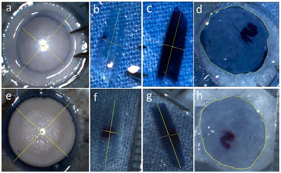
Background/Objectives: Intraoperative margin assessments of oral squamous cell carcinoma (SCC) are fundamentally limited by sampling errors and freezing artifacts inherent to standard frozen section analysis. We developed a mobile, multi-scale, wide field-of-view (FOV) swept-source optical coherence tomography/microscopy (SS-OCT/OCM) system operating in the Near-Infrared-II (NIR-II) window (1.68 μm) to provide a rapid, non-destructive, volumetric evaluation of excised oral mucosal tissues. Methods: To correlate optical images with histopathology, we engineered a custom 3D-printed tissue cassette that physically mitigates macroscopic shrinkage during scanning and subsequent tissue fixation. A three-axis motorized assembly extends the effective imaging FOV without compromising resolution, while a custom 3D multi-resolution pyramid stitching algorithm synthesizes wide-FOV mosaics. Results: The customized cassette enabled precise, one-to-one spatial correlation between optical volumes and histopathology sections. Crucially, a 3 × 3 mosaic scan acquired with a 10× objective balanced imaging resolution and acquisition time, providing sufficient structural clarity to visualize basement membrane loss—a hallmark of SCC invasion. Conclusions: This 1.68 μm, fully automatic, multiscale SS-OCT/OCM platform demonstrates the feasibility of serving as a rapid, three-dimensional imaging tool for potential future use as an adjunct to conventional frozen sections.
Full article

Figure 1


