applsci-logo

Journal Browser

Journal Browser

Advances of Biomedical Signal Processing for Disease Diagnosis, Prognosis or Severity Determination

A topical collection in Applied Sciences (ISSN 2076-3417). This collection belongs to the section "Biomedical Engineering".

Viewed by 51091

Editors


E-Mail Website
Guest Editor
Neural and Cognitive Engineering Group (gNeC), Automation and Robotics Center (CAR), Spanish National Research Council (CSIC), 28500 Arganda del Rey, Spain
Interests: Artificial intelligence and knowledge discovery in medicine; neuroengineering; cognitive science.

E-Mail Website
Guest Editor
Neural and Cognitive Engineering Group (gNeC), Automation and Robotics Center(CAR), Spanish National Research Council (CSIC), 28500 Arganda del Rey, Spain
Interests: machine learning and data mining; computational models of human behavior; human–machine interfaces
Special Issues, Collections and Topics in MDPI journals

Topical Collection Information

Dear Colleagues,

Despite the overwhelming advance of technology during the last two decades in all fields, and more concretely in medicine, clinicians still frequently diagnose and prognose by observation, either directly on the patient or indirectly through images or analytical parameters, with a significant subjectivity bias. A huge number of accessible sensors are available nowadays that provide fine-grained dynamical information on inner body and organ processes, different from the regular information used in clinical practice. The analysis of this information can provide objective, more robust, and accurate diagnostic and prognostic criteria, as well as better characterize the disease stage. However, most biomedical signals contain noise of different types, due to the low amplitude, as well as aggregated information from different concurrent sources. In this sense, advanced methods and techniques are needed to extract clinically meaningful information from the signals in order to fully exploit their diagnostic and prognostic potential.

Besides, clinicians have traditionally worked inside the boundaries of their own field. However, given that technology is becoming more and more present in clinical practice, the collaboration between clinicians, physicists and engineers seems mandatory. Moreover, this interdisciplinary collaboration might lead to more accurate and efficient medicine.

The aim of this Special Issue is to evidence the benefit of the interdisciplinary joint effort of Physics, Engineering and Medicine by bringing together works on advanced biomedical signal processing techniques that provide added value to the diagnosis, prognosis or stage determination of any disease or condition, either structural or functional.

Dr. José Ignacio Serrano
Dr. María Dolores del Castillo
Guest Editors

Manuscript Submission Information

Manuscripts should be submitted online at www.mdpi.com by registering and logging in to this website. Once you are registered, click here to go to the submission form. Manuscripts can be submitted until the deadline. All submissions that pass pre-check are peer-reviewed. Accepted papers will be published continuously in the journal (as soon as accepted) and will be listed together on the collection website. Research articles, review articles as well as short communications are invited. For planned papers, a title and short abstract (about 100 words) can be sent to the Editorial Office for announcement on this website.

Submitted manuscripts should not have been published previously, nor be under consideration for publication elsewhere (except conference proceedings papers). All manuscripts are thoroughly refereed through a single-blind peer-review process. A guide for authors and other relevant information for submission of manuscripts is available on the Instructions for Authors page. Applied Sciences is an international peer-reviewed open access semimonthly journal published by MDPI.

Please visit the Instructions for Authors page before submitting a manuscript. The Article Processing Charge (APC) for publication in this open access journal is 2400 CHF (Swiss Francs). Submitted papers should be well formatted and use good English. Authors may use MDPI's English editing service prior to publication or during author revisions.

Keywords

  • medical image
  • computer-vision-based diagnosis and prognosis
  • LPF-, ECoG-, EEG-, MEG-, NIRS-, ECG-, EMG- or IMU-processing
  • speech and sound
  • oculomotor signal
  • novel artificial intelligence
  • machine learning
  • non-linear biomedical signal processing
  • graph-based signal characterization
  • biomedical signal integration
Published Papers (15 papers)

2023

Jump to: 2021, 2020

13 pages, 2042 KiB  
Article
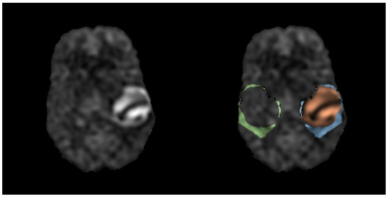
Histogram-Based Analysis of Low- and High-Grade Glioma and Its Surrounding Edema Using Arterial Spin Labeling Magnetic Resonance Imaging
by Thomas Lindner, Lasse Dührsen, Anna Andriana Kyselyova, Wiebke Entelmann, Luis Hau and Jens Fiehler
Appl. Sci. 2023, 13(19), 10581; https://doi.org/10.3390/app131910581 - 22 Sep 2023
Viewed by 609
Abstract
A glioma is a type of intra-axial brain tumor originating from the glial cells. Making up about one-third of all brain tumors, a timely diagnosis alongside correct grading and subsequent therapy planning is crucial. Magnetic Resonance Imaging is an established method for the [...] Read more.
A glioma is a type of intra-axial brain tumor originating from the glial cells. Making up about one-third of all brain tumors, a timely diagnosis alongside correct grading and subsequent therapy planning is crucial. Magnetic Resonance Imaging is an established method for the diagnosis of tumors. Arterial Spin Labeling (ASL) Perfusion Imaging allows for the non-contrast enhanced visualization of tumor hyper- or hypoperfusion. Commonly, cell swelling occurs around the tumor that causes edema, which subsequently puts healthy tissue at risk by potentially reducing regional perfusion. The patient collective in this study consists of 495 patients (501 scans) with histopathologically confirmed grade II-IV diffuse gliomas. The aim of this study was to evaluate the potential of histogram analysis of the ASL data to find biomarkers for the pathological diagnosis, grading, MGMT, and mutation status of the tumors as well as the analysis of tumor-surrounding edema. The analysis showed statistically significant results for the pathological diagnosis and grading but not for MGMT status or mutation. The differentiation between tumor and edema showed highly significant results yet did not show differences between edema and perfusion on the contralateral hemisphere. Full article
Show Figures

Figure 1

14 pages, 2064 KiB  
Article
Uncovering the Black Box of Coronary Artery Disease Diagnosis: The Significance of Explainability in Predictive Models
by Agorastos-Dimitrios Samaras, Serafeim Moustakidis, Ioannis D. Apostolopoulos, Elpiniki Papageorgiou and Nikolaos Papandrianos
Appl. Sci. 2023, 13(14), 8120; https://doi.org/10.3390/app13148120 - 12 Jul 2023
Cited by 1 | Viewed by 920
Abstract
In recent times, coronary artery disease (CAD) prediction and diagnosis have been the subject of many Medical decision support systems (MDSS) that make use of machine learning (ML) and deep learning (DL) algorithms. The common ground of most of these applications is that [...] Read more.
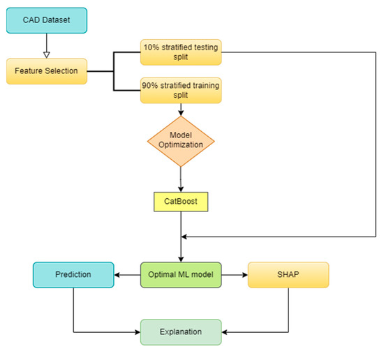
In recent times, coronary artery disease (CAD) prediction and diagnosis have been the subject of many Medical decision support systems (MDSS) that make use of machine learning (ML) and deep learning (DL) algorithms. The common ground of most of these applications is that they function as black boxes. They reach a conclusion/diagnosis using multiple features as input; however, the user is oftentimes oblivious to the prediction process and the feature weights leading to the eventual prediction. The primary objective of this study is to enhance the transparency and comprehensibility of a black-box prediction model designed for CAD. The dataset employed in this research comprises biometric and clinical information obtained from 571 patients, encompassing 21 different features. Among the instances, 43% of cases of CAD were confirmed through invasive coronary angiography (ICA). Furthermore, a prediction model utilizing the aforementioned dataset and the CatBoost algorithm is analyzed to highlight its prediction making process and the significance of each input datum. State-of-the-art explainability mechanics are employed to highlight the significance of each feature, and common patterns and differences with the medical bibliography are then discussed. Moreover, the findings are compared with common risk factors for CAD, to offer an evaluation of the prediction process from the medical expert’s point of view. By depicting how the algorithm weights the information contained in features, we shed light on the black-box mechanics of ML prediction models; by analyzing the findings, we explore their validity in accordance with the medical literature on the matter. Full article
Show Figures

Figure 1

2021

Jump to: 2023, 2020

20 pages, 19437 KiB  
Article
Reorganization of Resting-State EEG Functional Connectivity Patterns in Children with Cerebral Palsy Following a Motor Imagery Virtual-Reality Intervention
by Carlos Alberto Stefano Filho, José Ignacio Serrano, Romis Attux, Gabriela Castellano, Eduardo Rocon and Maria Dolores del Castillo
Appl. Sci. 2021, 11(5), 2372; https://doi.org/10.3390/app11052372 - 07 Mar 2021
Cited by 4 | Viewed by 2516
Abstract
Motor imagery (MI) has been suggested to provide additional benefits when included in traditional approaches of physical therapy for children with cerebral palsy (CP). Regardless, little is understood about the underlying neurological substrates that might justify its supposed benefits. In this work, we [...] Read more.
Motor imagery (MI) has been suggested to provide additional benefits when included in traditional approaches of physical therapy for children with cerebral palsy (CP). Regardless, little is understood about the underlying neurological substrates that might justify its supposed benefits. In this work, we studied resting-state (RS) electroencephalography (EEG) recordings of five children with CP that underwent a MI virtual-reality (VR) intervention. Our aim was to explore functional connectivity (FC) patterns alterations following this intervention through the formalism of graph theory, performing both group and subject-specific analyses. We found that FC patterns were more consistent across subjects prior to the MI-VR intervention, shifting along the anterior-posterior axis, post-intervention, for the β and γ bands. Additionally, group FC patterns were not found for the α range. Furthermore, intra-subject analyses reinforced the existence of large inter-subject variability and the need for a careful exploration of individual pattern alterations. Such patterns also hinted at a dependency between short-term functional plasticity mechanisms and the EEG frequency bands. Although our sample size is small, we provide a longitudinal analysis framework that can be replicated in future studies, especially at the group level, and whose foundation can be easily extended to verify the validity of our hypotheses. Full article
Show Figures

Figure 1

25 pages, 915 KiB  
Article
Ischemic Stroke Prediction by Exploring Sleep Related Features
by Jia Xie, Zhu Wang, Zhiwen Yu, Bin Guo and Xingshe Zhou
Appl. Sci. 2021, 11(5), 2083; https://doi.org/10.3390/app11052083 - 26 Feb 2021
Cited by 2 | Viewed by 2286
Abstract
Ischemic stroke is one of the typical chronic diseases caused by the degeneration of the neural system, which usually leads to great damages to human beings and reduces life quality significantly. Thereby, it is crucial to extract useful predictors from physiological signals, and [...] Read more.
Ischemic stroke is one of the typical chronic diseases caused by the degeneration of the neural system, which usually leads to great damages to human beings and reduces life quality significantly. Thereby, it is crucial to extract useful predictors from physiological signals, and further diagnose or predict ischemic stroke when there are no apparent symptoms. Specifically, in this study, we put forward a novel prediction method by exploring sleep related features. First, to characterize the pattern of ischemic stroke accurately, we extract a set of effective features from several aspects, including clinical features, fine-grained sleep structure-related features and electroencephalogram-related features. Second, a two-step prediction model is designed, which combines commonly used classifiers and a data filter model together to optimize the prediction result. We evaluate the framework using a real polysomnogram dataset that contains 20 stroke patients and 159 healthy individuals. Experimental results demonstrate that the proposed model can predict stroke events effectively, and the Precision, Recall, Precision Recall Curve and Area Under the Curve are 63%, 85%, 0.773 and 0.919, respectively. Full article
Show Figures

Figure 1

18 pages, 4192 KiB  
Article
Machine-Learning-Based Elderly Stroke Monitoring System Using Electroencephalography Vital Signals
by Yoon-A Choi, Sejin Park, Jong-Arm Jun, Chee Meng Benjamin Ho, Cheol-Sig Pyo, Hansung Lee and Jaehak Yu
Appl. Sci. 2021, 11(4), 1761; https://doi.org/10.3390/app11041761 - 17 Feb 2021
Cited by 24 | Viewed by 4440
Abstract
Stroke is the third highest cause of death worldwide after cancer and heart disease, and the number of stroke diseases due to aging is set to at least triple by 2030. As the top three causes of death worldwide are all related to [...] Read more.
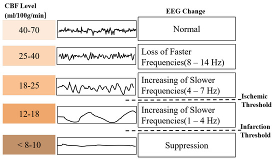
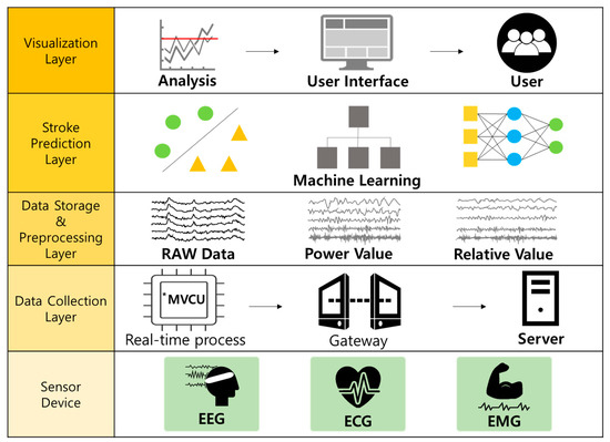
Stroke is the third highest cause of death worldwide after cancer and heart disease, and the number of stroke diseases due to aging is set to at least triple by 2030. As the top three causes of death worldwide are all related to chronic disease, the importance of healthcare is increasing even more. Models that can predict real-time health conditions and diseases using various healthcare services are attracting increasing attention. Most diagnosis and prediction methods of stroke for the elderly involve imaging techniques such as magnetic resonance imaging (MRI). It is difficult to rapidly and accurately diagnose and predict stroke diseases due to the long testing times and high costs associated with MRI. Thus, in this paper, we design and implement a health monitoring system that can predict the precursors of stroke diseases in the elderly in real time during daily walking. First, raw electroencephalography (EEG) data from six channels were preprocessed via Fast Fourier Transform (FFT). The raw EEG power values were then extracted from the raw spectra: alpha (α), beta (β), gamma (γ), delta (δ), and theta (θ) as well as the low β, high β, and θ to β ratio, respectively. The experiments in this paper confirm that the important features of EEG biometric signals alone during walking can accurately determine stroke precursors and occurrence in the elderly with more than 90% accuracy. Further, the Random Forest algorithm with quartiles and Z-score normalization validates the clinical significance and performance of the system proposed in this paper with a 92.51% stroke prediction accuracy. The proposed system can be implemented at a low cost, and it can be applied for early disease detection and prediction using the precursor symptoms of real-time stroke. Furthermore, it is expected that it will be able to detect other diseases such as cancer and heart disease in the future. Full article
Show Figures

Figure 1

14 pages, 1447 KiB  
Article
Can Trunk Acceleration Differentiate Stroke Patient Gait Patterns Using Time- and Frequency-Domain Features?
by Wei-Chun Hsu, Tommy Sugiarto, Ying-Yi Liao, Yi-Jia Lin, Fu-Chi Yang, Dueng-Yuan Hueng, Chi-Tien Sun and Kuan-Nien Chou
Appl. Sci. 2021, 11(4), 1541; https://doi.org/10.3390/app11041541 - 08 Feb 2021
Cited by 3 | Viewed by 2339
Abstract
This study classified the gait patterns of normal and stroke participants by using time- and frequency-domain features obtained from data provided by an inertial measurement unit sensor placed on the subject’s lower back (L5). Twenty-three participants were included and divided into two groups: [...] Read more.
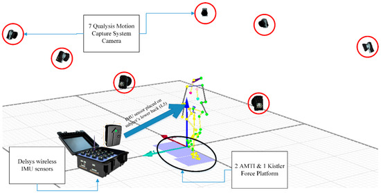
This study classified the gait patterns of normal and stroke participants by using time- and frequency-domain features obtained from data provided by an inertial measurement unit sensor placed on the subject’s lower back (L5). Twenty-three participants were included and divided into two groups: healthy group (young and older adults) and stroke group. Time- and frequency-domain features from an accelerometer were extracted, and a feature selection method comprising statistical analysis and signal-to-noise ratio (SNR) calculation was used to reduce the number of features. The features were then used to train four Support Vector Machine (SVM) kernels, and the results were subsequently compared. The quadratic SVM kernel had the highest accuracy (93.46%), as evaluated through cross-validation. Moreover, when different datasets were used on model testing, both the quadratic and cubic kernels showed the highest accuracy (96.55%). These results demonstrated the effectiveness of this study’s classification method in distinguishing between normal and stroke gait patterns, with only using a single sensor placed on the L5. Full article
Show Figures

Figure 1

11 pages, 1862 KiB  
Article
Cerebrovascular Reactivity Assessment during Carbon Dioxide Inhalation Using SPECT
by Yeong-Bae Lee and Chang-Ki Kang
Appl. Sci. 2021, 11(3), 1161; https://doi.org/10.3390/app11031161 - 27 Jan 2021
Cited by 1 | Viewed by 1438
Abstract
Background: Perfusion single-photon emission computed tomography (SPECT) using an acetazolamide is an important clinical tool used to assess cerebrovascular reactivity (CVR) in patients, but its use has been limited to clinical diagnostics. This study aimed to preliminarily evaluate the feasibility of perfusion SPECT [...] Read more.
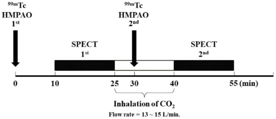
Background: Perfusion single-photon emission computed tomography (SPECT) using an acetazolamide is an important clinical tool used to assess cerebrovascular reactivity (CVR) in patients, but its use has been limited to clinical diagnostics. This study aimed to preliminarily evaluate the feasibility of perfusion SPECT using carbon dioxide (CO2). Methods: Ten healthy subjects participated in two consecutive SPECT scans using CO2 inhalation. To evaluate brain perfusion after preprocessing, the voxel-by-voxel CVR values were averaged in 13 subgroup regions of interest (ROIs) based on a template. Subsequently, averaged CVR values of each ROI were analyzed based on both cerebellar hemispheres. Results: CVR values in the eight subgroup ROIs, which included vermis, both insula/cingulate, and frontal cortices, showed significant changes (p < 0.05). CVR values were higher in vermis and right insula/cingulate by 3.34% and 3.15%, respectively. Conclusions: This study showed that quantitative SPECT with CO2 inhalation could be used to evaluate the voxel-based CVR in healthy subjects, which could be beneficial for elucidating induced hypercapnic states and for longitudinally investigating the healthy aging in brain vessels. Furthermore, the cerebrovascular hemodynamic parameters induced by CO2 could play an important role as a biomarker to evaluate treatment progress in patients with cerebrovascular disease. Full article
Show Figures

Figure 1

2020

Jump to: 2023, 2021

13 pages, 2317 KiB  
Article
Time Series Analysis Applied to EEG Shows Increased Global Connectivity during Motor Activation Detected in PD Patients Compared to Controls
by Ana María Maitín, Ramiro Perezzan, Diego Herráez-Aguilar, José Ignacio Serrano, María Dolores Del Castillo, Aida Arroyo, Jorge Andreo and Juan Pablo Romero
Appl. Sci. 2021, 11(1), 15; https://doi.org/10.3390/app11010015 - 22 Dec 2020
Cited by 2 | Viewed by 2555
Abstract
Background: Brain connectivity has shown to be a key characteristic in the study of both Parkinson’s Disease (PD) and the response of the patients to the dopaminergic medication. Time series analysis has been used here for the first time to study brain connectivity [...] Read more.
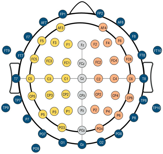
Background: Brain connectivity has shown to be a key characteristic in the study of both Parkinson’s Disease (PD) and the response of the patients to the dopaminergic medication. Time series analysis has been used here for the first time to study brain connectivity changes during motor activation in PD. Methods: A 64-channel EEG signal was registered during unilateral motor activation and resting-state in 6 non-demented PD patients before and after the administration of levodopa and in 6 matched healthy controls. Spectral entropy correlation, coherence, and interhemispheric divergence differences among PD patients and controls were analyzed under the assumption of stationarity of the time series. Results: During the motor activation test, PD patients showed an increased correlation coefficient (both hands p < 0.001) and a remarkable increase in coherence in all frequency range compared to the generalized reduction observed in controls (both hands p < 0.001). The Kullback­–Leibler Divergence (KLD) of the Spectral Entropy between brain hemispheres was observed to increase in controls (right hand p = 0.01; left hand p = 0.015) and to decrease in PD patients (right hand p = 0.02; left hand p = 0.002) with motor activation. Conclusions: Our results suggest that the oscillatory activity of the different cortex areas within healthy brains is relatively independent of the rest. PD brains exhibit a stronger connectivity which grows during motor activation. The levodopa mitigates this anomalous performance. Full article
Show Figures

Figure 1

21 pages, 1384 KiB  
Review
Machine Learning Approaches for Detecting Parkinson’s Disease from EEG Analysis: A Systematic Review
by Ana María Maitín, Alvaro José García-Tejedor and Juan Pablo Romero Muñoz
Appl. Sci. 2020, 10(23), 8662; https://doi.org/10.3390/app10238662 - 03 Dec 2020
Cited by 34 | Viewed by 5407
Abstract
Background: Diagnosis of Parkinson’s disease (PD) is mainly based on motor symptoms and can be supported by imaging techniques such as the single photon emission computed tomography (SPECT) or M-iodobenzyl-guanidine cardiac scintiscan (MIBG), which are expensive and not always available. In this review, [...] Read more.
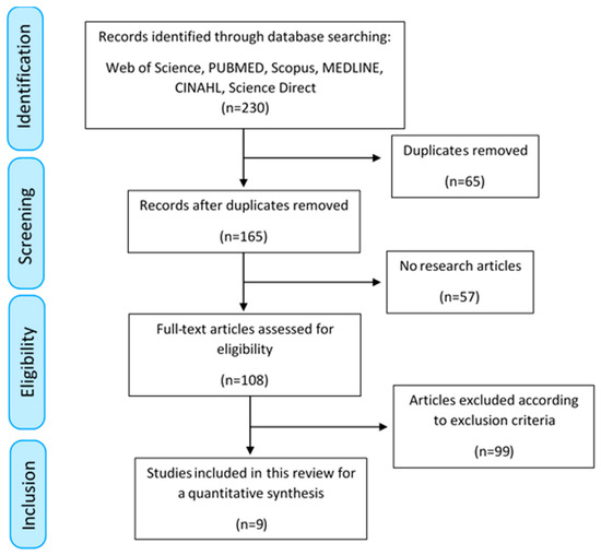
Background: Diagnosis of Parkinson’s disease (PD) is mainly based on motor symptoms and can be supported by imaging techniques such as the single photon emission computed tomography (SPECT) or M-iodobenzyl-guanidine cardiac scintiscan (MIBG), which are expensive and not always available. In this review, we analyzed studies that used machine learning (ML) techniques to diagnose PD through resting state or motor activation electroencephalography (EEG) tests. Methods: The review process was performed following Preferred Reporting Items for Systematic Reviews and Meta-Analyses (PRISMA) guidelines. All publications previous to May 2020 were included, and their main characteristics and results were assessed and documented. Results: Nine studies were included. Seven used resting state EEG and two motor activation EEG. Subsymbolic models were used in 83.3% of studies. The accuracy for PD classification was 62–99.62%. There was no standard cleaning protocol for the EEG and a great heterogeneity in the characteristics that were extracted from the EEG. However, spectral characteristics predominated. Conclusions: Both the features introduced into the model and its architecture were essential for a good performance in predicting the classification. On the contrary, the cleaning protocol of the EEG, is highly heterogeneous among the different studies and did not influence the results. The use of ML techniques in EEG for neurodegenerative disorders classification is a recent and growing field. Full article
Show Figures

Figure 1

20 pages, 1027 KiB  
Review
Bioelectrical Signals for the Diagnosis and Therapy of Functional Gastrointestinal Disorders
by Recep Avci, Kiara J.W. Miller, Niranchan Paskaranandavadivel, Leonard A. Bradshaw, Maggie-Lee Huckabee and Leo K. Cheng
Appl. Sci. 2020, 10(22), 8102; https://doi.org/10.3390/app10228102 - 16 Nov 2020
Cited by 15 | Viewed by 3604
Abstract
Coordinated contractions and motility patterns unique to each gastrointestinal organ facilitate the digestive process. These motor activities are coordinated by bioelectrical events, sensory and motor nerves, and hormones. The motility problems in the gastrointestinal tract known as functional gastrointestinal disorders (FGIDs) are generally [...] Read more.
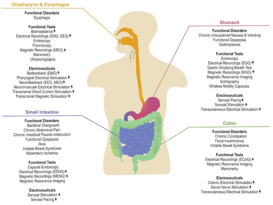
Coordinated contractions and motility patterns unique to each gastrointestinal organ facilitate the digestive process. These motor activities are coordinated by bioelectrical events, sensory and motor nerves, and hormones. The motility problems in the gastrointestinal tract known as functional gastrointestinal disorders (FGIDs) are generally caused by impaired neuromuscular activity and are highly prevalent. Their diagnosis is challenging as symptoms are often vague and difficult to localize. Therefore, the underlying pathophysiological factors remain unknown. However, there is an increasing level of research and clinical evidence suggesting a link between FGIDs and altered bioelectrical activity. In addition, electroceuticals (bioelectrical therapies to treat diseases) have recently gained significant interest. This paper gives an overview of bioelectrical signatures of gastrointestinal organs with normal and/or impaired motility patterns and bioelectrical therapies that have been developed for treating FGIDs. The existing research evidence suggests that bioelectrical activities could potentially help to identify the diverse etiologies of FGIDs and overcome the drawbacks of the current clinically adapted methods. Moreover, electroceuticals could potentially be effective in the treatment of FGIDs and replace the limited existing conventional therapies which often attempt to treat the symptoms rather than the underlying condition. Full article
Show Figures

Figure 1

18 pages, 1302 KiB  
Article
Coronary Artery Disease Detection by Machine Learning with Coronary Bifurcation Features
by Xueping Chen, Yi Fu, Jiangguo Lin, Yanru Ji, Ying Fang and Jianhua Wu
Appl. Sci. 2020, 10(21), 7656; https://doi.org/10.3390/app10217656 - 29 Oct 2020
Cited by 14 | Viewed by 3350
Abstract
Background: Early accurate detection of coronary artery disease (CAD) is one of the most important medical research areas. Researchers are motivated to utilize machine learning techniques for quick and accurate detection of CAD. Methods: To obtain the high quality of features used for [...] Read more.
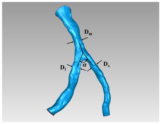
Background: Early accurate detection of coronary artery disease (CAD) is one of the most important medical research areas. Researchers are motivated to utilize machine learning techniques for quick and accurate detection of CAD. Methods: To obtain the high quality of features used for machine learning, we here extracted the coronary bifurcation features from the coronary computed tomography angiography (CCTA) images by using the morphometric method. The machine learning classifier algorithms, such as logistic regression (LR), decision tree (DT), linear discriminant analysis (LDA), k-nearest neighbors (k-NN), artificial neural network (ANN), and support vector machine (SVM) were applied for estimating the performance by using the measured features. Results: The results showed that in comparison with other machine learning methods, the polynomial-SVM with the use of the grid search optimization method had the best performance for the detection of CAD and had yielded the classification accuracy of 100.00%. Among six examined coronary bifurcation features, the exponent of vessel diameter (n) and the area expansion ratio (AER) were two key features in the detection of CAD. Conclusions: This study could aid the clinicians to detect CAD accurately, which may probably provide an alternative method for the non-invasive diagnosis in clinical. Full article
Show Figures

Figure 1

22 pages, 5546 KiB  
Article
Temporal Convolutional Network Connected with an Anti-Arrhythmia Hidden Semi-Markov Model for Heart Sound Segmentation
by Yibo Yin, Kainan Ma and Ming Liu
Appl. Sci. 2020, 10(20), 7049; https://doi.org/10.3390/app10207049 - 11 Oct 2020
Cited by 10 | Viewed by 2625
Abstract
Heart sound segmentation (HSS) is a critical step in heart sound processing, where it improves the interpretability of heart sound disease classification algorithms. In this study, we aimed to develop a real-time algorithm for HSS by combining the temporal convolutional network (TCN) and [...] Read more.
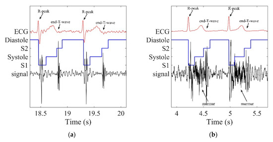
Heart sound segmentation (HSS) is a critical step in heart sound processing, where it improves the interpretability of heart sound disease classification algorithms. In this study, we aimed to develop a real-time algorithm for HSS by combining the temporal convolutional network (TCN) and the hidden semi-Markov model (HSMM), and improve the performance of HSMM for heart sounds with arrhythmias. We experimented with TCN and determined the best parameters based on spectral features, envelopes, and one-dimensional CNN. However, the TCN results could contradict the natural fixed order of S1-systolic-S2-diastolic of heart sound, and thereby the Viterbi algorithm based on HSMM was connected to correct the order errors. On this basis, we improved the performance of the Viterbi algorithm when detecting heart sounds with cardiac arrhythmias by changing the distribution and weights of the state duration probabilities. The public PhysioNet Computing in Cardiology Challenge 2016 data set was employed to evaluate the performance of the proposed algorithm. The proposed algorithm achieved an F1 score of 97.02%, and this result was comparable with the current state-of-the-art segmentation algorithms. In addition, the proposed enhanced Viterbi algorithm for HSMM corrected 30 out of 30 arrhythmia errors after checking one by one in the dataset. Full article
Show Figures

Figure 1

18 pages, 1072 KiB  
Article
Prosody-Based Measures for Automatic Severity Assessment of Dysarthric Speech
by Abner Hernandez, Sunhee Kim and Minhwa Chung
Appl. Sci. 2020, 10(19), 6999; https://doi.org/10.3390/app10196999 - 08 Oct 2020
Cited by 17 | Viewed by 3872
Abstract
One of the first cues for many neurological disorders are impairments in speech. The traditional method of diagnosing speech disorders such as dysarthria involves a perceptual evaluation from a trained speech therapist. However, this approach is known to be difficult to use for [...] Read more.
One of the first cues for many neurological disorders are impairments in speech. The traditional method of diagnosing speech disorders such as dysarthria involves a perceptual evaluation from a trained speech therapist. However, this approach is known to be difficult to use for assessing speech impairments due to the subjective nature of the task. As prosodic impairments are one of the earliest cues of dysarthria, the current study presents an automatic method of assessing dysarthria in a range of severity levels using prosody-based measures. We extract prosodic measures related to pitch, speech rate, and rhythm from speakers with dysarthria and healthy controls in English and Korean datasets, despite the fact that these two languages differ in terms of prosodic characteristics. These prosody-based measures are then used as inputs to random forest, support vector machine and neural network classifiers to automatically assess different severity levels of dysarthria. Compared to baseline MFCC features, 18.13% and 11.22% relative accuracy improvement are achieved for English and Korean datasets, respectively, when including prosody-based features. Furthermore, most improvements are obtained with a better classification of mild dysarthric utterances: a recall improvement from 42.42% to 83.34% for English speakers with mild dysarthria and a recall improvement from 36.73% to 80.00% for Korean speakers with mild dysarthria. Full article
Show Figures

Figure 1

19 pages, 2396 KiB  
Article
AI-Based Stroke Disease Prediction System Using Real-Time Electromyography Signals
by Jaehak Yu, Sejin Park, Soon-Hyun Kwon, Chee Meng Benjamin Ho, Cheol-Sig Pyo and Hansung Lee
Appl. Sci. 2020, 10(19), 6791; https://doi.org/10.3390/app10196791 - 28 Sep 2020
Cited by 46 | Viewed by 11102
Abstract
Stroke is a leading cause of disabilities in adults and the elderly which can result in numerous social or economic difficulties. If left untreated, stroke can lead to death. In most cases, patients with stroke have been observed to have abnormal bio-signals (i.e., [...] Read more.
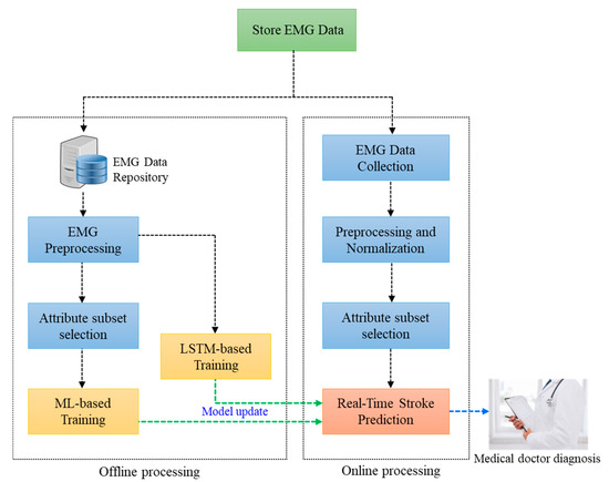
Stroke is a leading cause of disabilities in adults and the elderly which can result in numerous social or economic difficulties. If left untreated, stroke can lead to death. In most cases, patients with stroke have been observed to have abnormal bio-signals (i.e., ECG). Therefore, if individuals are monitored and have their bio-signals measured and accurately assessed in real-time, they can receive appropriate treatment quickly. However, most diagnosis and prediction systems for stroke are image analysis tools such as CT or MRI, which are expensive and difficult to use for real-time diagnosis. In this paper, we developed a stroke prediction system that detects stroke using real-time bio-signals with artificial intelligence (AI). Both machine learning (Random Forest) and deep learning (Long Short-Term Memory) algorithms were used in our system. EMG (Electromyography) bio-signals were collected in real time from thighs and calves, after which the important features were extracted, and prediction models were developed based on everyday activities. Prediction accuracies of 90.38% for Random Forest and of 98.958% for LSTM were obtained for our proposed system. This system can be considered an alternative, low-cost, real-time diagnosis system that can obtain accurate stroke prediction and can potentially be used for other diseases such as heart disease. Full article
Show Figures

Figure 1

17 pages, 2842 KiB  
Article
Continuous and Noninvasive Estimation of Right Ventricle Systolic Blood Pressure Using Heart Sound Signal by Deep Bidirectional LSTM Network
by Miao Wang, Hong Tang, Tengfei Feng and Binbin Guo
Appl. Sci. 2020, 10(16), 5466; https://doi.org/10.3390/app10165466 - 07 Aug 2020
Cited by 3 | Viewed by 2458
Abstract
Objective: Timely monitoring right ventricular systolic blood pressure (RVSBP) is helpful in the early detection of pulmonary hypertension (PH). However, it is not easy to monitor RVSBP directly. The objective of this paper is to develop a deep learning technique for RVSBP noninvasive [...] Read more.
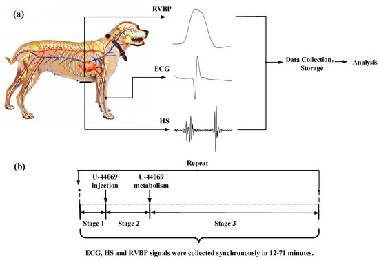
Objective: Timely monitoring right ventricular systolic blood pressure (RVSBP) is helpful in the early detection of pulmonary hypertension (PH). However, it is not easy to monitor RVSBP directly. The objective of this paper is to develop a deep learning technique for RVSBP noninvasive estimation using heart sound (HS) signals supported by (electrocardiography) ECG signals without complex features extraction. Methods: Five beagle dog subjects were used. The medicine U-44069 was injected into the subjects to induce a wide range of RVSBP variation. The blood pressure in right ventricle, ECG of lead I and HS signals were recorded simultaneously. Thirty-two records were collected. The relations between RVSBP and cyclic HS signals were modeled by the Bidirectional Long Short-Term Memory (Bi-LSTM) network. Results: The mean absolute error (MAE) ± standard deviation (SD) inside record was 1.85 ± 1.82 mmHg. It was 4.37 ± 2.49 mmHg across record but within subject. The corrective factors were added after training the Bi-LSTM network across subjects. Finally, the MAE ± SD from 12.46 ± 6.56 mmHg dropped to 6.37 ± 4.90 mmHg across subjects. Significance: Our work was the first to apply the Bi-LSTM network to build relations between the HS signal and RVSBP. This work suggested a noninvasive and continuous RVSBP estimation using the HS signal supported by the ECG signal by deep learning architecture without the need of healthcare professionals. Full article
Show Figures

Figure 1

Back to TopTop