

-
Crinophagy in Pancreatic Beta Cells: From Insulin Granule Turnover to Diabetes Pathogenesis -
Effects of Maternal Heartbeat Exposure on Neonatal Autonomic Regulation -
SHP-2 Expression Is a Positive Prognostic Biomarker in Non-Small-Cell Lung Carcinoma: Association with KRAS Mutation and Prolonged Survival
Journal Description
Pathophysiology
Pathophysiology
is an international, peer-reviewed, open access journal on the etiology, development, and elimination of pathological processes, published quarterly online by MDPI (since Volume 21, Issue 1 - 2020). The International Society for Pathophysiology (ISP) is affiliated with Pathophysiology, serving as its official journal. Society members receive discounts on the article processing charges.
- Open Access— free for readers, with article processing charges (APC) paid by authors or their institutions.
- High Visibility: indexed within ESCI (Web of Science), Scopus, PMC, PubMed, and other databases.
- Journal Rank: JCR - Q1 (Pathology) / CiteScore - Q1 (Pathology and Forensic Medicine)
- Rapid Publication: manuscripts are peer-reviewed and a first decision is provided to authors approximately 27.7 days after submission; acceptance to publication is undertaken in 2.9 days (median values for papers published in this journal in the first half of 2026).
- Recognition of Reviewers: Reviewers whose reports are timely and of high quality receive an APC discount voucher for a future publication in an MDPI journal. Become a reviewer.
Impact Factor:
3.5 (2025)
Latest Articles
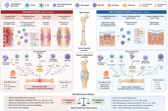
Exploratory Analysis of Bone Marrow IL-10 and Plasma IL-1RA in Acute Traumatic Versus Chronic Non-Traumatic Orthopedic Patients: A Prospective Observational Study
Pathophysiology 2026, 33(3), 71; https://doi.org/10.3390/pathophysiology33030071 - 21 Sep 2026
Abstract
►
Show Figures
Introduction: Inflammation is a central component of the biological processes involved in orthopedic interventions, whether performed for acute traumatic injuries or chronic non-traumatic conditions. Cytokines play major roles in modulating these responses; however, data describing cytokine activity in bone marrow, particularly during
[...] Read more.
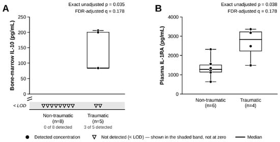
Introduction: Inflammation is a central component of the biological processes involved in orthopedic interventions, whether performed for acute traumatic injuries or chronic non-traumatic conditions. Cytokines play major roles in modulating these responses; however, data describing cytokine activity in bone marrow, particularly during orthopedic procedures, remain limited. This study aimed to examine variations in cytokine levels in traumatic and non-traumatic orthopedic surgeries by analyzing simultaneous bone marrow and plasma samples. Methods: This single-center prospective observational study with exploratory objectives included skeletally mature patients undergoing urgent surgery for acute traumatic fractures or planned surgery for chronic non-traumatic degenerative orthopedic conditions. Bone-marrow and peripheral plasma samples were collected simultaneously intraoperatively. All samples were tested in duplicate for interleukin-8 (IL-8), interleukin-6 (IL-6), interleukin-10 (IL-10), interleukin-4 (IL-4), interleukin-1 receptor antagonist (IL-1RA), and tumor necrosis factor-α (TNF-α) using developmental ELISA kits. Cytokine levels were compared between traumatic and non-traumatic groups and between bone-marrow and plasma samples. Results: Nineteen participants were included, providing 19 bone-marrow samples and 15 plasma samples, with the number of available measurements varying among cytokines. Bone-marrow IL-10 was detected in three of five available traumatic samples and in none of the eight available non-traumatic samples (unadjusted p = 0.035; false-discovery-rate-adjusted q = 0.178). Plasma IL-1RA concentrations were higher in traumatic than in non-traumatic patients (median 2828.00 versus 1269.00 pg/mL; unadjusted p = 0.038; q = 0.178). Bone-marrow IL-8 also showed higher concentrations in traumatic patients and approached the unadjusted significance threshold (p = 0.053; q = 0.178). No comparison remained statistically significant after FDR correction. Discussion: Bone-marrow IL-10 detection and plasma IL-1RA concentrations showed nominal between-group differences that did not remain statistically significant after multiple-comparison adjustment. Although preliminary and hypothesis-generating, these observations identify potentially informative local and systemic cytokine patterns and highlight the value of further investigating bone-marrow cytokine responses in orthopedic trauma.
Full article
Open AccessSystematic Review
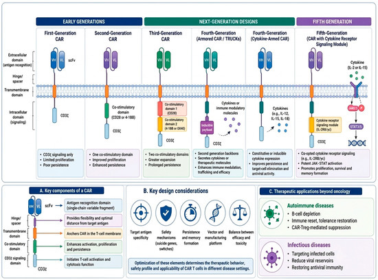
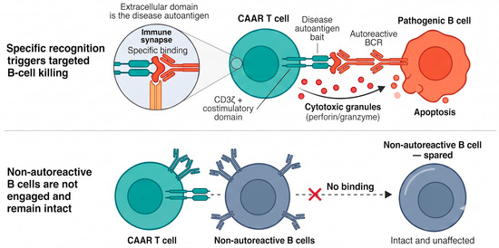
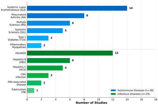
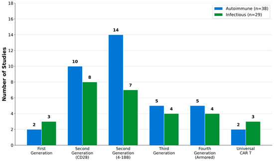
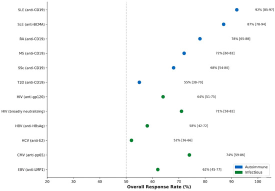
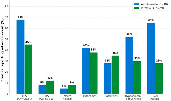
Beyond Oncology: Exploring the Expanding Role of CAR T Cell Therapy in Autoimmune and Infectious Diseases—A Systematic Review
by
Kawther Zaher and Jehan Alrahimi
Pathophysiology 2026, 33(3), 70; https://doi.org/10.3390/pathophysiology33030070 - 19 Sep 2026
Abstract
►▼
Show Figures
Background: Chimeric antigen receptor (CAR) T cell therapy has revolutionized the treatment of select hematologic malignancies and is increasingly being explored for non-oncological indications, including autoimmune diseases and persistent infectious diseases. This systematic review synthesized current evidence on CAR T cell and CAR-Treg
[...] Read more.
Background: Chimeric antigen receptor (CAR) T cell therapy has revolutionized the treatment of select hematologic malignancies and is increasingly being explored for non-oncological indications, including autoimmune diseases and persistent infectious diseases. This systematic review synthesized current evidence on CAR T cell and CAR-Treg approaches beyond oncology, with emphasis on therapeutic targets, translational and clinical outcomes, safety, and implementation barriers. Methods: The review followed PRISMA 2020 guidelines. PubMed/MEDLINE, Scopus, Web of Science, Cochrane Library, ClinicalTrials.gov, and major trial registries were searched for studies published from 1 January 2010 to 31 March 2026. Eligible studies included clinical, preclinical, and translational investigations of CAR T cell or CAR-Treg strategies in autoimmune or infectious diseases. Results: Because of heterogeneity in disease indications, CAR constructs, endpoints, and study designs, findings were synthesized descriptively. The strongest early clinical signals were observed with B cell-directed CAR T cell therapy for severe, refractory autoimmune diseases, particularly systemic lupus erythematosus, systemic sclerosis, and inflammatory myopathies. Infectious disease applications, mainly HIV and hepatitis B, showed preliminary safety, persistence, and partial antiviral activity, but durable pathogen eradication remains unproven. Conclusion: Overall, CAR T cell therapy beyond oncology is promising but remains preliminary, requiring standardized reporting, long-term safety monitoring, scalable manufacturing, and carefully defined risk–benefit thresholds. Human clinical evidence was interpreted separately from preclinical and mechanistic evidence, and clinical conclusions were based primarily on human studies.
Full article

Figure 1
Open AccessReview
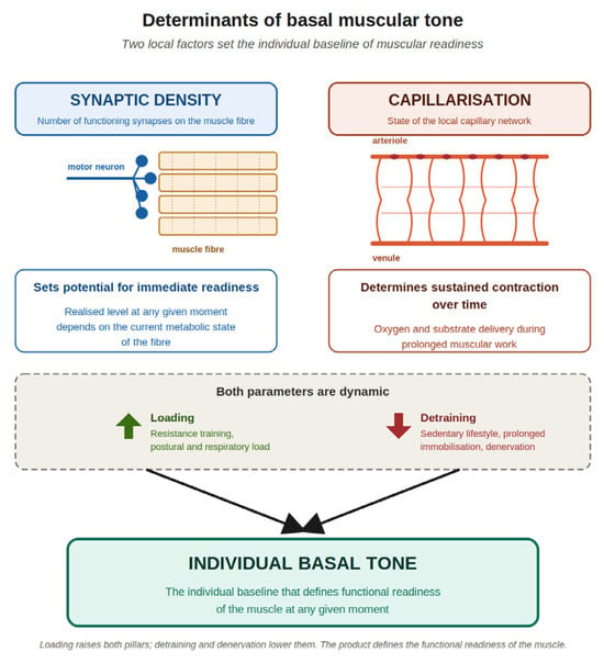
Basal Tone, Muscle Memory and Denervation: Pathophysiological Foundations of Abdominal Wall Hernia Development and Recovery
by
Zelimkhan G. M. Berikkhanov, Zakhar A. Akulov, Maria A. Sukhanova, Alexey L. Shestakov, Aleksey G. Kotelnikov, Andrey M. Nikolaev, Vadim S. Razumovsky, Evgeniy A. Tarabrin, Milena Yu. Ivanova and Sergey Yu. Muraviev
Pathophysiology 2026, 33(3), 69; https://doi.org/10.3390/pathophysiology33030069 - 14 Sep 2026
Abstract
►▼
Show Figures
Modern abdominal wall hernia surgery is centered on the biomechanics of the fascial-aponeurotic framework and on intra-abdominal pressure (IAP) control; neuromuscular regulation of the wall remains outside perioperative planning. This review reframes the abdominal wall as a coordinated neuromuscular system and outlines a
[...] Read more.
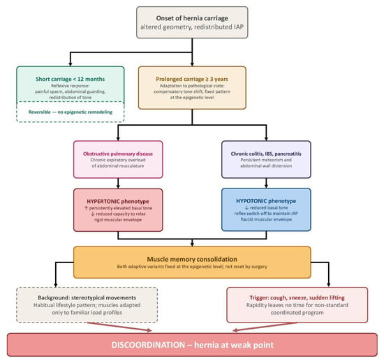
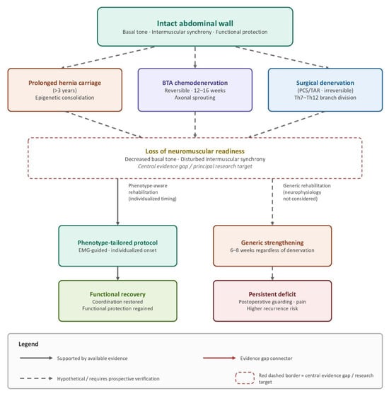
Modern abdominal wall hernia surgery is centered on the biomechanics of the fascial-aponeurotic framework and on intra-abdominal pressure (IAP) control; neuromuscular regulation of the wall remains outside perioperative planning. This review reframes the abdominal wall as a coordinated neuromuscular system and outlines a framework in which ventral herniation is hypothesized to represent a compensatory response to altered regulation of elevated IAP, while discoordinated contraction of the muscular layers is proposed as a potential predisposing factor. A structured literature search was performed in PubMed/MEDLINE, Embase and the Cochrane Library from inception to March 2026 across four thematic blocks: basal tone and innervation, spinal pathology and coordination, skeletal muscle memory, and electromyographic and clinical models of denervation. Synthesis followed SANRA criteria. This is not a formal systematic review or meta-analysis. Basal tone of the abdominal muscles is individual and depends on spinal innervation, lifestyle, comorbidity, and hernia duration; obstructive pulmonary disease yields a hypertonic phenotype, and persistent meteorism a hypotonic one. In a patient predisposed to herniation, elevated IAP may be partially accommodated through redistribution of abdominal contents; within the proposed framework, herniation is hypothesized to function as a pressure-relief mechanism. Discoordination of the muscular layers may reduce the capacity of the abdominal wall to adapt to rapid or unfamiliar loading, potentially contributing to hernia formation in predisposed patients. Six meta-analyses of preoperative botulinum toxin type A confirm lateral muscle elongation; none assessed basal-tone recovery after toxin washout. Each hernia patient presents with an individual neuromuscular phenotype shaped by basal tone, intermuscular coordination and muscle memory. The proposed framework complements mechano-fascial concepts with a neurophysiological dimension and provides a hypothesis-generating rationale for investigating phenotype-tailored prehabilitation and postoperative rehabilitation. Prospective electromyographic mapping with functional maneuvers is the immediate research priority.
Full article

Figure 1
Open AccessReview
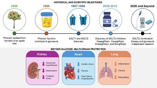
Dapagliflozin Beyond Glucose Lowering: Mechanisms of Renal and Systemic Protection
by
Madison L. Wise and Abdel A. Alli
Pathophysiology 2026, 33(3), 68; https://doi.org/10.3390/pathophysiology33030068 - 10 Sep 2026
Abstract
►▼
Show Figures
Sodium glucose cotransporter-2 inhibitors (SGLT2is) have rapidly evolved from glucose-lowering agents to multifaceted therapies with significant renoprotective and cardioprotective potential. Although originally developed to inhibit glucose reabsorption within the renal proximal tubule for the treatment of Type 2 diabetes mellitus (T2DM), growing evidence
[...] Read more.
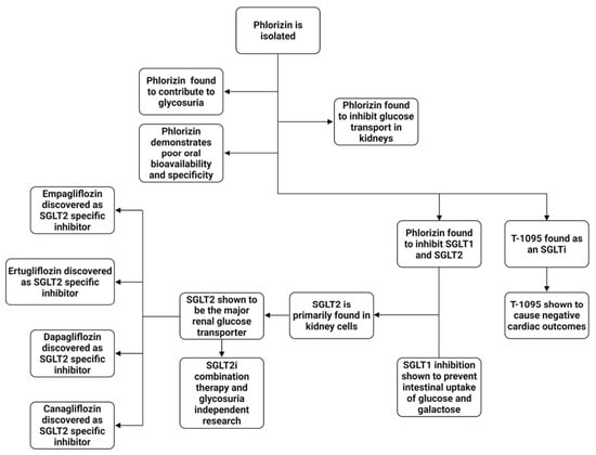
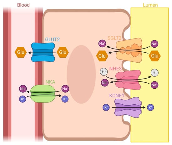
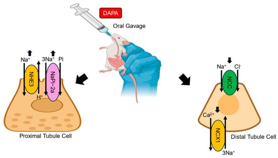
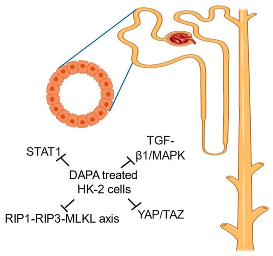
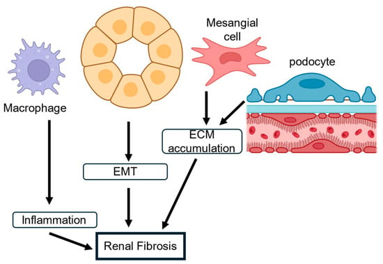
Sodium glucose cotransporter-2 inhibitors (SGLT2is) have rapidly evolved from glucose-lowering agents to multifaceted therapies with significant renoprotective and cardioprotective potential. Although originally developed to inhibit glucose reabsorption within the renal proximal tubule for the treatment of Type 2 diabetes mellitus (T2DM), growing evidence indicates that SGLT2is exert broad systemic actions extending beyond glycemic control. Among this drug class, dapagliflozin has emerged as a clinically important agent with pleiotropic effects involving renal hemodynamics, inflammatory signaling, mitochondrial function, fibrosis regulation, and cellular stress adaptation. This review outlines the historical progression from the discovery of phlorizin to the development of highly selective modern SGLT2 inhibitors while emphasizing mechanistic insights gained from experimental and clinical studies of dapagliflozin. In addition to the established effects on sodium–glucose transport, dapagliflozin modulates multiple epithelial transport proteins including NHE3, NaPi-2a, NCC, and NCX1, highlighting complex regulatory effects on sodium handling and tubular electrolyte transport. Emerging evidence further demonstrates that dapagliflozin suppresses inflammatory and profibrotic pathways involving YAP/TAZ, STAT1, TGF-β, NLRP3, and NF-KB signaling. Restoration of tubuloglomerular feedback, attenuation of oxidative stress, and preservation of mitochondrial function also appear to contribute substantially to the renoprotective actions of SGLT2 inhibition. Beyond the kidney, dapagliflozin and related SGLT2is exert cardioprotective effects through coordinated improvements in cardiac energetics, inflammatory regulation, and hemodynamic function. Emerging studies additionally suggest potential pulmonary benefits, including reductions in inflammatory signaling, pulmonary edema, and respiratory complications. Collectively, these findings support a shift in understanding SGLT2is from targeted metabolic therapies to broader regulators of cellular and organ function. Continued investigation into the glucose-independent mechanisms of dapagliflozin may reveal additional therapeutic applications across chronic metabolic, cardiovascular, and inflammatory diseases.
Full article

Graphical abstract
Open AccessReview
Placental Pathophysiology and Developmental Programming of Adult Cardiometabolic Risk: A Narrative Review of Pregnancy Exposures
by
Eleftherios Panteris, Ioanna Kakatsaki, Zoi Koukou, Charalambos Kolvatzis, Styliani Papanikolaou and Eleftheria Hatzidaki
Pathophysiology 2026, 33(3), 67; https://doi.org/10.3390/pathophysiology33030067 - 8 Sep 2026
Abstract
►▼
Show Figures
Non-communicable diseases remain the dominant cause of adult morbidity and mortality, yet cardiometabolic, vascular and renal risk may originate before birth. This structured narrative review critically evaluates human evidence linking pregnancy-related exposures, placental pathophysiology and later offspring health within a maternal–placental–fetal–life-course framework. Evidence
[...] Read more.
Non-communicable diseases remain the dominant cause of adult morbidity and mortality, yet cardiometabolic, vascular and renal risk may originate before birth. This structured narrative review critically evaluates human evidence linking pregnancy-related exposures, placental pathophysiology and later offspring health within a maternal–placental–fetal–life-course framework. Evidence published from 2020 to 2026 was emphasised, while seminal earlier cohorts were retained when they provided uniquely long follow-up or direct placental measurements. The most consistent associations concern maternal obesity, gestational diabetes, excessive gestational weight gain and hypertensive disorders, which are linked with higher offspring blood pressure, greater adiposity and adverse glucose–insulin profiles. Human placental studies implicate vascular malperfusion, altered nutrient transport, inflammatory and oxidative signalling, endocrine function and epigenetic regulation. Few studies, however, have measured the prenatal exposure, a specific placental phenotype and a long-term offspring outcome within the same longitudinal design; most proposed placental pathways are supported by convergent evidence or biological plausibility rather than demonstrated mediation. Preterm birth and fetal growth restriction are clinically observable, etiologically heterogeneous sentinel phenotypes, not obligatory mediators. Placental and offspring epigenetic signatures, telomere biology and mitochondrial function represent distinct domains of biological embedding; their causal and prognostic significance remains uncertain. Longitudinal studies integrating maternal exposures, placental histopathology and molecular phenotypes, fetal organ development, childhood cardiometabolic trajectories and adult clinical outcomes are needed to determine whether, and under what circumstances, the placenta plays an intermediary role rather than merely serving as a marker of an adverse pregnancy environment.
Full article

Figure 1
Open AccessReview
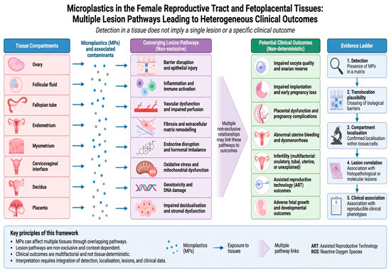
Microplastics in the Female Genital Tract and Fetoplacental Continuum: A Lesion-Based Framework for Pathophysiological Interpretation
by
Sayandeep K. Das, Prithviraj Karak, Afsona Parveen, Swastika N. Das and Savitri M. Nerune
Pathophysiology 2026, 33(3), 66; https://doi.org/10.3390/pathophysiology33030066 - 1 Sep 2026
Abstract
Background/Objectives: Microplastics (MPs) are increasingly reported in human reproductive tissues and fluids, but detection alone does not establish tissue injury or disease. Recent reviews have synthesised occurrence, reproductive toxicity, placental transfer, fertility, and pregnancy outcomes. The unresolved problem is that occurrence, model-response,
[...] Read more.
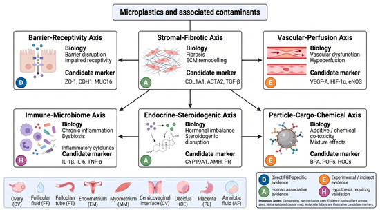
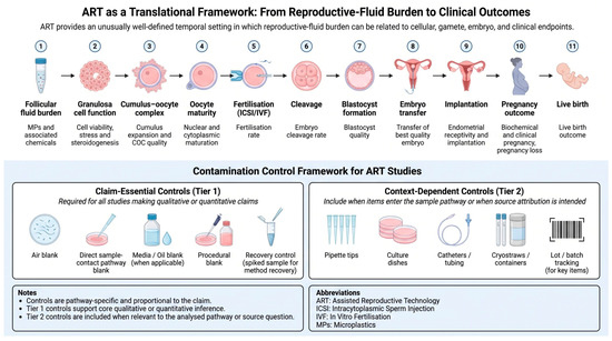
Background/Objectives: Microplastics (MPs) are increasingly reported in human reproductive tissues and fluids, but detection alone does not establish tissue injury or disease. Recent reviews have synthesised occurrence, reproductive toxicity, placental transfer, fertility, and pregnancy outcomes. The unresolved problem is that occurrence, model-response, lesion-comparison, clinical-association, and causal evidence are not interchangeable. This review asks what minimum evidence is required for an MP finding in the female genital tract (FGT) or fetoplacental continuum to become lesion-relevant and where inference must stop. Methods: We performed a critical narrative search of PubMed, Scopus, Web of Science, ScienceDirect, and Google Scholar from database inception through 10 July 2026. The final synthesis comprised 59 sources: 28 primary studies, 24 reviews or systematic syntheses, and 7 regulatory, consensus, or professional-guidance documents.. Methodological recommendations were graded as claim-essential, context-dependent, or exploratory and as routine, specialised, or research-intensive. Results: The distinct contribution is an FGT-specific mechanistic and claim-to-evidence architecture rather than another environmental or reproductive-toxicity catalogue. We propose an ordered, claim-calibrated evidence ladder linking external exposure, internal dose, reproductive-fluid burden, spatial localisation, lesion response, and clinically annotated phenotype. The framework specifies the minimum evidence for occurrence, localisation, lesion, clinical, and causal claims and provides explicit stopping and downgrading rules rather than treating all desirable measurements as mandatory. The FGT Lesion Atlas is a compartment-by-compartment map of prioritised, non-exhaustive endpoints and sampling requirements. The FGT Lesionome is a cross-compartment synthesis of recurring barrier-receptivity, stromal-fibrotic, vascular-perfusion, immune-microbiome, endocrine-steroidogenic, and particle-cargo axes. An integrated hypothesis-generating pathway links systemic or local exposure, internal and target-compartment burden, physicochemical conditioning in reproductive-fluid microenvironments, particle or particle-associated-constituent interaction with compartment-specific cells and matrices, the six response axes, physiological dysfunction, and clinically annotated outcomes. A reproductive-fluid model, an assisted-reproductive-technology sentinel sequence, and an FGT Microplastic Pathology Reporting Checklist rank study elements by evidentiary necessity, feasibility, and interpretive consequence. Conclusions: The framework is hypothesis-generating, not a validated causal map or a universal core outcome set. It separates essential validity safeguards from context-dependent best practices and exploratory endpoints. Human reproductive and fetoplacental disease causation remains unproven without contamination-controlled sampling, polymer confirmation, spatial co-localisation with lesions, temporality, exposure-response assessment, confounder control, replication, and appropriately powered, temporally informative clinical outcomes.
Full article
(This article belongs to the Section Cellular and Molecular Mechanisms)
►▼
Show Figures

Figure 1
Open AccessArticle
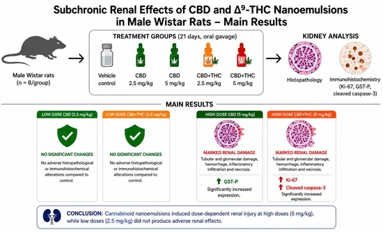
Subchronic Exposure to Low-Dose Cannabidiol (CBD) and Δ9-Tetrahydrocannabinol (Δ9-THC) Nanoemulsions Suggests Low Renal Toxicity: A Preliminary Study
by
Thiago Guedes Pinto, Barbara dos Anjos Rosario, Pedro Everson Alexandre de Aquino, Glauce Socorro de Barros Viana, Nágila Maria Pontes Silva Ricardo, Edilberto Rocha Silveira, Débora Hellen Almeida de Brito, Dávila Zampieri, Milena de Barros Viana and Daniel Araki Ribeiro
Pathophysiology 2026, 33(3), 65; https://doi.org/10.3390/pathophysiology33030065 - 1 Sep 2026
Abstract
►▼
Show Figures
Background/Objectives: The increasing therapeutic use of cannabinoid-based formulations highlights the need to better characterize their safety profile, particularly in organs involved in xenobiotic elimination, such as the kidneys. This study evaluated the renal effects of cannabidiol (CBD) and Δ9-tetrahydrocannabinol (Δ9
[...] Read more.
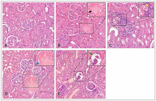
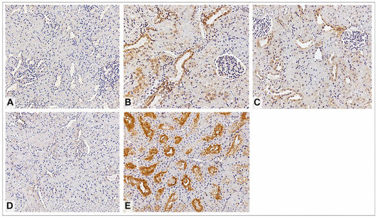
Background/Objectives: The increasing therapeutic use of cannabinoid-based formulations highlights the need to better characterize their safety profile, particularly in organs involved in xenobiotic elimination, such as the kidneys. This study evaluated the renal effects of cannabidiol (CBD) and Δ9-tetrahydrocannabinol (Δ9-THC) nanoemulsions following subchronic oral administration in male Wistar rats. Methods: Forty animals (n = 8/group) were randomly allocated into five groups: vehicle control (Licuri oil + Pluronic® F127), CBD 2.5 mg/kg, CBD 5 mg/kg, CBD+THC 2.5 mg/kg, and CBD+THC 5 mg/kg. Treatments were administered daily by oral gavage for 21 consecutive days. Kidney samples were analyzed by histopathology and immunohistochemistry (Ki-67, GST-P, and cleaved caspase-3). Results: High-dose groups (5 mg/kg) exhibited marked histopathological alterations, including tubular and glomerular damage, hemorrhagic areas, inflammatory infiltration, and necrosis. These groups also showed increased Ki-67 and cleaved caspase-3 immunoexpression compared to controls. GST-P expression was significantly increased only in the CBD 5 mg/kg group. No significant changes were observed in the low-dose groups. Conclusions: These findings suggest dose-dependent morphological and molecular alterations in renal tissue associated with cannabinoid nanoemulsion exposure under experimental conditions.
Full article

Graphical abstract
Open AccessReview
Role of miR-155 and miR-103 in Oxidative Stress in Cardiovascular Disease: A Narrative Review
by
Martina Cacciapuoti, Lucia Federica Stefanelli, Ilaria Caputo, Giulia Driussi, Monica Ceol, Giovanna Priante, Lorenzo A. Calò and Federico Nalesso
Pathophysiology 2026, 33(3), 64; https://doi.org/10.3390/pathophysiology33030064 - 25 Aug 2026
Abstract
Background/Objectives: Oxidative stress is a major contributor to the pathogenesis of cardiovascular diseases, including hypertension, ischemic cardiomyopathy, and heart failure. MicroRNAs (miRNAs) have been extensively investigated in various contexts, and some of them have been identified to play a role in cardiovascular
[...] Read more.
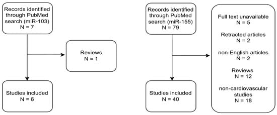
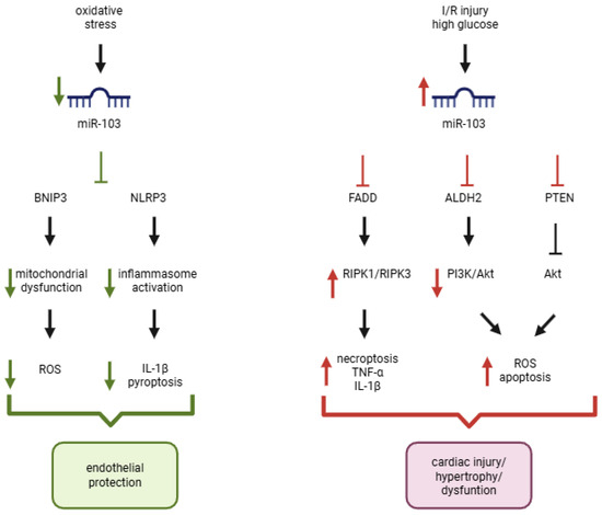
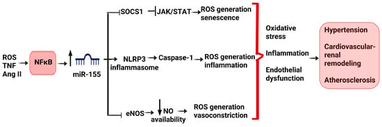
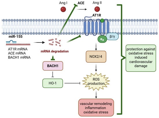
Background/Objectives: Oxidative stress is a major contributor to the pathogenesis of cardiovascular diseases, including hypertension, ischemic cardiomyopathy, and heart failure. MicroRNAs (miRNAs) have been extensively investigated in various contexts, and some of them have been identified to play a role in cardiovascular disease. This narrative review focuses on miR-103 and miR-155, two miRNAs implicated in the modulation of oxidative stress and cardiovascular remodeling. Methods: The following queries were used in PubMed since inception until May 2026: ((“miR-155” OR “microRNA-155” OR miR155) AND (“oxidative stress” OR ROS OR “reactive oxygen species”) AND (“cardiovascular disease” OR cardiovascular OR cardiac OR heart OR vascular)); ((“miR-103” OR “microRNA-103” OR miR103) AND (“oxidative stress” OR ROS OR “reactive oxygen species”) AND (“cardiovascular disease” OR cardiovascular OR cardiac OR heart OR vascular)). Results: A total of seven citations for miR-103 and 79 citations for miR-155 were identified. Reviews and papers about diseases other than those on cardiac/vascular involvement were excluded. miR-155 emerges as a potential regulator of inflammatory-redox signaling, whereas miR-103 appears more closely linked to cell fate and metabolic pathways. In both cases, available evidence supports a context-dependent role that challenges simplistic classification as pro- or antioxidant miRNAs. Conclusions: Available evidence suggests that both miR-103 and miR-155 are important regulators of oxidative stress-related pathways in cardiovascular disease. Nevertheless, the context-dependent effects observed across different cardiovascular disorders raise concerns regarding the safety of systemic miRNA modulation-based therapeutic strategies. Future studies should clarify the determinants of this context-dependent behavior and identify the specific conditions under which these miRNAs exert protective or harmful effects, which might pave the way for the development of miRNA-based therapeutic strategies targeting oxidative stress and cardiovascular remodeling.
Full article
(This article belongs to the Special Issue Cardiovascular Convergence: Bridging Basic, Translational and Clinical Research)
►▼
Show Figures

Figure 1
Open AccessReview
Assessment of Exercise Intolerance and Evaluation for HFpEF: A Literature Review of Pathophysiology, Diagnosis, and Management
by
Ali Moradi, Kurt Ramey, Kutiba Tabbaa, Abdullah Sahyouni, Kevin Sanchez Garcez, Shivanshu Kumar, Elaine Pan, Grant Barton, Olugbenga Oyesanmi and Robert Subbiondo
Pathophysiology 2026, 33(3), 63; https://doi.org/10.3390/pathophysiology33030063 - 20 Aug 2026
Abstract
Heart failure with preserved ejection fraction (HFpEF) is a type of heart failure in which the ejection fraction remains within the normal range (≥50%); however, patients still experience typical symptoms of heart failure. One of the most common manifestations in this population is
[...] Read more.
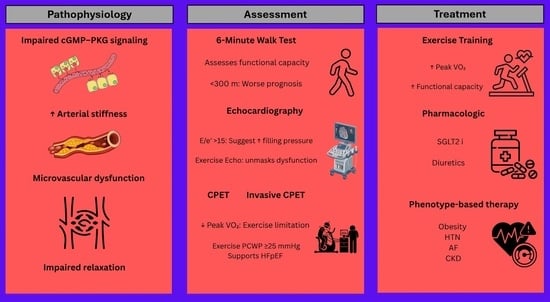
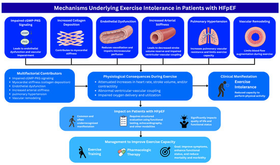
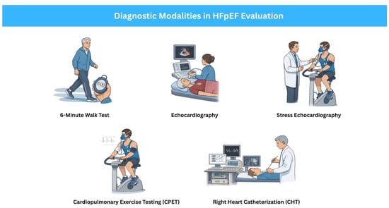
Heart failure with preserved ejection fraction (HFpEF) is a type of heart failure in which the ejection fraction remains within the normal range (≥50%); however, patients still experience typical symptoms of heart failure. One of the most common manifestations in this population is exercise intolerance, which in some cases may be the only presenting symptom. Exercise intolerance refers to a reduced capacity to perform physical activity. Several processes, including impaired cGMP–PKG signaling, increased collagen deposition, endothelial dysfunction, increased arterial stiffness, pulmonary hypertension, and vascular remodeling, contribute to the pathophysiology of HFpEF. The disruption of these fundamental physiological processes significantly impairs exercise capacity, leading to attenuated increases in heart rate, stroke volume, and/or contractility, along with abnormal ventricular–vascular coupling during exertion. When EI is suspected, a spectrum of diagnostic modalities—from simple, low-cost tools such as the 6 min walk test to advanced imaging—can be used to evaluate its presence and its association with HFpEF. When EI in HFpEF is diagnosed, management should focus on addressing this limitation to improve quality of life and reduce morbidity and mortality. Exercise training and pharmacological therapies, such as SGLT2 inhibitors, may be beneficial in this population. In conclusion, EI is a common manifestation of HFpEF that significantly impacts patients’ quality of life. Understanding its underlying mechanisms and addressing it appropriately are essential to improving patient outcomes.
Full article
(This article belongs to the Collection Feature Papers in Pathophysiology)
►▼
Show Figures

Graphical abstract
Open AccessReview
Erythropoiesis-Targeted Doping in Sports: From Improved Oxygen Transport to Cardiovascular Risk
by
Gabriela Chlebowska, Krzysztof Michalak, Łukasz Mazur and Wioletta Szczurek-Wasilewicz
Pathophysiology 2026, 33(3), 62; https://doi.org/10.3390/pathophysiology33030062 - 12 Aug 2026
Abstract
►▼
Show Figures
Erythropoiesis-targeted doping remains a major challenge for sports medicine because pharmacological and genetic manipulation of erythropoiesis can improve oxygen transport and endurance performance while increasing the risk of serious cardiovascular complications. Erythropoiesis-targeting strategies extend beyond recombinant erythropoietin (EPO) to include hypoxia-inducible factor prolyl
[...] Read more.
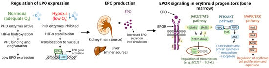
Erythropoiesis-targeted doping remains a major challenge for sports medicine because pharmacological and genetic manipulation of erythropoiesis can improve oxygen transport and endurance performance while increasing the risk of serious cardiovascular complications. Erythropoiesis-targeting strategies extend beyond recombinant erythropoietin (EPO) to include hypoxia-inducible factor prolyl hydroxylase inhibitors (HIF-PHIs), modified erythropoietin receptor (EPOR) agonists, transforming growth factor beta (TGF-β) signaling inhibitors, cytoprotective EPO derivatives, and gene- or cell-based approaches, and require complementary detection strategies based on direct analytical methods and the Athlete Biological Passport (ABP). Although they may enhance oxygen delivery and endurance performance, excessive stimulation of erythropoiesis may increase blood viscosity, impair vascular function, and elevate the risk of hypertension, thromboembolic complications, and other cardiovascular (CV) events. Erythropoiesis-targeted doping has evolved beyond recombinant EPO into a diverse group of pharmacological and genetic strategies that require increasingly sophisticated detection approaches. A thorough understanding of their molecular mechanisms and cardiovascular consequences is essential for improving anti-doping surveillance and protecting athlete health. This review summarizes current erythropoiesis-targeting agents, their mechanisms of action, detection strategies, cardiovascular risks, and implications for anti-doping practice.
Full article

Figure 1
Open AccessArticle
Reproductive and Cardiometabolic Characterization of a Letrozole- and High-Fat Diet-Induced PMOS-like Rat Model: An Experimental Study
by
Milica Milinkovic Sorgic, Aleksandar Matic, Vladimir Jakovljevic, Nikola Jovic, Sladjana Novakovic, Teodora Todorovic, Jovan Milosavljevic, Bozidar Pindovic, Jasmina Sretenovic, Petar Canovic, Jovana Jakovljevic Uzelac and Jovana Joksimovic Jovic
Pathophysiology 2026, 33(3), 61; https://doi.org/10.3390/pathophysiology33030061 - 11 Aug 2026
Abstract
Background/Objectives: Polyendocrine metabolic ovarian syndrome (PMOS) is a complex endocrine–metabolic disorder of multifactorial etiology, driven by interactions among hyperandrogenism, metabolic dysfunction, oxidative stress, and chronic low-grade inflammation that interact to promote reproductive dysfunction and increase risk of cardiometabolic complications. Although the combination of
[...] Read more.
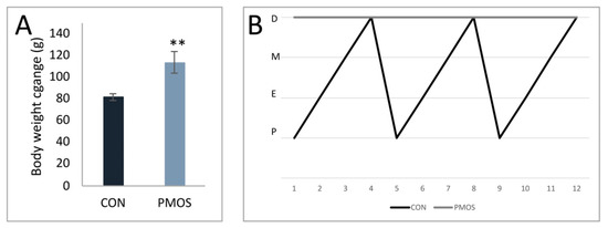
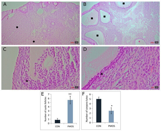
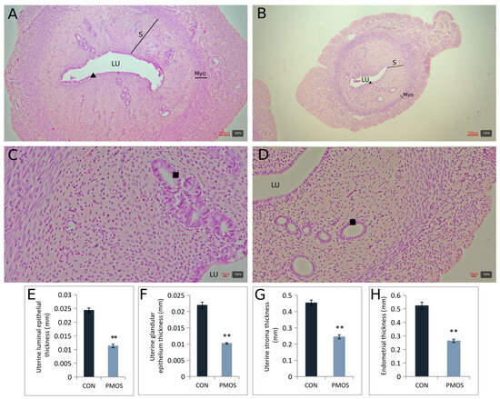
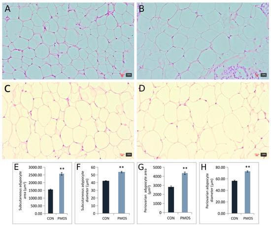
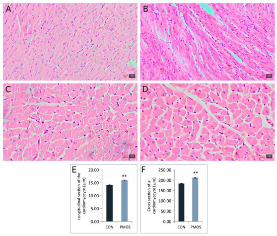
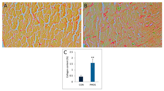
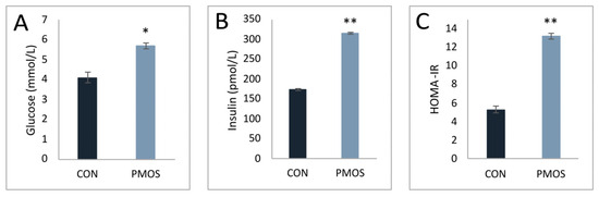
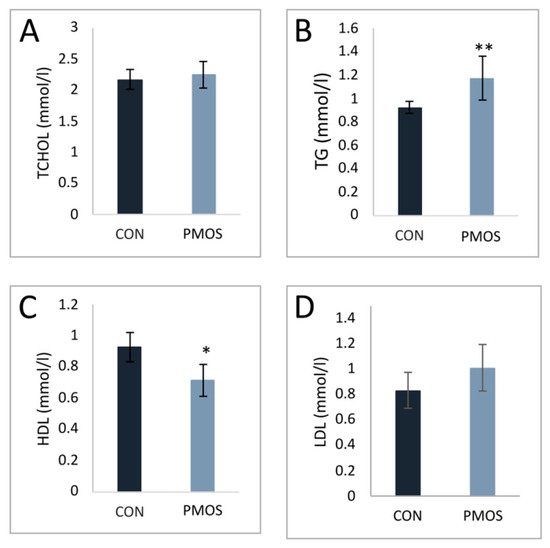
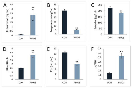
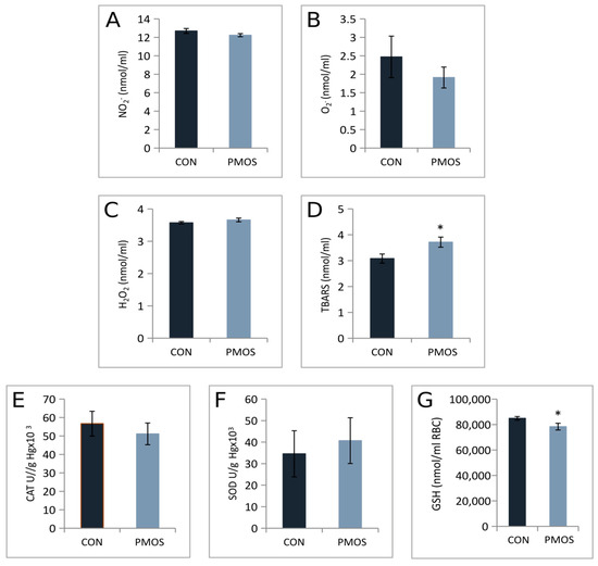
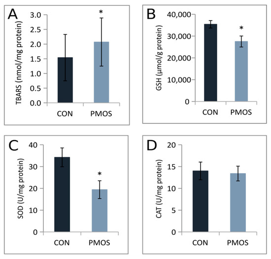
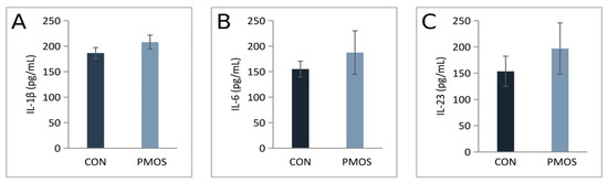
Background/Objectives: Polyendocrine metabolic ovarian syndrome (PMOS) is a complex endocrine–metabolic disorder of multifactorial etiology, driven by interactions among hyperandrogenism, metabolic dysfunction, oxidative stress, and chronic low-grade inflammation that interact to promote reproductive dysfunction and increase risk of cardiometabolic complications. Although the combination of letrozole and high-fat diet (LET + HFD) represents a widely used experimental approach for inducing a PMOS-like phenotype in rats, its multisystem pathophysiological features remain incompletely characterized. The present study aimed to characterize reproductive, cardiometabolic, hormonal, inflammatory, oxidative, and morphometric alterations associated with a LET + HFD-induced PMOS-like phenotype in rats. Methods: PMOS-like model was induced in rats over a 21-day period of orally administered letrozole combined with a high-fat diet. Results: Body weight gain was higher, while uterine weight was lower in the PMOS group compared with controls. Metabolic parameters suggested insulin resistance, while hormonal profiling revealed increased total testosterone and LH levels, accompanied by reduced FSH, estradiol, and progesterone levels. Elevated triglycerides and reduced HDL levels were also observed in the PMOS group. Morphometric analysis revealed numerous atretic and large thin-walled cystic follicles in the ovaries, accompanied by thinning of the uterine luminal and glandular epithelium, stromal layer, and hypoplasia of endometrial glands. Both the longitudinal diameter and cross-sectional area of cardiomyocytes were increased in the PMOS group, together with nuclear hypertrophy and enhanced myocardial collagen deposition. In addition, altered oxidative stress markers, including increased lipid peroxidation and reduced glutathione levels, were observed, whereas IL-6, IL-1β, and IL-23 were not significantly altered. Conclusions: The LET + HFD model reproduces key reproductive and cardiometabolic features of PMOS, extending beyond reproductive dysfunction to involve oxidative and structural alterations in multiple organs. These findings support its use for investigating PMOS pathophysiology and evaluating potential preventive and therapeutic strategies.
Full article
(This article belongs to the Section Metabolic Disorders)
►▼
Show Figures

Figure 1
Open AccessSystematic Review
A Comprehensive Meta-Analysis of the Cardiovascular Adverse Effects of Bispecific Antibody Therapy in Hematologic Malignancies
by
Derek Tai, Navneet Sandhu, Aren Dermarderosian, Norayr Mkrtchyan, Daniel Park, Vinisha Garg, Angel Nguyen and Mojtaba Akhtari
Pathophysiology 2026, 33(3), 60; https://doi.org/10.3390/pathophysiology33030060 - 11 Aug 2026
Abstract
►▼
Show Figures
Introduction: Bispecific antibodies (BsAbs) are a promising and potent therapeutic intervention for relapsed or refractory (R/R) hematologic malignancies, including acute lymphoblastic leukemia (ALL), multiple myeloma (MM), and B-cell lymphomas. These recombinant molecules are designed to target two distinct antigens or epitopes. While BsAbs
[...] Read more.
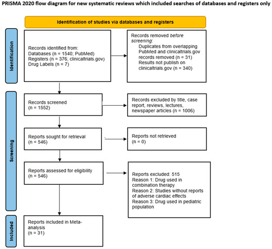
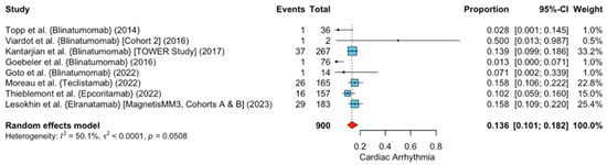
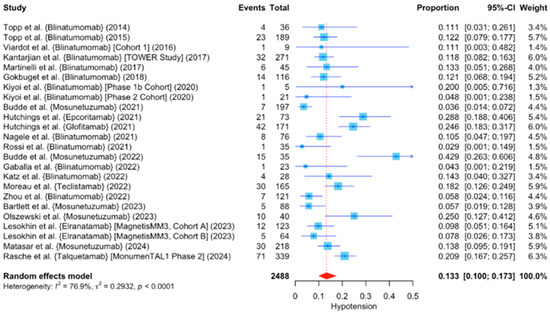
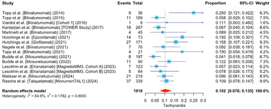
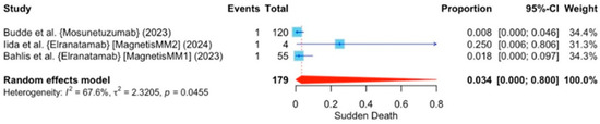
Introduction: Bispecific antibodies (BsAbs) are a promising and potent therapeutic intervention for relapsed or refractory (R/R) hematologic malignancies, including acute lymphoblastic leukemia (ALL), multiple myeloma (MM), and B-cell lymphomas. These recombinant molecules are designed to target two distinct antigens or epitopes. While BsAbs have shown significant efficacy in managing hematologic malignancies, toxicities must be carefully monitored and their safety profiles require further investigation. This meta-analysis investigates cardiovascular adverse effects associated with seven BsAbs across multiple clinical trials. Methods: A systematic literature review identified seven BsAbs, blinatumomab, elranatamab, epcoritamab, glofitamab, mosunetuzumab, talquetamab, and teclistamab, analyzing 25 clinical trials (Phase I–III) that included monotherapy regimens. The cardiovascular adverse events studied were arrhythmias, heart failure, blood pressure changes, and myocardial infarctions. A meta-analysis using the R meta package calculated proportions and 95% confidence intervals (CIs) and employed a random-effects model to assess heterogeneity. Results: Among 3215 patients, pooled analysis revealed significant findings with 13.6% cardiac arrhythmia (95% CI 10.1–18.2%, p = 0.05), 13.3% hypotension (95% CI 10–17.3%, p < 0.0001), 10.2% tachycardia (95% CI 7.6–13.5%, p = 0.0003), and 3.4% sudden death (95% CI 0–80%, p = 0.046). Less frequent events were hypertension (8%), acute myocardial infarction (1.5%), heart failure (1.4%), and atrial fibrillation (1.3%). Discussion: As BsAbs see increased use in R/R hematologic malignancies, cardiovascular complications must be closely monitored. This analysis highlights hypotension, cardiac arrhythmias, tachycardia, and sudden death as significant adverse effects. Although study heterogeneity and limited patient-level data may have influenced incidence estimates, these findings support routine cardiovascular monitoring and underscore the need for prospective studies to identify high-risk patients and optimize prevention and management strategies. Oncologists, cardiologists, and pharmacists should establish strategies for early detection and management to optimize patients’ outcomes.
Full article

Figure 1

Open AccessReview
The Microbiota–Gut–Brain Axis and Nutritional Interventions in Amyotrophic Lateral Sclerosis: Pathophysiological Mechanisms, Neuroinflammation, and Non-Motor Manifestations—Scoping Review
by
Elena Sanchis-Sanchis, José Enrique de la Rubia Ortí, David Sancho-Cantus, Cristina Cunha-Pérez and Jorge Casaña-Mohedo
Pathophysiology 2026, 33(3), 59; https://doi.org/10.3390/pathophysiology33030059 - 10 Aug 2026
Abstract
►▼
Show Figures
Amyotrophic Lateral Sclerosis (ALS) is a progressive neurodegenerative disorder in which systemic pathophysiological alterations significantly contribute to disease progression and non-motor manifestations, such as depression and anxiety. The microbiota–gut–brain axis represents a critical bidirectional pathway in which intestinal dysbiosis and epithelial barrier disruption
[...] Read more.
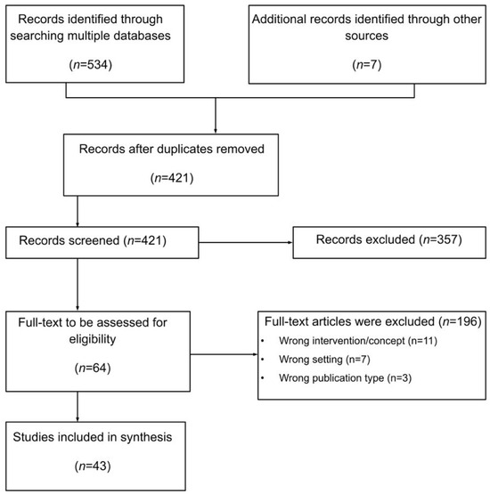
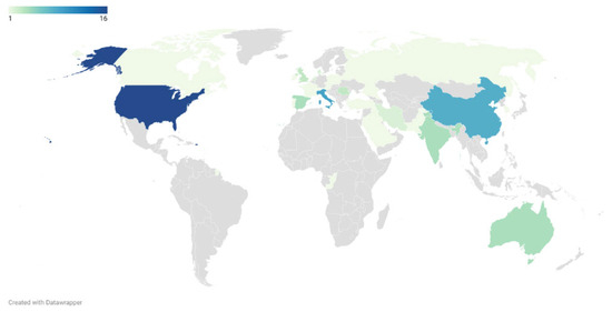
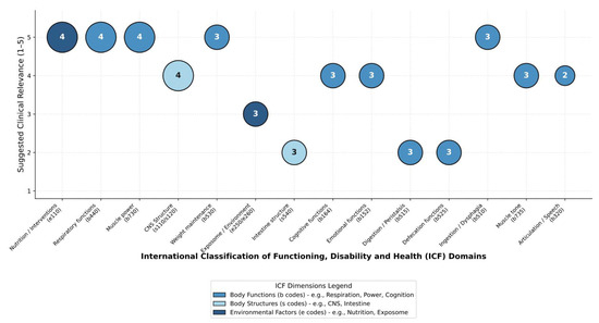
Amyotrophic Lateral Sclerosis (ALS) is a progressive neurodegenerative disorder in which systemic pathophysiological alterations significantly contribute to disease progression and non-motor manifestations, such as depression and anxiety. The microbiota–gut–brain axis represents a critical bidirectional pathway in which intestinal dysbiosis and epithelial barrier disruption catalyze central neuroinflammation. This scoping review synthesizes evidence from 43 empirical and analytical studies across 28 countries and maps the findings under the WHO International Classification of Functioning (ICF) framework. Pathophysiological data reveal a profound taxonomic shift in patients with ALS, characterized by severe depletion of neuroprotective, butyrate-producing genera (Akkermansia and Prevotella) and enrichment of pro-inflammatory Enterobacteriaceae. This dysbiotic state leads to structural damage to the intestinal mucosa, alteration of Paneth cells, and downregulation of tight junction proteins (zonulin), triggering a “leaky gut” phenomenon. Subsequent systemic translocation of lipopolysaccharides (LPS) induces TLR4-mediated endotoxemia, microglial hyperactivation, and accelerated motor neuron apoptosis. Conversely, therapeutic modulation via Fecal Microbiota Transplantation (FMT), psychobiotics, and metabolic interventions (ketogenic or Mediterranean diets) has demonstrated significant efficacy in restoring epithelial integrity, mitigating mitochondrial hypermetabolism, and reducing emotional distress. This review identifies a critical research gap in the microstructural characterization of the enteric nervous system in ALS. Incorporating microbiome-targeted biomarkers into clinical protocols is crucial for implementing a stratified, multi-systemic therapeutic strategy aimed at enhancing patient prognosis and psychological well-being.
Full article

Figure 1
Open AccessReview
Heart Meets Brain: Insights into Neurocardiac Pathophysiology
by
Berk Rasheed, Jacob Freed, Ruhi Parikh, Aman Singh and Krishna K. Singh
Pathophysiology 2026, 33(3), 58; https://doi.org/10.3390/pathophysiology33030058 - 10 Aug 2026
Abstract
The intricate relationship between the heart and the brain has transitioned from historical, cardio-centric theories to a modern, circuit-based, bi-directional framework known as neurocardiology. This review provides a comprehensive anatomical, physiological, and clinical synthesis of the bidirectional heart–brain axis, highlighting how disruptions in
[...] Read more.
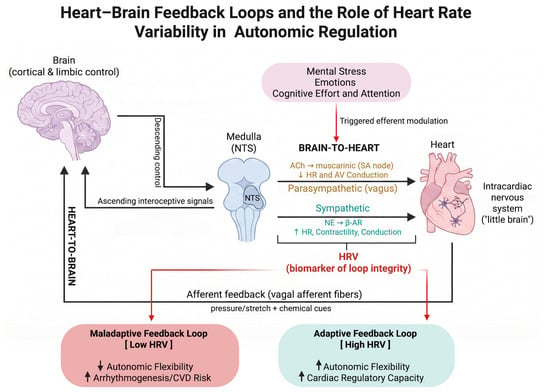
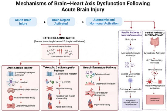
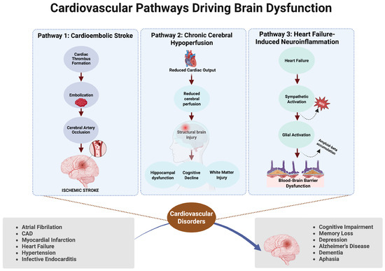
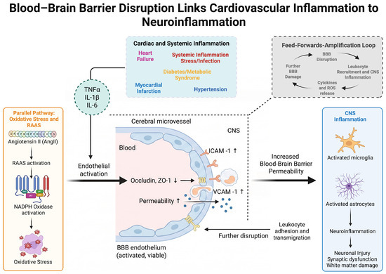
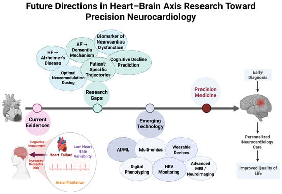
The intricate relationship between the heart and the brain has transitioned from historical, cardio-centric theories to a modern, circuit-based, bi-directional framework known as neurocardiology. This review provides a comprehensive anatomical, physiological, and clinical synthesis of the bidirectional heart–brain axis, highlighting how disruptions in one organ systematically impact the other. Structurally, the heart–brain axis operates as a closed-loop feedback network. Vagal parasympathetic control dominates at rest, driven by an asymmetrical fiber distribution where sensory afferents actively supply interoceptive data to the brainstem. Hemodynamically, cerebral blood flow delivery is linked to cardiac output in an age-dependent fashion with the related vascular dysfunction. Clinically, brain-to-heart pathologies like acute brain injury trigger life-threatening catecholamine surges, neuroinflammation, and myocardial stunning, including Takotsubo cardiomyopathy. Conversely, heart-to-brain pathways reveal that atrial fibrillation and heart failure can independently trigger cardioembolic stroke, long-term neuroinflammation, and a profound, progressive burden of cognitive decline. The heart and brain are inexorably linked through complex structural, mechanical, and paracrine pathways. Ameliorating clinical outcomes demands integrated cross-specialty diagnostic screening, digital rhythm monitoring, and holistic neurocardioprotective therapeutic strategies.
Full article
(This article belongs to the Special Issue Cardiovascular Convergence: Bridging Basic, Translational and Clinical Research)
►▼
Show Figures

Figure 1
Open AccessReview
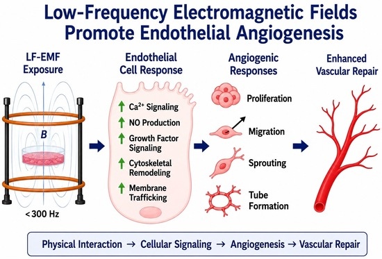
Time-Varying Low-Frequency Electromagnetic Fields and Endothelial Angiogenesis: From Early In Vitro Discoveries to Emerging Mechanisms
by
Linghong Li and Wayne F. Patton
Pathophysiology 2026, 33(3), 57; https://doi.org/10.3390/pathophysiology33030057 - 10 Aug 2026
Abstract
Background/Objectives: Time-varying low-frequency electromagnetic fields (LF-EMFs), including pulsed electromagnetic fields (PEMFs) and sinusoidal low-frequency electromagnetic fields, have been investigated as modulators of tissue repair and vascular remodeling. Although numerous studies report pro-angiogenic effects in endothelial systems, the underlying mechanisms remain incompletely understood. This
[...] Read more.
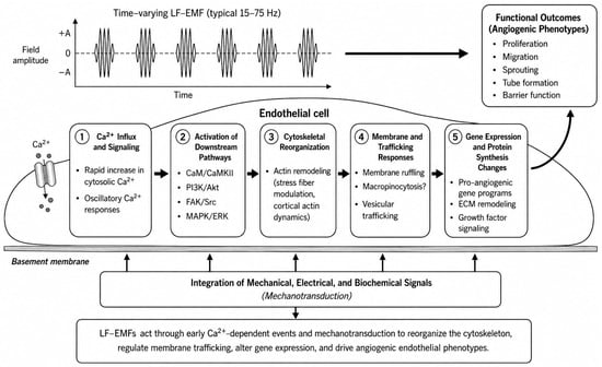
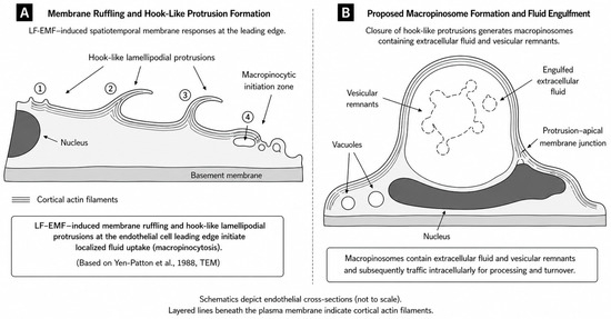
Background/Objectives: Time-varying low-frequency electromagnetic fields (LF-EMFs), including pulsed electromagnetic fields (PEMFs) and sinusoidal low-frequency electromagnetic fields, have been investigated as modulators of tissue repair and vascular remodeling. Although numerous studies report pro-angiogenic effects in endothelial systems, the underlying mechanisms remain incompletely understood. This review synthesizes evidence for LF-EMF-induced endothelial angiogenesis and evaluates emerging mechanistic explanations. Methods: Literature was identified through searches of PubMed, Web of Science, and Google Scholar through March 2026. Peer-reviewed studies emphasizing in vitro endothelial angiogenesis models were prioritized, with selected in vivo studies included when relevant to mechanistic interpretation. The review was conducted as a narrative synthesis rather than a systematic review or meta-analysis. Results: Experimental evidence indicates that LF-EMF exposure can enhance endothelial proliferation, migration, sprouting, tube formation, and survival across diverse model systems. Reported mechanisms include modulation of growth factor signaling, calcium-dependent pathways, nitric oxide production, cytoskeletal remodeling, and mechanotransduction. Reexamination of early ultrastructural studies suggests that membrane ruffling, vacuole formation, and vesicular structures observed following LF-EMF exposure may be interpreted within the framework of macropinocytosis and vesicular trafficking, providing a potential mechanistic link between electromagnetic stimulation and endothelial morphogenesis. Conclusions: Current evidence supports the conclusion that LF-EMFs can modulate key endothelial processes involved in angiogenesis and vascular remodeling. Membrane trafficking and macropinocytosis provide a biologically plausible framework linking historical observations with contemporary endothelial cell biology, although direct experimental validation is needed. Future progress will require standardized exposure paradigms and mechanistic studies using physiologically relevant angiogenesis models.
Full article
(This article belongs to the Section Cellular and Molecular Mechanisms)
►▼
Show Figures

Graphical abstract
Open AccessReview
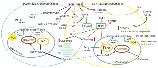
Oxidative Phosphorylation and Fatty Acid Oxidation Are Central to Mitochondrial Metabolism Rewiring in CML Stem/Progenitor Cell Survival
by
Jelena Milenkovic, Dijana Stojanovic, Branka Djordjevic, Sanja Velickovic, Vladana Stojiljkovic, Milica Veljkovic and Maja Milojkovic
Pathophysiology 2026, 33(3), 56; https://doi.org/10.3390/pathophysiology33030056 - 6 Aug 2026
Abstract
Background/Objectives: Quiescent leukemia stem cells (LSCs) are self-renewing, pluripotent cells that present a major obstacle to the successful curative treatment of chronic myeloid leukemia (CML). LSCs function independently of BCR::ABL1 signaling and persist following tyrosine kinase inhibitor treatment. The mechanisms enabling LSC
[...] Read more.
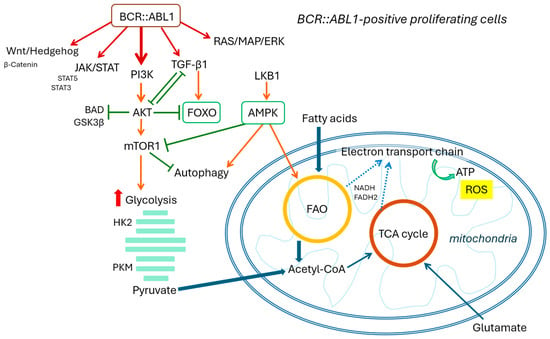
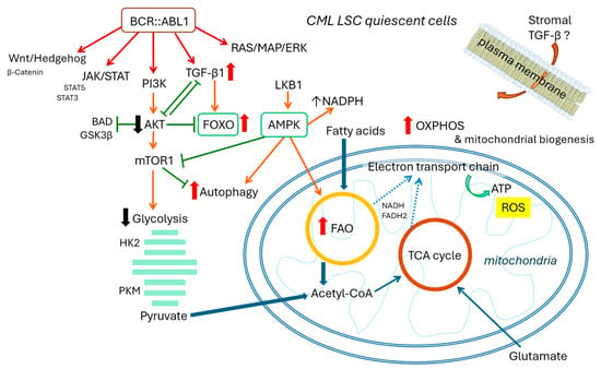
Background/Objectives: Quiescent leukemia stem cells (LSCs) are self-renewing, pluripotent cells that present a major obstacle to the successful curative treatment of chronic myeloid leukemia (CML). LSCs function independently of BCR::ABL1 signaling and persist following tyrosine kinase inhibitor treatment. The mechanisms enabling LSC survival are a central focus of current CML research. This review details the complex relationship between signaling pathways and discusses recent advancements in energy metabolism research within the pathogenesis of CML. Discussion: Energy metabolism is critical to the biology of CML LSCs. These cells depend on oxidative phosphorylation (OXPHOS) and mitochondrial homeostasis, utilizing fatty acid oxidation as their primary ATP source. Research highlights significant alterations in signaling networks, marked by a dynamic interplay among dominant pathways within the CML clone. While TGF-β-FOXO signaling maintains the self-renewal capacity of quiescent LSCs, proliferating mature CML cells rely heavily on glycolysis and the PI3K/Akt pathway. Furthermore, unique metabolic traits of LSCs underscore the impact of leukemic cell–microenvironment interactions in fostering a permissive niche. Conclusions: Fatty acid oxidation is critical to the survival and self-renewal of CML LSCs. This adaptation of mitochondrial function is closely linked to signaling alterations and entails an adjustment of mitochondrial respiration alongside stimulated OXPHOS. Emerging research unveils many potential targets within metabolic signaling that can be exploited to overcome these survival mechanisms, highlighting the disruption of mitochondrial energy support as a promising strategy to selectively eradicate CML LSCs.
Full article
(This article belongs to the Section Cellular and Molecular Mechanisms)
►▼
Show Figures

Graphical abstract
Open AccessArticle
Effects of Maternal Heartbeat Exposure on Neonatal Autonomic Regulation
by
Sara Puddu, Stefano Vicentin, Filippo Zemin, Paola Veronese and Elisa Cainelli
Pathophysiology 2026, 33(3), 55; https://doi.org/10.3390/pathophysiology33030055 - 3 Aug 2026
Abstract
►▼
Show Figures
Background: During intrauterine life, the fetus is continuously exposed to rhythmic sensory stimuli; among these, the maternal heartbeat is one of the earliest and most biologically salient, contributing to the early organization of physiological regulation. This exploratory longitudinal observational study examined whether exposure
[...] Read more.
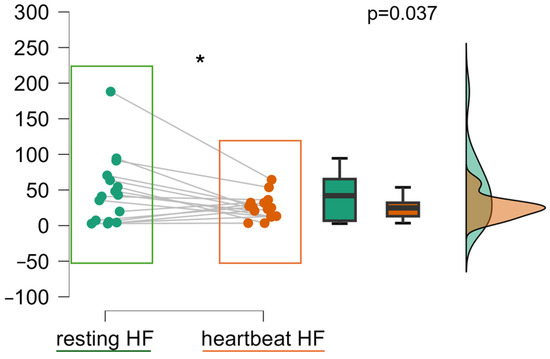
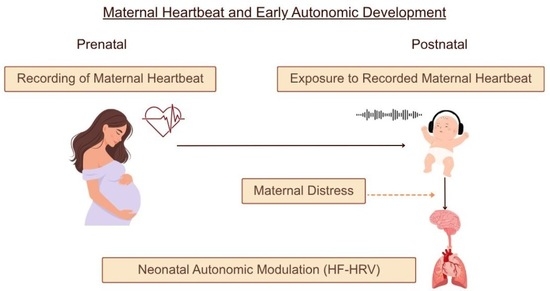
Background: During intrauterine life, the fetus is continuously exposed to rhythmic sensory stimuli; among these, the maternal heartbeat is one of the earliest and most biologically salient, contributing to the early organization of physiological regulation. This exploratory longitudinal observational study examined whether exposure to the maternal heartbeat influences neonatal autonomic activity and whether this response is moderated by prenatal maternal psychological distress. Methods: Sixteen dyads of mother–child were included in the analyses. Pregnant women were first assessed between 24 and 28 weeks of gestation: maternal distress was measured using the Depression Anxiety Stress Scales—21 (DASS-21), and maternal heartbeat recordings were obtained. At 3–4 weeks postpartum, neonates were exposed to recordings of their own mother’s heartbeat. Neonatal heart rate variability (HRV) was assessed using electrocardiography at rest and during the hearing of the maternal heartbeat. Results: Exposure to the heartbeat significantly reduced high-frequency (HF) HRV compared to baseline (p = 0.037), providing evidence for modulation of parasympathetic activity. Importantly, an interaction between condition and maternal distress was observed (p = 0.021), suggesting that neonatal autonomic responses differ as a function of maternal psychological state. Conclusions: Overall, these preliminary findings suggest that neonatal autonomic responses to maternal heartbeat exposure may not be uniform but may vary according to maternal psychological distress, highlighting the potential role of the maternal psychophysiological context in shaping early autonomic regulation.
Full article

Graphical abstract
Open AccessSystematic Review
The Pathophysiological Mechanisms of Glia in Animal Models of Chronic Orofacial Pain: A Systematic Review
by
Afonso Brito, Bruno Daniel Carneiro and Daniel Humberto Pozza
Pathophysiology 2026, 33(3), 54; https://doi.org/10.3390/pathophysiology33030054 - 21 Jul 2026
Abstract
►▼
Show Figures
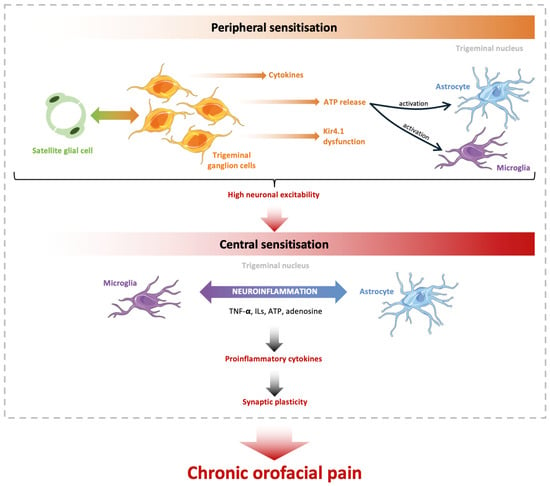
Background/Objectives: Chronic orofacial pain represents a heterogeneous group of disorders affecting the trigeminal system and remains difficult to treat due to its complex pathophysiology, which involves glial cells in the development and maintenance of persistent pain states. This systematic review aimed to
[...] Read more.
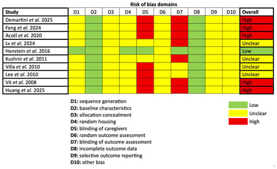
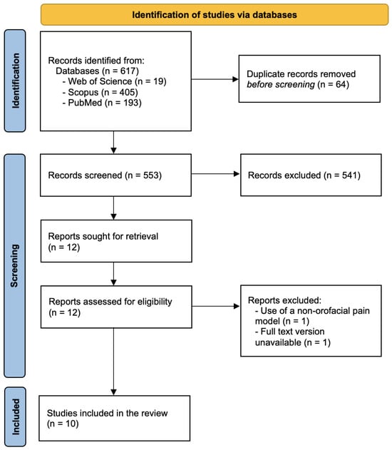
Background/Objectives: Chronic orofacial pain represents a heterogeneous group of disorders affecting the trigeminal system and remains difficult to treat due to its complex pathophysiology, which involves glial cells in the development and maintenance of persistent pain states. This systematic review aimed to synthesize evidence from animal studies investigating the pathophysiological role of glial cells in chronic orofacial pain. Methods: After registering on PROSPERO, a systematic search of the literature, based on PRISMA guidelines, was conducted to identify experimental animal studies evaluating glial involvement in orofacial pain models. After screening and eligibility assessment, 10 studies met the inclusion criteria. Data regarding experimental models, glial populations investigated, and pain-related behavioral outcomes were extracted and qualitatively synthesized. Results: Activation of satellite glial cells, microglia, and astrocytes was consistently associated with increased neuroinflammatory signaling and enhanced neuronal excitability within trigeminal pathways, demonstrating that both peripheral and central nervous tissues were involved. Several studies reported that pharmacological modulation of glial activity may reduce pain-related behaviors. Conclusions: Glial cells are key modulators of chronic orofacial pain through neuroimmune interactions that contribute to peripheral and central sensitization. Although these findings highlight promising therapeutic targets, further translational research is required to clarify their relevance for human pain conditions.
Full article

Figure 1

Open AccessCommunication
Are Sputum High Mobility Group Box 1 and D-Dimer Changes Relevant Markers of Tissue Damage and Fibrinolysis in Cystic Fibrosis?
by
Sante Di Gioia, Annalucia Carbone, Pamela Vitullo, Domenico Tierno, Domenico Larobina, Gabriele Grassi, Mario Grassi and Massimo Conese
Pathophysiology 2026, 33(3), 53; https://doi.org/10.3390/pathophysiology33030053 - 21 Jul 2026
Abstract
►▼
Show Figures
Background/Objectives: Cystic fibrosis (CF) is a genetic disease whose hallmarks include chronic inflammation of the airways causing tissue damage with increased levels of alarmins in the respiratory secretions, as well as activation of the coagulation/fibrinolytic systems that are linked to inflammation. However, no
[...] Read more.
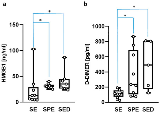
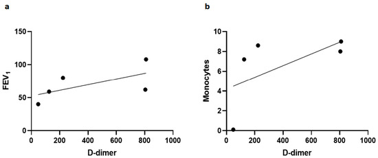
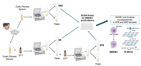
Background/Objectives: Cystic fibrosis (CF) is a genetic disease whose hallmarks include chronic inflammation of the airways causing tissue damage with increased levels of alarmins in the respiratory secretions, as well as activation of the coagulation/fibrinolytic systems that are linked to inflammation. However, no information is available about the fibrinolytic system and how fibrinolysis is involved in airway injury. On the road to understanding this issue, we determined levels of two key markers, High Mobility Group Box 1 (HMGB1), an alarmin, and D-dimers, associated with fibrin breakdown and airway inflammation, in the sputum from patients with cystic fibrosis. Methods: Sputum samples were collected from 13 individuals with CF and subjected to two different treatment protocols. In the first protocol, the sample was treated with dithiothreitol (DTT) and then centrifuged in order to collect the supernatant (SED). In the second protocol, the sample was centrifuged and the supernatant was obtained (SE). The pellet obtained was treated with DTT and then centrifuged in order to collect the supernatant (SPE). ELISA assays were performed on all samples. Results: HMGB1 and D-dimer levels were significantly lower in supernatants from sputum centrifuged before DTT treatment (SE) compared to those processed after DTT (SPE and SED). However, no significant difference was observed between SPE and SED samples for both markers. D-dimer levels in SED correlated positively with FEV1 and blood monocyte counts. Conclusions: Direct sputum processing with DTT may be a useful procedure for assessing biomarkers of inflammation and fibrinolysis in CF.
Full article

Graphical abstract
Open AccessFeature PaperArticle
SHP-2 Expression Is a Positive Prognostic Biomarker in Non-Small-Cell Lung Carcinoma: Association with KRAS Mutation and Prolonged Survival
by
Konstantinos Stamopoulos, Georgios Pilichos, Eleni Karatrasoglou, Penelope Korkolopoulou and Stratigoula Sakellariou
Pathophysiology 2026, 33(3), 52; https://doi.org/10.3390/pathophysiology33030052 - 18 Jul 2026
Abstract
Background/Objectives: Lung cancer tumors are the most frequent malignant tumors and the primary cause of cancer-related deaths. Despite the improvement in survival related to the implementation of immunotherapy, a significant proportion of patients fail to demonstrate satisfactory response. As a result, the
[...] Read more.
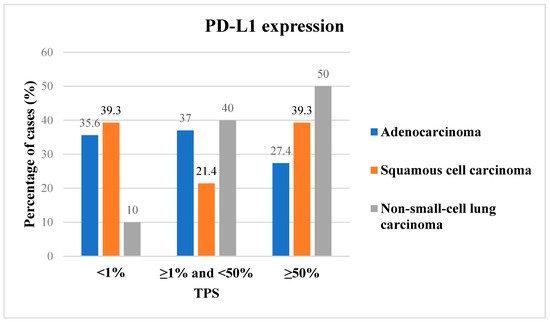
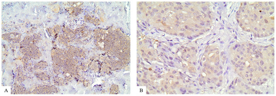
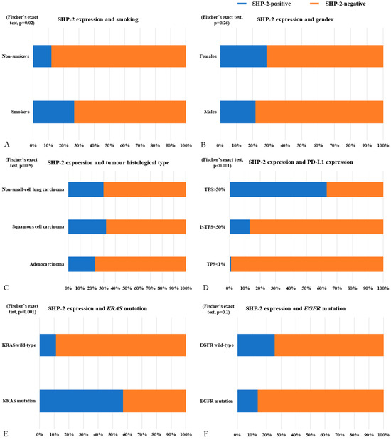
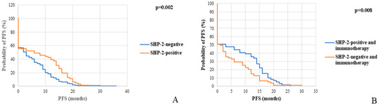
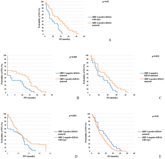
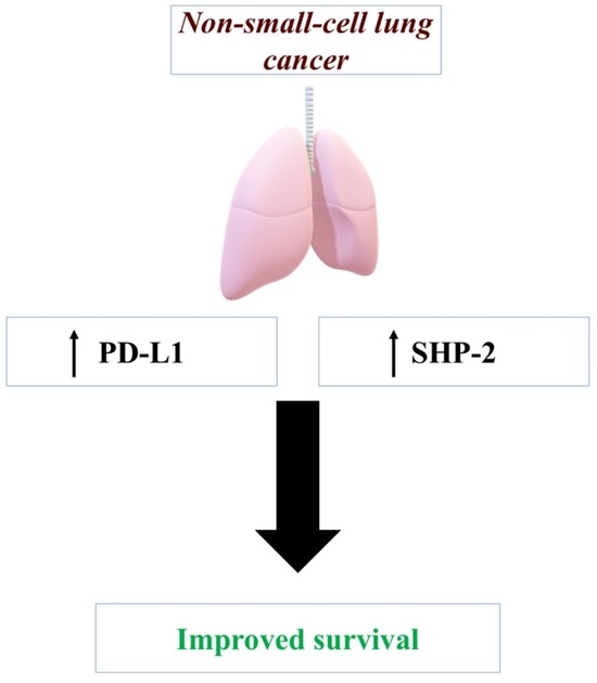
Background/Objectives: Lung cancer tumors are the most frequent malignant tumors and the primary cause of cancer-related deaths. Despite the improvement in survival related to the implementation of immunotherapy, a significant proportion of patients fail to demonstrate satisfactory response. As a result, the need for new biomarkers, which will predict patient response, as well as for novel therapeutic targets, is urgent. SHP-2 is an intracellular signal transduction molecule, modifying signal transduction of numerous intracellular cascades, acting primarily as an oncogene. In this study we analyzed the expression pattern of SHP-2 in a cohort of non-small-cell lung cancer cases and attempted to correlate it with available clinicopathological parameters. Methods: We performed immunohistochemistry for SHP-2 detection in a cohort of 258 NSCLC cases. H-score was estimated and statistical analyses correlated it with survival and other clinicopathological and molecular parameters. Results: SHP-2 expression was observed in 60 cases (23.3%), with an H-score ranging from 150 to 300. Positive samples were distributed among the three major histological subtypes, without statistically significant differences. Statistical analysis revealed significant positive correlation of SHP-2 expression with smoking, PD-L1 levels, and KRAS mutations. Moreover, positive SHP-2 immunohistochemistry was associated with better clinical outcome in both the total cohort and in the group of patients who received immunotherapy. Finally, the simultaneous presence of SHP-2 expression and KRAS mutation was correlated with prolonged survival. Conclusions: Our findings indicate that SHP-2 could be a useful prognostic factor and a very reliable biomarker in predicting response to immune checkpoint inhibitors.
Full article
(This article belongs to the Section Cellular and Molecular Mechanisms)
►▼
Show Figures

Graphical abstract
Highly Accessed Articles
Latest Books
E-Mail Alert
News
Topics
Topic in
Biomedicines, Clinics and Practice, Diseases, JCM, Pathophysiology, Diagnostics
Evolving Insights into Pathophysiology, Diagnosis, Therapy, and Prognosis in Acute Respiratory Distress Syndrome (ARDS): From Bench to Bedside—Third Edition
Topic Editors: Denise Battaglini, Patricia Rieken Macêdo Rocco, Raffaele MerolaDeadline: 31 March 2027
Special Issues
Special Issue in
Pathophysiology
Cardiovascular Convergence: Bridging Basic, Translational and Clinical Research
Guest Editors: Dragan Djuric, Vladimir JakovljevicDeadline: 30 November 2026
Special Issue in
Pathophysiology
Factors Modifying Response to Breast Cancer Treatment
Guest Editor: Robert Treat ChattertonDeadline: 5 December 2026
Special Issue in
Pathophysiology
Obstructive Sleep Apnea Phenotypes: First Decade of Exploration and Beyond
Guest Editors: Jonathan Steven Alexander, Oleg ChernyshevDeadline: 15 December 2026
Special Issue in
Pathophysiology
Inflammation as a Pathophysiological Link Between Obesity and Diabetes
Guest Editors: Omar Cauli, Oscar CaballeroDeadline: 31 December 2026
Topical Collections
Topical Collection in
Pathophysiology
Feature Papers in Pathophysiology
Collection Editor: Jonathan Steven Alexander