


Abstract
Background/Objectives: Oxidative stress is a major contributor to the pathogenesis of cardiovascular diseases, including hypertension, ischemic cardiomyopathy, and heart failure. MicroRNAs (miRNAs) have been extensively investigated in various contexts, and some of them have been identified to play a role in cardiovascular disease. This narrative review focuses on miR-103 and miR-155, two miRNAs implicated in the modulation of oxidative stress and cardiovascular remodeling. Methods: The following queries were used in PubMed since inception until May 2026: ((“miR-155” OR “microRNA-155” OR miR155) AND (“oxidative stress” OR ROS OR “reactive oxygen species”) AND (“cardiovascular disease” OR cardiovascular OR cardiac OR heart OR vascular)); ((“miR-103” OR “microRNA-103” OR miR103) AND (“oxidative stress” OR ROS OR “reactive oxygen species”) AND (“cardiovascular disease” OR cardiovascular OR cardiac OR heart OR vascular)). Results: A total of seven citations for miR-103 and 79 citations for miR-155 were identified. Reviews and papers about diseases other than those on cardiac/vascular involvement were excluded. miR-155 emerges as a potential regulator of inflammatory-redox signaling, whereas miR-103 appears more closely linked to cell fate and metabolic pathways. In both cases, available evidence supports a context-dependent role that challenges simplistic classification as pro- or antioxidant miRNAs. Conclusions: Available evidence suggests that both miR-103 and miR-155 are important regulators of oxidative stress-related pathways in cardiovascular disease. Nevertheless, the context-dependent effects observed across different cardiovascular disorders raise concerns regarding the safety of systemic miRNA modulation-based therapeutic strategies. Future studies should clarify the determinants of this context-dependent behavior and identify the specific conditions under which these miRNAs exert protective or harmful effects, which might pave the way for the development of miRNA-based therapeutic strategies targeting oxidative stress and cardiovascular remodeling.
1. Introduction
Oxidative stress is defined as the imbalance between the production and accumulation of pro-oxidant and antioxidant species [1,2]. Among reactive oxygen species (ROS), superoxide (O2−) is generated by several enzymatic sources, including NADPH oxidases, uncoupled nitric oxide synthase, xanthine oxidase, cytochrome P450, lipoxygenases, and cyclooxygenases. Due to its short half-life, O2− is rapidly converted into hydrogen peroxide (H2O2) by superoxide dismutase 1 and 2 (SOD1/2), a more stable and membrane-permeable ROS, which can further generate highly reactive hydroxyl radicals. Besides acting as signaling molecules, ROS can react with nitric oxide to form toxic reactive species such as peroxynitrite. To counterbalance oxidative stress, cells rely on antioxidant defenses, including enzymatic scavengers, such as superoxide dismutase and glutathione peroxidase, as well as adaptive mechanisms such as autophagy, whose effects may be either protective or harmful depending on the intensity and duration of oxidative stress [2,3,4]. While oxidants are essential for pathogen killing, their excessive production induces tissue damage. Failure of the antioxidant armamentarium to successfully moderate prooxidant species results in detrimental effects that contribute to multiple pathophysiological processes, such as aging, cancer, cardiovascular disease, kidney disease, and autoimmune diseases [1,5,6,7,8,9]. Given the high oxygen and metabolic demands of the heart, the cardiovascular system is particularly susceptible to oxidative stress. Oxidative stress plays a critical role in the pathogenesis of multiple cardiovascular diseases, such as hypertension, fibrotic and ischemic cardiomyopathy, diabetes, and pre-eclampsia, and is interconnected with inflammation and senescence [10,11,12].
MicroRNAs (miRNAs) are short non-coding RNA molecules, typically 18–22 nucleotides in length. More than 1000 miRNA genes are encoded in the human genome. The biosynthesis starts with transcription by RNA polymerase II and synthesis of the hairpin structure. This precursor miRNA then binds to Ran-GTP and exportin 5 and is finally exported to the cell cytoplasm. MiRNA precursors are then transformed into double-stranded fragments of about 22 nucleotides by Dicer. The RNA-induced silencing complex (RISC) then integrates with the mature guided transcript (miRNA). This RISC–miRNA complex directly targets newly synthesized mRNAs to undergo biogenesis inhibition or mRNA degradation [13] by interacting with the 3′ untranslated region (3′UTR) of target mRNAs. Through this interaction, they can suppress protein synthesis or promote mRNA degradation, thereby modulating multiple cellular processes [14,15,16]. An individual miRNA can bind to multiple transcripts, and a single transcript can be bound by various miRNAs, determining an intricate and dynamic posttranscriptional regulation [17]. Recent literature has shown that miRNAs can play a multifaceted role in the interplay between oxidative stress and cardiovascular disease [8,9].
Among these, miR-103 (also reported as miR-103-3p) belongs to the miR-103/107 family and is located on human chromosome 5 [18]. It is expressed in various cell types and has been described as an oncogenic miRNA because of its ability to enhance cellular proliferation in several cancers, including non-small cell lung cancer, breast cancer, colon cancer, and squamous cell carcinoma. Beyond its role in tumor biology, the miR-103/107 family has also been associated with hypertensive nephropathy, cardiovascular remodeling, oxidative stress, and metabolic disorders [18,19,20].
MiR-155 (also reported as mir-155-5p) is a highly conserved single-stranded non-coding RNA transcribed from the B-cell integration cluster (BIC) gene located on chromosome 21. miR-155-5p derives from the 5′ arm of the precursor miRNA hairpin and is the more biologically active form. Although it is predominantly expressed in hematopoietic cells, its expression has also been detected in fibroblasts, epithelial and reproductive tissues, as well as in the central nervous system [14]. This miRNA participates in a wide range of biological functions, such as immune and inflammatory responses, regulation of apoptosis and cell proliferation, maintenance of vascular homeostasis, hypertension, cardiovascular remodeling, and cancer [14,21]. Owing to its pleiotropic functions, miR-155 has emerged as a particularly complex regulator in cardiovascular pathophysiology [14,21,22].
MiR-103 and miR-155 have both been studied in oxidative stress in cardiovascular disease, and recent evidence suggests they may exert either protective or detrimental effects depending on the pathological context. This narrative review intentionally focuses on miR-103 and miR-155, as they provide an opportunity to discuss the complexity of miRNA-mediated regulation of oxidative stress in cardiovascular diseases and summarizes the available evidence from in vivo/in vitro and human-based studies of the role played by miR-103 and miR-155 in oxidative stress in cardiovascular disease.
2. Methods
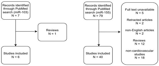
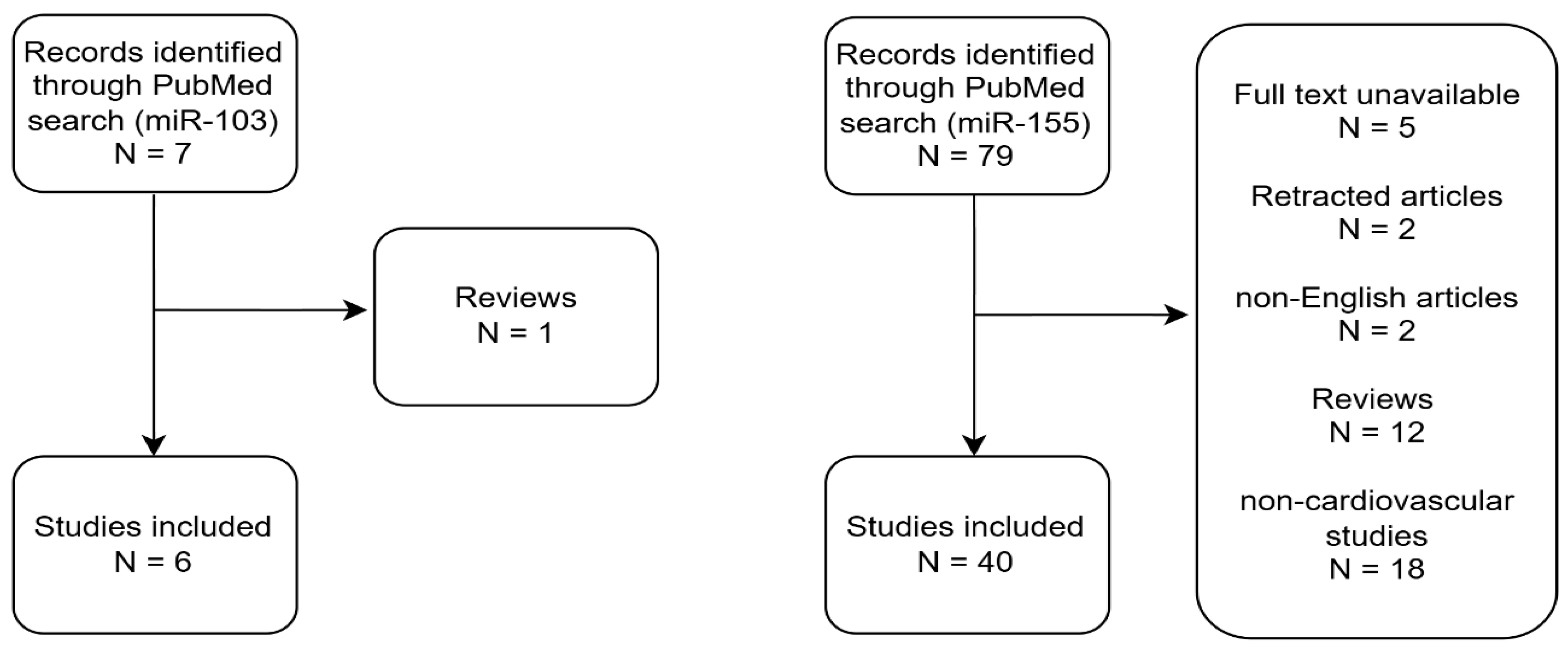
The following queries were used in PubMed since inception until 11 May 2026: ((“miR-155” OR “microRNA-155” OR miR155) AND (“oxidative stress” OR ROS OR “reactive oxygen species”) AND (“cardiovascular disease” OR cardiovascular OR cardiac OR heart OR vascular)); ((“miR-103” OR “microRNA-103” OR miR103) AND (“oxidative stress” OR ROS OR “reactive oxygen species”) AND (“cardiovascular disease” OR cardiovascular OR cardiac OR heart OR vascular)). Study selection was performed by one reviewer and independently verified by a second reviewer. Any discrepancies in opinion were settled by discussion and consensus between the two reviewers. The search produced a total of 7 citations for miR-103 and 79 citations for miR-155, two of which were recently retracted. Access was limited for 5 papers. Reviews and papers about diseases other than those with cardiac, vascular, or cardiovascular involvement were excluded. Only articles in English were fully read (Figure 1, Table 1 and Table 2). It should be noted that some reports analyze the miR-103/107 family collectively because of the high sequence homology and overlapping biological functions of the miRNAs. Such cases have been acknowledged and specified in the text.
Figure 1.
Flowcharts depicting study selection for miR-103 and miR-155.
3. Discussion
3.1. miR-103
MiR-103 has been particularly studied in the context of cardiac damage and function. Current evidence suggests a context-dependent role of miR-103 in cardiovascular oxidative stress, with both protective and detrimental effects being reported depending on the disease model and downstream molecular targets (Table 1).
Table 1.
Summary of Evidence about miR-103 Role in Oxidative Stress in Cardiovascular Disease.
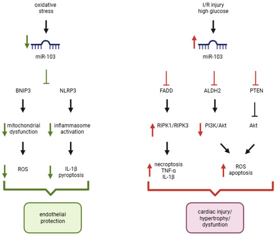
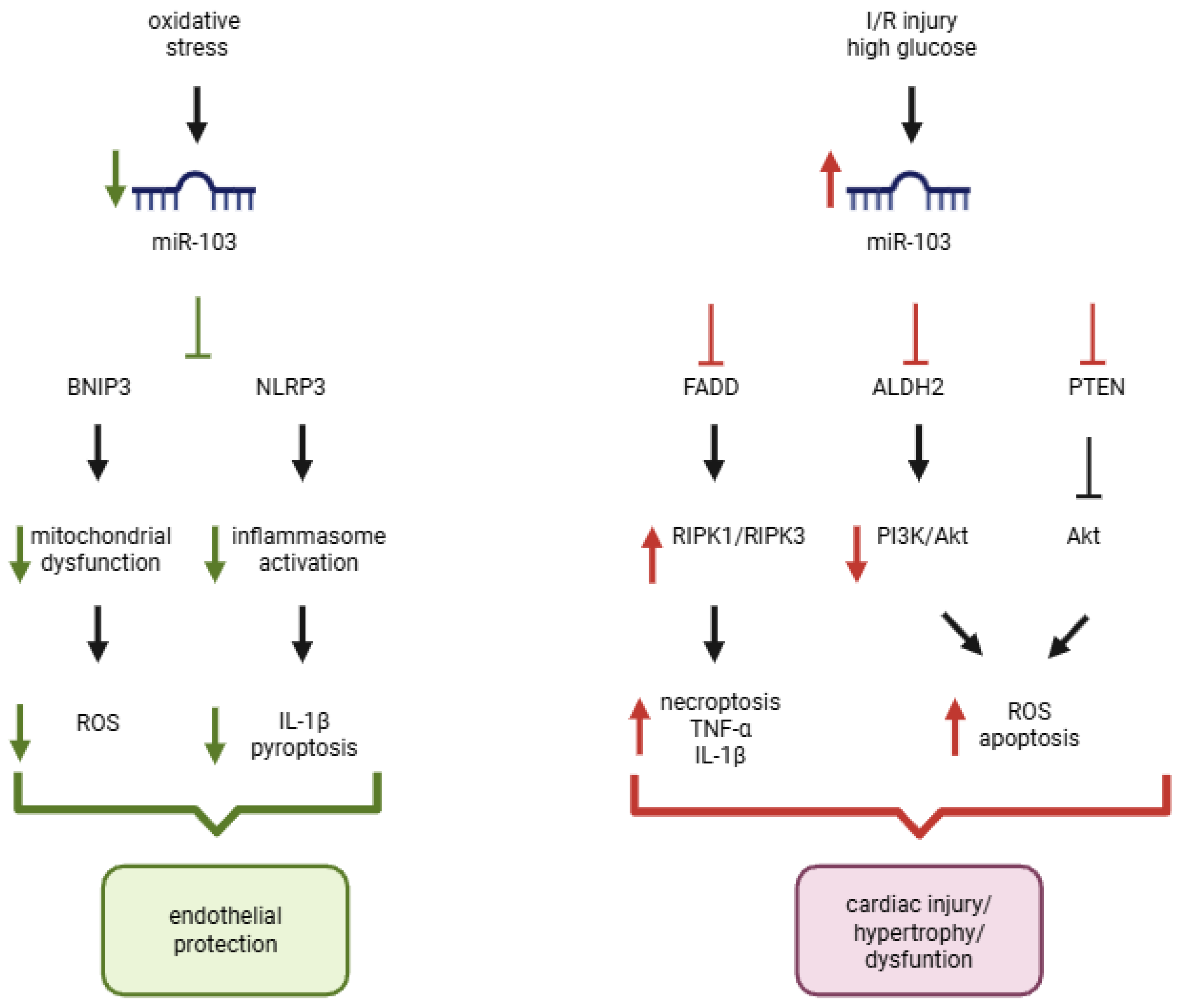
In cardiomyocytes from diabetic mice, high glucose increased miR-1033p levels together with ROS and reduced SOD. H19, a long non-coding RNA, worked as a sponge for miR-103-3p, thereby preventing the inhibition of ALDH2 (Aldehyde dehydrogenase 2), thus activating the PI3K/Akt protective pathway, ultimately resulting in reduced oxidative stress and apoptosis [23]. By reducing miR-103-3p, orientin, a flavonoid, exerted antioxidant effects [23]. Within the same pathway, in heart failure cardiomyocytes, miR-103 suppresses PTEN (phosphatase and tensin homolog), an inhibitor of AKT signaling [24], increasing ROS production and cardiac hypertrophy [24]. Interestingly, although miR-103 modulates different downstream targets across cardiovascular disorders, both studies converge on dysregulation of the PI3K/AKT signaling axis and oxidative stress pathways. In diabetic cardiomyopathy, miR-103-3p suppresses the antioxidant enzyme ALDH2, leading to impaired AKT-mediated cardioprotective signaling [23], whereas in pressure overload-induced heart failure, it targets PTEN, resulting in excessive AKT activation and maladaptive hypertrophic remodeling [24] (Figure 2).
Figure 2.
Context-dependent effects of miR-103 on cardiovascular oxidative stress. In endothelial cells exposed to oxidative stress, reduced miR-103 expression contributes to mitochondrial dysfunction and inflammasome activation through BNIP3 and NLRP3 signaling, whereas restoration of miR-103 attenuates ROS production, IL-1β release, and cell injury. Conversely, in pathological settings such as ischemia/reperfusion (I/R) injury, diabetic cardiomyopathy, and pressure overload-induced heart failure, increased miR-103/107 expression promotes inflammatory cell death, oxidative stress, and maladaptive remodeling by targeting FADD, ALDH2, and PTEN-dependent pathways. Collectively, current evidence indicates that miR-103 acts as a context-dependent regulator of oxidative stress and cell fate decisions in cardiovascular disease. Abbreviations: ALDH2: aldehyde dehydrogenase 2; BNIP3: BCL2 interacting protein 3; FADD: Fas-associated protein with death domain; IL-1β: interleukin-1 beta; I/R: ischemia/reperfusion; NLRP3: NOD-, LRR- and pyrin domain-containing protein 3; PTEN: phosphatase and tensin homolog; ROS: reactive oxygen species. Created in BioRender. Cacciapuoti, M. (2026) https://BioRender.com/1gyeils.
MiR-103 has also been studied in oxidative stress environments. In in vitro models of induced oxidative stress, mechanistic evidence demonstrated that H2O2 downregulated the expression of miR-103 in a time-dependent manner. Nevertheless, cells transfected with miR-103 showed increased viability and lower intracellular ROS formation in an H2O2-induced oxidative stress environment by targeting BNIP3 (Bcl2/adenovirus E1B 19 kDa interacting protein 3), a mitochondrial pro-apoptotic protein involved in mitochondrial dysfunction, oxidative stress, and cell death [20,25]. In endothelial coronary artery cells, the same pathway demonstrated that miR-103 inhibition aggravates pyroptosis through the NLRP3 inflammasome and is associated with higher levels of IL-1β expression. Conversely, in vitro models (cardiomyocytes) of induced oxidative stress have shown that exposure to high levels of H2O2 (500 μM) significantly increases miR-103/107 levels, which target FADD (Fas-associated death domain protein), thereby decreasing its protein expression and impairing its protective effect on necroptosis by preventing the formation of the complex RIPK1/RIPK3 (Receptor-interacting serine/threonine-protein kinases 1 and 3) [26].
Interestingly, the protective effects observed in endothelial oxidative injury contrast with findings from ischemia/reperfusion (I/R) models, where miR-103/107 appear to promote inflammatory cell death pathways. These findings are supported by mechanistic evidence based on loss-of-function and rescue experiments. In fact, Wang et al. observed that miR-103/107 was increased in murine models of I/R and was associated with higher levels of TNF-α and IL-1β and with worsening cardiac function. Finally, miR103/107 antagomir administration significantly reduced the necrosis area induced by H2O2 in murine models of ischemia/reperfusion (I/R) [26].
Human-based studies evaluating miR-103 in cardiovascular diseases are sporadic and provide poor observational evidence. MiR-103 levels were found to be lower in heart failure patients compared to healthy volunteers [27], but oxidative stress parameters were not assessed. Additionally, in a cohort of patients with Gitelman syndrome, a model of reduced oxidative stress, plasmatic miR-103 levels were higher than in controls [28].
Beyond cardiovascular disease, miR-103/107 have also emerged as key regulators of metabolic homeostasis through modulation of insulin sensitivity, adipocyte differentiation, and fatty acid β-oxidation via Caveolin-1-dependent pathways. These findings lead to the development of anti-miR-103/107-based treatment strategies for type 2 diabetes mellitus with Non-alcoholic Fatty Liver Disease (NAFLD)—dosed in a first-in-human phase I study by Regulus’s collaboration partner AstraZeneca—and non-alcoholic steatohepatitis (NASH) [28,29,30]. Nevertheless, whether modulation of miR-103 and miR-103/107 may represent a viable therapeutic strategy in cardiovascular diseases has not been investigated yet, given the context-dependent effects reported across different models of oxidative stress and cardiac injury. For now, no specific molecule targeting this miRNA is being studied as a potential therapy for cardiovascular diseases.
3.2. miR-155
Current findings do not support a univocal role for miR-155 in cardiovascular oxidative stress. Rather, its effects appear to vary according to the disease context, cellular environment, and downstream molecular targets involved (Table 2).
Table 2.
Summary of Evidence about miR-155 Role in Oxidative Stress in Cardiovascular Disease.
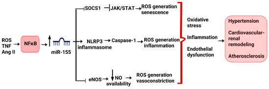
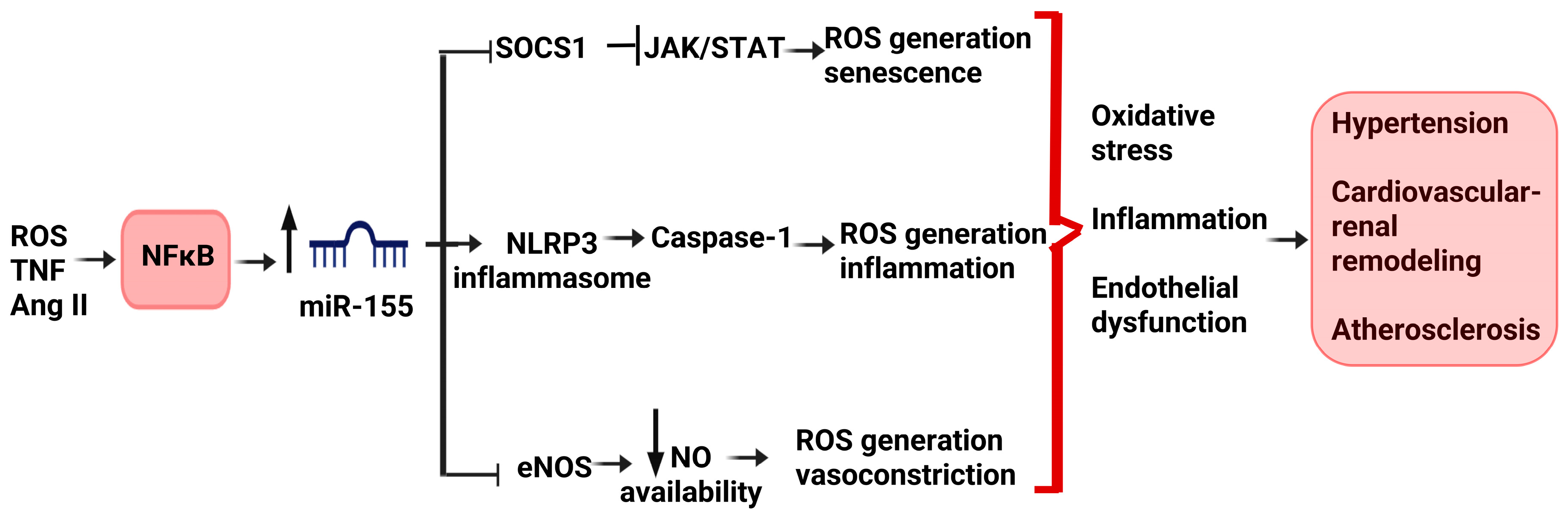
One of the main pathways linking miR-155 and oxidative stress in cardiovascular disease is that of NFκB. In fact, miR-155 biogenesis can be induced by NFκB, but miR-155 targets can also lead to NFκB activation, thereby contributing to oxidative stress and inflammation (Figure 3).
Figure 3.
Proposed NFκB-dependent pro-oxidant signaling mediated by miR-155 in cardiovascular disease. Oxidative and inflammatory stimuli, including ROS, TNF-α, and Ang II, activate NFκB, which promotes miR-155 expression. Increased miR-155 contributes to oxidative stress and vascular injury through multiple mechanisms, including suppression of SOCS1 with subsequent activation of JAK/STAT signaling, stimulation of the NLRP3 inflammasome pathway, and inhibition of eNOS, resulting in reduced nitric oxide bioavailability. Collectively, these pathways promote inflammation, endothelial dysfunction, cellular senescence, and ROS generation, ultimately contributing to hypertension, vascular remodeling, and atherosclerosis. Abbreviation: Ang II: angiotensin II; eNOS: endothelial nitric oxide synthase; JAK/STAT: Janus kinase/signal transducer and activator of transcription; miR-155: microRNA-155; NFκB: nuclear factor kappa B; NO: nitric oxide; ROS: reactive oxygen species; SOCS1: suppressor of cytokine signaling 1; TNF-α: tumor necrosis factor alpha. Created in BioRender. Cacciapuoti, M. (2026) https://BioRender.com/14u2rob.
MiR-155 was broadly studied in atherogenesis [15]. MiR-155 expression was found to be induced by oxLDL (but not native LDL) in macrophages, by worsening oxidative stress conditions. Moreover, miR-155 promoted ROS production in oxLDL-induced Raw264.7 cells [47]. These findings were also supported by two more studies conducted in endothelial cells, where anti-miR-155-5p attenuated oxidative stress (demonstrated by SOD expression reduction), and anti-miR-155-5p increased NO production, respectively, thereby contrasting endothelial dysfunction [37,64]. MiR-155 was demonstrated to promote vascular smooth muscle cell proliferation by inhibiting MST-2, which was identified as a target of miR-155, thus favoring inflammation and oxidative stress [46]. In human VSMCs (vascular smooth muscle cells), miR-155 played a proatherogenic role in studies based on pharmacologic interventions: miR-155 inhibition abolished the effects of salusin-β on inflammation, monocyte adhesion, and ROS production in VSMCs [45]. In ApoE-/- mice, treatment with Shexiang Tongxin dropping pill (STDP)—a traditional Chinese medicine treatment for angina pectoris—was associated with a reduction in oxidative stress and inflammation and was accompanied by a decrease in miR-155-5p levels [51].
In endothelial cells from aortas derived from gestational hypoxia offspring rats, a model of pre-eclampsia, miR-155-5p levels were elevated and associated with increased expression of NADPH oxidase 2 and ROS generation, as well as impaired NO synthesis. In this context, a miR-155-5p inhibitor reduced ROS production, suggesting a causal role in pre-eclampsia pathogenesis [36]. Aspirin is a preventative strategy in pre-eclampsia. In HUVECs, aspirin was reported to inhibit the TNF-α induced miR-155 biogenesis by suppressing NFκB activation, thereby reducing endothelial dysfunction, vasoconstriction, and inflammation [48]. In HUVECs, Korean red ginseng extract delayed senescence, reduced oxidative stress, and blocked NFκB-dependent miR-155-5p biogenesis, resulting in increased eNOS expression [49], and the same pathway was confirmed in hypertensive rats [39]. In indoxyl sulfate-treated human aortic cells mimicking uremic vascular calcifications, NFκB activation induced miR-155-5p biogenesis along with an increase in Matrix Gla Protein and an increase in ROS production, both reversed by NFκB inhibition [38]. Finally, miR-155-5p was elevated in hypertensive mice’ renal arteries and endothelial cells and was accompanied by increased ROS, NFκB activation, and decreased eNOS [39].
Regarding cardiomyopathies in cardiomyocytes transferred with extracellular vesicles derived from an ischemia–reperfusion (IR) environment, the expression of miR-155-5p was significantly increased and contributed to ferroptosis by targeting and inhibiting the Nfe2l2 pathway, which regulates the expression of cytoprotective and antioxidative genes [31]. Addition of miR-155-5p mimics also facilitated oxidative stress with a reduction in NADPH levels and an increase in MDA production [31]. In mice models of fibrotic cardiomyopathy, miR-155 levels were associated with oxidative stress and activation of the HIF-1-α and TGF-β signaling pathways, while treatment with apigenin, an antioxidant flavonoid molecule, decreased miR-155-5p levels [44]. Finally, ferroptosis and oxidative stress are pathological mechanisms involved in cardiac damage in SARS-CoV-2 infection, and observational evidence based on bioinformatic analysis suggested that miR-155-5p may play a role too [61].
Oxidative stress triggered by hyperglycemia, insulin resistance, and hyperlipidemia is a pivotal contributor to endothelial dysfunction, which ultimately leads to high-mortality-burdened macro- and microvascular complications in diabetes [11]. Research by Zhang et al. observed that in HUVECs exposed to a high-glucose environment, miR-155 promoted ROS generation by targeting the SIRT1/Nrf2/HO-1 pathway and induced epithelial–mesenchymal transition, suggesting a role of this miRNA in the difficult wound-healing process in diabetes [34]. In ovariectomized diabetic mice, observational results showed that miR-155 expression was enhanced in the macrophages along with an increase in ROS, cell apoptosis, cardiac hypertrophy, and fibrosis in the heart [41]. The administration of antagomiR-155 covalently conjugated with a gold nanoparticle (AuNP) successfully delivered the nucleic acids into the macrophages, increasing M2 macrophages over M1 proliferation and reducing inflammation and cardiac damage, making it a promising strategy for improving cardiac function [41].
MiR-155 was also identified to intervene in the SOCS1 signaling pathway within the context of diabetic damage and aging. SOCS1 was confirmed to be a target of miR-155 also in endothelial cells, leading to cell cycle arrest (by increasing p21) and ROS production, ultimately resulting in a pro-senescence effect [33]. Another study, though conducted in diabetic kidney disease, confirmed that a miR-155-5p inhibitor decreases the JAK/STAT1 signaling pathway responsible for inflammation and oxidative stress in ApoE-/- diabetic mice by upregulating SOCS1 [71].
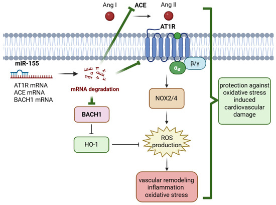
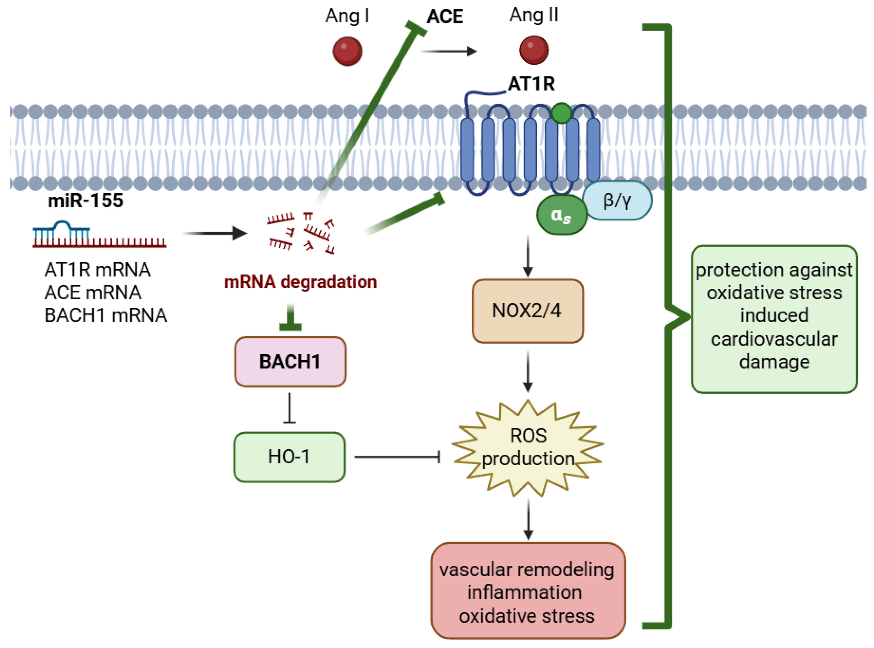
On the other hand, an opposite role of miR-155 in the RAAS axis and Ang II-induced oxidative stress and cardiovascular remodeling in hypertension was proposed (Figure 4). A robust proof based on functional studies comes from the paper by Dupont et al., who pointed out miR-155 as the most down-regulated miRNA in vascular aging in mice’ smooth muscle cells with intact mineralocorticoid receptors [32].
Figure 4.
Proposed antioxidant and vasculoprotective effects of miR-155 through modulation of the renin–angiotensin system and BACH1 signaling. Increased miR-155 expression promotes the degradation of AT1R, ACE, and BACH1 mRNAs. Downregulation of AT1R attenuates Ang II-mediated signaling, reducing NOX2/4 activation, ROS generation, and downstream vascular inflammation, oxidative stress, and remodeling. In parallel, repression of BACH1 relieves its inhibitory effect on heme oxygenase-1 (HO-1), enhancing antioxidant defenses and further limiting ROS accumulation. Collectively, these mechanisms contribute to the protective role of miR-155 against oxidative stress-induced cardiovascular damage. Abbreviations: ACE: Angiotensin-converting enzyme; Ang II: angiotensin II; AT1R: angiotensin II receptor type 1; BACH1: BTB and CNC homology 1; HO-1: heme oxygenase-1; NOX2: NADPH oxidase 2; NOX4: NADPH oxidase 4; ROS: reactive oxygen species. Created in BioRender. Cacciapuoti, M. (2026) https://BioRender.com/2hlhmsy.
In mesenteric vessels of mice, a decrease in miR-155 was associated with a concomitant increase in MR mRNA with aging. Restoration of miR-155 in smooth muscle cells of aged MR-intact mice reduced Cav1.2, the pore-forming L-type Calcium Channel (LTCC), and Agtr1 mRNA and attenuated AT II-induced vasoconstriction and oxidative stress [32]. Wu et al. observed that in VSMC cells of spontaneously hypertensive rats, miR-155 mimic inhibited ACE expression and reduced ROS production along with IL-1β and TNF-α expression, thereby impairing VSMC migration and oxidative stress, which contribute to maladaptive vascular remodeling in hypertension and related organ damage. However, the same effect was not detected after exogenous Ang II administration, suggesting ACE could be a direct target of miR-155-5p [40]. Tong et al. confirmed the inhibitory role of miR-155 in VSMC migration and oxidative stress by reporting that exogenous miR-155-5p administration reduced NOX2 and NOX4 protein expression and activity in VSMCs from spontaneously hypertensive rats by knocking down BACH1 expression, a transcriptional repressor of the cytoprotective enzyme heme oxygenase-1 (HO-1) [35]. Conversely, BACH1, which is also expressed on chromosome 21, was not identified as a target of miR-155 in the process of senescence in endothelial cells derived from mouse aortas in the study of He et al. [33].
Bernard-Harrison et al. reported that in Dahl salt-sensitive mice under a high-salt diet, miR-155 serum and aortic levels were lower than in Dahl salt-sensitive mice under a low-salt diet, while AT1R levels were higher, together with p-MYPT1/MYPT-1 and proteinuria, suggesting a deleterious role of low levels of miR-155 [53]. In spontaneously hypertensive rats, miR-155 was instead elevated, while AT1R levels and proteinuria were decreased, suggesting that miR-155 might contribute to keeping AT1R levels low, thereby limiting Ang II-oxidative stress-mediated kidney damage despite hypertension [53]. However, no causal relationship can be inferred from these results. Song et al. observed that treatment with a soybean-derived antihypertensive (LSW) reduced oxidative stress and inflammation while increasing miR-155-5p compared to Ang-II- and control-treated HUVECs [50]. Moreover, in the context of myocardial injury, miR-155 was increased and played a regulatory role in oxidative stress-induced necrosis in cardiomyocyte progenitor cells by targeting receptor-interacting protein 1 (RIP1) [43]. Finally, treatment-based evidence shows that in HUVECs and PBMCs, miR-155 was up-regulated along with antioxidant genes’ expression as a result of hydroxyurea, an antioxidant agent prescribed in sickle-cell anemia [56].
MiR-155 was also evaluated in a broad number of small patient cohorts affected by cardiovascular diseases. However, the available evidence is largely associative, with studies reporting heterogeneous and sometimes opposing associations with oxidative stress. For example, miR-155 was observed to be up-regulated in placentas from pre-eclamptic women vs. controls and affects cysteine-rich protein 61 (CYR61), a significant premature angiogenesis component in gestation [72]. Another example is Friedrich’s ataxia patients, where a polymorphic miR-155 binding site in AGTR1 was associated with cardiac hypertrophy, while in CD14 + monocytes from patients affected by coronary disease, miR-155 expression was up-regulated [47]. Additionally, a decrease in miR-155 expression was detected with aging [73]. Serum miR-155 predicted the BP treatment response to MR antagonists in elderly humans [32]. Oxidative stress development is also associated with cigarette smoking. Interestingly, miR-155 levels were statistically increased right after smoking in a cohort of smokers [37]. Duisenbeck et al. reported that in diabetic patients, miR-155 levels were higher than in healthy subjects and positively correlated with HbA1c and glycemia. However, no significant difference was detected in diabetic patients with macrovascular complications. Intriguingly, the same authors proposed a predictive model for the diagnosis of diabetes in obese subjects, based on the combination of miR-210, miR-155,-5p glutathione peroxidase, lipid peroxidation, and BMI [59], suggesting a close relationship between obesity, diabetes, and oxidative stress. The relationship between miR-155-3p, glucose levels, and oxidative stress parameters was confirmed in a study conducted in metabolic syndrome patients [58]. Moawad et al. compared coronary heart disease patients with healthy subjects and showed higher levels of miR-155-5p along with higher MDA and lower SOD levels in the first group [60]. Furthermore, in a cohort of patients with Gitelman syndrome, plasma miR-155 levels were higher than in controls, proposing a role of this miRNA in their well-established protection against Ang II-induced cardiovascular remodeling [28]. Finally, given the location of the miR-155 gene on chromosome 21, multiple studies in Down syndrome (DS)—a population burdened by a higher prevalence of cardiovascular disease and higher risk for chemotherapy cardiotoxicity—have been conducted [21]. MiR-155 expression was not different in ex vivo studies conducted on heart samples from DS heart donors vs. non-DS heart donors. However, in non-DS donors, there was a positive association between BACH1 mRNA levels and miR-155 expression. This suggested that in the myocardium of euploid subjects, increasing levels of the inhibitor miR-155 would be necessary to target increasing levels of the BACH1 mRNA transcript. In contrast, the lack of a significant association between BACH1 mRNA and miR-155 levels in samples from donors with DS was proposed to be indicative of a disruption in this layer of control against oxidative stress in DS [67]. However, the lack of functional studies does not allow us to consider a causal relationship between miR-155 and oxidative stress in these studies.
Overall, evidence on the role of miR-155 in oxidative stress in cardiovascular disease is broad and heterogeneous, suggesting that miR-155 may function as a redox rheostat rather than as a purely pro- or antioxidant mediator. Based on the above-mentioned evidence, no certain conclusions can be drawn about a protective or harmful role of this miRNA in cardiovascular disease.
So far, only therapeutics based on miR-155 antagonists have been developed, mostly in cancer fields [15]. One example is cobomarsen, which was moved into phase II for T-cell leukemia/lymphoma [74]. Moreover, MRG-107 is an antagomir of miR-155 aimed at inhibiting the activity of miR-155 in immune mechanisms and inflammation in amyotrophic lateral sclerosis (ALS), with promising results in preclinical models [75]. In fibrosis research, local injection of antagomiR-155 was demonstrated to inhibit the Wnt/β catenin and AKT signaling pathways, thereby reducing fibrosis [76]. Regarding cardiovascular research, knocking out miR-155 in atherogenic apolipoprotein E knockout (ApoE KO, or ApoE-/-) mice resulted in the establishment of the first metabolically healthy obesity (MHO) mouse model with decreased aortic atherosclerosis, increased obesity, white adipose tissue hypertrophy, and non-alcoholic fatty liver disease but without insulin resistance [77]. On the other hand, promoting the expression of miR-155 was proposed as an effective strategy to regulate blood pressure in hypertensive disorders [15]. Nevertheless, no therapeutic strategy based on miR-155 has yet reached a clinical stage.
4. Conclusions and Future Directions
Although miR-103 and miR-155 are both involved in oxidative stress pathways, miR-155 emerges as a potential regulator of inflammatory-redox signaling, whereas miR-103 appears more closely linked to cell fate and metabolic pathways. In both cases, available evidence supports a context-dependent role that challenges a simplistic classification as pro- or antioxidant miRNAs.
Future studies should clarify whether the divergent pro- and antioxidant effects depend primarily on cell type, disease stage, inflammatory milieu, or compensatory adaptive responses. For example, most of the reported effects related to oxidative stress have been demonstrated in acute settings, and studies on long-standing mimic/inhibition of specific miRNAs are lacking. A substantial proportion of the available literature remains associative, with limited mechanistic validation through gain- and loss-of-function approaches. In addition, only a few validated targets of miR-155 and miR-103 within the context of oxidative stress were identified. The in vitro models of oxidative stress were mostly artificially induced via H2O2 exposure and therefore only partially mimic the real oxidative conditions of cells. Moreover, oxidative stress was assessed differently across the studies. Importantly, studies based on humans are the minority, are characterized by low standardization, and predominantly yield indirect evidence linking miR-155 to oxidative stress. Most human-based studies are on small cohorts of patients, and longitudinal clinical investigations are currently lacking. Finally, clinical studies investigating miR-103’s relationship with oxidative stress are not available yet. It should be noted that a limitation of this review is that the literature search was restricted to the PubMed database, which may have resulted in the omission of studies indexed exclusively in other databases.
The clinical development of anti-miR-103 and anti-miR-155 therapies has been pursued almost exclusively in non-cardiovascular conditions. However, to the best of our knowledge, no therapeutic approach for cardiovascular disease has yet progressed beyond the preclinical stage. The marked heterogeneity observed across the studies described in this review suggests that the biological effects of miR-103 and miR-155 in cardiovascular disorders depend on specific experimental and pathological context. These findings raise concerns about the possibility of a systemic inhibition or overexpression of miR-103 or miR-155, which could produce different, or even opposite, biological effects depending on the target tissue, cell type, and inflammatory microenvironment. Consequently, a therapeutic strategy that is beneficial in one cardiovascular setting may suppress protective responses or exacerbate pathological mechanisms in another. Future translational studies should therefore focus on developing tissue- and cell-specific delivery systems while carefully evaluating off-target effects, pharmacokinetics, dosing regimens, immunogenicity, and long-term safety. Such knowledge will not only improve our understanding of the pathophysiological role of miR-103 and miR-155 in cardiovascular disease but may also pave the way for the development of novel miRNA-based therapeutic strategies targeting oxidative stress and cardiovascular remodeling.
Author Contributions
Conceptualization, M.C. (Martina Cacciapuoti) and L.A.C.; methodology, M.C. (Martina Cacciapuoti); investigation, M.C. (Martina Cacciapuoti), L.F.S., I.C., G.D., M.C. (Monica Ceol), G.P., F.N. and L.A.C.; resources, L.A.C.; writing—original draft preparation, M.C. (Martina Cacciapuoti) and L.A.C.; writing—review and editing, M.C. (Martina Cacciapuoti) and L.A.C.; supervision, L.A.C. All authors have read and agreed to the published version of the manuscript.
Funding
This study was funded in part by the Department of Medicine (DIMED), University of Padua Research Grant, Decreto Rep. n. 269/2025, Prot. 11128, 29/10/2025 to G.D.
Institutional Review Board Statement
Not applicable.
Informed Consent Statement
Not applicable.
Data Availability Statement
No new data were created or analyzed in this study. Data sharing is not applicable to this article.
Conflicts of Interest
The authors declare no conflicts of interest.
References
- Pizzino, G.; Irrera, N.; Cucinotta, M.; Pallio, G.; Mannino, F.; Arcoraci, V.; Squadrito, F.; Altavilla, D.; Bitto, A. Oxidative Stress: Harms and Benefits for Human Health. Oxid. Med. Cell. Longev. 2017, 2017, 8416763. [Google Scholar] [CrossRef] [Scilit]
- Ravarotto, V.; Bertoldi, G.; Innico, G.; Gobbi, L.; Calò, L.A. The Pivotal Role of Oxidative Stress in the Pathophysiology of Cardiovascular-Renal Remodeling in Kidney Disease. Antioxidants 2021, 10, 1041. [Google Scholar] [CrossRef] [Scilit]
- Basso, A.; Baldini, P.; Bertoldi, G.; Driussi, G.; Caputo, I.; Bettin, E.; Cacciapuoti, M.; Calò, L.A. Oxidative stress reduction by icodextrin-based glucose-free solutions in peritoneal dialysis: Support for new promising approaches. Artif. Organs 2024, 48, 1031–1037. [Google Scholar] [CrossRef] [Scilit]
- Basso, A.; Cacciapuoti, M.; Stefanelli, L.F.; Nalesso, F.; Calò, L.A. Glucose-Free Solutions Mediated Inhibition of Oxidative Stress and Oxidative Stress-Related Damages in Peritoneal Dialysis: A Promising Solution. Life 2024, 14, 1173. [Google Scholar] [CrossRef] [Scilit]
- Sugamura, K.; Keaney, J.F., Jr. Reactive oxygen species in cardiovascular disease. Free. Radic. Biol. Med. 2011, 51, 978–992. [Google Scholar] [CrossRef] [Scilit]
- Pietrangelo, D.; Lopa, C.; Litterio, M.; Cotugno, M.; Rubattu, S.; Lombardi, A. Metabolic Disturbances Involved in Cardiovascular Diseases: The Role of Mitochondrial Dysfunction, Altered Bioenergetics and Oxidative Stress. Int. J. Mol. Sci. 2025, 26, 6791. [Google Scholar] [CrossRef] [Scilit] [PubMed]
- Sgarabotto, L.; Ravarotto, V.; Stefanelli, L.F.; Cacciapuoti, M.; Davis, P.A.; Nalesso, F.; Calò, L.A. Oxidants and Cardiorenal Vascular Remodeling—Insights from Rare Genetic Tubulopathies: Bartter’s and Gitelman’s Syndromes. Antioxidants 2023, 12, 811. [Google Scholar] [CrossRef] [Scilit]
- Lee, Y.; Im, E. Regulation of miRNAs by Natural Antioxidants in Cardiovascular Diseases: Focus on SIRT1 and eNOS. Antioxidants 2021, 10, 377. [Google Scholar] [CrossRef] [Scilit]
- Abolhasani, S.; Ahmadi, Y.; Fattahi, D.; Rostami, Y.; Chollou, K.M. microRNA-Mediated Regulation of Oxidative Stress in Cardiovascular Diseases. J. Clin. Lab. Anal. 2025, 39, e70017. [Google Scholar] [CrossRef] [Scilit]
- Ma, J.; Chen, X. Advances in pathogenesis and treatment of essential hypertension. Front. Cardiovasc. Med. 2022, 9, 1003852. [Google Scholar] [CrossRef] [Scilit]
- An, Y.; Xu, B.-T.; Wan, S.-R.; Ma, X.-M.; Long, Y.; Xu, Y.; Jiang, Z.-Z. The role of oxidative stress in diabetes mellitus-induced vascular endothelial dysfunction. Cardiovasc. Diabetol. 2023, 22, 237. [Google Scholar] [CrossRef] [Scilit] [PubMed]
- Yun, H.R.; Singh, M.K.; Han, S.; Ranbhise, J.S.; Ha, J.; Kim, S.S.; Kang, I. Integrating Senescence and Oxidative Stress in Cardiac Disease. Int. J. Mol. Sci. 2025, 26, 11917. [Google Scholar] [CrossRef] [Scilit]
- Ali, F.; Shen, A.; Islam, W.; Saleem, M.Z.; Muthu, R.; Xie, Q.; Wu, M.; Cheng, Y.; Chu, J.; Lin, W.; et al. Role of MicroRNAs and their corresponding ACE2/Apelin signaling pathways in hypertension. Microb. Pathog. 2022, 162, 105361. [Google Scholar] [CrossRef] [Scilit]
- Peters, L.J.F.; Floege, J.; Biessen, E.A.L.; Jankowski, J.; van der Vorst, E.P.C. MicroRNAs in Chronic Kidney Disease: Four Candidates for Clinical Application. Int. J. Mol. Sci. 2020, 21, 6547. [Google Scholar] [CrossRef] [Scilit]
- Zhao, X.; Wang, P. Interpretation of the molecular mechanism and therapeutic potential of microRNA-155 in cardiovascular and cerebrovascular diseases (Review). Int. J. Mol. Med. 2025, 57, 10. [Google Scholar] [CrossRef] [Scilit]
- Bátkai, S.; Thum, T. MicroRNAs in Hypertension: Mechanisms and Therapeutic Targets. Curr. Hypertens. Rep. 2011, 14, 79–87. [Google Scholar] [CrossRef] [Scilit]
- Logan, S.M.; Gupta, A.; Wang, A.; Levy, R.J.; Storey, K.B. Isoflurane and low-level carbon monoxide exposures increase expression of pro-survival miRNA in neonatal mouse heart. Cell Stress Chaperon. 2021, 26, 541–548. [Google Scholar] [CrossRef] [Scilit]
- Qi, H.; Ren, J.; E, M.; Zhang, Q.; Cao, Y.; Ba, L.; Song, C.; Shi, P.; Fu, B.; Sun, H. MiR-103 inhibiting cardiac hypertrophy through inactivation of myocardial cell autophagy via targeting TRPV3 channel in rat hearts. J. Cell. Mol. Med. 2019, 23, 1926–1939. [Google Scholar] [CrossRef] [Scilit]
- Lu, Q.; Ma, Z.; Ding, Y.; Bedarida, T.; Chen, L.; Xie, Z.; Song, P.; Zou, M.-H. Circulating miR-103a-3p contributes to angiotensin II-induced renal inflammation and fibrosis via a SNRK/NF-κB/p65 regulatory axis. Nat. Commun. 2019, 10, 2145. [Google Scholar] [CrossRef] [Scilit]
- Xu, M.-C.; Gao, X.-F.; Ruan, C.; Ge, Z.-R.; Lu, J.-D.; Zhang, J.-J.; Zhang, Y.; Wang, L.; Shi, H.-M. miR-103 Regulates Oxidative Stress by Targeting the BCL2/Adenovirus E1B 19 kDa Interacting Protein 3 in HUVECs. Oxidative Med. Cell. Longev. 2015, 2015, 489647. [Google Scholar] [CrossRef] [Scilit]
- Papadopoulos, K.I.; Papadopoulou, A.; Aw, T.C. Beauty and the beast: Host microRNA-155 versus SARS-CoV-2. Hum. Cell 2023, 36, 908–922. [Google Scholar] [CrossRef] [Scilit]
- Nemecz, M.; Alexandru, N.; Tanko, G.; Georgescu, A. Role of MicroRNA in Endothelial Dysfunction and Hypertension. Curr. Hypertens. Rep. 2016, 18, 87. [Google Scholar] [CrossRef] [Scilit]
- Wang, X.; Xiong, X.; Jiang, W.; Xu, S.; Li, J. Orientin Alleviates Oxidative Stress And Apoptosis In Diabetic Cardiomyopathy Via The Lncrna H19/Mir-103-3p/ALDH2/PI3K/AKT Axis. Arq. Bras. Cardiol. 2025, 122, e20240885. [Google Scholar] [CrossRef] [Scilit]
- Zhang, X.; Yuan, S.; Liu, J.; Tang, Y.; Wang, Y.; Zhan, J.; Fan, J.; Nie, X.; Zhao, Y.; Wen, Z.; et al. Overexpression of cytosolic long noncoding RNA cytb protects against pressure-overload-induced heart failure via sponging microRNA-103-3p. Mol. Ther. Nucleic Acids 2022, 27, 1127–1145. [Google Scholar] [CrossRef] [Scilit]
- Wang, Y.; Song, X.; Li, Z.; Liu, N.; Yan, Y.; Li, T.; Sun, W.; Guan, Y.; Li, M.; Yang, Y.; et al. MicroRNA-103 Protects Coronary Artery Endothelial Cells against H2O2-Induced Oxidative Stress via BNIP3-Mediated End-Stage Autophagy and Antipyroptosis Pathways. Oxidative Med. Cell. Longev. 2020, 2020, 15. [Google Scholar] [CrossRef] [Scilit]
- Wang, J.-X.; Zhang, X.-J.; Li, Q.; Wang, K.; Wang, Y.; Jiao, J.-Q.; Feng, C.; Teng, S.; Zhou, L.-Y.; Gong, Y.; et al. MicroRNA-103/107 Regulate Programmed Necrosis and Myocardial Ischemia/Reperfusion Injury Through Targeting FADD. Circ. Res. 2015, 117, 352–363. [Google Scholar] [CrossRef] [Scilit]
- Ellis, K.L.; Cameron, V.A.; Troughton, R.W.; Frampton, C.M.; Ellmers, L.J.; Richards, A.M. Circulating microRNAs as Candidate Markers to Distinguish Heart Failure in Breathless Patients. Eur. J. Heart Fail. 2013, 15, 1138–1147. [Google Scholar] [CrossRef] [Scilit]
- Cacciapuoti, M.; Caputo, I.; Driussi, G.; Davis, P.A.; Nalesso, F.; Calò, L.A. Higher expression of miR-155 and miR-103 in patients with Gitelman’s syndrome: Potential implications for Angiotensin II-induced cardiovascular remodeling in hypertension. Kidney Blood Press. Res. 2026, 51, 606–613. [Google Scholar] [CrossRef] [Scilit]
- Zhang, M.; Tang, Y.; Tang, E.; Lu, W. MicroRNA-103 represses hepatic de novo lipogenesis and alleviates NAFLD via targeting FASN and SCD1. Biochem. Biophys. Res. Commun. 2020, 524, 716–722. [Google Scholar] [CrossRef] [Scilit]
- Huang, Y. Preclinical and Clinical Advances of GalNAc-Decorated Nucleic Acid Therapeutics. Mol. Ther. Nucleic Acids 2017, 6, 116–132. [Google Scholar] [CrossRef] [Scilit]
- Ge, X.; Meng, Q.; Liu, J.; Wang, E.; Liu, X.; Shi, S.; Gong, X.; Liu, Z.; Han, W.; Zhou, X. Cardiac extracellular vesicles aggravate cardiomyocyte ferroptosis in myocardial ischemia-reperfusion injury via miR-155-5p-Nfe2l2 signaling. Biochim. Biophys. Acta (BBA)-Gen. Subj. 2026, 1870, 130912. [Google Scholar] [CrossRef] [Scilit]
- DuPont, J.J.; McCurley, A.; Davel, A.P.; McCarthy, J.; Bender, S.B.; Hong, K.; Yang, Y.; Yoo, J.-K.; Aronovitz, M.; Baur, W.E.; et al. Vascular mineralocorticoid receptor regulates microRNA-155 to promote vasoconstriction and rising blood pressure with aging. J. Clin. Investig. 2016, 1, e88942. [Google Scholar] [CrossRef] [Scilit]
- He, J.; Zhang, B.; Zhang, H.; Tu, Q.; Chen, X.; Qiu, Y.; Liu, Z.; Xia, W.; Wu, X.; Tao, J. M1 Macrophage is a Novel Potential Trigger for Endothelial Senescence: Role of Exosomal miR-155 Targeting SOCS1 Signal. Hum. Mutat. 2025, 18, 6771390. [Google Scholar] [CrossRef] [Scilit]
- Zhang, Y.; Hei, F.; Xiao, Y.; Liu, Y.; Han, J.; Hu, D.; Wang, H. Acidic fibroblast growth factor inhibits reactive oxygen species-induced epithelial–mesenchymal transdifferentiation in vascular endothelial cells via the miR-155-5p/SIRT1/Nrf2/HO-1 pathway to promote wound healing in diabetic mice. Burn. Trauma 2024, 12, tkae010. [Google Scholar] [CrossRef] [Scilit]
- Tong, Y.; Zhou, M.-H.; Li, S.-P.; Zhao, H.-M.; Zhang, Y.-R.; Chen, D.; Wu, Y.-X.; Pang, Q.-F. MiR-155-5p Attenuates Vascular Smooth Muscle Cell Oxidative Stress and Migration via Inhibiting BACH1 Expression. Biomedicines 2023, 11, 1679. [Google Scholar] [CrossRef] [Scilit]
- Zhao, M.; Lei, J.; Deng, F.; Zhao, C.; Xu, T.; Ji, B.; Fu, M.; Wang, X.; Sun, M.; Zhang, M.; et al. Gestational Hypoxia Impaired Endothelial Nitric Oxide Synthesis Via miR-155-5p/NADPH Oxidase/Reactive Oxygen Species Axis in Male Offspring Vessels. J. Am. Heart Assoc. 2024, 13, e032079. [Google Scholar] [CrossRef] [Scilit]
- Frati, G.; Forte, M.; di Nonno, F.; Bordin, A.; Chimenti, I.; Picchio, V.; Cavarretta, E.; Stanzione, R.; Bianchi, F.; Carnevale, R.; et al. Inhibition of miR-155 Attenuates Detrimental Vascular Effects of Tobacco Cigarette Smoking. J. Am. Heart Assoc. 2020, 9, e017000. [Google Scholar] [CrossRef] [Scilit]
- He, X.; Wang, Z.; Wei, L.; Cheng, X.; Chen, L.; Gao, F.; Jiang, H. Indoxyl sulfate promotes osteogenic differentiation of vascular smooth muscle cells by miR-155-5p-dependent downregulation of matrix Gla protein via ROS/NF-κB signaling. Exp. Cell Res. 2020, 397, 112301. [Google Scholar] [CrossRef] [Scilit]
- Wang, X.; Han, W.; Zhang, Y.; Zong, Y.; Tan, N.; Zhang, Y.; Li, L.; Liu, C.; Liu, L. Soluble Epoxide Hydrolase Inhibitor t-AUCB Ameliorates Vascular Endothelial Dysfunction by Influencing the NF-κB/miR-155-5p/eNOS/NO/IκB Cycle in Hypertensive Rats. Antioxidants 2022, 11, 1372. [Google Scholar] [CrossRef] [Scilit]
- Wu, N.; Ye, C.; Zheng, F.; Wan, G.-W.; Wu, L.-L.; Chen, Q.; Li, Y.-H.; Kang, Y.-M.; Zhu, G.-Q. MiR155-5p Inhibits Cell Migration and Oxidative Stress in Vascular Smooth Muscle Cells of Spontaneously Hypertensive Rats. Antioxidants 2020, 9, 204. [Google Scholar] [CrossRef] [Scilit]
- Jia, C.; Chen, H.; Wei, M.; Chen, X.; Zhang, Y.; Cao, L.; Yuan, P.; Wang, F.; Yang, G.; Ma, J. Gold nanoparticle-based miR155 antagonist macrophage delivery restores the cardiac function in ovariectomized diabetic mouse model. Int. J. Nanomed. 2017, 12, 4963–4979. [Google Scholar] [CrossRef] [Scilit]
- Liu, Y.; Pan, Q.; Zhao, Y.; He, C.; Bi, K.; Chen, Y.; Zhao, B.; Chen, Y.; Ma, X. MicroRNA-155 Regulates ROS Production, NO Generation, Apoptosis and Multiple Functions of Human Brain Microvessel Endothelial Cells Under Physiological and Pathological Conditions. J. Cell. Biochem. 2015, 116, 2870–2881. [Google Scholar] [CrossRef] [Scilit]
- Liu, J.; van Mil, A.; Vrijsen, K.; Zhao, J.; Gao, L.; Metz, C.H.G.; Goumans, M.-J.; Doevendans, P.A.; Sluijter, J.P.G. MicroRNA-155 prevents necrotic cell death in human cardiomyocyte progenitor cells via targeting RIP1. J. Cell. Mol. Med. 2010, 15, 1474–1482. [Google Scholar] [CrossRef] [Scilit]
- Wang, F.; Zhang, J.; Niu, G.; Weng, J.; Zhang, Q.; Xie, M.; Li, C.; Sun, K. Apigenin inhibits isoproterenol-induced myocardial fibrosis and Smad pathway in mice by regulating oxidative stress and miR-122-5p/155-5p expressions. Drug Dev. Res. 2022, 83, 1003–1015. [Google Scholar] [CrossRef] [Scilit]
- Sun, H.-J.; Zhao, M.-X.; Liu, T.-Y.; Ren, X.-S.; Chen, Q.; Li, Y.-H.; Kang, Y.-M.; Zhu, G.-Q. Salusin-β induces foam cell formation and monocyte adhesion in human vascular smooth muscle cells via miR155/NOX2/NFκB pathway. Sci. Rep. 2016, 6, 23596. [Google Scholar] [CrossRef] [Scilit]
- Yang, Z.; Zheng, B.; Zhang, Y.; He, M.; Zhang, X.-H.; Ma, D.; Zhang, R.-N.; Wu, X.-L.; Wen, J.-K. miR-155-dependent regulation of mammalian sterile 20-like kinase 2 (MST2) coordinates inflammation, oxidative stress and proliferation in vascular smooth muscle cells. Biochim. Biophys. Acta (BBA)-Mol. Basis Dis. 2015, 1852, 1477–1489. [Google Scholar] [CrossRef] [Scilit]
- Tian, F.-J.; An, L.-N.; Wang, G.-K.; Zhu, J.-Q.; Li, Q.; Zhang, Y.-Y.; Zeng, A.; Zou, J.; Zhu, R.-F.; Han, X.-S.; et al. Elevated microRNA-155 promotes foam cell formation by targeting HBP1 in atherogenesis. Cardiovasc. Res. 2014, 103, 100–110. [Google Scholar] [CrossRef] [Scilit]
- Kim, J.; Lee, K.-S.; Kim, J.-H.; Lee, D.-K.; Park, M.; Choi, S.; Park, W.; Kim, S.; Choi, Y.K.; Hwang, J.Y.; et al. Aspirin prevents TNF-α-induced endothelial cell dysfunction by regulating the NF-κB-dependent miR-155/eNOS pathway: Role of a miR-155/eNOS axis in preeclampsia. Free. Radic. Biol. Med. 2017, 104, 185–198. [Google Scholar] [CrossRef] [Scilit]
- Kim, T.-H.; Kim, J.-Y.; Bae, J.; Kim, Y.-M.; Won, M.-H.; Ha, K.-S.; Kwon, Y.-G.; Kim, Y.-M. Korean Red ginseng prevents endothelial senescence by downregulating the HO-1/NF-κB/miRNA-155-5p/eNOS pathway. J. Ginseng Res. 2021, 45, 344–353. [Google Scholar] [CrossRef] [Scilit]
- Song, T.; Lv, M.; Zhou, M.; Huang, M.; Zheng, L.; Zhao, M. Soybean-Derived Antihypertensive Peptide LSW (Leu–Ser–Trp) Antagonizes the Damage of Angiotensin II to Vascular Endothelial Cells through the Trans-vesicular Pathway. J. Agric. Food Chem. 2021, 69, 10536–10549. [Google Scholar] [CrossRef] [Scilit]
- Xiong, M.; Jia, C.; Cui, J.; Wang, P.; Du, X.; Yang, Q.; Zhu, Y.; Wang, W.; Zhang, T.; Chen, Y. Shexiang Tongxin dropping pill attenuates atherosclerotic lesions in ApoE deficient mouse model. J. Ethnopharmacol. 2015, 159, 84–92. [Google Scholar] [CrossRef] [Scilit]
- Cai, R.; Hao, Y.; Liu, Y.-Y.; Huang, L.; Yao, Y.; Zhou, M.-S. Tumor Necrosis Factor Alpha Deficiency Improves Endothelial Function and Cardiovascular Injury in Deoxycorticosterone Acetate/Salt-Hypertensive Mice. BioMed Res. Int. 2020, 2020, 3921074. [Google Scholar] [CrossRef] [Scilit]
- Harrison-Bernard, L.M.; Raij, L.; Tian, R.X.; Jaimes, E.A. Genetically conditioned interaction among microRNA-155, alpha-klotho, and intra-renal RAS in male rats: Link to CKD progression. Physiol. Rep. 2024, 12, e16172. [Google Scholar] [CrossRef] [Scilit]
- Costantino, S.; Paneni, F.; Lüscher, T.F.; Cosentino, F. MicroRNA profiling unveils hyperglycaemic memory in the diabetic heart. Eur. Heart J. 2015, 37, 572–576. [Google Scholar] [CrossRef] [Scilit]
- Muñoz-Pacheco, P.; Ortega-Hernández, A.; Miana, M.; Cachofeiro, V.; Fernández-Cruz, A.; Gómez-Garre, D. Ezetimibe inhibits PMA-induced monocyte/macrophage differentiation by altering microRNA expression: A novel anti-atherosclerotic mechanism. Pharmacol. Res. 2012, 66, 536–543. [Google Scholar] [CrossRef] [Scilit]
- Santana, S.S.; Pitanga, T.N.; de Santana, J.M.; Zanette, D.L.; Vieira, J.d.J.; Yahouédéhou, S.C.M.A.; Adanho, C.S.A.; Viana, S.d.M.; Luz, N.F.; Borges, V.M.; et al. Hydroxyurea Scavenges Free Radicals and Induces the Expression of Antioxidant Genes in Human Cell Cultures Treated with Hemin. Front. Immunol. 2020, 11, 1488. [Google Scholar] [CrossRef] [Scilit]
- Nguyen, D.D.N.; Zain, S.M.; Kamarulzaman, M.H.; Low, T.Y.; Chilian, W.M.; Pan, Y.; Ting, K.N.; Hamid, A.; Kadir, A.A.; Pung, Y.-F. Intracellular and exosomal microRNAome profiling of human vascular smooth muscle cells during replicative senescence. Am. J. Physiol. Circ. Physiol. 2021, 321, H770–H783. [Google Scholar] [CrossRef] [Scilit]
- Khedr, N.F.; Zahran, E.S.; Ebeid, A.M.; Melek, S.T.; Werida, R.H. Effect of green coffee on miR-133a, miR-155 and inflammatory biomarkers in obese individuals. Diabetol. Metab. Syndr. 2024, 16, 256. [Google Scholar] [CrossRef] [Scilit]
- Duisenbek, A.; Pérez, M.D.A.; Pérez, M.; Benitez, J.M.A.; Pérez, V.R.P.; Ortega, J.G.; Ussipbek, B.; Yessenbekova, A.; López-Armas, G.C.; Ablaikhanova, N.; et al. Unveiling the Predictive Model for Macrovascular Complications in Type 2 Diabetes Mellitus: microRNAs Expression, Lipid Profile, and Oxidative Stress Markers. Int. J. Mol. Sci. 2024, 25, 11763. [Google Scholar] [CrossRef] [Scilit]
- Moawad, A.M.; Awady, S.; Ali, A.A.E.R.; Abdelgwad, M.; Belal, S.; Taha, S.H.N.; Mohamed, M.I.; Hassan, F.M. Phthalate Exposure and Coronary Heart Disease: Possible Implications of Oxidative Stress and Altered miRNA Expression. Chem. Res. Toxicol. 2024, 37, 723–730. [Google Scholar] [CrossRef] [Scilit]
- Saghati, A.A.; Sharifi, Z.; Hatamikhah, M.; Salimi, M.; Talkhabi, M. Unraveling the relevance of SARS-CoV-2 infection and ferroptosis within the heart of COVID-19 patients. Heliyon 2024, 10, e36567. [Google Scholar] [CrossRef] [Scilit]
- Kelly, M.; Bagnall, R.D.; Peverill, R.E.; Donelan, L.; Corben, L.; Delatycki, M.B.; Semsarian, C. A polymorphic miR-155 binding site in AGTR1 is associated with cardiac hypertrophy in Friedreich ataxia. J. Mol. Cell. Cardiol. 2011, 51, 848–854. [Google Scholar] [CrossRef] [Scilit]
- Kim, S.-M.; Lim, S.-M.; Yoo, J.-A.; Woo, M.-J.; Cho, K.-H. Consumption of high-dose vitamin C (1250 mg per day) enhances functional and structural properties of serum lipoprotein to improve anti-oxidant, anti-atherosclerotic, and anti-aging effects via regulation of anti-inflammatory microRNA. Food Funct. 2015, 6, 3604–3612. [Google Scholar] [CrossRef] [Scilit]
- Chen, H.; Gao, M.Y.L.; Zhang, L.; He, F.L.; Shi, Y.K.; Pan, X.H.; Wang, H. MicroRNA-155 affects oxidative damage through regulating autophagy in endothelial cells. Oncol. Lett. 2019, 17, 2237–2243. [Google Scholar] [CrossRef] [Scilit]
- Liu, M.; Saredy, J.; Zhang, R.; Shao, Y.; Sun, Y.; Yang, W.Y.; Wang, J.; Liu, L.; Drummer, C.; Johnson, C.; et al. Approaching Inflammation Paradoxes—Proinflammatory Cytokine Blockages Induce Inflammatory Regulators. Front. Immunol. 2020, 11, 554301. [Google Scholar] [CrossRef] [Scilit]
- Constantin, A.; Comarița, I.K.; Alexandru, N.; Filippi, A.; Bojin, F.; Gherghiceanu, M.; Vîlcu, A.; Nemecz, M.; Niculescu, L.S.; Păunescu, V.; et al. Stem cell-derived extracellular vesicles reduce the expression of molecules involved in cardiac hypertrophy—In a model of human-induced pluripotent stem cell-derived cardiomyocytes. Front. Pharmacol. 2022, 13, 1003684. [Google Scholar] [CrossRef] [Scilit]
- Hefti, E.; Quiñones-Lombraña, A.; Redzematovic, A.; Hui, J.; Blanco, J.G. Analysis of mtDNA, miR-155 and BACH1 expression in hearts from donors with and without Down syndrome. Mitochondrial DNA Part A 2014, 27, 896–903. [Google Scholar] [CrossRef] [Scilit]
- Jia, C.; Xiong, M.; Wang, P.; Cui, J.; Du, X.; Yang, Q.; Wang, W.; Chen, Y.; Zhang, T. Notoginsenoside R1 Attenuates Atherosclerotic Lesions in ApoE Deficient Mouse Model. PLoS ONE 2014, 9, e99849. [Google Scholar] [CrossRef] [Scilit]
- Liu, D.; Han, L.; Wu, X.; Yang, X.; Zhang, Q.; Jiang, F. Genome-wide microRNA changes in human intracranial aneurysms. BMC Neurol. 2014, 14, 188. [Google Scholar] [CrossRef] [Scilit]
- Witvrouwen, I.; Mannaerts, D.; Ratajczak, J.; Boeren, E.; Faes, E.; Van Craenenbroeck, A.H.; Jacquemyn, Y.; Van Craenenbroeck, E.M. MicroRNAs targeting VEGF are related to vascular dysfunction in preeclampsia. Biosci. Rep. 2021, 41, BSR20210874. [Google Scholar] [CrossRef] [Scilit]
- Prieto, I.; Kavanagh, M.; Jimenez-Castilla, L.; Pardines, M.; Lazaro, I.; del Real, I.H.; Flores-Muñoz, M.; Egido, J.; Lopez-Franco, O.; Gomez-Guerrero, C. A mutual regulatory loop between miR-155 and SOCS1 influences renal inflammation and diabetic kidney disease. Mol. Ther. Nucleic Acids 2023, 34, 102041. [Google Scholar] [CrossRef] [Scilit]
- Zhang, Y.; Diao, Z.; Su, L.; Sun, H.; Li, R.; Cui, H.; Hu, Y. MicroRNA-155 contributes to preeclampsia by down-regulating CYR61. Am. J. Obstet. Gynecol. 2010, 202, 466.e1–466.e7. [Google Scholar] [CrossRef] [Scilit]
- Hooten, N.N.; Abdelmohsen, K.; Gorospe, M.; Ejiogu, N.; Zonderman, A.B.; Evans, M.K. microRNA Expression Patterns Reveal Differential Expression of Target Genes with Age. PLoS ONE 2010, 5, e10724. [Google Scholar] [CrossRef] [Scilit]
- Anastasiadou, E.; Seto, A.G.; Beatty, X.; Hermreck, M.; Gilles, M.-E.; Stroopinsky, D.; Pinter-Brown, L.C.; Pestano, L.; Marchese, C.; Avigan, D.; et al. Cobomarsen, an Oligonucleotide Inhibitor of miR-155, Slows DLBCL Tumor Cell Growth In Vitro and In Vivo. Clin. Cancer Res. 2021, 27, 1139–1149. [Google Scholar] [CrossRef] [Scilit]
- Seyhan, A.A. Trials and Tribulations of MicroRNA Therapeutics. Int. J. Mol. Sci. 2024, 25, 1469. [Google Scholar] [CrossRef] [Scilit] [PubMed]
- Yan, Q.; Chen, J.; Li, W.; Bao, C.; Fu, Q. Targeting miR-155 to Treat Experimental Scleroderma. Sci. Rep. 2016, 6, 20314. [Google Scholar] [CrossRef] [Scilit]
- Virtue, A.; Johnson, C.; Lopez-Pastraña, J.; Shao, Y.; Fu, H.; Li, X.; Li, Y.-F.; Yin, Y.; Mai, J.; Rizzo, V.; et al. MicroRNA-155 Deficiency Leads to Decreased Atherosclerosis, Increased White Adipose Tissue Obesity, and Non-alcoholic Fatty Liver Disease. J. Biol. Chem. 2017, 292, 1267–1287. [Google Scholar] [CrossRef] [Scilit]
Disclaimer/Publisher’s Note: The statements, opinions and data contained in all publications are solely those of the individual author(s) and contributor(s) and not of MDPI and/or the editor(s). MDPI and/or the editor(s) disclaim responsibility for any injury to people or property resulting from any ideas, methods, instructions or products referred to in the content. |
© 2026 by the authors. Licensee MDPI, Basel, Switzerland. This article is an open access article distributed under the terms and conditions of the Creative Commons Attribution (CC BY) license.