Abstract
Parkia platycephala has emerged as a promising medicinal species, attracting growing scientific interest due to its richness in bioactive compounds such as phenolic acids, flavonoids, tannins, saponins, terpenes, and lectins, present across different parts of the plant. Studies to date have highlighted its varied biological activities, including antioxidant, anti-inflammatory, antimicrobial, and antitumoral properties. Additionally, these compounds have been shown to increase the efficacy of conventional therapeutic drugs and reduce resistance to their effects. Given the breadth and relevance of these findings, compiling and systematizing the available data on P. platycephala is essential. A comprehensive synthesis not only facilitates a better understanding of the plant’s phytochemical and pharmacological potential but also lays the groundwork for the development of innovative, multi-target therapeutic strategies. This effort is particularly relevant in addressing current challenges in treating infections, inflammatory disorders, and cancer, thereby reinforcing the species’ value as a strategic resource for future drug discovery and integrative medicine.
1. Introduction
Infectious diseases can be defined as clinical conditions arising from the invasion and subsequent replication of pathogens (e.g., viruses, bacteria, fungi, or parasites) within the host []. In cases where these agents persist, it is observed that they evade immune detection and clearance, resulting in sustained inflammation due to the continuous release of pro-inflammatory mediators and the activation of pattern recognition receptors (PRRs) by pathogen-associated molecular patterns (PAMPs). This response is initiated by the presence of toxins, oncoproteins, or growth factors [].
Infectious agents have been demonstrated to play a significant role in carcinogenesis, which can be attributed to a number of factors. These include the ability of infectious agents to induce chronic inflammation, as well as the capacity to promote genetic alterations through genomic insertion. In addition, infectious agents have been found to be capable of modulating oncogene expression and facilitating immune evasion [].
Furthermore, the presence of multiple infectious agents has been shown to intensify the inflammatory environment, resulting in elevated cytokine levels and increased reactive oxygen and nitrogen species (ROS). These molecules have been demonstrated to induce oxidative damage to DNA, thereby compromising genomic stability and promoting mutagenesis. It is evident that the persistent inflammatory microenvironment promotes a recurrent cycle of tissue injury and repair, which in turn selects cells that possess adaptive mutations that confer survival and proliferative advantages, thereby ultimately promoting neoplastic transformation [].
Rudolf Virchow pioneered the concept of a correlation between chronic inflammation and cancer progression in 1863, which was based on his observations of leukocytes in tumor lesions. Support for this hypothesis was subsequently provided by further evidence that identified inflammation as a carcinogenic risk factor. The induction of inflammation by non-infectious agents can be facilitated by the activation of pattern recognition receptors (PRRs), which are capable of recognizing pathogen-associated molecular patterns (PAMPs). These PAMPs are recognized in situations where toxins, oncoproteins, or growth factors are present [].
The continuous cycle of tissue injury and repair is characterized by cell death and subsequent compensatory proliferation, which is driven by the selection of clones bearing adaptive mutations. These mutations confer survival advantages and facilitate the expansion of malignantly infected populations [].
Confronted with the challenge of combating infectious diseases, contemporary therapeutic strategies encounter significant limitations. These limitations encompass the systemic toxicity of antimicrobial agents, the inefficient distribution of drugs to target tissues, and the mounting concern of microbial resistance []. Despite the demonstrable efficacy of many of these agents, their use is often limited by the occurrence of adverse effects at therapeutically adequate doses. Additionally, it has been demonstrated that an accelerated rate of metabolism can impede the ability to sustain effective concentrations of pharmaceutical agents at the site of infection, consequently undermining the potential for favorable clinical outcomes [].
At the same time, therapeutic interventions for inflammatory diseases encounter considerable challenges, primarily due to the complexity of accurately directing pharmaceutical agents to the affected regions. The non-selective systemic distribution of pharmaceuticals has been demonstrated to result in the exposure of healthy tissues to therapeutic compounds. This phenomenon can potentially precipitate adverse effects and impose constraints on the escalation of administrable doses. Additionally, the enhanced vascular permeability, a prevalent feature in inflamed regions, can enable the permeation of drug delivery systems, such as liposomes. However, this process can concomitantly trigger the uncontrolled dispersion of pharmaceutical agents, thereby compromising their retention at the intended location. The toxicity of systemic administration of anti-inflammatory agents is a considerable impediment, given the propensity of these substances to induce severe adverse effects, thereby restricting their utilization over prolonged periods or at high doses [].
Concerning cancer, conventional treatments comprise surgical procedures, chemotherapy, radiotherapy, and immunotherapy. These treatments, however, demonstrate limited efficacy, affecting both malignant and normal cells, which results in adverse effects in patients []. Tumoral resistance is a significant challenge in cancer treatments, with malignant cells often developing adaptations, including increased expression of efflux pumps, resulting in the expulsion of drugs from the cell interior, and alterations in signaling pathways that impede the therapeutic response. The presence of cellular heterogeneity and the intricate characteristics of the tumor microenvironment present further challenges. These factors impede the efficacious and homogeneous administration of medications, consequently influencing clinical outcomes [].
Natural products represent a promising area of research for the treatment and prevention of diseases due to their chemical diversity and biochemical and pharmacological properties, which enable them to target multiple cellular pathways []. The utilization of plant-derived natural compounds as a source for the discovery of novel bioactive drugs represents a promising avenue for research. The scientific evidence suggests that these compounds have the potential to mitigate the adverse effects inherent to cancer treatments and to stimulate the immune system, thereby contributing to enhanced disease control [].
It is noteworthy that natural sources, encompassing plants, fungi, and microorganisms, have exhibited remarkable efficacy in this regard. A substantial proportion (approximately 60% in inflammation) of contemporary drugs used to treat cancer, infection, and inflammation are derived from these natural compounds, and such drugs frequently exhibit a reduced incidence of adverse effects when compared to conventional therapeutic approaches [,].
The Parkia genus belongs to the Fabaceae family and includes around 34 species, of which 9 are usually used in folk medicine for the treatment of medical conditions such as diabetes, hypertension, diarrhea, wounds, cough, chronic hemorrhoids, conjunctivitis, and measles. Among all species, P. biglobosa, P. speciosa, P. javanica, P. bicolor, P. biglandulosa, P. filicoidea, and P. clappertoniana are the most studied. Secondary metabolites, including terpenoids, phenolic acids, flavonoids (both aglycones and glycosides), and volatile compounds, have been isolated form Parkia sp. and display several pharmacological activities. Extracts, isolated compounds, and seed lectins were described to possess anticancer, antimicrobial, antihypertensive, antiulcer, antidiabetic, anti-inflammatory, antioxidant, hepatoprotective, and antidiarrheal properties [].
In this paper, the authors aim to draw attention to Parkia platycephala as a source of bioactive compounds and highlight its antioxidant, anti-inflammatory, antimicrobial, and antitumor properties.
2. Morphological and Phytochemical Characterization of Parkia platycephala
Parkia platycephala Benth, (Family Fabaceae; Subfamily Caesalpinioideae, clade Mimosoideae) (PPs) is one of 17 species endemic in Brazil, popularly known as “faveira”, “faveira-preta”, “visgueiro”, “fava-de-boi”, “sabiú”, or “fava-de-bolota” [,,]. It is a tree, endemic in the Cerrado biome, characterized by a savanna-like woody vegetation. This tree is found in the states of Tocantins, Maranhão, and Piauí, the northeastern region of Brazil [,]. The species of the Fabaceae family are being investigated as a source of natural compounds with therapeutic potential, distinguished by multiple advantages, such as adaptability, phenotypic plasticity, and high levels of genetic diversity [,,].
This important forage species is distinguished by its ability to reach altitudes of 20–30 m []. It possesses compound leaves and a capitular inflorescence, comprising flowers that typically exhibit a spectrum of red hues, ranging from red to dark red. In Maranhão, the flowers of P. plathycephala can be seen between June and August, and the fruit can be seen in September. Anthesis occurs in the late afternoon []. On occasion, the coloration may extend to include yellow or white shades. Reflecting highly specialized adaptive strategies for survival in ecosystems characterized by recurrent drought and fire regimes, the species, which has a thick, corky, and highly fire-resistant bark [], enters its flowering phase at the height of the dry season []. The flowers are divided into three types based on their different functions: those in the apical zone are fertile, with the function of reproduction, those in the plane immediately below are nectar secretors, while those at the base are sterile [].
The fruits, dark and leathery in appearance, are characterized by their leguminous nature and are attached to elongated peduncles. The ripening process of these fruits results in the exudation of a viscous resin [].
In the realm of botanical adaptations, the capacity to withstand environmental variations assumes a pivotal role. A salient illustration of this phenomenon is the remarkable resilience of the seeds of P. platycephala, which exhibit a substantial capacity to withstand extensive temperature variations, thus enhancing their ability to thrive in challenging ecological environments [].
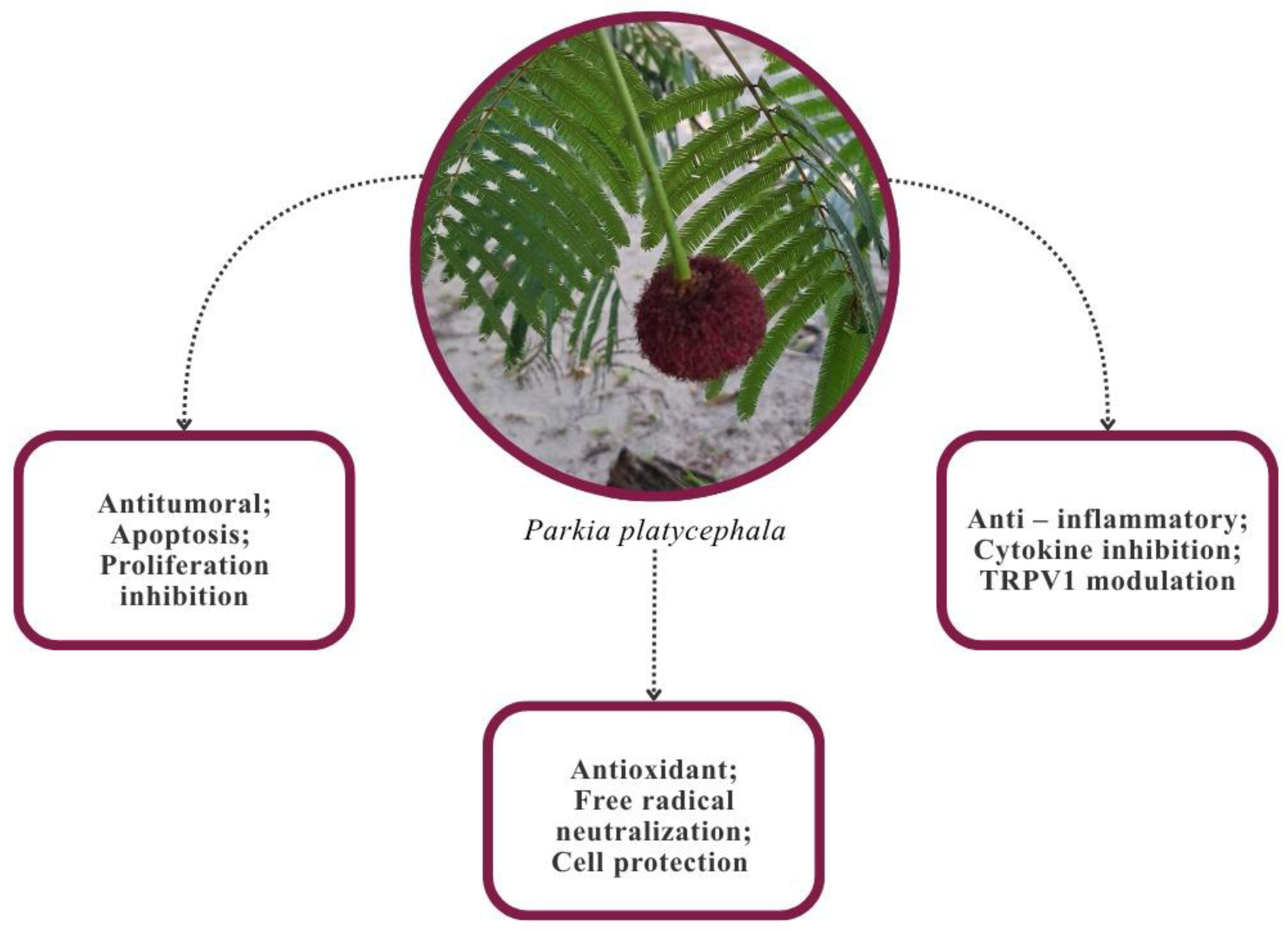
Phytochemical investigations have revealed the presence of total phenolics, flavonoids, flavones, phytosterols, condensed tannins, saponins, and terpenes in bio-extracts and leaf fractions, which possess bioactive properties [,,,,,,,] (Table 1 and Figure 1).
Table 1.
Principal bioactive compounds of PPs and their biological activities.
Figure 1.
Primary pharmacological activities and mechanisms of action of Parkia platycephala [,,,,,,,]. Authors’ photograph; Melonio, M.
3. Toxicity
Although the literature highlights the chemical potential of Parkia platycephala, toxicity studies involving extracts from different plant parts are essential to ensure its safe use (Table 2). These studies aim to identify possible adverse effects, particularly those related to carcinogenicity, cardiotoxicity, and dermatological reactions [].
Table 2.
Toxicity profile of extracts from different parts of Parkia platycephala.
3.1. Subacute and Systemic Toxicity
The toxicity of the crude ethanolic extract from P. platycephala leaves was evaluated in female Wistar rats subjected to oral administration of doses of 250, 500, and 1000 mg/kg for approximately 30 days. The results indicated a possible anti-estrogenic effect and reproductive toxicity, evidenced by a reduction in the proestrus phase, prolongation of diestrus, and decreased uterine weight. No morphological alterations or significant lesions were observed in the main organs analyzed, suggesting that the extract presents signs of moderate systemic toxicity [].
3.2. Subacute and Systemic Toxicity
The toxicity of different parts of P. platycephala was investigated using various extracts. The hexane extract from leaves and seeds was classified as non-toxic, with IC50 values above 4000 µg/mL. Conversely, the ethanolic seed extract was classified as moderately toxic, with IC50 values between 100 and 500 µg/mL [].
In a murine model, oral administration of 2 g/kg of the extract caused no clinical signs of toxicity or mortality after 72 h. Furthermore, the ethanolic leaf extract showed no acute toxicity (up to 1000 mg/kg) and no cytotoxicity in rat erythrocytes (100 µg/mL) [].
Overall, the available data indicate that the evaluated extracts of P. platycephala present low-to-moderate toxicity, suggesting relative safety for in vivo preclinical studies.
There are not many studies on the toxicity of Parkia species, nor are there any that directly compare the toxicity of different species. Studies conducted with P. biglobosa indicate that extracts from the bark of this plant have antioxidant and anti-inflammatory properties, with toxicity results in Artemia salina and rat models suggesting that they are safe [].
4. Biological Activities of Parkia platycephala
4.1. Antioxidant and Anti-Inflammatory Activity
In P. platycephala, the plant protein lectin (PPL) stands out as an important bioactive protein, known for its ability to bind to glucose and mannose. It is composed of three β-prism domains, each of which contains a different carbohydrate recognition domain (CRD) []. The PPL has been categorized as part of the Jacalin-related lectin (JRL) group, and it has been established that this plays an important role in the plant’s defense system against phytopathogenic threats, responding to both biotic and abiotic factors [].
Observations have revealed that Parkia exhibits the capability to impede the movement of leukocytes. According to the researchers, this process, known as migration, is a key component of the inflammatory response, initiated by direct or indirect exposure to chemotactic agents, suggesting that the PPL might influence the inflammatory response. Leukocyte migration to the affected tissue was inhibited [], suggesting a possible influence on the production or activity of pro-inflammatory cytokines such as TNF-α and IL-1β. The PLL has been identified as a highly promising anti-inflammatory agent. This is due to its dual capacity to modulate the TRPV1 channel in addition to interfering with immune responses. The PPL has been demonstrated to bind selectively to mannose residues on the N-glycans of TRPV1, resulting in receptor desensitization, which in turn leads to a reduction in neuronal excitability and the inhibition of prostaglandin and inflammatory cytokine release. At the same time, its ability to bind to glycans on macrophages and mast cells promotes membrane stabilization and blocks Toll-like receptor-dependent (TLR-dependent) pathways. This, in turn, limits the ability of inflammatory cells to be recruited and activated at the location of the injury [].
The high antioxidant activity exhibited by hydroalcoholic extracts from the leaves and bark of P. platycephala is linked to the presence of high levels of phenolic compounds, such as gallic, ellagic, and ferulic acids, as well as the flavonoid kaempferol [], all of which are well-studied antioxidant compounds. The function of these bioactive molecules is predominantly the transfer of electrons to free radicals, which serves to stabilize them and reduce the oxidation of cell components [].
The extracts demonstrated a remarkable capacity to sequester ROS; they have also been observed to stimulate the activity of antioxidant enzymes within cells and to impede the process of lipid peroxide formation. This, in turn, serves to reinforce the structural integrity of the cell. A number of studies have shown that the organic extract of leaves of the plant under discussion has the capacity to enhance the activity of various enzymes found within cells. Among these enzymes, catalase has been shown to be of particular relevance in terms of its role in neutralizing ROS. The antioxidant properties of the substance have been demonstrated to mitigate oxidative stress and to prevent lipid peroxidation, which is critical to promoting the protective mechanisms of cells against damage from harmful agents [].
Notably, P. platycephala has garnered attention for its fruit, which exhibits remarkable antioxidant activity. This property is chiefly ascribed to the presence of polyphenols, especially condensed tannins, in abundance. The antioxidative efficacy of these bioactive substances is closely associated with their capacity to scavenge free radicals, consequently impeding oxidative processes implicated in the biomolecules, such as lipids and proteins. The antioxidant effect is attributed to phenolic compounds’ ability to donate electron or hydrogen atoms, neutralize free radicals, and impede chain reactions that could compromise cellular membrane stability [].
The high antioxidant efficacy of P. platycephala extracts from the flowers is attributable to the substantial presence of phenolic metabolites, specifically phenolic acids and flavonoids. The antioxidant properties of these substances are primarily attributable to their ability to scavenge free radicals, thus effectively impeding the propagation of oxidation reactions. These properties are further augmented by the strong affinity exhibited by these metabolites towards polar solvents, thereby facilitating the extraction of highly reducing substances from aqueous environments. Consequently, the noteworthy antioxidant effect exhibited by P. platycephala floral extracts is attributed to the synergistic integration of electron donation, the capture of free radicals, and the stabilization of ROS [,].
P. platycephala, particularly in the form of seeds, has been demonstrated to inhibit the enzyme acetylcholinesterase, which catalyzes the degradation of the neurotransmitter acetylcholine. The inhibitory effect helps to increase the accessibility of acetylcholine at neuronal synapses, improving nerve transmission and potentially attenuating the symptoms of diseases such as Alzheimer’s. The anticholinesterase properties of the plant further reinforce its therapeutic potential, suggesting that its bioactive compounds may act synergistically to protect neurons and combat oxidative stress [].
In the context of research into natural therapeutic agents, P. platycephala has been identified as a promising candidate, primarily due to its ability to modulate antioxidant and anti-inflammatory mechanisms (Table 3) [].
Table 3.
Antioxidant and anti-inflammatory proprieties of P. platycephala.
Other studies were conducted on specific Parkia species concerning their antioxidant and anti-inflammatory potential. The anti-inflammatory and antioxidant activities of P. biglobosa bark extracts seem to be related to the content of chlorogenic acid, catechin, caffeine, epicatechin, and cichoric acid [].
4.2. Antimicrobial Activity
The lectin and extracts obtained from P. platycephala have been shown to possess significant antimicrobial activity, exhibiting effects against a variety of bacterial and fungal infections. Extracts obtained from P. platycephala, which are aqueous and hydroalcoholic in nature, have been shown to exhibit a rich phytochemical profile. This profile encompasses a range of compounds, including anthraquinones, tannins, saponins, catechins, and lactones, with a particular emphasis on sesquiterpene lactones [].
These findings are further corroborated by phytochemical analyses, which demonstrate a close association between these secondary metabolites and the antimicrobial properties observed in the extracts. The effects of these compounds are primarily exerted through the processes of cell wall complexation, enzyme inhibition, and the sequestration of essential metal ions. Collectively, these metabolites are capable of inhibiting bacterial growth, mainly Gram-positive strains. This finding underscores their potential for use in the development of novel therapeutic agents.
The isolated PPL has been the focus of particular studies due to its ability to enhance the efficacy of antibiotics. Despite the fact that the PPL does not demonstrate clinically significant direct antibacterial activity when tested against Staphylococcus aureus, Escherichia coli, and Pseudomonas aeruginosa, it nevertheless significantly potentiates the effect of gentamicin, thereby lowering the minimum inhibitory concentration (MIC) values required to inhibit S. aureus and E. coli []. The adjuvant action of combining P. platycephala extracts or the PPL with antibiotics may allow for reduced antibiotic dosages, thereby minimizing antibiotic-related toxicity and delaying the development of bacterial resistance [].
PPL-2, which is extracted from the seeds of P. platycephala, exhibited bacteriostatic activity in in vitro assays. The inhibitory potency was found to be contingent upon the bacterial cell wall type, with substantial disparities observed between Gram-positive and Gram-negative strains, potentially attributable to the structural heterogeneity of surface-exposed glycans. The present findings lend support to the theory that PPL-2 specifically interacts with components of the bacterial membrane, thereby disrupting its stability and interfering with cell division mechanisms [,].
Bark extracts of P. platycephala demonstrate a phytochemical profile characterized by tannins, saponins, flavonoids, alkaloids, and steroids, the presence of which is responsible for their antimicrobial activity (Table 4) [,,,,,,,,,,,,].
Table 4.
Antimicrobial activity of Parkia platycephala.
4.3. Antitumor Activity
Lectins isolated from P. platycephala, especially those extracted from the leaves, have demonstrated significant antiproliferative potential (Table 5). These proteins have been shown to affect the viability of various tumor cell lines, including hepatocellular carcinoma, leukemia, colorectal adenocarcinoma, lung, liver, and melanoma cells [,,,,,,,,,,,,,,,,,]. This anticancer activity is believed to be closely related to the high affinity of lectins for glycoconjugates, particularly glycoproteins and glycolipids, which are frequently overexpressed or aberrantly glycosylated on the membranes of cancer cells. By selectively binding to these altered carbohydrate moieties, P. platycephala lectins can interfere with key cellular process-es, such as receptor-mediated signaling, endocytosis, and cell–cell communication. This interaction may lead to the disruption of mitogenic signaling pathways and the induction of cell cycle arrest, particularly at the G0/G1 or G2/M checkpoints, ultimately inhibiting cancer cell proliferation []. Moreover, some studies suggest that these lectins may also trigger apoptosis via mitochondrial depolarization, caspase activation, and modulation of pro- and anti-apoptotic proteins. In addition to their direct cytotoxic effects, lectins may also exert immunomodulatory activity by enhancing the recognition and elimination of tumor cells by the immune system []. Altogether, these findings underscore the therapeutic potential of P. platycephala lectins as promising bio-tools in cancer research and drug development.
Table 5.
Antitumor activity of Parkia platycephala: bioactive compounds and mechanisms of action.
Morphological changes consistent with apoptosis induction, including chromatin condensation and nuclear fragmentation, have been shown to occur when tumor cells are exposed to these lectins []. Together, these observations support the idea that P. platycephala lectins not only inhibit cell proliferation but also promote apoptosis of malignant cells.
In addition to their antiproliferative effects, these agents have been observed to modulate signaling pathways, induce apoptosis and autophagy, and stimulate the production of ROS, ultimately resulting in oxidative stress and cell death []. The aforementioned properties underscore the significance of leaf lectins from the Parkia genus as bioactive compounds, exhibiting considerable promise in the development of novel cancer therapeutic interventions.
Moreover, natural compounds have demonstrated significant cell cycle modulation potential in tumor cells, such as HT-29 (colorectal) and PC3 (prostate) cancer cell lines, which may be a key factor in regulating neoplastic cell growth [,,]. A recent research study utilizing extracts of plant bark and flowers, along with methanolic or ethanolic extracts derived from the bark or flowers of various plant species, has indicated that the bioactive compounds present in these plant components may impact regulatory cell cycle mechanisms, thereby hindering the progression of cancer cells []. Specifically, these compounds, which included isolated flavonoids and complex plant extracts were observed to inhibit regulatory proteins such as cyclins and cyclin-dependent kinases (CDKs) during the transition between the G1 and S phases of the cell cycle [,]. This inhibition can cause the cell cycle to slow down or even stop, preventing the cancer cells from growing uncontrollably.
However, in the case of Parkia platycephala, it is crucial to acknowledge that, within the scope of our study, the bark extracts were the only ones to manifest noteworthy biological activities, including antioxidant and anticholinesterase properties. Conversely, the flower extracts failed to exhibit significant antiproliferative or cytotoxic effects []. This study highlights the necessity for further research to elucidate the effects of Parkia platycephala extracts, particularly those derived from the flowers, on cell cycle progression and cancer cell proliferation.
5. Applications of Parkia platycephala in Animal Feed
Nutritional studies on animal feeding involving P. platycephala are reported in the literature, using this species as an alternative, partial, or total feed source for ruminants [,]. This is due to several factors, such as its abundant availability during dry seasons, the reduced costs compared to conventional animal feed, and its high nutritional potential [,,].
5.1. Nutritional Potential
The fruits of P. platycephala stand out for their nutritional potential, as studies have reported a high protein content []. Nutritional assessments of P. platycephala pods revealed 11.10% crude protein and 72.5% total digestible nutrients []. They also present high levels of dry matter (77.25%), soluble carbohydrates (69.26%) [], and non-fibrous carbohydrates (68.53%) [,,].
Pods offer potential as a rapidly fermentable energy source in ruminant diets because they are more easily degraded by ruminal microorganisms, supply energy more quickly [,], do not compromise animal performance [], and are associated with weight gain, increased production of short-chain fatty acids (such as butyrate), and reduced internal fat [,]. In a study with lambs, due to the tannin content of pods, the introduction of pods into their diet modulated biohydrogenation and improved the fatty acid profile and lipid oxidation, with a consequent improvement in the organoleptic and nutritional quality of the meat [].
Thus, it can be concluded that the use of P. platycephala in animal feed can improve the organoleptic and nutritional quality of meat, reduce production costs, and lessen the environmental impact [,].
5.2. Application of P. platycephala Pods
The pods of P. plathycephala, which are found on the plant in September and October in Brazil, are a very rich and cheaper nutritional source for animal feed [,]. Moreover, their nutritional impact on animal feed can extend throughout the year, as the beans can be dried and stored and then fed to animals [,].
Studies involving the addition of 30% P. platycephala pod meal combined with 1.5% urea to elephant grass (Pennisetum purpureum Schum) silage resulted in improved fermentative characteristics and chemical composition. There was an increase in dry matter (29.92%) and crude protein (20.92%) contents, and a reduction in neutral detergent fiber (51.34%) and acid detergent fiber (30.27%). The silage became more palatable, with better utilization by ruminal microorganisms, increased intake, and improved animal performance [].
Lactating goats fed with P. platycephala pod meal showed no impairment in dry matter intake (~1450–1570 g/day) or overall performance. A significant increase in serum calcium (10.57 mg/dL) was observed, which is important for lactation and the maintenance of stable hematological parameters [].
The replacement of up to 50% of maize with P. platycephala pods in the diet of confined sheep showed excellent nutritional values without significantly compromising performance [,,]. Similarly, equivalent proportions of commercial concentrate and ground pods produced superior nutritional responses compared to the exclusive use of pods [].
5.3. Application of P. platycephala Seeds
P. platycephala seeds have a promising bromatological profile for zootechnical applications, characterized by a high crude protein content (32.2% dry basis), a significant lipid content (13.4%), and high dietary fiber (52.3%), as well as providing considerable energy density (1048 kJ/100 g).
The seeds have also been evaluated as an alternative food source for tambatinga (Colossoma macropomum × Piaractus brachypomus). The results indicated that the lectin present in these seeds has antinutritional effects [], evidenced by reduced weight gain, specific growth rate, and relative weight gain, along with decreased glycogen levels and lower muscle protein content. The authors also noted that the seed meal could be used in fish and other animal feed if the lectin was inactivated [].
Thus, different parts of P. platycephala can be used in the feed of various species due to their regional availability, high nutritional value, and lower cost compared to other feed sources [,,].
6. Conclusions
This detailed review corroborates the pharmacological significance of P. platycephala, confirming its potential as a source of bioactive compounds and highlighting its promise as a source of novel, multi-target therapeutic agents.
The authors are aware that there are few reviews on plants of the Parkia genus. With regard to P. platycephala, Fernandes et al. published a review article in 2023 on toxicity and biological activities, but only on compounds from bark and flowers. Thus, to our knowledge, this will be the first extensive review on P. plathycephala regarding its composition, biological activities, toxicity, and potential use in humans and animals.
Remarkably, substances extracted from P. platycephala have shown the ability to promote apoptosis, suppress cell proliferation, and neutralize ROS. Furthermore, these compounds have demonstrated the capacity to improve the effectiveness of antibiotics, highlighting their potential to address significant unmet medical needs, particularly in cases of drug-resistant infections and cancers that do not respond to current treatments.
Nevertheless, further rigorous investigation is required to translate these experimental findings into clinical application. This includes standardizing extraction protocols, comprehensively elucidating mechanisms of action, and systematically validating safety and efficacy in both preclinical and clinical settings.
In the context of an expanding global interest in the search for innovative, natural-based therapies, P. platycephala emerges as a significant source of pharmacological innovation. It is imperative that future interdisciplinary studies encompassing phytochemistry, pharmacology, molecular biology, and clinical sciences are conducted to ensure the comprehensive realization of its therapeutic potential. Such efforts will not only facilitate the advancement of evidence-based medicine but also contribute to the enhancement of public health outcomes.
Based on all the activities described, and the implications for human and animal health and nutrition, P. platychephala adds value to the “One Health” concept. In addition, the use of various parts of the plant for different purposes has an important impact on sustainability and the optimization of natural resource use.
Author Contributions
Conceptualization, F.C. and R.M.; writing—original draft preparation, J.N.; writing—review and editing, J.N., M.C.M., C.Q.d.R., F.C., and R.M. All authors have read and agreed to the published version of the manuscript.
Funding
This study was financed in part by the Coordenação de Aperfeiçoamento de Pessoal de Nível Superior – Brazil (CAPES)–Finance Code 001, PROCAD-AM [88887.472618/2019-00 (C.Q.d.R.)] and CNPq [PPBio-: 441189/2023-7 (C.Q.d.R.)].
Data Availability Statement
No new data were created or analyzed in this study. Data sharing is not applicable to this article.
Acknowledgments
Herbário MAR for plant identification.
Conflicts of Interest
The authors declare no conflicts of interest.
References
- Li, D.; Wu, M. Pattern recognition receptors in health and diseases. Signal Transduct. Target. Ther. 2021, 6, 291. [Google Scholar] [CrossRef]
- Greten, F.R.; Grivennikov, S.I. Inflammation and Cancer: Triggers, Mechanisms, and Consequences. Immunity 2019, 51, 27–41. [Google Scholar] [CrossRef]
- Reuter, S.; Gupta, S.C.; Chaturvedi, M.M.; Aggarwal, B.B. Oxidative stress, inflammation, and cancer: How are they linked? Free Radic. Biol. Med. 2010, 49, 1603–1616. [Google Scholar] [CrossRef]
- Lee, Y.T.; Tan, Y.J.; Oon, C.E. Molecular targeted therapy: Treating cancer with specificity. Eur. J. Pharmacol. 2018, 834, 188–196. [Google Scholar] [CrossRef] [PubMed]
- Khan, S.U.; Fatima, K.; Aisha, S.; Malik, F. Unveiling the mechanisms and challenges of cancer drug resistance. Cell Commun. Signal. 2024, 22, 109. [Google Scholar] [CrossRef] [PubMed]
- Newman, D.J.; Cragg, G.M. Natural Products as Sources of New Drugs over the Nearly Four Decades from 01/1981 to 09/2019. J. Nat. Prod. 2020, 83, 770–803. [Google Scholar] [CrossRef]
- Yuan, H.; Ma, Q.; Ye, L.; Piao, G. The Traditional Medicine and Modern Medicine from Natural Products. Molecules 2016, 21, 559. [Google Scholar] [CrossRef]
- Saleh, M.S.M.; Jalil, J.; Zainalabidin, S.; Asmadi, A.Y.; Mustafa, N.H.; Kamisah, Y. Genus Parkia: Phytochemical, Medicinal Uses, and Pharmacological Properties. Int. J. Mol. Sci. 2021, 22, 618. [Google Scholar] [CrossRef]
- de Oliveira Silva, D.Y.B.; de Farias, S.G.G.; Dias, P.C.; e Silva, R.B.; Dias, B.A.S. Phenotypic and genotypic evaluation of Parkia platycephala families: A proposal for pre-selection. Crop Breed. Appl. Biotechnol. 2023, 23, e42792314. [Google Scholar] [CrossRef]
- Chaves, S.R.; dos Santos, R.R.; da Silva, A.L.G. Reproductive biology of Parkia platycephala Benth (Legumi-nosae, Caesalpinioideae, clado mimosoide). Braz. J. Dev. 2020, 6, 79442–79458. [Google Scholar] [CrossRef]
- Fernandes, R.M.; Rodrigues, M.A.M.; Cardoso, C.A.L.; Alves, D.R.; Morais, S.M.; Panontin, J.F.; Scapin, E. Phytocomponents, Evaluation of Anticholinesterase Activity and Toxicity of Hydroethanolic Extracts of Parkia platy-cephala Benth. J. Braz. Chem. Soc. 2022, 33, 1414–1422. [Google Scholar]
- Fernandes, R.M.N.; Cardoso, C.A.L.; Alves, D.R.; Morais, S.M.; Scapin, E. Parkia from Cerrado: Phytochemical bioprospection, toxicity and in vitro bioactivities of bark and flower extracts. Braz. J. Biol. 2023, 83, e275733. [Google Scholar] [CrossRef] [PubMed]
- Costa, I.C.G.; Damasceno, F.C.; Menegatti, A.C.O.; Vieira Júnior, G.M.; Chaves, M.H.; de França Ferreira, É.L. Antiradical effect and phytochemical characterisation of the leaves of Parkia platycephala Benth. Nat. Prod. Res. 2024, 39, 5403–5407. [Google Scholar] [CrossRef] [PubMed]
- Fernandes, H.B.; Silva Fv BPassos, F.F.; SBezerra, R.D.; Chaves, M.H.; Oliveira, F.A.; Meneses Oliveira, R.C. Gastroprotective effect of the ethanolic extract of Parkia platycephala Benth. leaves against acute gastric lesion models in rodents. Biol. Res. 2010, 43, 451–457. [Google Scholar] [CrossRef]
- de Moura, R.; Fernandes, N. Bioprospecção Fitoquímica, Toxicidade e Atividades Antioxidante e Anticolinesterásica de Extratos da Parkia Platycephala (Benth.). Doutorado em Biodiversidade e Biotecnologia da Amazônia Legal, Programa De Pos-Graduação Em Biodiversidade E Biotecnologia-Rede Bionorte, Universidade Federal Do Tocantins, Tocantins, Brazil, 2023. [Google Scholar]
- Sá Santos, M.M.; da Silva, F.M.P.; da Silva, J.F.M.; Pimenta, R.S. Phytochemistry and antibacterial activity of aqueous and hydroalcoholic extracts of three medicinal plants against food pathogens. Acta Sci. Biol. Sci. 2018, 40, e36674. [Google Scholar] [CrossRef]
- Oliveira, A.F.; Junior, L.M.C.; Lima, A.S.; Silva, C.R.; Ribeiro, M.N.; Mesquista, J.W.; Rocha, C.Q.; Tangerina, M.M.; Vilegas, W. Anthel-mintic activity of plant extracts from Brazilian savanna. Vet. Parasitol. 2017, 236, 121–127. [Google Scholar] [CrossRef]
- Falya, Y.; Sumiwi, S.A.; Levita, J. Mini Review: Toxicity Study of Plant Extracts. IOSR J. Pharm. Biol. Sci. 2020, 15, 25–32. [Google Scholar]
- Costa, B.A.; Sousa, A.S.; Borges, M.S.; Damasceno, D.C.; Costa-Silva, J.H. Toxicidade sistêmica e reprodutiva induzida pelo extrato etanólico de Parkia platycephala em ratas Wistar. Rev. Bras. Farmacogn. 2013, 23, 920–926. [Google Scholar] [CrossRef]
- Fernandes, R.M.N.; Rodrigues, M.A.M.; Panontin, J.F.; Alves, D.R.; Morais, S.M.; Soares, I.M.; Scapin, E. Chemical Investigation, Toxic Potential and Acetylcholinesterase Inhibitory Effect of Parkia platycephala Leaf and Seed Extracts. J. Med. Plants Res. 2021, 15, 401–412. [Google Scholar] [CrossRef]
- Dangnon, B.; Dah-Nouvlessounon, D.; Hoteyi, S.M.I.; Sina, H.; Tomescu, J.A.; Akpo, K.J.-M.; Sangare-Oumar, M.M.; Adjanohoun, A.; Babalola, O.O.; Vamanu, E.; et al. Gastroprotective, Antioxidant, Anti-Inflammatory, and Toxicological Evaluation of Stem Bark Extracts of Vitellaria paradoxa and Parkia biglobosa. Pharmaceuticals 2025, 18, 1184. [Google Scholar] [CrossRef]
- Mann, K.; Farias, C.M.S.A.; del Sol, F.G.; Santos, C.F.; Grangeiro, T.B.; Nagano, C.S.; Cavada, B.S.; Calvete, J.J. The amino-acid sequence of the glucose/mannose-specific lectin isolated from Parkia platycephala seeds reveals three tandemly arranged jacalin-related domains. Eur. J. Biochem. 2001, 268, 4414–4422. [Google Scholar] [CrossRef] [PubMed]
- Bari, A.U.; Santiago, M.Q.; Osterne, V.J.S.; Pinto-Junior, V.R.; Pereira, L.P.; Silva-Filho, J.C.; Debray, H.; Rocha, B.A.M.; Delatorre, P.; Teixeira, C.S.; et al. Lectins from Parkia biglobosa and Parkia platycephala: A comparative study of structure and biological effects. Int. J. Biol. Macromol. 2016, 92, 194–201. [Google Scholar] [CrossRef]
- Souza, M.A.; Carvalho, F.C.; Ruas, L.P.; Ricci-Azevedo, R.; Roque-Barreira, M.C. The immunomodulatory effect of plant lectins: A review with emphasis on ArtinM properties. Glycoconj. J. 2013, 30, 641–657. [Google Scholar] [CrossRef]
- Cavada, B.S.; Osterne, V.J.S.; Oliveira, M.V.; Pinto-Junior, V.R.; Silva, M.T.L.; Bari, A.U.; Lima, L.D.; Lossio, C.F.; Nascimento, K.S. Reviewing Mimosoideae lectins: A group of under explored legume lectins. Int. J. Biol. Macromol. 2020, 154, 159–165. [Google Scholar] [CrossRef]
- Aladesanmi, A.J. Tetrapleura tetraptera: Molluscicidal activity and chemical constituents. Afr. J. Tradit. Complement. Altern. Med. 2007, 4, 23–36. [Google Scholar] [CrossRef]
- Batista Gomes, P.G.; Raposo Santos, M.; de Araújo, G.G.L.; Parente, H.N.; de Oliveira Maia Parente, M.; de Moura Zanine, A.; de Jesus Ferreira, D.; Santos, E.M.; Gois, G.C.; de Sousa Santos, F.N.; et al. Parkia platycephala Replacing Ground Corn in the Diet of Confined Lambs: Intake, Digestibility, Ingestive Behaviour, Rumen Fermentation and Carcass Yield. Arch. Anim. Nutr. 2024, 78, 381–397. [Google Scholar] [CrossRef] [PubMed]
- Konozy, E.H.E.; Osman MEfadil, M. Plant lectin: A promising future anti-tumor drug. Biochimie 2022, 202, 136–145. [Google Scholar] [CrossRef]
- Dai, J.; Mumper, R.J. Plant Phenolics: Extraction, Analysis and Their Antioxidant and Anticancer Properties. Molecules 2010, 15, 7313–7352. [Google Scholar] [CrossRef]
- Boeing, J.S.; Barizão, É.O.; e Silva, B.C.; Montanher, P.F.; de Cinque Almeida, V.; Visentainer, J.V. Evaluation of solvent effect on the extraction of phenolic compounds and antioxidant capacities from the berries: Application of principal component analysis. Chem. Cent. J. 2014, 8, 48. [Google Scholar] [CrossRef]
- Silva, R.R.S.; Silva, C.R.; Santos, V.F.; Barbosa, C.R.S.; Muniz, D.F.; Santos, A.L.E.; Santos, M.H.C.; Rocha, B.A.M.; Batista, K.L.R.; Costa-Júnior, L.M.; et al. Parkia platycephala lectin enhances the antibiotic activity against multi-resistant bacterial strains and inhibits the development of Haemonchus contortus. Microb. Pathog. 2019, 135, 103629. [Google Scholar] [CrossRef]
- Carvalho, A.F.U.; Farias, D.F.; da Rocha-Bezerra, L.C.B.; de Sousa, N.M.; Cavalheiro, M.G.; Fernandes, G.S.; Brasil, I.C.F.; Maia, A.A.B.; de Sousa, D.O.B.; Vasconcelos, I.M.; et al. Preliminary Assessment of the Nutritional Composition of Underexploited Wild Legumes from Semi-Arid Caatinga and Moist Forest Environments of Northeastern Brazil. J. Food Compos. Anal. 2011, 24, 487–493. [Google Scholar] [CrossRef]
- Parasuraman, S. Toxicological screening. J. Pharmacol. Pharmacother. 2011, 2, 74. [Google Scholar] [CrossRef]
- Yau, T.; Dan, X.; Ng, C.C.W.; Ng, T.B. Lectins with Potential for Anti-Cancer Therapy. Molecules 2015, 20, 3791–3810. [Google Scholar] [CrossRef] [PubMed]
- Jiang, Q.L.; Zhang, S.; Tian, M.; Zhang, S.Y.; Xie, T.; Chen, D.Y.; Chen, Y.J.; He, J.; Liu, J.; Ouyang, L.; et al. Plant lectins, from ancient sugar-binding proteins to emerging anti-cancer drugs in apoptosis and autophagy. Cell Prolif. 2015, 48, 17–28. [Google Scholar] [CrossRef] [PubMed]
- Bailon-Moscoso, N.; Cevallos-Solorzano, G.; Romero-Benavides, J.C.; Orellana, M.I.R. Natural Compounds as Modulators of Cell Cycle Arrest: Application for Anticancer Chemotherapies. Curr. Genom. 2017, 18, 106. [Google Scholar] [CrossRef] [PubMed]
- Lu, X.; Jae, I.J.; Han, J.C.; Do, Y.L.; Hyun, S.L.; Hyang, S.C.; Dae, Y.K.; Park, J.H.Y. Fisetin inhibits the activities of cyclin-dependent kinases leading to cell cycle arrest in HT-29 human colon cancer cells. J. Nutr. 2005, 135, 2884–2890. [Google Scholar] [CrossRef]
- Pasban-Aliabadi, H.; Sobhani, V.; Esmaeili-Mahani, S.; Najafipour, H.; Askari, A.; Jalalian, H. Effects of Baneh (Pistacia atlantica) Gum on Human Breast Cancer Cell Line (MCF-7) and Its Interaction with Anticancer Drug Doxorubicin. Iran. J. Pharm. Res. 2019, 18, 1959–1966. [Google Scholar]
- Silva, J.H.O.; Monteiro, V.F.C.; Moura, P.H.A. Diversidade de plantas no Cerrado brasileiro: Um enfoque em Parkia platycephala. In Ciências Botânicas; Monteiro, V.F.C., Moura, P.H.A., Eds.; Atena Editora: Ponta Grossa, Brazil, 2021; pp. 79–94. [Google Scholar]
- Alves, A.A.; Sales, R.; Neiva, J.; Medeiros, A.; Braga, A.; Azevedo, A. Degradabilidade ruminal in situ de vagens de faveira (Parkia platycephala Benth.) em diferentes tamanhos de partículas. Arq. Bras. Med. Vet. Zootec. 2007, 59, 1045–1051. [Google Scholar] [CrossRef]
- Araújo, A.R.; Rodriguez, N.M.; Rogério, M.C.P.; Borges, I.; Saliba, E.O.S.; Santos, S.A.; Pompeu, R.C.F.F.; Fernandes, F.E.P.; Monteiro, J.P.; Muir, J.P. Nutritional Evaluation and Productivity of Supplemented Sheep Grazing in Semiarid Rangeland of Northeastern Brazil. Trop. Anim. Health Prod. 2019, 51, 957–966. [Google Scholar] [CrossRef]
- Vilela, G.; Pinheiro, G.; Castro, L.; Santos, F.; Oliveira, J.; Parente, M.; Parente, M.; Costa, J.; Barbosa, S.; Silva, S. Pontencial Nutricional Da Parkia Platycephala Na Qualidade Da Carne E Características De Carcaça De Pequenos Ruminantes. In Agropecuária e Meio Ambiente: Uma Visão Integrada; Cardoso, A., Borges, S., Silva, S., Andrade, F., Eds.; Editora Científica Digital: Guarujá, Brazil, 2025. [Google Scholar] [CrossRef]
- Batista, I.L.; Araújo, M.J.; Marques, C.A.T.; Carvalho, F.J.V.; Jácome, D.D.S.; Oliveira, R.L. Effects of Parkia platycephala on Feeding Behavior, Rumen Health, Blood Markers, and Physiological Responses of Lactating Goats. Rev. Bras. Zootec. 2020, 49, e20200096. [Google Scholar] [CrossRef]
- da Silva, L.R.F.; Alves, A.A.; Vasconcelos, V.R.; Santos do Nascimento, H.T.; Filho, M.A.M. Valor Nutritivo da Vagem de Faveira (Parkia platycephala Benth.) para Ruminantes. Rev. Bras. De Zootec. 1999, 41, 1065–1069. [Google Scholar] [CrossRef]
- Costa, L.A.; Araújo, M.J.; Edvan, R.L.; Bezerra, L.R.; Sousa, A.R.; Viana, F.J.C.; Dias-Silva, T.P. Chemical Composition, Fermentative Characteristics, and In Situ Ruminal Degradability of Elephant Grass Silage Containing Parkia platycephala Pod Meal and Urea. Trop. Anim. Health Prod. 2020, 52, 3481–3492. [Google Scholar] [CrossRef]
- Da Silva, R.C.; Teixeira, C.S.; Pretto, A.; Costa, T.S.; de Siqueira, J.C.; Pantoja, B.T.S.; Baldisserotto, B.; Lopes, J.M. Antinutritional Effect of Lectin from Faveira (Parkia platycephala) Seeds in Tambatinga (Colossoma macropomum × Piaractus brachypomus). Bol. Inst. Pesca 2022, 48, e745. [Google Scholar] [CrossRef]
- Silva, L.R.F.; Alves, A.A.; Vasconcelos, V.R.; do Nascimento, H.T.S.; Filho, M.A.M. Nutritive Value of Diets Containing Pods of Faveira (Parkia platycephala Benth.) for Confined Finishing Sheep. Rev. Bras. Zootec. 2012, 41, 1065–1069. [Google Scholar] [CrossRef]
- Lima, J.R.L.; Rodrigues, R.C.; Sousa, G.O.C.; de Jesus, A.P.R.; da Costa, A.C.; Fonseca, A.S.R.; Cabral, L.d.S.; Costa, C.d.S.; de Oliveira, J.S.; da Silva, E.C. Performance of Grazing Sheep Kept on Tamani Grass Supplemented with Parkia platycephala Pods Replacing Corn in Multiple Supplements. N. Z. J. Agric. Res. 2025, 68, 515–531. [Google Scholar] [CrossRef]
- Sousa, G.O.C.; Rodrigues, R.C.; Lima, J.R.L.; de Jesus, A.P.R.; da Costa, A.C.; Andrade, A.C.; Rodrigues, M.M.; da Silva, E.C.; Santos, A.M.d.P.; Oliveira, P.L.R.; et al. Parkia platycephala in Multiple Supplements for Sheep Grazing on Tamani Grass Pastures: Effects on Pasture Productivity, Structural Characteristics, and Ingestive Behaviour. N. Z. J. Agric. Res. 2025, 68, 1901–1914. [Google Scholar] [CrossRef]
- Porto, D.L.; de Santana Arauco, A.M.; Boechat, C.L.; de Oliveira Silva, A.; Moitinho, M.R.; de Farias, S.G.G. Arbuscular Mycorrhizal Fungi on the Initial Growth and Nutrition of Parkia platycephala Benth. under Water Stress. Cerne 2020, 26, 66–74. [Google Scholar] [CrossRef]
Disclaimer/Publisher’s Note: The statements, opinions and data contained in all publications are solely those of the individual author(s) and contributor(s) and not of MDPI and/or the editor(s). MDPI and/or the editor(s) disclaim responsibility for any injury to people or property resulting from any ideas, methods, instructions or products referred to in the content. |
© 2025 by the authors. Licensee MDPI, Basel, Switzerland. This article is an open access article distributed under the terms and conditions of the Creative Commons Attribution (CC BY) license (https://creativecommons.org/licenses/by/4.0/).