Abstract
Understanding the impact of subaerial biofilms (SABs) on geomaterial weathering is crucial for the sustainable conservation of cultural heritage belements. As SABs can either exacerbate or mitigate sudden thermal or hygroscopic stress, it is essential to identify their protective or deteriorative role on a case-by-case basis. This study aims to validate the utility of infrared thermography in examining the impact of biofilms on stone and exploring the water-related behaviors of porous materials colonized by SABs.
1. Introduction
The study and preservation of stone cultural heritage elements are of utmost importance due to their historical, artistic, and cultural significance. Many microorganisms, such as bacteria, fungi, and algae, adeptly colonize lithic materials, forming complex microbial communities at the stone/air interface known as subaerial biofilms (SABs) [1]. SABs offer favorable conditions to their inhabitants, promoted by a self-produced extracellular polymeric matrix (EPM). This EPM contributes to the three-dimensional structure of biofilms and facilitates energy and matter exchanges among microorganisms, the substrate, and the surrounding environment [2]. While SABs on stone cultural heritage elements have traditionally been associated with biodeterioration [3], recent research has revealed their potential neutral or even protective effects on the underlying surfaces in specific circumstances [4]. A recent study by Villa et al. examined the impact of desiccated SABs on the water exchange dynamics between the substrate and the environment; the findings revealed that these SABs not only induce a delay in water evaporation but also act as a protective barrier against rapid water uptake [5]. This research emphasizes the importance of conducting case-specific individual investigations on the influence of SABs on underlying substrates for the conservation of stone monuments. To effectively address this issue, it becomes imperative to develop analytical methodologies that are easy, fast, cost-effective, and non-invasive. These methodologies should be versatile enough to be employed in both laboratory and field settings. While there are numerous methods available to study SABs from a microbiological perspective, only a limited number of methods exist to investigate the interplay between SABs and their surroundings [6]. The utilization of passive IRT for investigating moisture-induced deterioration in building materials is extensively documented in the scientific literature [7,8,9]. There are a limited number of studies that have applied IRT to investigate SABs. These studies primarily highlight the potential of IRT for the early detection of biofilm during the initial stages of surface colonization [10], monitoring biofilm formation in conjunction with sample degradation [11], and mapping the weathering process [12]. Within an alternative framework, IRT has been employed to assess the heterogeneity of soil moisture dynamics and microbial activity during rewetting processes, monitoring short-term hygrothermal events [13].
The aim of this study is to establish a rapid laboratory protocol based on IRT for examining the influence of SABs on the dynamics of moisture exchange between stone surfaces and the surrounding environment in view of the goal of expanding this methodology for use in conducting field measurements.
2. Materials and Methods
In order to investigate the suitability of IRT for studying the impact of SABs on water dynamics, a laboratory experiment was conducted under controlled environmental conditions. The experiment involved the utilization of mono- (Synechocystis PCC 6803) and dual-species (Synechocystis PCC 6803 and Escherichia coli ATCC 25404) SABs on limestone specimens (H × W × T = 7.5 cm × 2.5 cm × 1 cm) grown in a drip flow reactor as reported by [14]. After 10 days, SABs reached maturity, and three replicates of each type (MSB1-3 and DSB1-3) were carefully removed from the bioreactors, along with control specimens without biofilms (WR0). These specimens were then acclimated for 12 days under a relative humidity of 77%, a temperature of 20 °C, and natural light exposure. To investigate humidity dynamics with or without SABs, wetting methods and IRT analysis were used. Through the Spilling Drop Test (SDT), wettability, surface roughness, and porosity were evaluated by observing a drop’s shape over time [15]. The Evaporative Thermal Index (ETI) was utilized by dividing the difference between the moist and dry surface temperatures by the dry surface temperature, yielding valuable information about the evaporation process when applying liquid water to the samples [16]. SDT was performed following the methodology described by Melada et al. [17]. Thermograms were taken immediately after the deposition of deionized water, cropped to include the drop and background, and limited to the first 25 s. For segmentation purposes, thresholding using Yen’s method was applied to each image, effectively separating the drop from the background [18]. Quantitative estimates of the drop’s area were computed from segmented binary images. For the study of the ETI, deionized water was sprayed onto the samples, and evaporation activity was monitored using IRT at 80 cm distance from the sample with 20 Hz frame-rate. The measurements were conducted in a climatic chamber, with the following parameters: temperature set at 25 °C, relative humidity set at 70%, and a controlled ventilation rate of 1 m/s. The ETI was calculated for samples with SABs and for the control without it, as reported in [16]. The study employed an AVIO 550Pro infrared camera, which operates in the long-wave (from 8 μm to 14 μm) wavelength range and captures images with dimensions of 480 × 640 pixels. Subsequent processing of the data was performed via developing scripts in Python 3.9.
3. Results and Conclusions
This section outlines the initial results concerning SABs’ influence on moisture exchange, utilizing infrared thermography (IRT). Specifically, the results are presented for samples with SABs (mono-species MSB1 and dual-species DSB2) and one SAB-free limestone sample (control, WR0), which are representative of the overall sample set. Figure 1a clearly demonstrates that when a water droplet is deposited on the biofilm, it does not spread and remains visible for a time interval longer than the duration of the experiment. This observation indicates the presence of a surface condition that hinders the droplet’s spreading. In contrast, the droplet placed on the limestone (WR0) expands by 400% relative to the moment of deposition (Figure 1a) and is rapidly absorbed by the material, as indicated by the decrease in thermal contrast shown in Figure 1b.
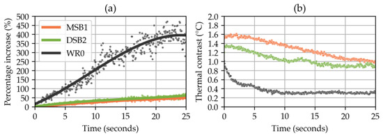
Figure 1.
(a) The temporal evolution of the drop size, expressed as a percentage increase relative to the instant of deposition. Scatter points represent measurements, while the line is a third-degree polynomial fit. (b) The temporal variation in thermal contrast between the drop and the underlying surface.
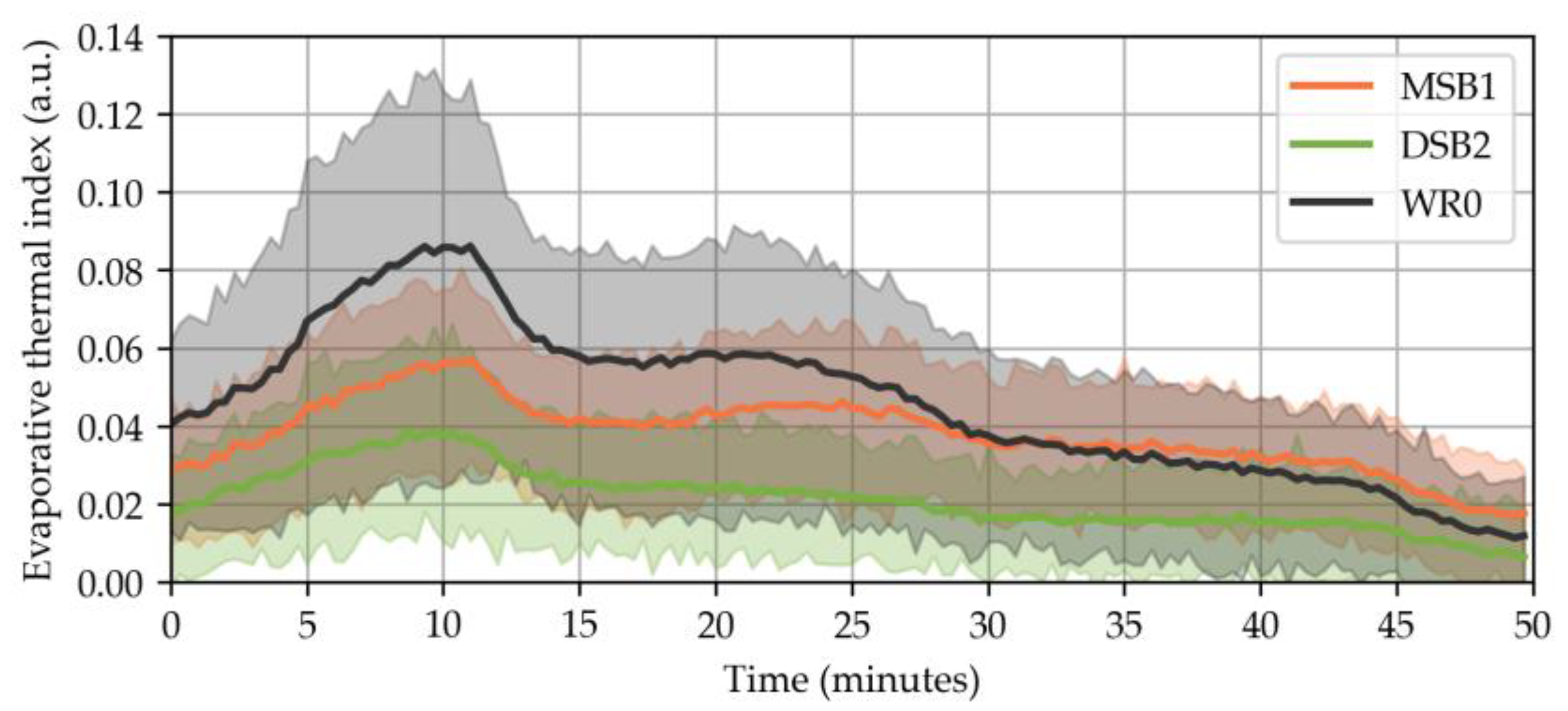
Figure 2 provides valuable insights into the effects of moisture-related stress on monuments. By analyzing the ETI, we can understand how SABs respond to moisture, considering the environmental conditions, biofilm phenotype, and substrate characteristics. The ETI reflects the moisture content in a shallow layer, with higher values indicating a higher rate of evaporation (increased evaporative cooling) when the same amount of water is present under similar conditions [16]. This implies that the water present on the surface either does not penetrate into the stone through capillary action or becomes bound within the EPS of SABs, while still being able to evaporate freely.

Figure 2.
The image illustrates the change in the ETI over time after the application of deionized water spray on laboratory-grown subaerial biofilm (SAB) colonies on limestone, comparing it to limestone without biofilm. The experiments were conducted under controlled conditions. The error band represents the temperature range of wet areas compared to the average temperature of dry areas.
The study’s results indicate that both SDT and the ETI successfully analyze the moisture exchange dynamics between the SABs, the stone, and the environment in laboratory settings. Extending these methods to field studies and laboratory-scale experiments under different environmental conditions will advance our understanding of SABs’ behavior and contribute to effective SAB management at cultural heritage sites.
Author Contributions
Conceptualization, J.M., F.V., N.L. and M.G.; methodology, J.M. and N.L.; software, J.M.; validation, I.B., E.C., D.R. and A.M.; formal analysis, J.M.; investigation, I.B., E.C., D.R. and A.M.; resources, F.V. and N.L.; data curation, J.M., I.B. and A.M.; writing—original draft preparation, I.B. and A.M.; writing—review and editing, J.M., N.L., M.G. and F.V.; visualization, J.M.; supervision N.L., M.G. and F.V. All authors have read and agreed to the published version of the manuscript.
Funding
This research received no external funding.
Institutional Review Board Statement
Not applicable.
Informed Consent Statement
Not applicable.
Data Availability Statement
Data are available on request.
Acknowledgments
This research was partially supported by the Italian Ministry of Education, Universities, and Research (MIUR) through the project “Dipartimenti di Eccellenza 2018–2022” (WP4—Risorse del Patrimonio Culturale) awarded to the Dipartimento di Scienze della Terra “A. Desio” of the Università degli Studi di Milano.
Conflicts of Interest
The authors declare no conflict of interest.
References
- Vázquez-Nion, D.; Rodríguez-Castro, J.; López-Rodríguez, M.C.; Fernández-Silva, I.; Prieto, B. Subaerial Biofilms on Granitic Historic Buildings: Microbial Diversity and Development of Phototrophic Multi-Species Cultures. Biofouling 2016, 32, 657–669. [Google Scholar] [CrossRef] [PubMed]
- Villa, F.; Cappitelli, F. The Ecology of Subaerial Biofilms in Dry and Inhospitable Terrestrial Environments. Microorganisms 2019, 7, 380. [Google Scholar] [CrossRef] [PubMed]
- Li, Y.-H.; Gu, J.-D. A More Accurate Definition of Water Characteristics in Stone Materials for an Improved Understanding and Effective Protection of Cultural Heritage from Biodeterioration. Int. Biodeterior. Biodegrad. 2022, 166, 105338. [Google Scholar] [CrossRef]
- Favero-Longo, S.E.; Viles, H.A. A Review of the Nature, Role and Control of Lithobionts on Stone Cultural Heritage: Weighing-up and Managing Biodeterioration and Bioprotection. World J. Microbiol. Biotechnol. 2020, 36, 100. [Google Scholar] [CrossRef] [PubMed]
- Villa, F.; Ludwig, N.; Mazzini, S.; Scaglioni, L.; Fuchs, A.L.; Tripet, B.; Copié, V.; Stewart, P.S.; Cappitelli, F. A Desiccated Dual-Species Subaerial Biofilm Reprograms Its Metabolism and Affects Water Dynamics in Limestone. Sci. Total Environ. 2023, 868, 161666. [Google Scholar] [CrossRef]
- Negi, A.; Sarethy, I.P. Microbial Biodeterioration of Cultural Heritage: Events, Colonization, and Analyses. Microb. Ecol. 2019, 78, 1014–1029. [Google Scholar] [CrossRef] [PubMed]
- Bison, P.; Cadelano, G.; Ferrarini, G.; Girotto, M.; Gomez Serito, M.; Peron, F.; Volinia, M. Evaluation of Moisture Diffusion by IR Thermography. Eng. Proc. 2021, 8, 23. [Google Scholar] [CrossRef]
- Melada, J.; Gargano, M.; Veronese, I.; Ludwig, N. Does Electro-Osmosis Work in Moisture Damage Prevention? Applicability of Infrared-Based Methods to Verify Water Distribution under Electric Fields. J. Cult. Herit. 2018, 31, S38–S45. [Google Scholar] [CrossRef]
- Garrido, I.; Lagüela, S.; Sfarra, S.; Madruga, F.J.; Arias, P. Automatic Detection of Moistures in Different Construction Materials from Thermographic Images. J. Therm. Anal. Calorim. 2019, 138, 1649–1668. [Google Scholar] [CrossRef]
- Eyssautier-Chuine, S.; Mouhoubi, K.; Reffuveille, F.; Bodnar, J.-L. Thermographic Imaging for Early Detection of Biocolonization on Buildings. Build. Res. Inf. 2020, 48, 856–865. [Google Scholar] [CrossRef]
- Ramachandran, D.; George, R.P.; Vishwakarma, V.; Kamachi Mudali, U. Strength and Durability Studies of Fly Ash Concrete in Sea Water Environments Compared with Normal and Superplasticizer Concrete. KSCE J. Civ. Eng. 2017, 21, 1282–1290. [Google Scholar] [CrossRef]
- Pappalardo, G.; Mineo, S.; Caliò, D.; Bognandi, A. Evaluation of Natural Stone Weathering in Heritage Building by Infrared Thermography. Heritage 2022, 5, 2594–2614. [Google Scholar] [CrossRef]
- Schwarz, K.; Heitkötter, J.; Heil, J.; Marschner, B.; Stumpe, B. The Potential of Active and Passive Infrared Thermography for Identifying Dynamics of Soil Moisture and Microbial Activity at High Spatial and Temporal Resolution. Geoderma 2018, 327, 119–129. [Google Scholar] [CrossRef]
- Villa, F.; Pitts, B.; Lauchnor, E.; Cappitelli, F.; Stewart, P.S. Development of a Laboratory Model of a Phototroph-Heterotroph Mixed-Species Biofilm at the Stone/Air Interface. Front. Microbiol. 2015, 6, 1251. [Google Scholar] [CrossRef] [PubMed]
- Ludwig, N.; Rosina, E.; Sansonetti, A. Evaluation and Monitoring of Water Diffusion into Stone Porous Materials by Means of Innovative IR Thermography Techniques. Measurement 2018, 118, 348–353. [Google Scholar] [CrossRef]
- Tavukçuoğlu, A.; Grinzato, E. Determination of Critical Moisture Content in Porous Materials by IR Thermography. Quant. InfraRed Thermogr. J. 2006, 3, 231–245. [Google Scholar] [CrossRef]
- Melada, J.; Arosio, P.; Gargano, M.; Ludwig, N. Automatic Thermograms Segmentation, Preliminary Insight into Spilling Drop Test. Quant. InfraRed Thermogr. J. 2023, 1–15. [Google Scholar] [CrossRef]
- Sezgin, M.; Sankur, B. Survey over Image Thresholding Techniques and Quantitative Performance Evaluation. J. Electron. Imaging 2004, 13, 146–165. [Google Scholar] [CrossRef]
Disclaimer/Publisher’s Note: The statements, opinions and data contained in all publications are solely those of the individual author(s) and contributor(s) and not of MDPI and/or the editor(s). MDPI and/or the editor(s) disclaim responsibility for any injury to people or property resulting from any ideas, methods, instructions or products referred to in the content. |
© 2023 by the authors. Licensee MDPI, Basel, Switzerland. This article is an open access article distributed under the terms and conditions of the Creative Commons Attribution (CC BY) license (https://creativecommons.org/licenses/by/4.0/).