Abstract
Identifying a suitable textile electrode that would be durable and assist in recording high-quality bio-signal quality is crucial in the production of medical devices. Therefore, this study is aimed at comparing the time domain characteristics of silver-plated-polyamide-embroidered cotton (SPEC), copper-nickel-plated polyester (CNP), and stainless-steel-fabric (SSF) dry textile electromyography (EMG) electrodes through principal component analysis (PCA). The standard silver/silver chloride (Ag/AgCl) gel electrode was considered as the reference for all the test textile electrodes mentioned above. The EMG signal was measured by activation of the bicep and tibialis anterior muscles, and the time domain features such as root mean square (RMS) voltage, average rectified value (ARV) voltage, signal to noise ratio (SNR), kurtosis, and skewness were extracted from the EMG signal. The SSF electrode outperformed CNP and SPEC electrodes. Each textile electrode exhibited signal-to-noise ratio (SNR) values comparable to that of the standard electrode. The SNR values were 24.38 dB, 17.72 dB, 15.55 dB, and 13.30 dB for Ag/AgCl, SSF, CNP and SPEC electrodes, respectively. The performance of all the conductive textile electrodes was comparable to that of Ag/AgCl. However, the gel electrode required skin preparation and exhibited short-term stability, whereas, textile electrode materials were long-lasting and could be used for biological signal monitoring at home without the assistance of medical professionals.
1. Introduction
Healthcare home devices have improved as a result of the advancement in medical technology and the design of smart devices. In today’s society, it is critical to keep track of a patient’s health status at home, which is made possible through smart textile technology. Medical information is available in the form of biological signals such as the electrocardiogram (ECG), electromyogram (EMG), and electroencephalogram (EEG) [1,2]. Electromyography (EMG) is one of the widely used diagnostic techniques to examine the electrical activity of striated muscles in terms of voltage and assess the health condition of the neuron groups that stimulate these muscles [3]. The quality of EMG signals recorded in clinical applications is directly correlated with the use of high-grade EMG electrodes. Recent developments in reusable textile sensing systems have enormous advantages, as wearable devices embedded with surface EMG have opened up new opportunities in the field of rehabilitation and professional sports. This has enabled the collection of data in real-world conditions, such as home settings or athletes’ outdoor practices supplanting the use of gelled electrodes [4,5,6,7]. Evaluating the various types of conductive-textile electrodes for EMG bio-signal sensing can disclose critical information about a patient’s health and can be assessed at home compared to traditional electrodes. When detecting and recording the EMG signal, two main factors which influence the fidelity of the signal are to be considered. The first is the SNR, which is the ratio of the power of the signal component to the power of the noise component. In general, noise is defined as electrical signals that are not part of the desired EMG signal. The other factor is the distortion of the signal, meaning that the relative contribution of any frequency component in the EMG signal should not be altered. The continuous long-term EMG monitoring with better signal quality is important for early detection and monitoring of electrical activity of striated muscles before the patient devolve into a series of complications. Ernest N. Kamavuako et al. compared embroidered electrodes with gel electrodes and found that embroidered EMG electrodes are a cost-effective and good substitute for the sensors used in contemporary myoelectric prosthesis [8]. However, there is insufficient research on embroidered electrodes and their comparison with conventional electrodes in terms of (1) myoelectric control of the affected muscles as a treatment, (2) study of the variation of EMG signal over time caused by natural biological fluctuations, (3) the durability of the fabric electrodes for long-term use, and (4) the discomfort of fastening an electrode in the forearm at some pressure (e.g., 20 mm). The effects of ideal electrode size and the pressure exerted by the fabric worn over the electrodes on sEMG signals were investigated by Siyeon Kim, Sojung Lee, and colleagues [9].
Comparative research on the performance of electrode textile materials confined to muscle areas is difficult to find. Aljoscha Hermann et al. investigated pants embedded with EMG sensors that used various stainless steel electrodes to assess quadricep and hamstring activity [10]. Based on different sleeve patterns, Gozde Goncu-Berk et al. conducted comparative research on the electrical characteristics of embroidered and conventional EMG electrodes. On their investigation they claimed that their embroidery textile electrode had a lower electrical resistance and a higher SNR (dB) than conventional electrodes; however, the reproducibility and durability of the embroidery materials employed were not addressed in their studies [11]. Because of ‘strict’ regulations on the use of electronic parts, the functionalization of electronics incorporated into textiles must be improvised and evolved as needed. Electronic textiles have been rapidly improving in recent years, and new capabilities essential to our daily lives are projected to be made available through electronic textiles [12]. Paiva, A. and Carvalho et al. studied the comparison and performance evaluation among different types of knitted structures combining knit loops with float and tuck loops, as well as different conductive yarns for the same knit combination, for the development of dry textile electrodes so as to improve its reapplication [13]. Wearable textiles are a hot topic nowadays, and the quest for textile electrodes with the best performance evaluations is currently trending.
In this study, we devised many methods to evaluate the best conductivity of different fabrics. The major strategy is to select the best-performing textile electrode from three various types of conductive textile electrodes to be incorporated into a smart garment. Better performing textile electrodes would be used as EMG sensors for continuous signal monitoring. The optimized anatomical and physiological positioning of the electrode must be investigated because the electrode position has a significant effect on the performance evaluation. The skeletal muscle is the only one in the human body that shows voluntary movements, so we chose the biceps brachii and tibialis anterior muscles for this comparison study. The evaluation was made based on their optimal performance in EMG signal monitoring under static and dynamic conditions.
2. Materials and Methods
Figure 1 outlines the general approach adopted for the electrode development, feature extraction and analysis. Three types of woven conductive textile electrodes: (1) silver-plated polyamide e-embroidered cotton (SPEC), (2) copper-nickel-plated polyester (CNP), and (3) stainless steel fabric (SSF) was used in this study. The SPEC electrodes were created using the Ink/Stitch program, which is an embroidery plugin for Inkscape, and then transferred to a computerized knitted textile embroidery machine (Brother670E). The electrodes were created with a stitch length of 1.5 mm, and a conductive, silver-colored polyester fabric made of copper-nickel-plated polyester [14], and stainless steel electro-conductive fabric with a square resistance of 20.3 ohms and 966 GSM were prepared [15]. All the conductive fabrics were developed with a conductive dimensional area of 20 mm × 20 mm, as shown in Figure 2a–c. A conductive snap button was added to the back of each electrode to serve as the connection point between the sensing area and the BIOPAC system connector clips. The snap was stitched in such that it did not make any direct contact with the skin. Resistance was measured from the clip to the edge of the electrode [14]. Data were collected using the BIOPAC MP360 EMG data acquisition module (BIOPAC Systems, Inc., Goleta, CA, USA).
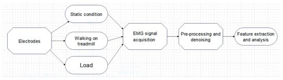
Figure 1.
General methodology of the proposed study.
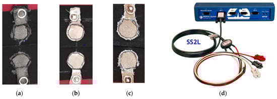
Figure 2.
EMG Textile electrodes: (a) woven embroidered textile electrode; (b) silver+ copper-plated conductive fabric; (c) stainless steel electro-conductive fabric; (d) Biopac.
Different physiology, sex, and other inter-individual differences were considered while selecting the subjects for this study. Data collection and analysis for all electrodes were performed using the BIOPAC MP360 EMG data acquisition module (BIOPAC Systems, Inc., Goleta, CA, USA). The positive and negative terminals of the BIOPAC wires were connected to the two snap elements (Figure 2d) attached to the conductive textile. The ground terminal from the BIOPAC was attached to an additional elastic fabric containing a single snap button. The placement of the differential electrode (positive-negative terminal pair) and the ground were placed apart by a distance of 20 cm for the biceps (near wrist region) and 10 cm for the tibialis (below the region of interest). The inter-electrode distance (IED) of the two electrodes constituting the differential electrode was 2 cm.
2.1. Human Subject Testing
The subjects, aged from 20 to 35 years old, used the bicep and tibialis muscles. The protocol of this study was approved by Jimma University, Jimma Technology Institutes of Institutional Doctoral School Review Board of Materials Science and Engineering. All subjects were given written informed consent and provided permission for the publication of photographs for scientific and educational purposes. The mounted configuration of the electrodes for EMG measurement from biceps brachii and tibialis anterior is as shown in Figure 3a,b. Each sEMG signal was recorded for two minutes with a sampling rate of 2000 Hz.
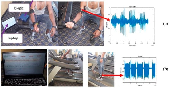
Figure 3.
sEMG measurement setup showing the specified electrode locations to evaluate muscle response at relaxation and contraction from biceps brachii at isometric conditions (a); During treadmill walking experiment on participants using the developed textile electrodes for tibialis anterior (b). The corresponding acquired EMG signals during these experiment is also shown on the side.
Performance evaluation of each electrode was based on the signal recorded under isometric conditions, such as the contraction and relaxation cycles of the biceps brachii muscles while exercising with loads of 2, 4, and 6 kg. For the tibialis anterior, treadmill-based walking and running experiments were performed. Five minutes after the application of the electrodes, the recording was begun to avoid a change in the signal quality due to the sweat effect. Figure 4 and Figure 5 shows some of the acquired signals that depicts the different stages of the activity involving different weights or loads (Figure 4) and treadmill exercises (Figure 5). Principal component analysis was carried out on the time domain features extracted from the EMG signal obtained from each electrode.
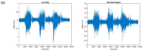
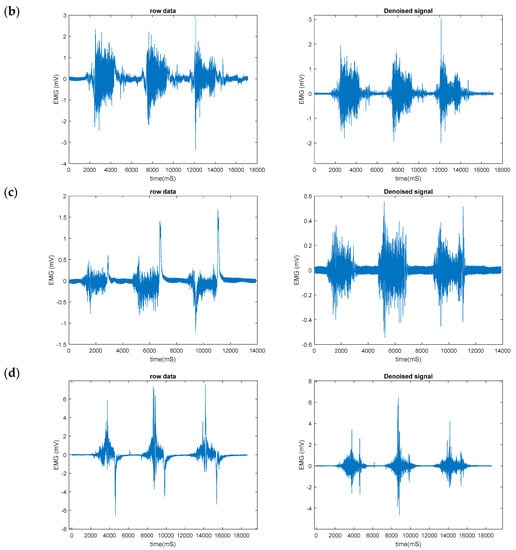
Figure 4.
sEMG raw signal (left) and denoised signal (right) collected from biceps using (a) functional (Ag/AgCl) electrode, (b) copper platted polyester electrode (CNP) (c) stainless-steel electrode (SSF) and (d) silver-plated embroidered electrode (SPEC).
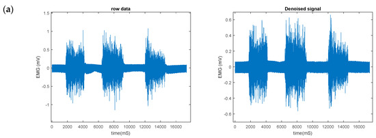
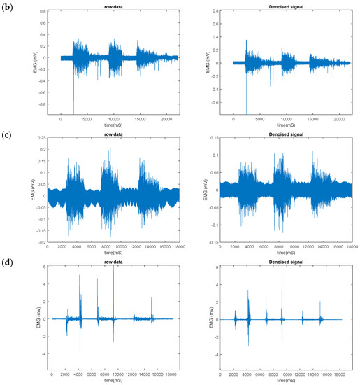
Figure 5.
sEMG raw signal (left) and denoised signal (right) collected from tibialis using (a) functional (Ag/AgCl) electrode, (b) copper-plated polyester electrode (CNP) (c) stainless-steel electrode (SSF) and (d) silver-plated embroidered electrode (SPEC).
2.2. EMG Feature Extraction
Six different types of time domain statistical features were selected for extraction from the preprocessed EMG signal and evaluated for different type of textile electrode on the two muscle groups. These are root mean square (RMS), average rectified value (ARV), variance, standard deviation, kurtosis, and skewness. All of these features were derived from the wavelet denoised signals. The RMS feature represents the square root of the mean power of the EMG signal. This feature is related to non-fatigue muscle contraction and constant force [16].
2.3. Principal Component Analysis (PCA)
A within-subject design was selected to reduce the impact of signal variability due to different physiology, sex, and other inter-individual differences. A multivariate analysis was performed a priori to determine the number of subjects necessary to observe a statistically significant change. To determine the differences between traditional and developed textile electrodes, we included the kurtosis feature in this study. With a single-variate analysis, we would have required almost 24 subjects to see significant inter-electrode differences, but by adopting multivariate analysis, we can infer the same through a smaller number of subjects, as shown in the score plot (Figure 6 and Figure 7).
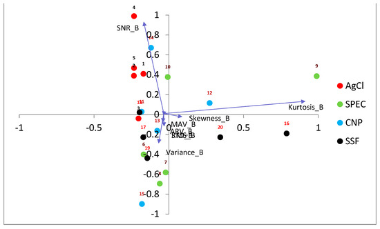
Figure 6.
Biplot for EMG features obtained from the biceps. The scores of each electrode, including the SPEC, CNP, and SSF electrodes, are shown in filled circles, and the loading vectors responsible for cluster separation are shown in blue arrows.
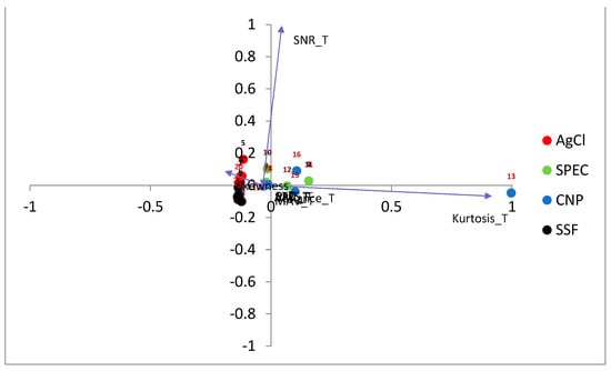
Figure 7.
Biplot for EMG features obtained from the tibialis. The scores of each electrode, including the SPEC, CNP, and SSF electrodes, are shown in filled circles, and the loading vectors responsible for cluster separation are shown in blue arrows. The feature (loading vector) pointing towards the right side, where the clusters of CNP and SPEC are spread out, i.e., ‘kurtosis’ is attributed as the contributor for separation from the clusters of SSF or AgCl.
3. Results
After assessing better signal quality through static trial experiments, several test sessions were conducted under dynamic conditions to acquire the complete experimental data. Signal quality for clinical electrodes and different types of textile electrodes were evaluated in confined movement scenarios. The electrical conductivity of the electrodes is one of the factors that affect the ability of the electrodes to acquire a biosignal.
Textile-based EMG electrodes were placed bilaterally on the tibialis anterior (TA), biceps brachii muscles with minimal skin preparations. In this research work, the evaluation of different types of dry textiles electrodes, such as embroidered, stainless steel, and polyester, were investigated for sEMG applications. Their side-by-side comparison with clinical Ag/AgCl electrodes in terms of SNR, EMG feature extraction, and signal correlation was performed for two different muscle groups, including biceps brachii and tibialis anterior (Table 1). Stainless-steel textile electrodes and Ag/AgCl showed high similarity for most of the extracted parameters compared to polyester and embroidered electrode. SNR values, on the other hand, reveal that the quality of acquired signals is similar for all types of electrodes, with the ratio of their SNRs of polyester textiles being the minimum at 84% in comparison to SNR of Ag/AgCl. In terms of the similarity of signal morphology, cross-correlation up to a maximum of 98% and an average of ~96% was achieved.

Table 1.
Comparison of SNR for muscle groups under different load.
3.1. EMG Signal Acquired Using Textile Electrodes
EMG signals were collected using the developed textile electrodes and the conventional Ag/AgCl electrode for comparison via the asynchronous method, i.e., all types of electrodes were fixed at the same place, but the EMG signal was acquired at different times. The textile electrodes were fastened on the skin using an elastic strap that has an adjustable length to keep the electrodes in the correct position. On the other hand, the commercial wet Ag/AgCl electrodes were attached to the skin with their self-adhesive pads. Three female and three male volunteer students from the Jimma University, Jimma Technology Institute, were recruited for this study. Each participant was allowed to partake in this study only after being briefed, reading, and voluntarily agreeing to the informed consent form. All the collected EMG signals were analyzed using the software EMG viewer manager. The protocol of this study was approved by Jimma University, Jimma Technology Institutes of Institutional Review Board. All subjects were given written informed consent and provided permission for the publication of photographs for scientific and educational purposes.
3.2. Pre-Processing
The feature extraction of EMG signals under three different conditions was analyzed using a MATLAB Simulink environment, visual inspection was performed during data capture, and digital signal processing was performed afterward (Mathworks, Natick, MA, USA). Before the extraction of important features, the recorded EMG signals were denoised using a high-pass filter (wavelet denoising techniques) with a high-pass cutoff frequency of 20 Hz, which removed the low-frequency trends. Following this, the high-frequency artifacts were removed using a low-pass filter (wavelet denoising techniques) with a cutoff frequency of 500 Hz. Figure 4 and Figure 5 shows the original and denoised signals obtained from both biceps brachii. and tibialis anterior. The better denoising method is selected after different types of filtering methods (Butterworth, Equripple, infinite impulse response, finite impulse response filter, and wavelet multiresolution analysis filter) were evaluated. Wavelet multiresolution analysis denoising performed better with improved SNR.
Biplot for EMG features obtained from Tibialis by AgCl and embroidery. Figure 6 shows that the features from, AgCl and stainless-steel electrodes (SFS) are clustered adjacent to each other. However, the clusters of embroidery (SPEC) and polyester (CNP) were dispersed and showed significant intra-subject variations for the tibialis muscle.
Inferring biplot for EMG features obtained from biceps we can see that magnitude and direction of projection for loading variables. Figure 7 shows that the features from all the electrodes, AgCl, stainless steel, embroidery and polyester, showed significant intra-subject variations. The variations have resulted from the ‘slippage effect’ observed during the experimental measurements.
4. Discussion
The EMG measured from six subjects shows that the three types of textile electrodes recorded good signals with reasonable stability when placed on different muscles. While previous studies have evaluated single textile electrodes with respect to the clinical electrode Ag/AgCl, and performed experiments on a specific muscle type and position [17], here we have used two muscle types and multiple (three) textile electrodes. Comparing different types of textile electrodes among each other allows us to analyze the results statistically, removing observational bias and adding statistical significance to this study.
Regarding the material type used for electrode design, it is stated that organic polymers such as polyproline are low-weight, flexible and inexpensive. But they have low and unstable electrical conductivity when compared to inorganic metals such as stainless steel, silver, and cupper-nickel composite used in this study. This implies the need for the development of hybrid materials combining inorganic and organic components to achieve both stretchability and conductivity. In 2009 a study was conducted on eight different textile electrodes and five textile conductive materials [4], showing that the impedance of the electrode depends on the proportion of the conductive material present within the textile. Different yarn materials and manufacturing processes also influence the contact impedance property [3]. Albeit different manufacturing techniques, namely embroidery, knitting, and weaving exists, in this study, we used knitting for manufacturing both the textile electrodes (except CNP, which was woven) and the substrate fabric material, on which the textile electrode was cut and sewn (stitching).
Previously, Ozberk Ozturk et al. studied the performance evaluation of sEMG with wearable graphene textiles electrodes against clinical Ag/AgCl electrodes [18]. In this study, we used the same parameters, such as electrode configuration, size, shape and inter-electrode distance for all the EMG signal acquisition using different textile electrodes. It has been reported in earlier studies that the EMG signal is highly affected by the size of the electrode. According to SENIAM, electrode size is defined as the size of the conductive area of a SEMG electrode [19]. In 2016, a study on the comparison of different sizes of circular electrodes concluded that larger electrodes performed better [20]. The results also showed that an increase in the area will decrease resistance. Similarly, another study compared three rectangular electrodes of different sizes [21] and concluded that larger dry electrodes achieved similar performance to that of the conventional electrodes. In 2019, a comparative study on textile electrodes with 24 mm and 10 mm diameters [22] showed that the larger electrode exhibited low skin-to-electrode impedance. Unlike the other studies, this work revealed that large electrodes are prone to high crosstalk effects among measured signals. A more recent study carried out in 2020 on six different sizes of textile electrodes ranging from 5 mm up to 30 mm in diameter [9] showed that electrodes >20 mm exhibited lower baseline noise levels and greater SNR. However, the electrode with a 20 mm diameter exhibited a better SNR value than the 30 mm one. The result clearly indicates that larger electrodes (>20 mm) show lower impedance but on much larger electrodes (>25 mm), we observe more cross talks. In all these studies and similar related works, it was shown that the shape of the electrodes has less effect on the SEMG signal. While using larger electrodes, the inter electrode distance (IED) will also increase in order to avoid overlapping of electrodes. According to SENIAM, inter-electrode distance is defined as the center-to-center distance between the conductive areas of two bipolar electrodes. Studies show that increasing IED will increase crosstalk. It has been suggested that unstable signal recordings due to tendon and motor endplate effects can be avoided by adjusting the inter-electrode distance. Therefore, the researchers in that work prepared a circular textile electrode with a 20 mm diameter and used 30 mm of IED in a bipolar electrode configuration. The bipolar electrode configuration, which uses two electrodes, is the most commonly used electrode configuration for sEMG, unlike monopolar and multipolar configurations [23].
Subsequent to this work, we would like to integrate the textile electrodes into a garment to assess the effect of motion artifacts during movement. As reported by Alper Cömert and Jari Hyttine, a challenge for any textile electrodes is maintaining stable skin contact during movement [24]. In our case, all our developed electrodes strapped to a conformal fit enabled the recording of clean signals, even when hands were moved during load lift for the biceps muscle or when running/walking on a treadmill for the tibialis muscle. The stainless-steel electrode performed better than embroidered or polyester electrodes; further, its performance was comparable to the reference Ag/AgCl electrode used in clinical settings. The ridges present in the SFS electrode would have assisted in maintaining better skin contact by pressing it onto the skin and preventing its slippage from its initial position. However, the silver-coated thread used for embroidery electrodes is softer when touched than the stainless steel or polyester electrode. The soft surface property of a textile electrode is much desired for its enhanced and long-term wearability, suitable for continuous signal measurement. Additionally, the spiking of artificial sweat on the textile electrodes before signal measurement will also enhance the signal quality [25].
5. Conclusions
In this article, we compared the EMG signals acquired from the biceps brachii and tibialis anterior muscle using different types of textiles electrodes. For all the test electrodes, the conventional Ag/AgCl electrode was used as the reference standard. Morphological analysis of the time domain signal revealed that, despite smaller amplitude, the fabricated embroidered woven textile electrodes are capable of measuring the EMG signals similar to that of stainless steel and copper-nickel-plated polyester electro-conductive fabric for the same electrode size and shape. We can also conclude that the stainless-steel embroidered textile electrodes have a good measuring performance under isometric conditions, allowing the measurement of EMG signals with morphological features similar to that of the standard electrodes. This suggests that the embroidery textile electrodes may be used as a reliable alternative to stainless steel, copper-nickel-plated electro-conductive fabric, and conventional silver chloride. The overall noise content did not differ substantially among the electrodes and did not compromise the sensitivity of the textile electrode’s signals. Further studies are in progress to evaluate the frequency content of the signals, which may vary due to the different impedance characteristics of the electrodes. Furthermore, the amplitude and the phase features at different frequencies provide more insight into the dynamic activities of the muscle.
A comparison of EMG signals recorded from healthy and diseased/ patient individuals with the best-performed textile electrode device is in progress. Proof-of-concept prototypes will be developed with applications for tracking health monitoring on patients. Globally more than one million people are suffering from muscle related disabilities. With a large population of disabled people worldwide, many assistive technologies and techniques are in great demand to improve the quality of life for such people. The World Health Organization (WHO) has proposed an action plan that emphasizes the development of assistive technology [26]. Our study, which used the three textile electrodes on two different muscle types, is in line with meeting the demand of the growing need for improvements in wearable health monitoring technology.
Author Contributions
B.B.E. designed and conducted the experiment, analyzed results, and wrote the paper; B.M. drafted the outline and analyzed experimental results; B.M. edited the paper; J.K. helped with experimental work and results analysis; and L.V.L. supervised and administered the project. All authors have read and agreed to the published version of the manuscript.
Funding
The research and APC were funded by the NASCERE project.
Institutional Review Board Statement
The study was conducted in accordance with the Declaration of Helsinki, and approved by the Institutional Review Board (or Ethics Committee) of Jimma Institute of technology (Ref No: MSC/JIT/16/2014 dated on 24 February 2022).
Informed Consent Statement
Informed consent was obtained from all subjects involved in the study.
Data Availability Statement
The data presented in this study are available in this article.
Acknowledgments
The authors would like to express appreciation for the support from the NASCERE project and Ghent University. The NASCERE project was sponsored with funds from the Ethiopian government. The study’s design, data collection, analysis, or interpretation; the preparation of the paper; and the choice to publish the findings were all made independently of the funders.
Conflicts of Interest
The authors declare no conflict of interest.
References
- Albulbul, A. Evaluating Major Electrode Types for Idle Biological Signal Measurements for Modern Medical Technology. Bioengineering 2016, 3, 20. [Google Scholar] [CrossRef] [PubMed]
- Ankhili, A.; Tao, X.; Cochrane, C.; Koncar, V.; Coulon, D.; Tarlet, J.-M. Comparative Study on Conductive Knitted Fabric Electrodes for Long-Term Electrocardiography Monitoring: Silver-Plated and PEDOT:PSS Coated Fabrics. Sensors 2018, 18, 3890. [Google Scholar] [CrossRef] [PubMed]
- Moloney, P.B.; Lefter, S.; Ryan, A.M.; Jansen, M.; Bermingham, N.; McNamara, B. The Diagnostic Yield of Electromyography at Detecting Abnormalities on Muscle Biopsy: A Single Center Experience. Neurodiagn. J. 2021, 61, 86–94. [Google Scholar] [CrossRef] [PubMed]
- Perego, P.; Taherinejad, N.; Caon, M. (Eds.) Wearables in Healthcare: Second EAI International Conference, HealthWear 2020, Virtual Event, December 10–11, 2020: Proceedings; Lecture notes of the Institute for Computer Sciences, Social Informatics and Telecommunications Engineering; Springer: Cham, Switzerland, 2021; ISBN 978-3-030-76066-3. [Google Scholar]
- Analia, R.; Sulistyo, V.A.; Iglesias, T.M.; Susanto; Pamungkas, D.S.; Jamzuri, E.R. The Reusable Electrode of EMG Sensor for Capturing The Calf Muscle Activities. In Proceedings of the 2020 3rd International Conference on Applied Engineering (ICAE), Batam, Indonesia, 7 October 2020; IEEE: Manhattan, NY, USA, 2020; pp. 1–4. [Google Scholar]
- Cerone, G.L.; Botter, A.; Vieira, T.; Gazzoni, M. Design and Characterization of a Textile Electrode System for the Detection of High-Density SEMG. IEEE Trans. Neural Syst. Rehabil. Eng. 2021, 29, 1110–1119. [Google Scholar] [CrossRef] [PubMed]
- da Fonseca, P.F.P.; Borgonovo-Santos, M.; Catarino, A.; Correia, M.V.; Vilas-Boas, J.P. Characterization of Textile Electrodes for EMG Measurements: Impedance and Signal Morphology. Corpoconsciência 2021, 25, 221–235. [Google Scholar] [CrossRef]
- Kamavuako, E.N.; Brown, M.; Bao, X.; Chihi, I.; Pitou, S.; Howard, M. Affordable Embroidered EMG Electrodes for Myoelectric Control of Prostheses: A Pilot Study. Sensors 2021, 21, 5245. [Google Scholar] [CrossRef] [PubMed]
- Kim, S.; Lee, S.; Jeong, W. EMG Measurement with Textile-Based Electrodes in Different Electrode Sizes and Clothing Pressures for Smart Clothing Design Optimization. Polymers 2020, 12, 2406. [Google Scholar] [CrossRef] [PubMed]
- Hermann, A.; Senner, V. EMG-Pants in Sports: Concept Validation of Textile-Integrated EMG Measurements. In Proceedings of the 8th International Conference on Sport Sciences Research and Technology Support, Budapest, Hungary, 5–6 November 2020; SCITEPRESS-Science and Technology Publications: Budapest, Hungary, 2020; pp. 197–204. [Google Scholar]
- Guvenc Tuna, B.; Goncu-Berk, G. Understanding the Effect of Clothing Pattern on E-Textile Electromyography (EMG) Electrode Performance. In Pivoting for the Pandemic; Iowa State University Digital Press: Ames, IA, USA, 2020. [Google Scholar] [CrossRef]
- Ehrmann, G.; Ehrmann, A. Electronic Textiles. Encyclopedia 2021, 1, 115–130. [Google Scholar] [CrossRef]
- Paiva, A.; Carvalho, H.; Catarino, A.; Postolache, O.; Postolache, G. Development of Dry Textile Electrodes for Electromiography a Comparison between Knitted Structures and Conductive Yarns. In Proceedings of the 2015 9th International Conference on Sensing Technology (ICST), Auckland, New Zealand, 8–10 December 2015; IEEE: Manhattan, NY, USA, 2015; pp. 447–451. [Google Scholar]
- Vojtech, L.; Neruda, M.; Reichl, T.; Dusek, K.; de la Torre Megías, C. Surface Area Evaluation of Electrically Conductive Polymer-Based Textiles. Materials 2018, 11, 1931. [Google Scholar] [CrossRef] [PubMed]
- Tseghai, G.B.; Malengier, B.; Nigusse, A.B.; Langenhove, L.V. Development and Evaluation of Resistive Pressure Sensors from Electro-Conductive Textile Fabric. In Second International Forum on Textiles for Graduate Students; Tianjin Polytechnic University: Tianjin, China, 2018. [Google Scholar]
- Spiewak, C. A Comprehensive Study on EMG Feature Extraction and Classifiers. Open Access J. Biomed. Eng. Biosci. 2018, 1, 17–26. [Google Scholar] [CrossRef]
- Sumner, B.; Mancuso, C.; Paradiso, R. Performances Evaluation of Textile Electrodes for EMG Remote Measurements. In Proceedings of the 2013 35th Annual International Conference of the IEEE Engineering in Medicine and Biology Society (EMBC), Osaka, Japan, 3–7 July 2013; IEEE: Manhattan, NY, USA, 2013; pp. 6510–6513. [Google Scholar]
- Ozturk, O.; Yapici, M.K. Surface Electromyography With Wearable Graphene Textiles. IEEE Sensors J. 2021, 21, 14397–14406. [Google Scholar] [CrossRef]
- Hermens, H.J. Development of Recommendations for SEMG Sensors and Sensor Placement Procedures. J. Electromyogr. Kinesiol. 2000, 10, 361–374. [Google Scholar] [CrossRef] [PubMed]
- Shafti, A.; Manero, R.B.R.; Borg, A.M.; Althoefer, K.; Howard, M.J. Embroidered Electromyography: A Systematic Design Guide. IEEE Trans. Neural Syst. Rehabil. Eng. 2016, 25, 4320. [Google Scholar] [CrossRef] [PubMed]
- An, X.; Tangsirinaruenart, O.; Stylios, G.K. Investigating the Performance of Dry Textile Electrodes for Wearable End-Uses. J. Text. Inst. 2019, 110, 151–158. [Google Scholar] [CrossRef]
- Pani, D.; Achilli, A.; Spanu, A.; Bonfiglio, A.; Gazzoni, M.; Botter, A. Validation of Polymer-Based Screen-Printed Textile Electrodes for Surface EMG Detection. IEEE Trans. Neural Syst. Rehabil. Eng. 2019, 27, 1370–1377. [Google Scholar] [CrossRef] [PubMed]
- Pfingst, B.E.; Franck, K.H.; Xu, L.; Bauer, E.M.; Zwolan, T.A. Effects of Electrode Configuration and Place of Stimulation on Speech Perception with Cochlear Prostheses. JARO 2001, 2, 87–103. [Google Scholar] [CrossRef] [PubMed]
- Cömert, A.; Hyttinen, J. A Motion Artifact Generation and Assessment System for the Rapid Testing of Surface Biopotential Electrodes. Physiol. Meas. 2015, 36, 1. [Google Scholar] [CrossRef] [PubMed]
- Leonhardt, S.; Zimmermann, N.; Kranen, P.; Kensche, D.; Muller, E.; Quix, C. Influence of Contact Pressure and Moisture on the Signal Quality of a Newly Developed Textile ECG Sensor Shirt. In Proceedings of the 2008 5th International Summer School and Symposium on Medical Devices and Biosensors, Hong Kong, China, 1–3 June 2008; IEEE: Manhattan, NY, USA, 2008; pp. 256–259. [Google Scholar]
- Gupta, A.; Sayed, T.; Garg, R.; Shreyam, R. Emg Signal Analysis of Healthy and Neuropathic Individuals. IOP Conf. Ser. Mater. Sci. Eng. 2017, 225, 012128. [Google Scholar] [CrossRef]


Disclaimer/Publisher’s Note: The statements, opinions and data contained in all publications are solely those of the individual author(s) and contributor(s) and not of MDPI and/or the editor(s). MDPI and/or the editor(s) disclaim responsibility for any injury to people or property resulting from any ideas, methods, instructions or products referred to in the content. |
© 2023 by the authors. Licensee MDPI, Basel, Switzerland. This article is an open access article distributed under the terms and conditions of the Creative Commons Attribution (CC BY) license (https://creativecommons.org/licenses/by/4.0/).