Abstract
The aim of this study was to investigate the antioxidant properties in vitro of three different vanillic dimmers (Compounds 1a–c). They were synthesized through an oxidative coupling strategy in good yields. The targeted compounds were found to be highly active for the total antioxidant assay, as well as for the lipid peroxidation test. All investigated compounds exhibited superior or comparable antioxidant capacity in comparison to precursor vanillin, proving that oxidative coupling is a great strategy to increase the antioxidant behavior of vanillin derivatives.
1. Introduction
Oxidative stress is defined as an imbalance between the production of reactive oxygen species and an organism’s ability to face their action by antioxidant protection [1,2]. This imbalance has been linked to numerous chronic affections, especially cardiovascular and oncological diseases [3,4]. Adding low concentrations of antioxidants can effectively quench the reactive oxygen species before they attack biomolecules, causing damage [5]. Vanillin is one of the most popular natural flavoring agents and is widely used in the food and cosmetic industries, and its antioxidant properties have been extensively studied [6]. Nowadays, synthetic antioxidants are increasingly in demand [7]. Thus, it is important to create synthetic compounds derived from natural molecules that could increase the antioxidant activity of their natural analogues. Hence, in this work, we use oxidative coupling as a strategy to improve, through functionalization, the antioxidant behavior of vanillin. For this series of compounds, we evaluated their antioxidant activity in vitrothrough three different assays.
2. Materials and Methods
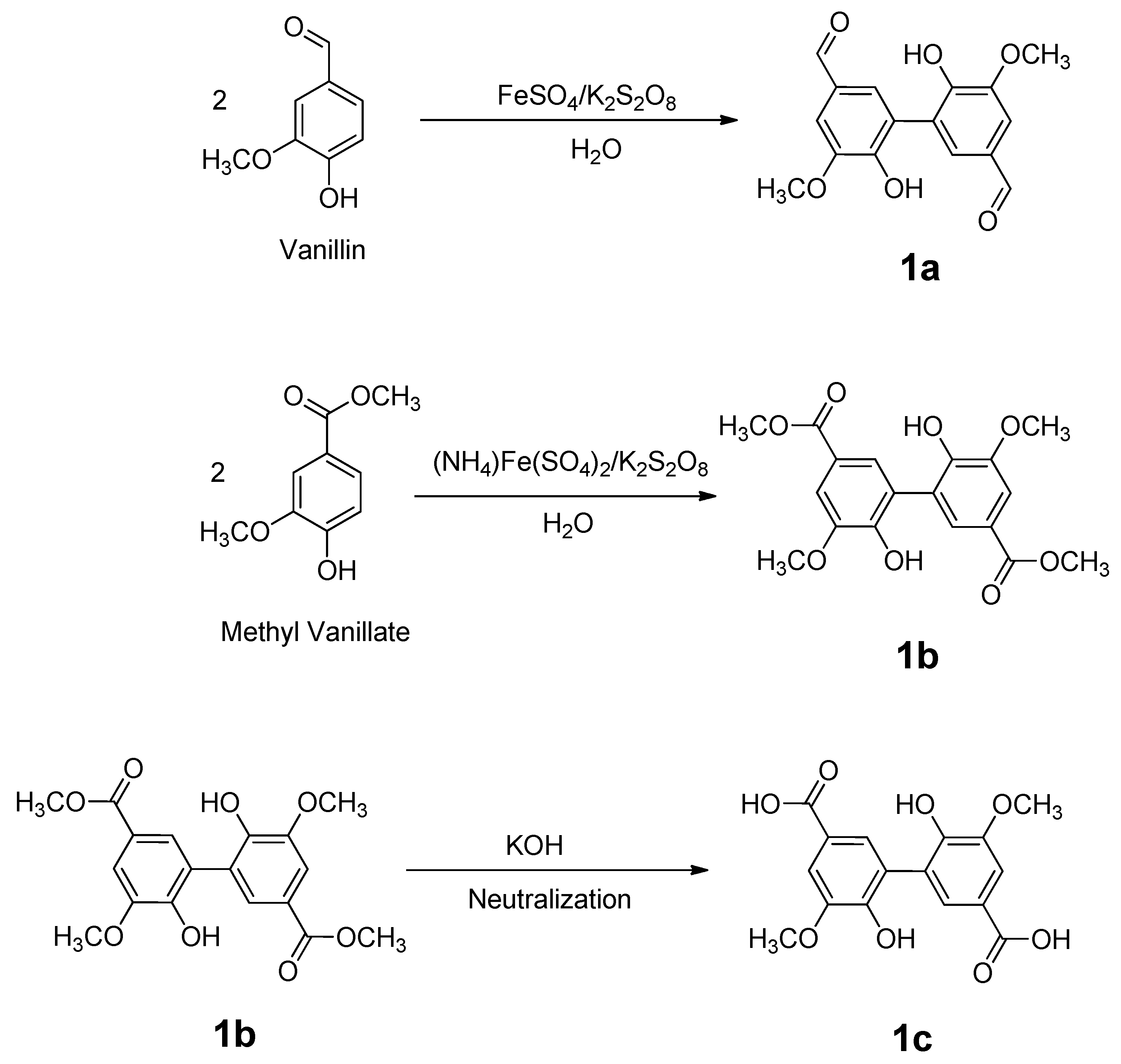
2.1. Synthesis of Vanillic Dimers
Oxidative coupled vanillic dimers 1a–c were prepared as depicted in Scheme 1 according to methods found in the literature [8,9,10].
Scheme 1.
Preparation of compounds 1a–c.
To obtain compound 1a, 0.89 mmol of FeSO4 and 12.51 mmol K2S2O8 were added under stirring to an aqueous solution of vanillin (23.33 mmol). The mixture was heated at 50 °C for 120 h until the reaction was complete as indicated by TLC assay. After cooling at room temperature, the crude product was filtered and washed with cold ethanol and dried in a vacuum oven at 40 °C for 24 h.
For the preparationof 1b, 6 mmol of methyl vanillate was dissolved in 200 mL of distilled water, and the mixture was heated until total dissolution. Once the ester was dissolved, 0.3 mmol of (NH4)2Fe(SO4)2 and 3.0 mmol of K2S2O8 were added, the heating was kept for 3 min, and then the heating was turned off. It was kept stirring for another 30 min. The product was filtered and washed with 300 mL hot water and 300 mL cold water, and dried in a vacuum oven at 40 °C for 24 h.
For the synthesis of compound 1c, 1.37 mmol of 1b was suspended in a THF:water mixture (15 mL, 1:1 v:v), and 13.7 mmol of KOH pellets were added. The resulting two phases were heated to 70 °C and stirred vigorously for16 h.
After cooling to room temperature, the organic phase was removed and the aqueous phase was acidified with a HCl solution (6 M) until pH = 1 was reached. The solid product was collected, washed with water until neutral, and dried in a vacuum oven at 40 °C for 24 h.
2.2. Evaluation of Antioxidant Properties
Compounds 1a–c synthesized were studied for their antioxidant properties using three assays with differentoxidative potential evaluation mechanisms.
Stock solutions (10 mg/mL) of targeted compounds 1a–c were prepared in DMSO. For all the experiments, different dilutions of these solutions in ethanol were performed.
2.2.1. Reducing Power Method (RP)
In this method, described by Oyaizu [11], a colored complex is formed with K3Fe(CN)6, CCl3COOH, and FeCl3. An increasein the absorbance of the reaction mixture indicates the reducing power of the samples. Briefly, 1 mL of the targeted compound (20–50 μg/mL) was placed in a tube and was mixed with 3 mL of buffer phosphate (pH 6.6) and 3 mL of an aqueous solution of K3Fe(CN)6 (1% w/v). The resulting mixture was incubated at 50 °C for 20 min, followed by the addition of 2.5 mL ofCCl3COOH (10% w/v). Finally, 2.5 mL of this mixture was collected and mixed with 2.5 mL of distilled water and 0.5 mL of FeCl3 (0.1% w/v). The absorbance was measured at 700 nm against a blank sample overtime.
2.2.2. Nitric Oxide Scavenging Activity
Sodium nitroprusside is known to decompose in aqueous solutions at pH 7.2 producing NO radicals. The method described by Marcocci et al. [12] was used, and it is based onthe decomposition of sodium nitroprusside at a physiological pH, producing nitric oxide radicals. Under aerobic conditions, the NO radical reacts with oxygen to produce stable products, the quantities of which can be determined using Griess reagent. For this, 2 mL of 10 mM sodium nitroprusside dissolved in 0.5 mL phosphate buffer (pH 7.4) is mixed with 0.5 mL of sample at various concentrations (0.4–1.0 mg/mL). The mixture is then incubated at 25 °C. After 150 min of incubation, 0.5 mL of incubated solution is withdrawn and mixed with 0.5 mL of Griess reagent. (0.5 g of sulphanilamide, 1.25 mL orto-phosphoric acid, and 0.05 g N-1-(Naphthyl)ethyl-enediamine). The mixture is then incubated at room temperature for 30 min, and its absorbance is measured at 546 nm. The amount of nitric oxide radical inhibition is calculated following Equation (1).
where Ac is the absorbance of the control and As is the absorbance of the sample.
Inhibition of NO radical (%) = [Ac − As)/Ac × 100
2.2.3. Thiobarbituric Acid Reactive Substances Method (TBARS)
The TBARS assay is used to evaluate lipid peroxidation. In this test, malondialdehyde (MDA) is measured, which results from the oxidation of lipid substrates. At low pH and high temperature (100 °C), MDA binds with thiobarbituric acid to form a red complex that can be measured at 532 nm. The increased amount of the red pigment formed correlates with the oxidative rancidity of the lipid.
The method of Ottolenghi [13] with modifications was used. A volume of 4 mL of the targeted compound (1 mg/mL) was placed in a tube along with 4.1 mg of 2.52% oleic acid in absolute ethanol, 8 mL of 0.05 M phosphate buffer (pH 7.0), and 3.9 mL of water. The mixture was placed in an oven at 40 °C in the dark. To 1 mL of this solution, 2 mL of trichloroacetic acid (20%) and 2 mL of thiobarbituric acid solution (0.67%w/v) were added and then incubated. The mixture was placed in a boiling water bath for 10 min, and the absorbance was measured at 532 nm. The antioxidant activity was determined using Equation (2).
where Ac is the absorbance of the control and As is the maximum absorbance of the sample on the same day.
Inhibition (%) = [(Ac − As)/Ac] × 100%
3. Results
3.1. Preparation of Vanillic Dimers
The preparation of targeted vanillic dimmers 1a–c using oxidative coupling as a strategy was easily achieved. The three synthesized compounds were solid, and their yields, m.p., and NMR data are in accordance with reference methods [8,9,10].
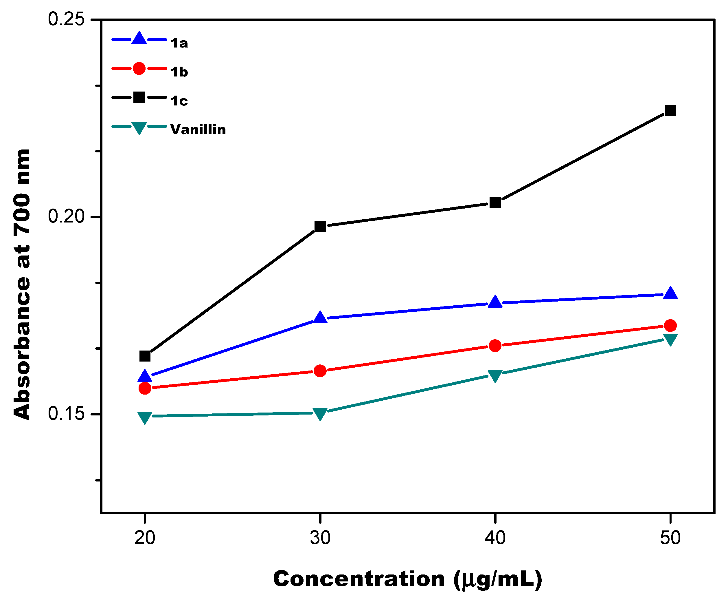
3.2. Antioxidant Properties
The antioxidant properties of the obtained compounds 1a–c were tested. The results of the reducing power assay are shown in Figure 1 and Figure 2. These results show that vanillic dimers 1a–c have reduction potentials higher than their natural analogue vanillin.
Figure 1.
Reducing power of compounds 1a–c (20 μg/mL) versus time.
Figure 2.
Reducing power of compounds 1a–c at different concentrations (20–50 μg/mL) (t = 120 min).
Additionally, it can be observed that the antioxidant activity measured by this method was correlated to the concentration of vanillic dimers. The highest absorbance values were observed when compound 1c was used.
Subsequently, the nitric oxide scavenging activity was evaluated. As it can be observed in Figure 3, all compounds including vanillin presented similar inhibitory percentages indicating the effective scavenging nitric oxide activity of these compounds.
Figure 3.
Nitric oxide scavenging activity of compounds 1a–c and vanillin at different concentrations (0.4–1.0 mg/mL).
Additionally, the antioxidant properties of compounds 1a–c were evaluated using the thiobarbituric acid reactive substance method (Figure 4). The oxidation of oleic acid was investigated, showing that in presence of compounds 1a and 1c, the inhibition percentages of the oxidation products are higher than their natural analogue vanillin. This proved that these compounds present inhibitory activity towards lipid peroxidation.
Figure 4.
Antioxidant properties of compounds 1a–c evaluated using the thiobarbituric acid reactive substance method.
4. Conclusions
In this study, the synthesis and antioxidant activity of three different vanillic dimers aredescribed. All investigated compounds exhibited superior antioxidant capacity in comparison to precursor vanillin, proving that oxidative coupling is a great strategy to increase the antioxidant activity of vanillin derivatives. Among the three dimers studied, compound 1c possesses the best antioxidant properties among the studied dimers.
Author Contributions
Conceptualization, C.A.F. and L.G.G.; methodology, L.G.G.; formal analysis, A.P.R., L.G.G. and C.M.O.; investigation, C.A.F.; resources, C.A.F.; data curation, A.P.R. and V.A.G.; writing—original draft preparation, L.G.G.; writing—review and editing, C.A.F.; supervision, V.A.G. All authors have read and agreed to the published version of the manuscript.
Funding
This research was funded by Universidad Nacional del Litoral Capital Semilla 2019 Grant.
Institutional Review Board Statement
Not applicable.
Informed Consent Statement
Not applicable.
Data Availability Statement
Not applicable.
Conflicts of Interest
The authors declare no conflict of interest. The funders had no role in the design of the study; in the collection, analyses, or interpretation of data; in the writing of the manuscript; or in the decision to publish the results.
References
- Betteridge, D.J. What is Oxidative Stress? Metabolism 2000, 49, 3–8. [Google Scholar] [CrossRef] [PubMed]
- Burton, G.J.; Jauniaux, E. Oxidative stress. Best. Pract. Res.Clin. Obstet. Gynaecol. 2011, 25, 287–299. [Google Scholar] [CrossRef] [PubMed]
- Pizzino, G.; Irrera, N.; Cucinotta, M.; Pallio, G.; Mannino, F.; Arcoraci, V.; Squadrito, F.; Altavilla, D.; Bitto, A. Oxidative Stress: Harms and Benefits for Human Health. Oxid. Med. Cell. Longev. 2017, 2017, 8416763. [Google Scholar] [CrossRef] [PubMed]
- Cao, G.; Cutler, R.G. High concentration of antioxidants may not improve defense against oxidative stress. Arch. Gerontol. Geriatr. 1993, 17, 189–201. [Google Scholar] [CrossRef] [PubMed]
- Dubois-Deruy, E.; Peugnet, V.; Turkieh, A.; Pinet, F. Oxidative Stress in Cardiovascular Diseases. Antioxidants 2020, 9, 864. [Google Scholar] [CrossRef] [PubMed]
- Tai, A.; Sawano, T.; Yazama, F.; Ito, H. Evaluation of antioxidant activity of vanillin by using multiple antioxidant assays. Biochim. Biophys. Acta 2011, 1810, 170–177. [Google Scholar] [CrossRef] [PubMed]
- Wojcik, M.; Burzynska-Pedziwiatr, I.; Wozniak, L.A. A Review of Natural and Synthetic Antioxidants Important for Health and Longevity. Curr. Med. Chem. 2010, 17, 3262–3288. [Google Scholar] [CrossRef] [PubMed]
- Delomenede, M.; Bedos-Belval, F.; Duran, H.; Vindis, C.; Baltas, M.; Nègre-Salvayre, A. Development of Novel Antiatherogenic Biaryls: Design, Synthesis and Reactivity. J. Med. Chem. 2008, 51, 3171–3181. [Google Scholar] [CrossRef] [PubMed]
- De Vasconcelos, D.B.; Lima, A.N.; Philot, E.A.; Scott, A.L.; Ferreira Boza, I.A.; de Souza, A.R.; Morgon, N.H.; Ximenes, V.F. Methyl divanillate: Redox properties and binding affinity with albumin of an antioxidant and potential NADPH oxidase inhibitor. RSC Adv. 2019, 9, 19983–19992. [Google Scholar] [CrossRef] [PubMed]
- Scholtes, J.F.; Trapp, O. Inducing Enantioselectivity in a Dynamic Catalyst by Supramolecular Interlocking. Angew. Chem. Int. Ed. 2019, 58, 6306–6310. [Google Scholar] [CrossRef] [PubMed]
- Oyaizu, M. Studies on products of browning reactions: Antioxidant activities of products of browning reaction prepared from glucosamine. J. Nutrit. 1986, 44, 307–315. [Google Scholar]
- Marcocci, I.; Marguire, J.J.; Droy-lefaiz, M.T.; Packer, L. The nitric oxide scavenging properties of Gingko bioba extract. Biochem. Biophys. Res. Commun. 1994, 201, 748–755. [Google Scholar] [CrossRef] [PubMed]
- Ottolenghi, A. Interaction of ascorbic acid and mitochrondria lipids. Arch. Biochem. Biophys. 1959, 79, 355–363. [Google Scholar] [CrossRef]
Disclaimer/Publisher’s Note: The statements, opinions and data contained in all publications are solely those of the individual author(s) and contributor(s) and not of MDPI and/or the editor(s). MDPI and/or the editor(s) disclaim responsibility for any injury to people or property resulting from any ideas, methods, instructions or products referred to in the content. |
© 2022 by the authors. Licensee MDPI, Basel, Switzerland. This article is an open access article distributed under the terms and conditions of the Creative Commons Attribution (CC BY) license (https://creativecommons.org/licenses/by/4.0/).