Abstract
We present the performance and application of a commercial off-the-shelf Si PIN diode (Hamamatsu S14605) as a charged particle detector in a compact ion beam system (IBS) capable of generating D–D and p–B fusion charged particles. This detector is inexpensive, widely available, and operates in photoconductive mode under a reverse bias voltage of 12 V, supplied by an A23 battery. A charge-sensitive preamplifier (CSP) is mounted on the backside of the detector’s four-layer PCB and powered by two ±3 V lithium batteries (A123). Both the detector and CSP are housed together on the vacuum side of the IBS, facing the fusion target. The system employs a CF-2.75-flanged DB-9 connector feedthrough to supply the signal, bias voltage, and rail voltages. To mitigate the high sensitivity of the detector to optical light, a thin aluminum foil assembly is used to block optical emissions from the ion beam and target. Charged particles generate step responses at the preamplifier output, with pulse rise times in the order of 0.2 to 0.3 µs. These signals are recorded using a custom-built data acquisition unit, which features an optical fiber data link to ensure the electrical isolation of the detector electronics. Subsequent digital signal processing is employed to optimally shape the pulses using a CR-RCn filter to produce Gaussian-shaped signals, enabling the accurate extraction of energy information. Performance results indicate that the detector’s baseline RMS ripple noise can be as low as 0.24 mV. Under actual laboratory conditions, the estimated signal-to-noise ratios (S/N) for charged particles from D–D fusion—protons, tritons, and helions—are approximately 225, 75, and 41, respectively.
1. Introduction
The Si PIN diode and other electronics-based charged particle detectors have gained prominence in recent years for fusion particle detection [1,2,3]. Dr. Ramirez-Jimenez et al. has designed and tested similar PIN photodiodes for detecting charged particles in the range of 4–13 MeV [4]. Unlike solid-state nuclear track detectors (SSNTDs) such as CR-39, PIN detectors provide a real-time, high-fidelity method for particle identification and energy measurement. With an appropriate preamplifier and pulse-shaping amplifier, a high energy resolution can be achieved, with full-width-half-maximum (FWHM) energy resolutions of 10–20 keV for Am-241 alpha-particles reported in the literature [5]. In fusion environments, proper shielding against optical light and the protection of electronics from electromagnetic interference (EMI) are essential for reliable operation. To address these challenges, Alpha Ring International developed a compact fusion demonstration system that uses an H+ or D+ ion beam to produce D–D and p–11B fusion through the beam-target method [1]. The system allows for the precise control of beam parameters (e.g., energy, current, pulse length), creating a highly controlled environment for testing and calibrating detectors with real fusion particles. This study focuses on the Hamamatsu S14605 PIN diode [6], selected for its low cost and ready availability. As a standalone module, the diode requires integration with a custom-designed preamplifier PCB for optimal performance (Figure 1). To reduce costs and maintain a compact form factor, we developed a custom data acquisition system integrated with the detector and preamplifier. The result is a self-contained unit that can be controlled via a USB-UART port, providing a versatile and cost-effective solution for fusion particle detection.
Figure 1.
(a) Detector system hardware stack showing how the Si PIN detector is mounted in vacuum to observe fusion charged particles from a beam-target experiment. Both the target and foil assembly are attached to a standard CF-2.75 vacuum cube and sealed with viton gaskets. (b) Back side of the PIN detector containing all of the preamplifier electronics components. (c) Front side with only the PIN detector exposed to charged particles.
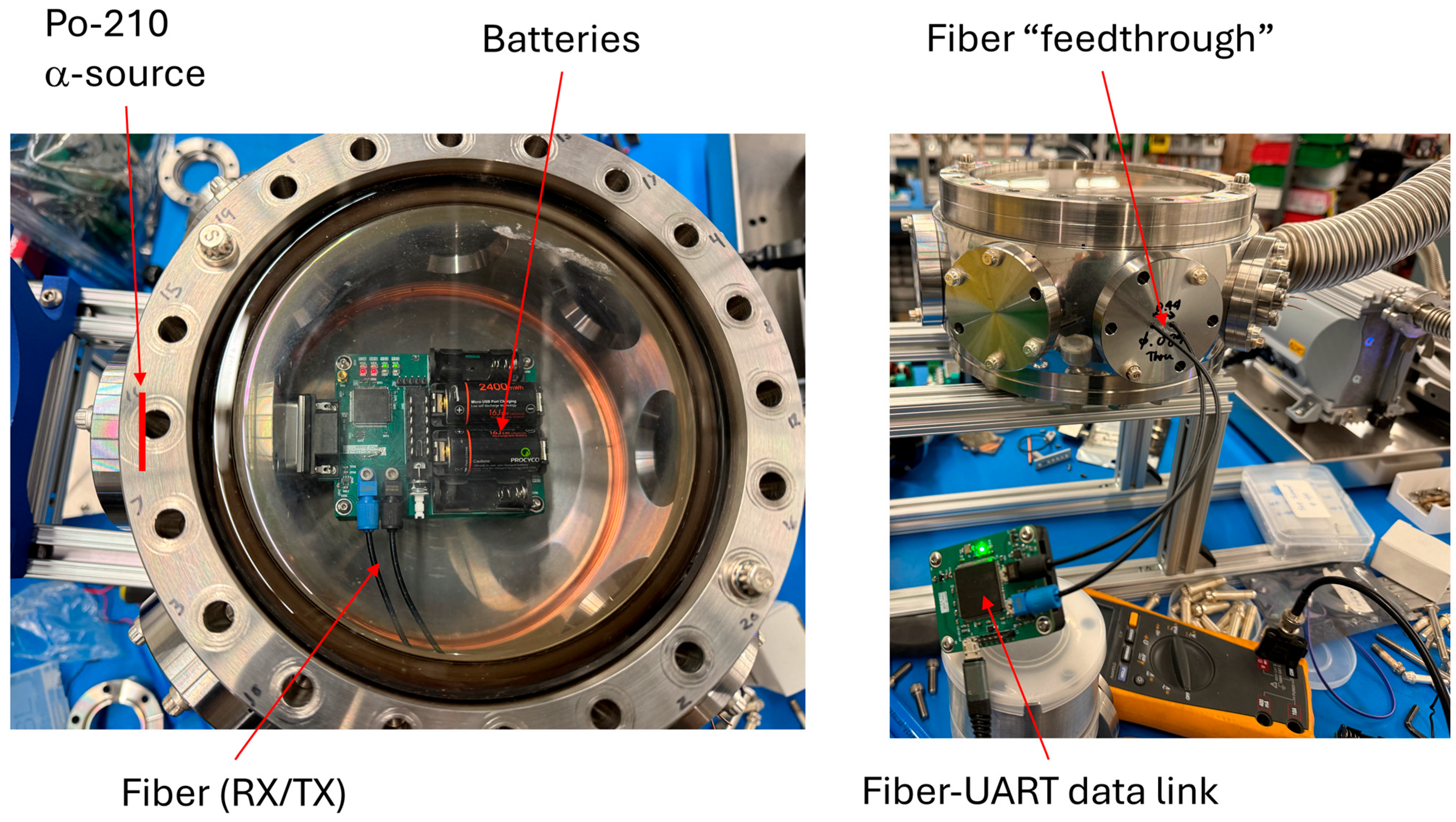
2. Detector System Design
The S14605 Si PIN detector has an active area of 9 mm × 9 mm with a nominal depletion depth of 500 μm. A reverse bias is crucial to obtain a good detector rise time and low baseline noise. Additionally, the pulse rise time is comparatively faster with a detector bias due to the presence of a sweeping electric field to carry away the charges quickly. The detector can operate with a bias voltage as high as +150 V; however, we have achieved adequate performance operating with a bias voltage of +12 V, which can be easily supplied with an A23 battery [7].
The PIN detector readout circuit (Figure 2) utilizes a dual CMOS-input op-amp (OPA1678) as the charge-coupled preamplifier (CSP) and a subsequent gain = 2 non-inverting buffer. Using a feedback capacitor (CF) of 5 pF, the ideal charge-to-voltage gain V/Q = 2 /CF is 400 mV/pC [8]. In a silicon detector, the average energy required to create one electron–hole pair is approximately 3.6 eV [9]. Thus, for a typical alpha-particle from Am-241 of 5.486 MeV, we would expect a signal pulse height of ~98 mV. However, stray capacitance from the PCB traces as well as charge leakage from the detector will limit this voltage. Therefore, it is necessary to test the performance using an actual charged particle source to determine the charge-to-voltage gain. This op-amp exhibits very low voltage and current noise, which is crucial for the charge-sensitive nature of the detector. Based on the specification sheet [10], the 1/f voltage noise knee frequency is ~1 kHz, voltage noise density is ~4.5 nV/Hz1/2, and current noise density is ~3 fA/Hz1/2. The low knee frequency is ideal for the detection of the fast rise time pulses, as the noise occupancy at the higher frequency is minimized. Figure 2 shows the circuit diagram of the readout electronics. Note that the rail voltages are set to ±2.5 V, supplied by positive and negative low-dropout (LDO) linear regulators [11,12]. These regulators require a voltage drop of less than 100 mV under typical operating currents of a few mA. The input of the regulators is powered from 3 V Li batteries (A123) [13]. These batteries offer a very high charge capacity for their size, so data acquisition can operate for a long time before replacement of the batteries. Due to the high impedance of the PIN diode in reverse bias, the A123 12 V biasing battery does not consume much charge and typically lasts much longer than the 3 V batteries powering the electronics.
Figure 2.
Electronic schematic of the Si−PIN detector and preamplifier.
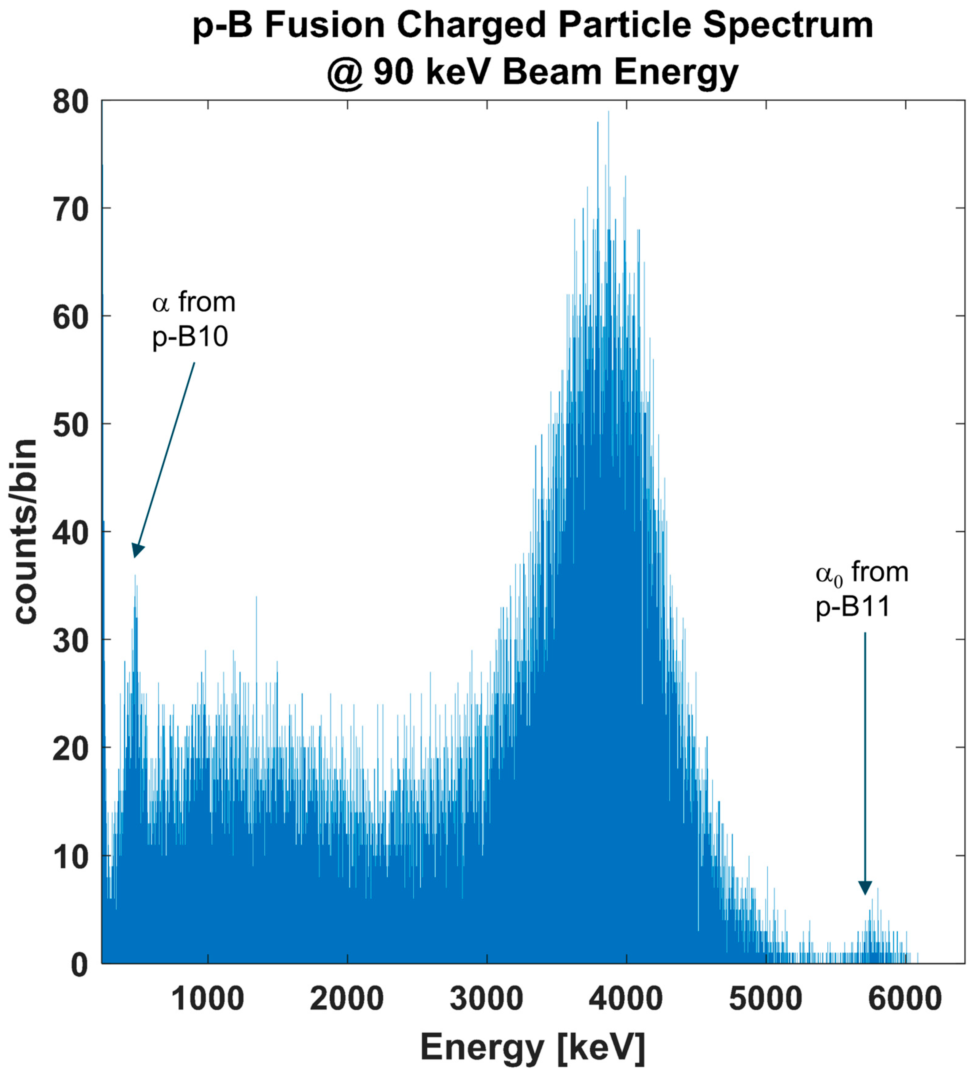
3. Signal Analysis Method and Results
The raw preamplifier signals exhibit a fast rise time of approximately 0.2 to 0.3 μs, followed by a long decay time determined by the RC time constant of the feedback capacitor and resistor on the first operational amplifier, which is approximately 100 μs. To measure the rise time of the preamplifier signal, we use the Picoscope 5444D [14] USB Oscilloscope with a sampling rate of 250 MS/s at a 12-bit resolution. To process these signals, discrete-time filtering methods based on the CR-RC⁴ configuration, as described in [1], are employed. Alternatively, other digital filtering methods, such as trapezoidal filtering, could be implemented and will be explored in future work. A typical pulse-shaping time of 0.5 to 2 µs is used, balancing signal integrity with the minimization of pile-up events. This filtering approach is effective for observing fusion products at lower ion beam energies, where beam-induced noise from secondary electrons and X-rays is minimal. Unlike Si PIN detectors that are designed to observe X-rays [15], our detector utilizes a significantly higher feedback capacitance, making it less responsive to individual X-ray events. However, in the ion beam-target environment, where bremsstrahlung X-rays dominate due to back-streaming ions and secondary electron emissions, the cumulative X-ray effect on the detector becomes evident and increases with applied voltage. As shown in Figure 3, during beam-on time (as indicated by Channel C), there is a quantifiable increase in the overall signal baseline on Channel A, which is the PIN detector signal. At a beam energy of 30 keV and an instantaneous beam current of approximately 0.6 mA, the observed baseline offset is around 1 mV. This represents a fourfold increase relative to the RMS baseline noise level, which is approximately 0.24 mV. This baseline offset is likely a result of X-rays generated by ion beam-induced mechanisms. It is also shown that at the instant the ion beam is turned off, the offset is removed. Therefore, the high pass portion of the digital filter is essential in this case as it effectively suppresses the slower responding X-ray signal induced by the beam.
Figure 3.
An example PIN detector signal pulse (blue) is shown for an ion beam operating at 30 keV with an instantaneous current of 0.6 mA. The ion beam is externally triggered by an LVTTL signal (green), which is overlaid on the detector waveform for reference. During the beam−on period, an offset of approximately 1 mV is induced on the detector signal. The baseline RMS ripple is measured to be around 0.24 mV. The waveform corresponds to a detected 3.02 MeV proton.
However, at higher beam energies, the induced noise becomes significant, often comparable in magnitude to the charged particle signal. For example, Figure 4 presents the detection of the p–11B alpha-particle signal from a system operating at 90 keV, 5 kHz, and with a 5% duty cycle. In this scenario, the fast-rising alpha-particle signal is embedded within a periodic noise signal induced by the beam. While the alpha-particles can still be distinguished from noise based on their rise time constant, the resulting spectrum exhibits a pronounced noise peak in the low-energy region (Figure 5).
Figure 4.
Waveform of the Si PIN detector signal from the preamplifier operating in a beam-target experiment with a nominal beam energy of 90 keV, a 5% duty cycle, and a 5 kHz rep. rate. The alpha pulse can be seen superimposed on top of the ion beam pulse noise. With CR−RC filtering, it is not possible to deconvolute the alpha pulse from the noise.
Figure 5.
This spectrum was taken over an 8 h period. The spectrum matches one that was taken from a more traditional accelerator facility studying the p–11B reaction [16]. Additionally, due to the lower accelerating potential, the p–10B mono-energetic alpha can be observed. In addition, we also observe the α0 from the coincident alpha of Be-8.
To address this, a CR²-RC⁴ discrete-time filtering method is applied using the same pulse-shaping time constant. This approach yields a significantly cleaner filtered bipolar signal. The resulting energy spectrum displays a drastically reduced noise peak (Figure 5), enabling the detection of the low-energy p–10B alpha peak. The 10B(p,α)⁷Be reaction has a Q-value of 1.1 MeV, imparting an alpha-particle kinetic energy of 0.7 MeV. Based on SRIM stopping power calculations for a 0.8 µm foil [16], the observed peak shift is consistent with the calculated energy spectrum. To our knowledge, this is the first time the proton–boron fusion energy spectrum has been obtained using a natural abundance boride target, clearly showing the p–10B reaction alpha-particle. In contrast, previous measurements of the alpha-particle energy spectrum, such as those reported in [17], were conducted at an H⁺ energy of 675 keV, which would have masked the p–10B alpha-particle peak due to scattered beam protons.
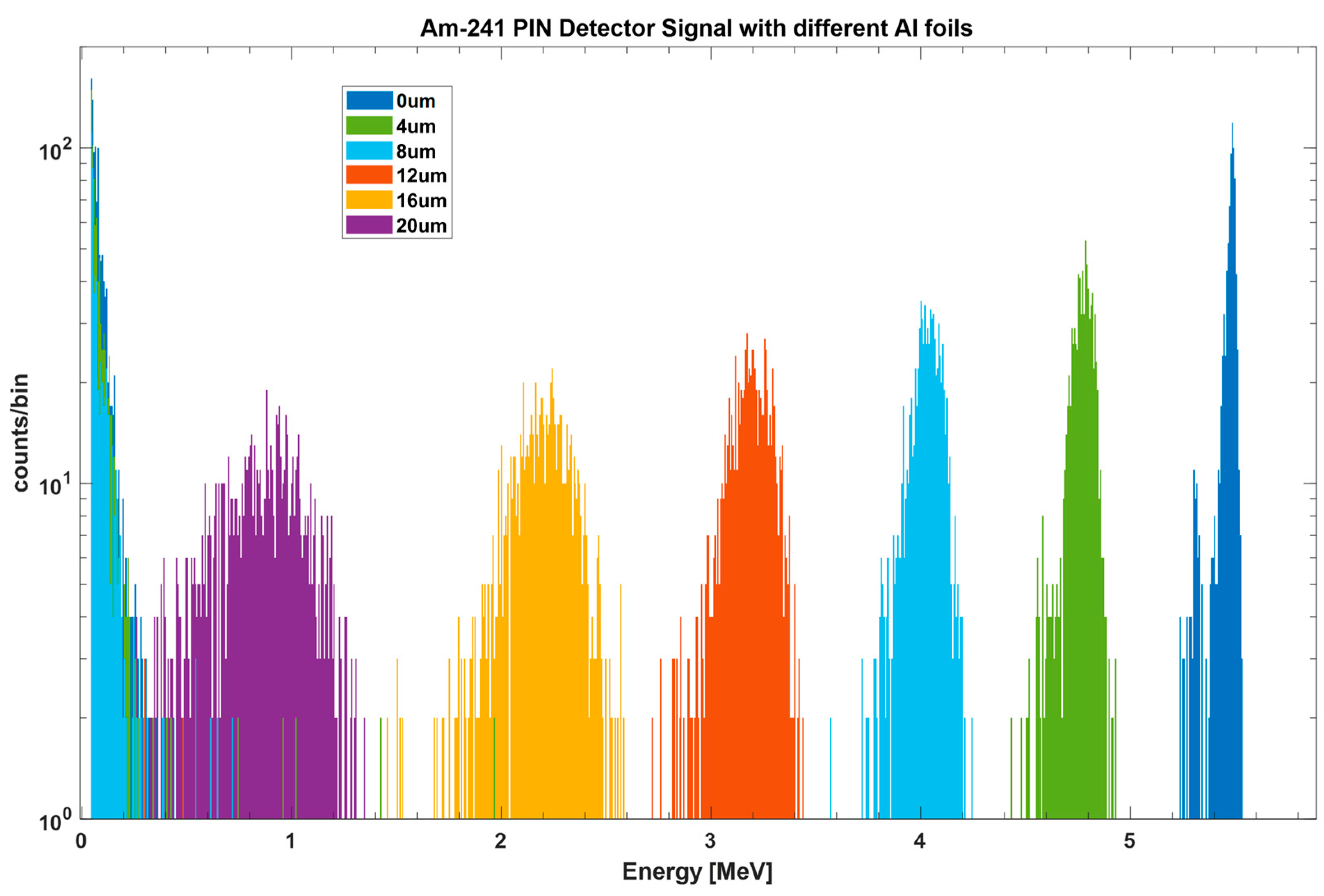
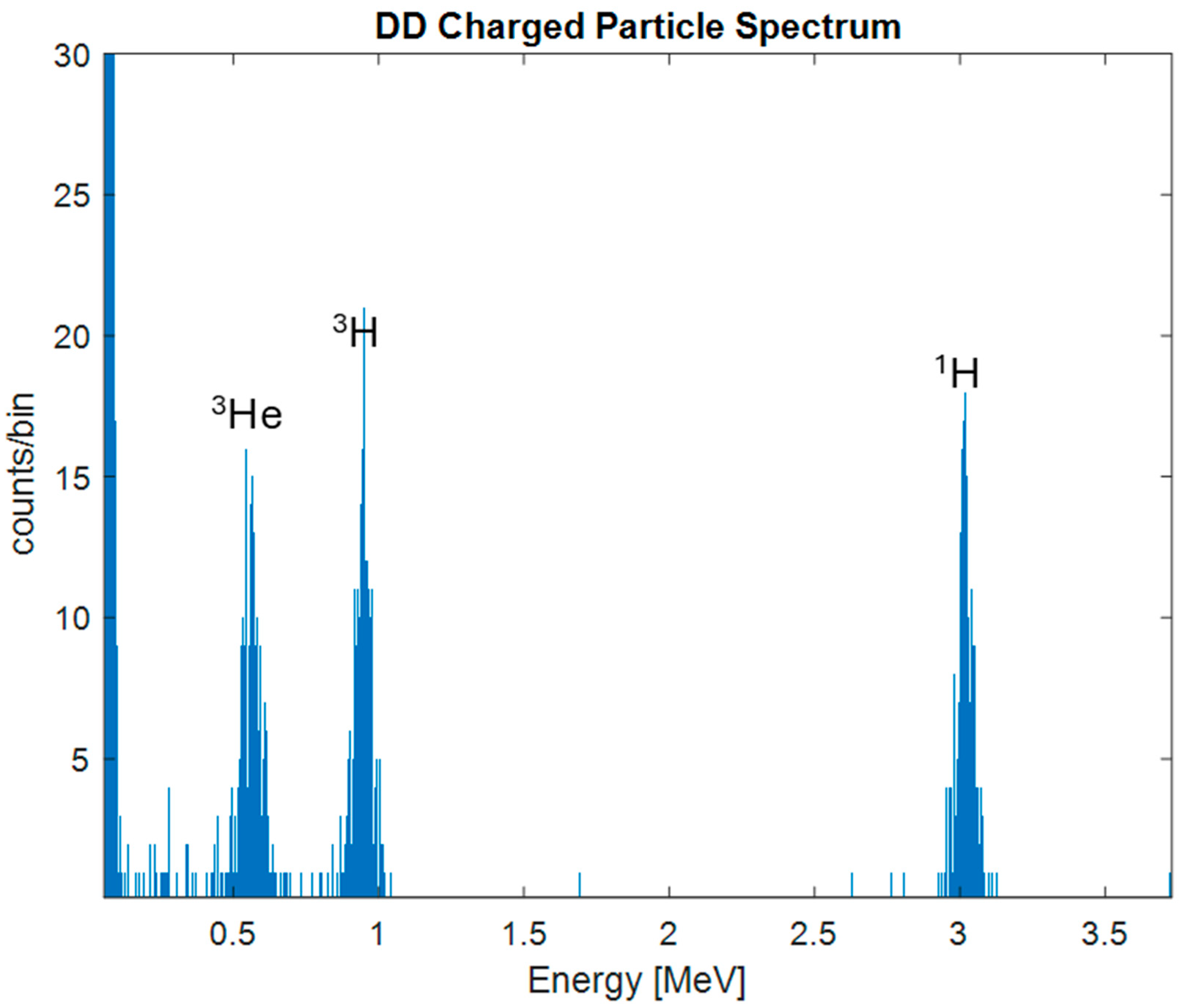
3.1. Am-241 Calibration
We used the Am-241 alpha source to calibrate the linearity of the detector. Using different thicknesses of aluminum foils between 0 and 20 μm, we were able to attenuate the energy of the alphas to energies from 5.486 MeV down to ~1 MeV. Figure 6 shows the energy spectrum for the different thickness cases. The results were obtained using the CR-RC4 filtering with a time constant of 4 μs, as described in the previous section. The “no foil” (0 μm) case was used to calibrate the 5.486 MeV Am-241 energy. For this case, the peak resolution (FWHM) was approximately 39 keV. Table 1 compares the SRIM calculation with the measured peak energy using the detector, showing a strong agreement between the calculated and measured values.
Figure 6.
Am-241 calibration of the Si PIN detector using various foil thicknesses. The FWHM of the unattenuated Am-241 peak is approximately 39 keV.
Table 1.
Attenuated Am-241 alpha energy for different foil thicknesses. The second column shows the calculated energy using SRIM; the third column shows the energy at the right edge of the peak measured with the PIN detector.
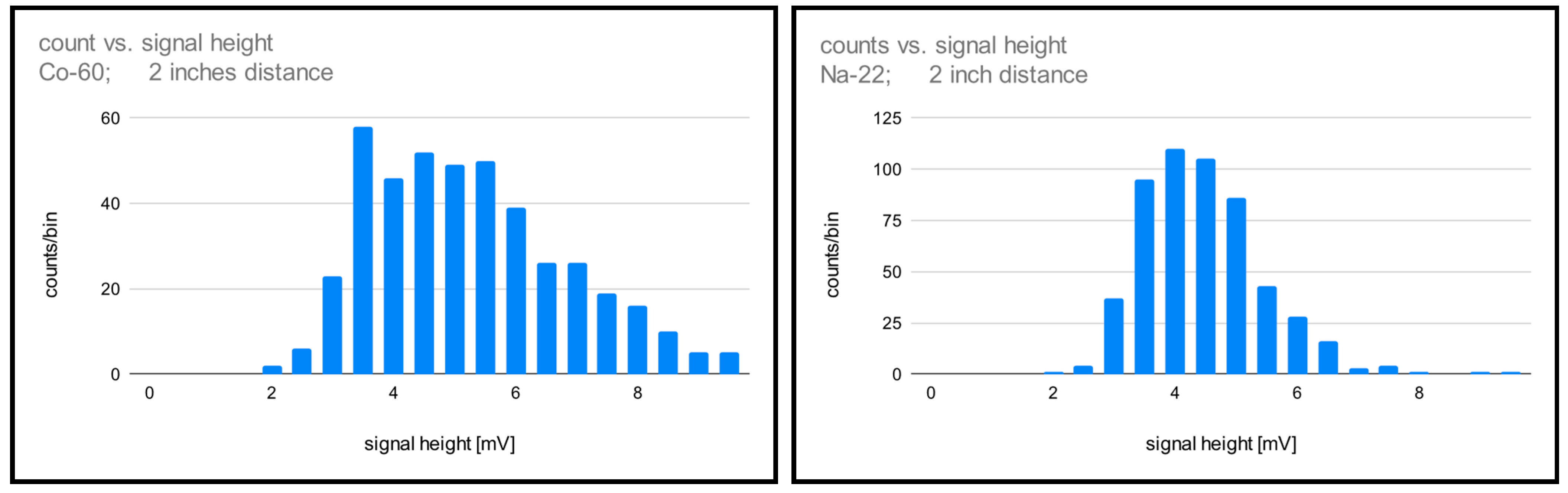
3.2. Effect of Gamma Rays on Si PIN Detector
We also evaluate the response of the PIN detector to gamma radiation by placing it two inches away from calibrated Co-60 and Na-22 check sources. At the time of the experiment, the sources had nominal activities of approximately 0.35 µCi and 0.12 µCi, respectively. The Co-60 source emits gamma rays at energies of 1.17 MeV and 1.33 MeV, while the Na-22 source emits a 511 keV gamma photon from positron annihilation. Signal pulses from these interactions are successfully observed and recorded. Figure 7 shows the histogram of pulse heights from the Co-60 and Na-22 sources. The observed spread in the signal’s energy distribution can be attributed primarily to Compton scattering, which dominates at this photon energy. In Compton interactions, only a portion of the gamma-ray energy is deposited in the detector, leading to incomplete energy absorption. This results in a broad energy distribution, as varying amounts of energy are transferred depending on the scattering angle. The count rates observed for the Co-60 and Na-22 sources are 44.3 and 126.4 counts per minute, respectively. We note that the count rate for the Co-60 source is lower even though it has higher activity than the Na-22 source, this is because the Compton scattering cross-section decreases with increasing energy according to the Klein–Nishina formula [18]. As a final check, we place a Cd-109 source with a nominal activity of 0.14 μCi at less than 4 mm distance from the detector and observe no detectable signal pulses. Given that this source emits photons primarily at 88 keV, as well as characteristic Ag-109 X-rays at 22 and 25 keV, the lack of a detectable response indicates that the detector is largely insensitive to single-event X-ray interactions under these conditions.
Figure 7.
Pulse height distribution of the PIN detector in response to two gamma-ray sources, measured at the output of the charge-coupled amplifier, as shown in Figure 2. The sources are placed 2 inches away from the detector with a nominal activity of 0.35 µCi (Co-60) and 0.12 µCi (Na-22). A large spread in energy is observed, likely due to Compton scattering.
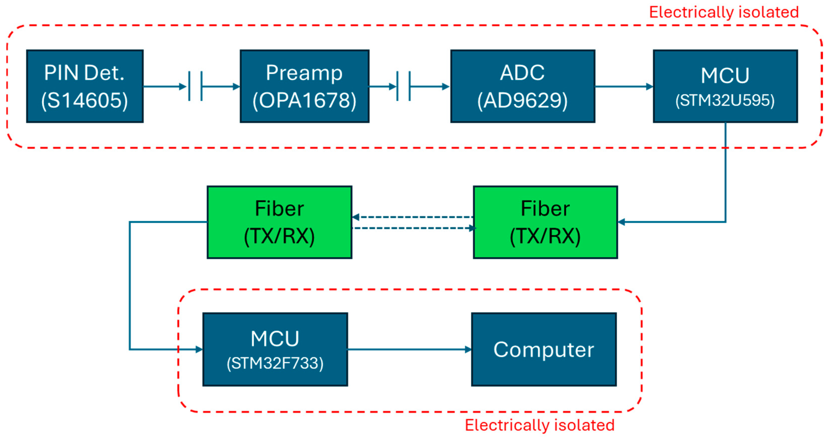
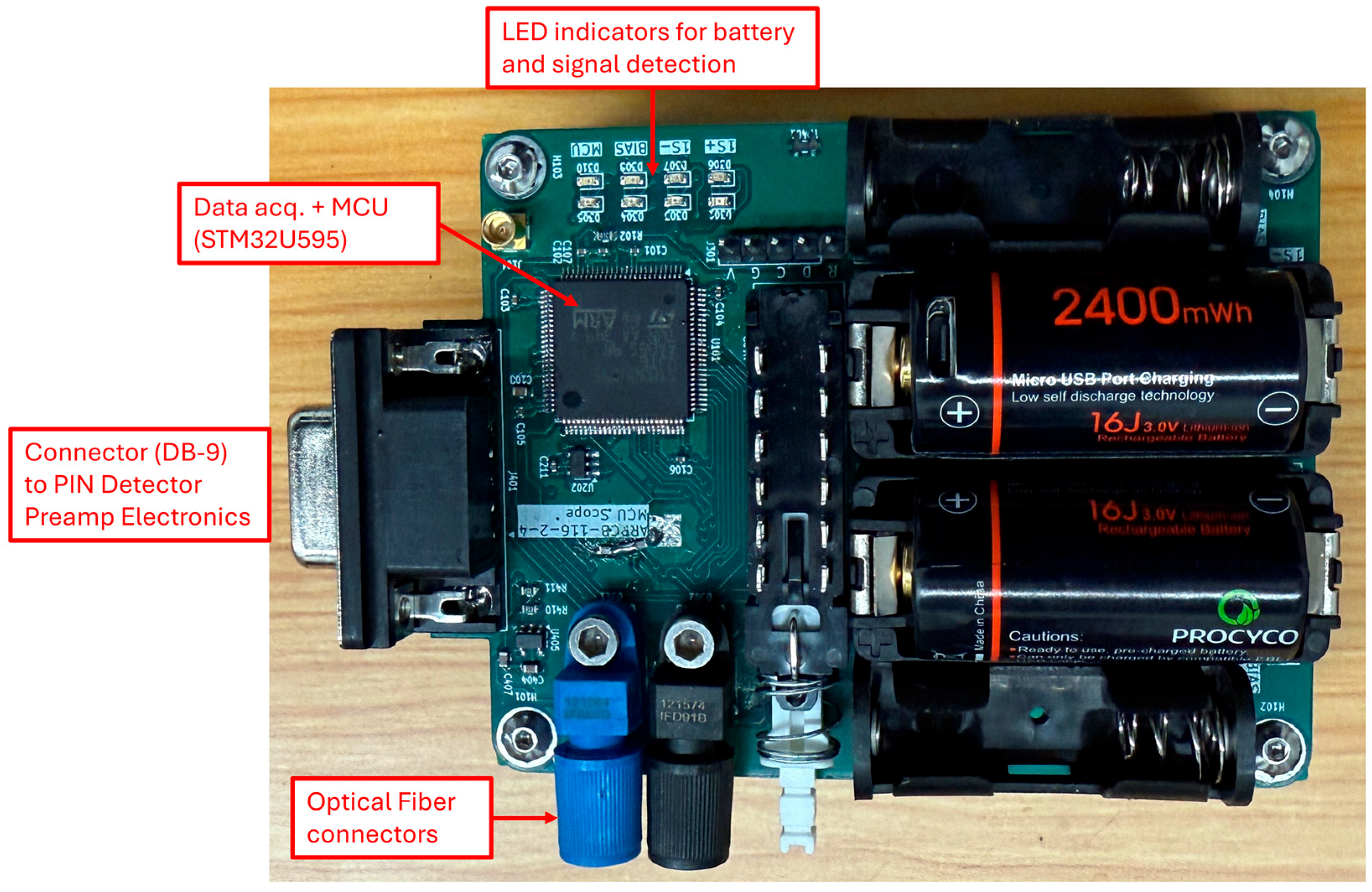
4. Data Acquisition Module
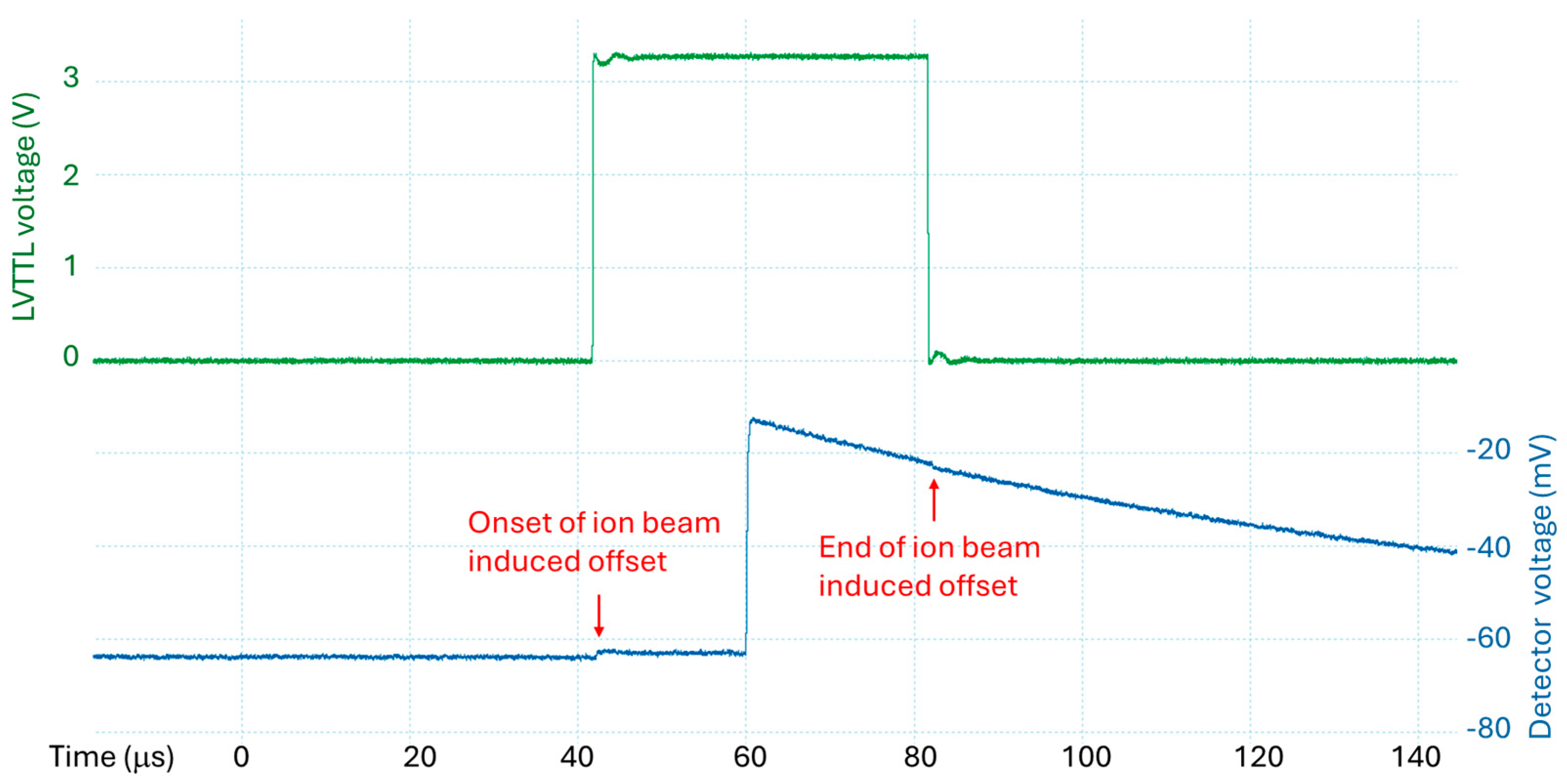
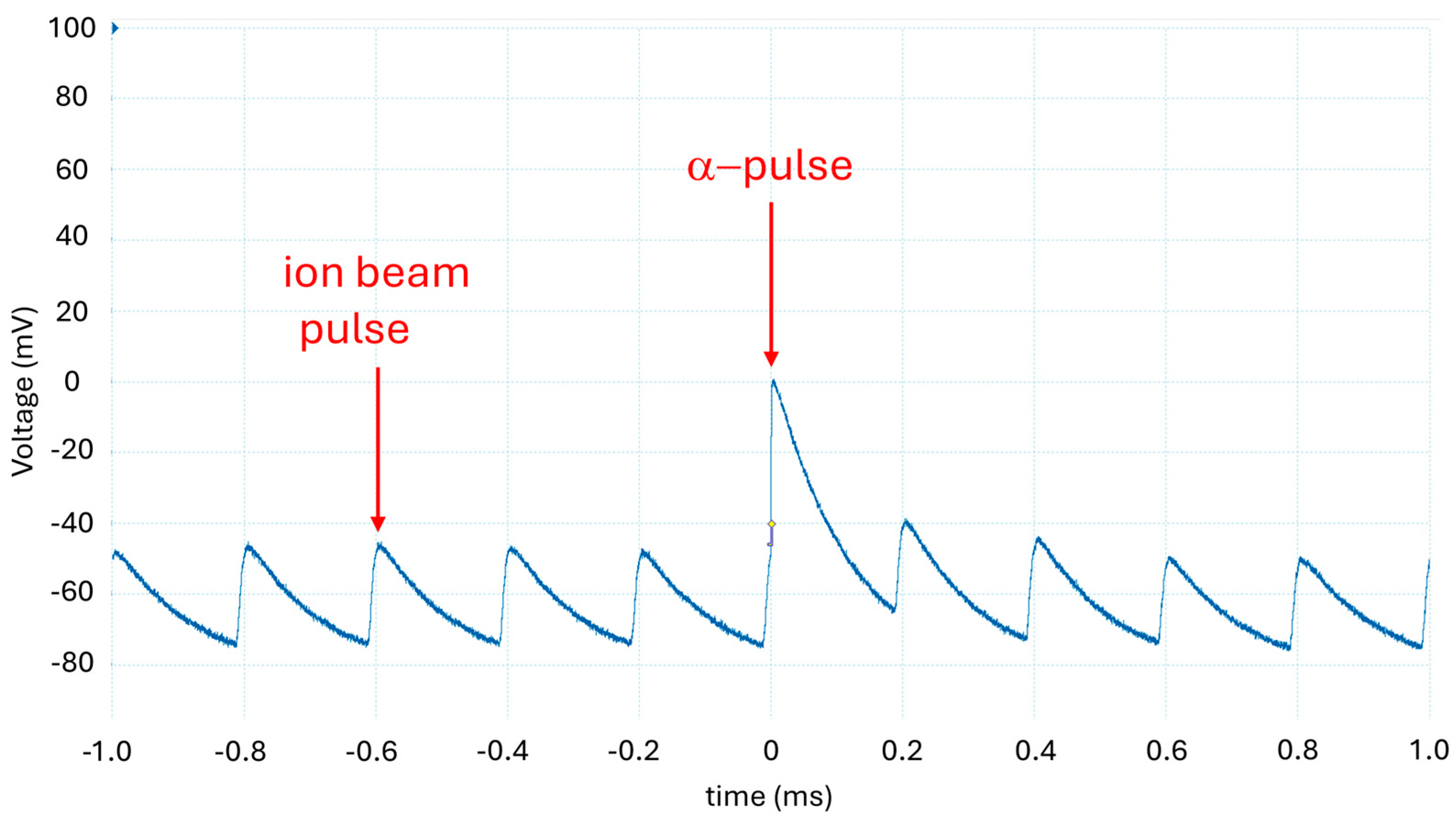
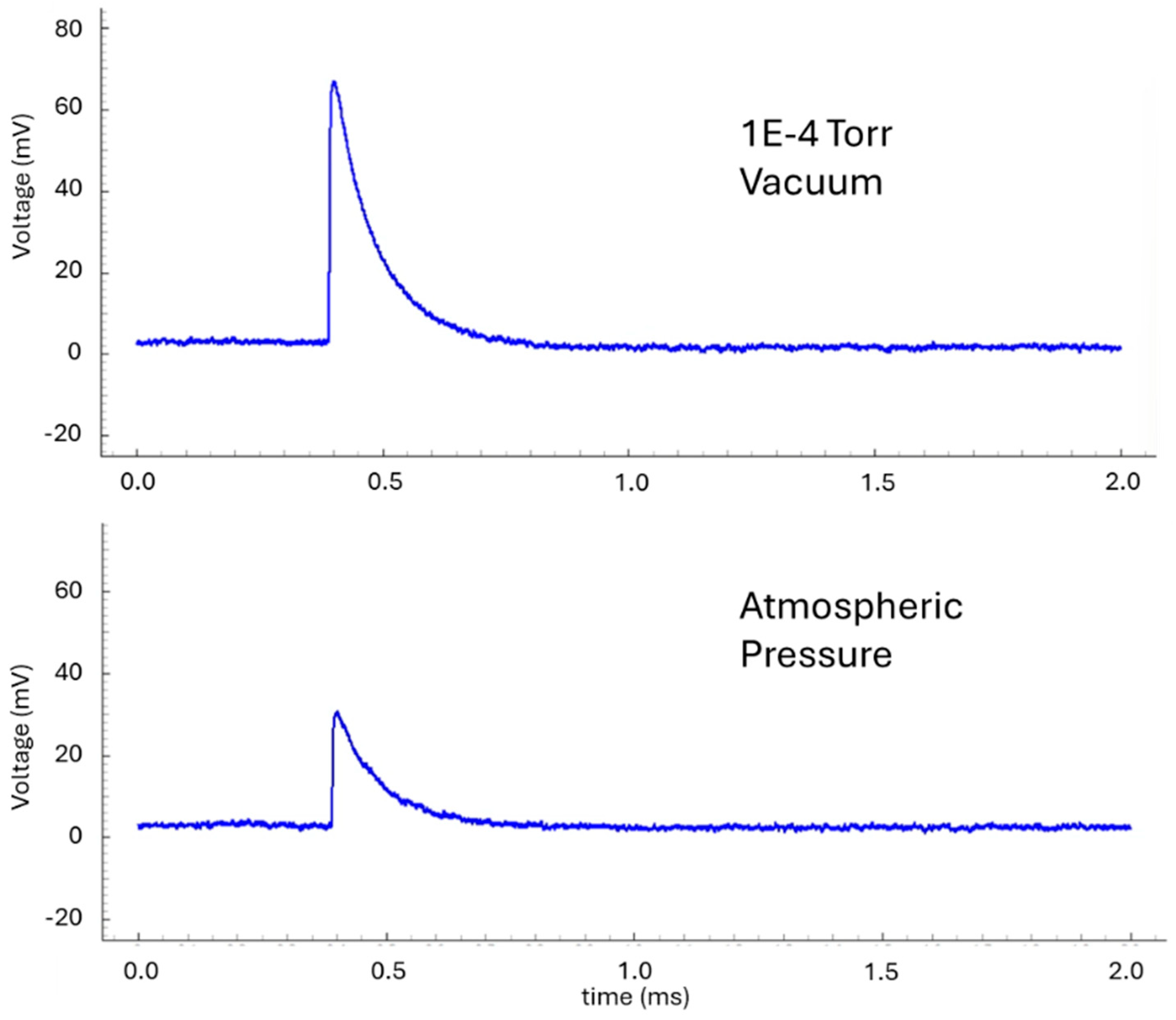
The data acquisition module is developed by ARI to ensure the entire system is as compact and electrically isolated as possible. A 20 MS/s analog-to-digital converter (Analog Devices AD9629) captures the signal from the PIN detector’s preamp output with a 12-bit resolution and range of ±1.8 V. The signals are then processed by a high-performance microcontroller unit (STM32U595), operating at a nominal clock speed of 160 MHz with excellent power efficiency. Additionally, operating at ±1.8 V instead of the typical ±3.3 V further reduces power consumption. Data are transmitted from the data acquisition module to a separate module using optical fiber connections. This separate module converts the optical signal to USB-UART, enabling communication with a computer. Although real-time streaming is limited by the high sampling rate and the bandwidth constraints of the USB connection, at low count rates we can buffer the signal and transfer the data in chunks to minimize latency. Additionally, the system supports a triggered acquisition mode, allowing data to be captured at a fixed interval following an external trigger, which can facilitate synchronization with external events. There are LEDs on the PCB to indicate both battery status and signal detection, providing an effective way to monitor system status without needing to operate the computer.
The data transfer workflow is illustrated in Figure 8. The top and bottom sections of the system are electrically isolated, with fiber optics serving as the data link between them. This isolation ensures low-noise operation of the PIN detector and the associated data acquisition electronics. For optical communication, we use a standard plastic optical fiber (POF) with a 1000 µm core, 2.2 mm cladding, and connectors sourced from Industrial Fiber Optics Inc. (Tempe, AZ, USA) [19]. The system employs an LED (IF-E91D) and a photodetector (IF-D91B) operating in the infrared wavelength range at around 870 nm. This configuration is selected for its cost-effectiveness and high baud rate capability, supporting data transfer speeds of up to 20 Mbps over 3 m. For our device, we define RX as transmission from the PIN detector to the computer and TX as transmission from the computer to the PIN detector. Currently, the RX line operates at 5 Mbps to ensure signal reliability, while the TX line operates at a much slower 50 kbps, as it only carries acknowledgements and configuration data. The lower speed helps reduce the cost of the signal driving components.
Figure 8.
Signal flow chart of the Si PIN detector system with optical fiber datalink communication. This allows the entire detection system, including data processing, to be isolated from the receiving computer.
The integrated PIN detector with a data acquisition module is utilized in the ARI-IBS to detect charged particles from D–D fusion. The ion beam operates at a nominal voltage of 30 keV, with a beam duty cycle of 5% and a current of 0.05 mA. This output is low enough to ensure user safety from excessive radiation exposure, while still producing sufficient fusion events to be reliably detected by the PIN detector. A typical D–D charged particle spectrum is shown in Figure 9, where the three distinct charged particles (proton, triton, and helion) from D–D fusion are clearly visible. To generate the spectrum, each pulse from the preamplifier signal is processed using a standard CR-RC⁴ filter. The third peak in the processed data is normalized to the D–D proton energy of 3.02 MeV. The noise threshold of the spectrum is determined to be approximately 13.4 keV, based on the previously discussed baseline RMS ripple. This threshold is used as the reference for calculating the signal-to-noise (S/N) ratios. Thus, the S/N ratios for the proton, triton, and helion signals are approximately 225, 75, and 41, respectively. It is worth noting that the helion signal, which has a nominal peak energy of 0.82 MeV, experiences attenuation due to the 0.8 µm aluminum foil used to block optical emissions. This attenuation reduces the detected energy by several hundred keV, resulting in a final measured energy of approximately 560 keV. Similarly, the triton signal is also slightly attenuated.
Figure 9.
Typical D–D charged particle spectrum obtained using the Si PIN detector with an integrated data acquisition system. The beam-target system was operated at a 30 keV beam energy with a 5% duty cycle and a nominal current of 0.05 mA.
Operation in Vacuum
For future applications, we envision using this device to measure charged particles within large fusion devices. In such environments, the unit must be capable of operating reliably under vacuum conditions, including both the electronics and the batteries. To validate its performance, we tested the entire ARPCB-117 assembly (Figure 10) in an 8-inch vacuum chamber. The assembly, which included the PIN detector preamplifier and Li-ion A123 batteries, was connected and positioned such that the PIN detector faced a Po-210 alpha source mounted on one of the chamber ports as a calibration test source. Optical fibers were used to transmit data between the atmospheric and vacuum sides, routed through a CF-2.75 flange. To ensure vacuum compatibility, the TX and RX fibers were securely epoxied in place with high-vacuum-compatible epoxy. The unit was fully electrically isolated from the chamber, which minimized conducted noise pickup and enhanced signal integrity.
Figure 10.
ARPCB-116 assembly detailing the various components. The unit is electrically isolated with its own battery supply and communicates via optical fiber.
We used a standard vacuum pumping system consisting of a turbomolecular pump (Agilent TwisTorr FS 84) backed by a dry diaphragm roughing pump (Agilent MD1) to achieve high vacuum conditions in the chamber (Figure 11). After approximately 2 h, the chamber reached a quasi-ultimate vacuum of ~ 1.6 × 10⁻⁵ Torr. The system was maintained under high vacuum conditions for about 6 h, during which no abnormalities were observed in the batteries or other electronic components. The vacuum remained stable, consistently below 2 × 10⁻⁵ Torr.
Figure 11.
Testing of the Si PIN detector and data acquisition system in a high vacuum chamber. The optical fiber datalink was passed through to the atmospheric side to connect with the USB-UART module, which was then connected to the computer.
Using the PIN detector preamplifier electronics, we successfully observed pulses from the Po-210 alpha source. In high vacuum, the alpha-particle signal exhibited a peak height of approximately 70 mV, consistent with expectations as the particles experienced no attenuation in the absence of a gas medium. For comparison, under atmospheric conditions, the peak height was reduced to approximately 30 mV due to attenuation from the 2–3 cm of air the particles needed to travel through (Figure 12). Additionally, we tested the system at a gas pressure of 2 Torr and found no significant difference in signal characteristics compared to those observed at ~1 × 10⁻⁴ Torr. This demonstrates the robustness of the detector and electronics under varying low-pressure conditions. It is worth noting that when cycling the unit between atmospheric and vacuum pressure, one should use a bias voltage well below the maximum recommended +150 V to eliminate the risk of Paschen curve breakdown.
Figure 12.
Si PIN detector signal from Po-210 under different operating pressures of 1 × 10⁻⁴ Torr (top) and atmospheric pressure (bottom). Note the lower peak height observed for the atmospheric pressure signal due to the particle energy being attenuated by the atmospheric gas medium.
5. Conclusions
We have developed a compact PIN detector with integrated electronics, specifically optimized for high-energy particle detection. The system has been successfully used to detect alpha particles from radioactive sources (e.g., Am-241, Po-210) as well as charged particles from D–D and p–B fusion reactions. Engineered with a strong emphasis on noise suppression and immunity, the detector delivers excellent signal-to-noise performance. The data link between the detector electronics and the control computer utilizes optical fiber communication, providing complete electrical isolation. This setup enables reliable data transmission over 10 m without any degradation in the detector’s electrical performance. Furthermore, the entire assembly, including the battery-powered electronics, has been rigorously tested in both high-vacuum (<1 × 10⁻⁴ Torr) and rough-vacuum environments (~2 Torr). Results confirm the system’s robustness and reliable operation in vacuum conditions for extended periods, making it well-suited for experimental applications. In future work, we plan to explore and characterize the effect of X-rays on the detector. However, based on our results with the ion beam system so far, we have not observed a significant impact on signal integrity. This compact PIN detector offers a valuable complement to other detection methods, such as solid-state nuclear track detectors (SSNTDs), for accurately verifying the presence and characteristics of fusion products.
6. Patents
Allan X. Chen and Benjamin F. Sigal have the patent APPARATUS AND METHOD FOR CHARGED PARTICLE DETECTION USING PIN DETECTOR AND ASSOCIATED DATA ACQUISITION ELECTRONICS pending to Alpha Ring International Limited.
Author Contributions
Conceptualization, A.X.C. and A.Y.W.; methodology, A.X.C. and B.F.S.; software, B.F.S.; validation, A.X.C., B.F.S., A.G., J.M., M.S., N.A. and K.-J.X.; formal analysis, A.X.C. and J.M.; investigation, A.X.C., B.F.S., A.G., J.M., M.S., N.A. and K.-J.X.; resources, A.X.C.; data curation, A.X.C. and B.F.S.; writing—original draft preparation, A.X.C.; writing—review and editing, A.X.C., B.F.S. and J.M.; visualization, A.X.C.; supervision, A.X.C.; project administration, A.X.C.; funding acquisition, A.X.C. and A.Y.W. All authors have read and agreed to the published version of the manuscript.
Funding
This research was fully funded by Alpha Ring International Limited.
Data Availability Statement
The original contributions presented in this study are included in the article. Further inquiries to be directed to the corresponding author.
Acknowledgments
The authors are grateful for discussions with and support from Roger Falcone, Richard Petrasso, Jyhpyng Wang, Kosta Yanev, Gianluca Gregori, Qiong Wang, Naomi Mitchell, Yuxing Wang, Chia-Yi Chen, Nai-Wei Liu, Chih-Jui Hsieh, Ming-Cheng Jheng, Benjamin Wrixon, Fay Li, Peter Liu, Paul Chau, Nathan Eschbach, Mason Guffey, David Chu, Peter Hsieh, Wilson Wu, Charles Wu, David Noriega, Ryan Yan, and Belinda Mei. This work was conducted by scientists and engineers as employees of Alpha Ring International Limited and its affiliated companies. The study was funded by Alpha Ring International Limited, which supports the education and training of a fusion industry workforce. The funding from Alpha Ring International Limited did not influence the scientific integrity or the results of the study.
Conflicts of Interest
All authors are employed by Alpha Ring International Limited at the time of their contribution to the research. The authors declare that this study received funding from Alpha Ring International Limited. The funder was not involved in the study design, collection, analysis, interpretation of data; the writing of this article; or the decision to submit it for publication.
References
- Chen, A.X.; Liu, N.-W.; Gunn, A.; Su, Z.; Sigal, B.F.; Salazar, M.; Abdalla, N.; Chen, J.; Wong, A.Y.; Wang, Q. Compact Ion Beam System for Fusion Demonstration. Phys. Open 2024, 21, 100234. [Google Scholar] [CrossRef]
- Magee, R.M.; Ogawa, K.; Tajima, T.; Allfrey, I.; Gota, H.; McCarroll, P.; Ohdachi, S.; Isobe, M.; Kamio, S.; Klumper, V.; et al. First measurements of p11B fusion in a magnetically confined plasma. Nat. Commun. 2023, 14, 955. [Google Scholar] [CrossRef]
- Ramirez-Jimenez, F.J.; Mondragon-Contreras, L.; Cruz-Estrada, P. Application of PIN diodes in Physics Research. AIP Conf. Proc. 2006, 857, 395–406. [Google Scholar] [CrossRef]
- Ramirez-Jimenez, F.J.; Aguilera, E.F.; Lopez-Callejas, R.; Benitez-Read, J.S.; Pacheco-Sotelo, J. A novel application of a PIN diode-preamplifier set for the measurement of charged particles. Nuc. Instr. Meth. Phys. Res. A 2005, 545, 721–726. [Google Scholar] [CrossRef]
- Gál, N.; Hrubčín, L.; Šagátová, A.; Vanko, G.; Kováčová, E.; Za’ko, B. High-resolution alpha-particle detector based on Schottky barrier 4H-SiC detector operated at elevated temperatures up to 500 °C. Appl. Surf. Sci. 2023, 635, 157708. [Google Scholar] [CrossRef]
- Hamamatsu Large-Area Si PIN Photodiode for Direct Radiation Detection. Available online: https://www.hamamatsu.com/us/en/product/optical-sensors/photodiodes/si-photodiodes/S14605.html (accessed on 12 May 2025).
- Energizer A23 Battery Data Sheet. Available online: https://data.energizer.com/pdfs/a23.pdf (accessed on 12 May 2025).
- Amptek A250 Charge Sensitive Preamplifier Application Note. Available online: https://www.amptek.com/internal-products/a250-charge-sensitive-preamplifier-application-note (accessed on 12 May 2025).
- Riegler, W. Particle Detectors—Summer Student Lectures 2010. Available online: https://indico.cern.ch/event/91742/attachments/1096757/1564539/L5_solid_calo_pid_riegler.pdf (accessed on 12 May 2025).
- OPA167x Low-Distortion Audio Operational Amplifiers. Available online: https://www.ti.com/lit/ds/symlink/opa1678.pdf (accessed on 12 May 2025).
- ADP7182 −28 V, −200 mA, Low Noise, Linear Regulator. Available online: https://www.analog.com/en/products/adp7182.html (accessed on 12 May 2025).
- Linear Regulators (LDO)|NCP160, LDO Regulator, 250 mA, Ultra-High PSRR, Ultra-Low Noise. Available online: https://www.onsemi.com/products/power-management/linear-regulators-ldo/ncp160 (accessed on 12 May 2025).
- Duracell CR123A Lithium/Manganese Dioxide Battery Data Sheet. Available online: https://www.duracell.com/wp-content/uploads/2021/06/CR123_US.pdf (accessed on 12 May 2025).
- Picoscope 5000 Series—Flexible Resolution USB Oscilloscope. Available online: https://www.picotech.com/oscilloscope/5000/flexible-resolution-oscilloscope (accessed on 12 May 2025).
- Ramirez-Jimenez, F.J.; Lopez-Callejas, R.; Cerdeira-Altuzarra, A.; Benitez-Read, J.S.; Estrada-Cueto, M.; Pacheco-Sotelo, J.O. PIN diode-preamplifier set for the measurement of low-energy g- and X-rays. Nuc. Instr. Meth. Phys. Res. A 2003, 497, 577–583. [Google Scholar] [CrossRef]
- Ziegler, J.F. SRIM-2003. Nuc. Instr. Meth. Phys. Res. Sec. B 2004, 219–220, 1027–1036. [Google Scholar] [CrossRef]
- Stave, S.; Ahmed, M.W.; France, R.H., III; Henshaw, S.S.; Müller, B.; Perdue, B.A.; Prior, R.M.; Spraker, M.C.; Weller, H.R. Understanding the B11 (p, α) αα reaction at the 0.675 MeV resonance. Phys. Lett. B 2011, 696, 26–29. [Google Scholar] [CrossRef]
- Klein, O.; Nishina, Y. Über die Streuung von Strahlung durch freie Elektronen nach der neuen relativistischen Quantendynamik von Dirac. Z. Physik 1929, 52, 853–868. [Google Scholar] [CrossRef]
- Industrial Fiber Optics, Inc. Available online: https://i-fiberoptics.com/ (accessed on 12 May 2025).
Disclaimer/Publisher’s Note: The statements, opinions and data contained in all publications are solely those of the individual author(s) and contributor(s) and not of MDPI and/or the editor(s). MDPI and/or the editor(s) disclaim responsibility for any injury to people or property resulting from any ideas, methods, instructions or products referred to in the content. |
© 2025 by the authors. Licensee MDPI, Basel, Switzerland. This article is an open access article distributed under the terms and conditions of the Creative Commons Attribution (CC BY) license (https://creativecommons.org/licenses/by/4.0/).