Article

An Engineered Cargo-Transport Molecular Motor Composed of a Kinesin Monomer and a Diffusing Microtubule-Associated Protein

Laboratory of Soft Matter Physics, Institute of Physics, Chinese Academy of Sciences, Beijing 100190, China
Biophysica 2025, 5(3), 26; https://doi.org/10.3390/biophysica5030026
Submission received: 16 May 2025 / Revised: 23 June 2025 / Accepted: 24 June 2025 / Published: 2 July 2025

Abstract

An engineered molecular motor composed of an ATP-dependent kinesin-1 monomer and an ATP-independent diffusing microtubule-associated protein is proposed, and its dynamics are studied theoretically. It is shown that the engineered motor can move directionally on microtubules towards the plus end, bearing great potential for applications in therapeutics or nanorobotics. The engineered motor can have an unloaded velocity similar to the wild-type kinesin-1 dimer, can take a mechanical (either forward or backward) step by hydrolyzing an ATP molecule under any load, and can generate the maximum force that is about half of that generated by the wild-type kinesin-1 dimer.

1. Introduction

Linear molecular motors constitute an important group of proteins or artificial macromolecules that can move directionally and continuously on their linear tracks [1,2,3,4,5,6,7,8,9,10]. Kinesin-1 protein is a typical one of those molecular motors [1,2,3,4,5,6,11]. Unless otherwise pointed out, kinesin-1 is simply called kinesin. A wild-type (WT) kinesin is structurally a homodimer with two identical motor domains (also called heads) being connected together by a common long coiled-coil stalk through their flexible neck linkers (NLs) [12]. After binding to the track, microtubule, by hydrolyzing ATP molecules, the WT kinesin dimer can step continuously towards the microtubule plus end with a high velocity of about (0.6~1) μm/s under no load and can generate the maximal force of about (6~8) pN [4,13,14,15,16].
Apart from the cargo transport by the WT kinesin dimers, the cargo transport by engineered kinesin motors, such as multiple coupled truncated kinesin monomers, has also been studied extensively [17,18,19,20,21,22,23,24,25]. In particular, the experimental data showed that the cargo transport by two or multiple coupled kinesin monomers with short truncated stalks under no load behaves nearly as efficiently as that by one or multiple kinesin dimers [20,21]. The subsequent theoretical studies explained how the two coupled kinesin monomers with the short truncated stalks have similar unloaded velocity to that of a kinesin dimer [25]. By contrast, the theoretical studies showed that the two coupled kinesin monomers with long stalks can only make a very inefficient movement with an unloaded velocity much smaller than that with the short stalks [25]. In addition, the design of the artificial molecular motor that can move directionally on the linear track has also been an interesting subject [8,9,10,26]. The engineered and artificial molecular motors bear great potential for applications in therapeutics or nanorobotics [10]. The purpose of this work is to design a new engineered motor that can move directionally with a high velocity on microtubule.
Previous theoretical and experimental studies indicated that the WT kinesin dimer shows tight chemomechanical coupling under a near-zero load [27,28,29]. However, prior theoretical studies indicated that the WT kinesin dimer could not show the tight chemomechanical coupling under a medium or a large backward load resisting its forward stepping [29]. Here, the tight chemomechanical coupling is referred to shows that the hydrolysis of an ATP induces (or is tightly coupled with) a mechanical (either a forward or a backward) step. An interesting issue is to engineer the kinesin to make it become the tight coupling under any load, which is also the purpose of this work.
Here, an engineered motor composed of a kinesin monomer and an ATP-independent diffusing microtubule-associated protein (MAP) is proposed, where the single MAP can diffuse in an unbiased manner along a microtubule filament with a large diffusion constant. Both the kinesin monomer and MAP have short truncated stalks that are connected to a stiff scaffold (see the inset of Figure 1). The proposal is inspired by previous studies showing that the two coupled kinesin monomers with the short truncated stalks can move as efficiently as a WT kinesin dimer [20,21,25]. To compare the dynamic performance of this engineered motor with that of the WT kinesin dimer, the dynamics of the engineered motor are studied theoretically. It is shown that even for the kinesin monomer having symmetrical interacting potential with the tubulin heterodimer along the microtubule axis, with the single monomer incapable of moving directionally, the engineered motor can move directionally towards the microtubule plus end with the tight chemomechanical coupling. The engineered motor can attain a velocity that is similar to that of the WT kinesin dimer under no load and can generate the maximal force that is reduced by only about two times relative to the WT kinesin dimer.

2. The Engineered Motor

2.1. Description

The engineered motor is composed of a kinesin monomer and an MAP. The kinesin monomer, for instance, includes a motor domain (aa1–324), flexible NL (aa325–338) and truncated stalk (aa339–350), as used before [20,21], where the stalk has a short length (about 5 nm) (see the inset of Figure 1). MAP can be the Ndc80 complex [30,31,32], the tail domain of kinesin-14 [32,33,34,35], or the head of Ase1-related protein PRC1 [36,37], also with the truncated stalk of a short length. Specifically, the truncated kinesin-14 HSET tail (aa1–230) can be used [34]. Both the stalk of kinesin monomer and that of MAP are connected fixedly to the stiff scaffold (see the inset of Figure 1), which can be achieved experimentally with the method used in Ref. [20]. Due to the short length, it is approximately considered that the stalks cannot be stretched and bent elastically. Here, we consider that the distance between the two connecting points of the stalk on the scaffold, l0, is equal to (m + 1/2)d, where m ( 1) is an integer and d = 8 nm is the repeat period of tubulins on the microtubule filament (see the lower panel of Figure 1a). Note that for a kinesin dimer with the two heads binding to microtubule, the two NLs have a total length of d = 8 nm, and each NL has a length of d/2 = 4 nm. To make the NL have a near-zero stretching force when the kinesin monomer in the engineered motor moves from the local tubulin to the adjacent tubulin, several amino acids with a net neutral charge can be inserted into the C-terminal portion of the NL, as done before [38].
The kinesin monomer has the following characteristics, which are summarized on the basis of structural, experimental, and all-atom molecular dynamics simulation studies [39,40,41,42,43,44,45,46,47,48,49,50,51,52,53] [see Section S1 in Supplementary Information (SI) for detailed descriptions and references]. (1) The monomer in nucleotide-free and ATP (ATP representing both ATP and ADP.Pi) states binds strongly to the microtubule, inducing large conformational changes of the local tubulin, whereas in the ADP state, it binds weakly to the microtubule, inducing little conformational changes of the microtubule. After the monomer transition from ATP to the ADP state, the local tubulin can still retain the large conformational changes for a short time tr (with the timescale of 10 μs) when the ADP monomer has a much weaker affinity (Ew1) for the local tubulin than an affinity (Ew2) for other tubulins without the large conformational changes. In time tr, the local tubulin returns elastically to the normally unchanged form, with its affinity for the ADP monomer changing to Ew2. (2) In the ATP state, the monomer can have a large change in its conformation, with its NL docking in the forward (plus-ended) orientation, relative to that in ADP or the nucleotide-free state. (3) The monomer with its NL in the forward orientation has a much higher rate of ATP transition to ADP than with its NL not in the forward orientation, and the rate constants of the ATPase activity are independent of the force on the NL.
MAP has the characteristic that it can diffuse in an unbiased manner along a microtubule filament with a large diffusion constant D (>0.1 μm2 s−1) [31,32,33,34], equivalent to a large forward or backward stepping rate equal to D/d2 (>1500 s−1). The rapid diffusion of MAP requires overcoming the small energy barrier between the two adjacent tubulins [32,33], because under no energy barrier, the diffusion constant is estimated as D = kBT/6πηr = 77 μm2 s−1, where kB is the Boltzmann constant, T = 310 K is the cell temperature, η = 0.01 g cm−1 s−1 is the viscosity [54], and r = 3 nm is the radius of MAP estimated from the number of residues of kinesin-14 tail [34] or from the structure of Ncd80 [55].

2.2. Chemomechanical Pathway

On the basis of the characteristics of the kinesin monomer and MAP, the movement of the engineered motor can be illustrated in Figure 1 (with m = 1). We start with the monomer in the ATP state binding strongly to tubulin IV and MAP binding to tubulin III, where the monomer has the large change in its conformation relative to that in nucleotide-free state and its NL docks in the forward orientation (lower panel of Figure 1a). MAP cannot diffuse forward to the occupied tubulin IV. (For the case of m > 1, MAP also cannot diffuse to the plus-ended adjacent unoccupied tubulin because this requires stretching the monomer’s NL to a length of 12 nm, which is impossible.) MAP can diffuse backward to tubulin II by overcoming the small energy barrier from the NL docking and large change in the conformation of the monomer [45,46,56] (upper panel of Figure 1a). In the upper panel of Figure 1a, MAP cannot diffuse to tubulin I because this requires stretching the monomer’s NL to a length of 12 nm. Thus, in Figure 1a, due to the large diffusion constant of MAP and the large rate of the large conformational change associated with NL docking of the ATP monomer, the system can transition rapidly between the two states (lower and upper panels), with the two states being in dynamic equilibrium.
Consider ATP transition to ADP in the monomer in the lower panel of Figure 1a. The monomer can detach from the local tubulin IV by overcoming very weak affinity Ew1, and due to the short stalk keeping the detached monomer close to the microtubule, the detached monomer binds rapidly (with a timescale of 1 μs that is shorter than tr) to the adjacent tubulin V with affinity Ew2 (Figure 1b). Alternatively, by overcoming Ew1, the monomer can diffuse from the local tubulin IV to tubulin V within time tr without detaching from the microtubule (Figure 1b). Since ADP release from the monomer bound to microtubule is a non-rate-limiting step of the ATPase activity [51], it is approximately considered that the rate of ADP release from the microtubule-bound monomer is much higher than that of ATP transition to ADP. From experimental data showing that the kinesin in the ADP state can slip with a low rate of only about 5.6 step/s under a backward load of 2 pN [50], and that the ADP release from the microtubule-bound monomer has a high rate of about (250–300) s−1 [51], it is noted that the ADP monomer having affinity Ew2 to tubulin IV in Figure 1b would be most probably kept still at tubulin IV until ADP release. However, due to the large diffusion constant in Figure 1b, MAP can shuttle rapidly between tubulins III and IV. After ADP release, ATP binds (Figure 1c). The state of Figure 1c is the same as that of Figure 1a, except that in Figure 1c, the engineered motor makes a forward step.
Consider ATP transition to ADP in the monomer in the upper panel of Figure 1a. By overcoming Ew1, the monomer can move from the local tubulin IV to the adjacent tubulin III with affinity Ew2 within time tr (Figure 1d) (noting that the monomer cannot move to tubulin V because this requires stretching the NL to a length of 12 nm). In Figure 1d, the ADP monomer would be most probably kept still at tubulin III, but MAP can shuttle rapidly between tubulins I and II. After ADP release, ATP binds (Figure 1e). The state of Figure 1e is the same as that of Figure 1a, except that in Figure 1e, the engineered motor makes a backward step.
It is evident that whether the interaction potential of the kinesin monomer in ADP state with microtubule is symmetrical or not along the microtubule axis the pathway of Figure 1 is applicable. In contrast to the WT kinesin dimer that moves forward in the hand-over-hand manner, with the two heads acting as the leading one alternatively [57,58,59] (see Section S2 and Figure S1 in SI), the engineered motor moves forward in the inchworm manner, with the kinesin monomer always acting as the leading one. The pathway of Figure 1 corresponds to the engineered motor having the configuration of the kinesin monomer in the plus-end position of MAP. For the engineered motor having the configuration of the kinesin monomer in the minus-end position of MAP, we have a similar pathway, but with MAP always acting as the leading one (see Figure S2 in SI).

3. Results and Discussion

Let k(+) and k(–) (<< k(+)) represent the rate of ATP transition to ADP of the monomer with and without the forward NL orientations, respectively. Let kD represent the rate of ADP release from the ADP monomer bound to microtubule. Since ADP release from the ADP monomer bound to the microtubule is a non-rate-limiting step of the ATPase activity [51], it is approximately considered that the rate of ADP release from the ADP monomer bound to the microtubule is much larger than that of ATP transition to ADP, with kD >> k(+). Let E0 represent the energy change of the large conformational change and NL docking of the ATP monomer relative to the ADP monomer.
In Figure 1a, the energy of the system in the state of the upper panel is increased by about Δ E = E 0 F d compared to that of the lower panel, where F is the backward load (in units of pN) and d = 8 nm is the repeat period of tubulins on a filament. Thus, in Figure 1a, the probability of the system in the state of the lower panel has the form
P NL = exp β E 0 F d exp β E 0 F d + 1
where β 1 = k B T , with kB being the Boltzmann constant and T the absolute temperature. The velocity, v, forward-to-backward stepping ratio, r, and mean number of ATPs consumed per mechanical (either forward or backward) step, N, for the engineered motor have forms (see Section S3 in SI)
v = P NL k ( + ) 1 P NL k ( ) d
r = P NL k ( + ) 1 P NL k ( )
N = 1
From Equations (1)–(4), it is seen that the values of three parameters, k(+), k(–), and E0, are required, which can be determined by fitting the available experimental data on load dependencies of velocity and stepping ratio of the kinesin dimer to the corresponding equations. Based on the chemomechanical pathway of the kinesin dimer (Figure S1), the equations for the velocity and stepping ratio of the kinesin dimer versus load are provided by Equations (S25)–(S27) (see Section S4 in SI). Using Equations (S25)–(S27) and by taking k(+) = 108 s–1 (Table 1), k(–) = 3 s–1 (Table 1), E0 = 3 kBT (Table 1), and λ = 0.485, where λ is the energy-splitting factor [defined in Equation (S27)], the theoretical results of the velocity and stepping ratio versus backward load are in quantitative agreement with the available experimental data for the kinesin dimer (Figure 2a,b, dashed lines for the theoretical results and dots for the experimental data from Carter and Cross [16]). Moreover, using Equations (S27) and (S31) (see Section S4), the theoretical results of the mean number of ATPs consumed per mechanical (either forward or backward) step of the kinesin dimer versus backward load are shown in Figure 2c (dashed line).
Now, we use Equations (1)–(3) and parameter values listed in Table 1 to calculate the velocity v and stepping ratio r of the engineered motor versus load F, with the results being shown in Figure 2a,b (solid lines). The results for the load dependence of the mean number of ATPs consumed per mechanical step, N, as provided by Equation (4), are shown in Figure 2c (solid line). From Figure 2a, it is interesting to see that under no load the velocity of the engineered motor is the same as that of the WT kinesin dimer. This implies that an ATPase-dependent kinesin monomer plus an ATPase-independent diffusing protein can provide the same velocity as the two ATPase-dependent kinesin heads in the dimer. From Figure 2a,b, it is seen that the engineered motor can provide a maximal force (or stall force) of about 3.4 pN, which is about half of the maximal force of about 7 pN generated by the WT kinesin dimer. More interestingly, from Figure 2c, it is seen that the engineered motor exhibits the tight chemomechanical coupling, with an ATP consumed for a mechanical step under any load. By contrast, only under a near-zero load does the WT kinesin dimer exhibits the nearly tight coupling, with approximately an ATP consumed for a mechanical step, which is consistent with the available experimental data [27,28,29], whereas under a high backward load, the WT kinesin dimer exhibits the non-tight coupling, with multiple ATPs consumed for a mechanical step, as presented before [29]. In other words, for the engineered motor, the decrease of the velocity with the increase of the backward load arises from the decrease of the ATPase rate, while the chemomechanical coupling efficiency (defined as the reciprocal of the number of ATPs consumed per mechanical step) is independent of the load. By contrast, for the WT kinesin dimer, the decrease of the velocity with the increase of the backward load arises from the decrease of the coupling efficiency, while the ATPase rate is independent of the load.
It is expected that if the stalks of the kinesin monomer and MAP in the engineered motor have long lengths, and the distance between the two connecting points of the stalk on the scaffold, l0, is larger than 2d, both the unloaded velocity and stall force of the engineered motor would be much smaller than those shown in Figure 2, similar to those analyzed before for the two coupled kinesin-1 or 3 monomers with the long stalks versus with the short truncated stalks [25]. These results on the velocity of the engineered motor with the long stalks being much smaller than that with the short stalks are consistent with recent experimental results showing that the monomeric kinesin-3 KLP-6, with its motor domain bound to one tubulin and its tail domain bound to the distant tubulin on microtubule, can move directionally with a low velocity, where the tail domain can diffuse with a large diffusion constant [60].

4. Concluding Remarks

An engineered motor composed of an ATP-dependent kinesin monomer and an ATP-independent MAP is proposed. For the case of the symmetrical potential of a monomer interacting with a tubulin along the filament direction, the single monomer cannot move directionally on microtubules. Interestingly, we show here that by coupling with the unbiased-diffusing MAP, the single kinesin monomer can move directionally and efficiently. The unloaded velocity of the engineered motor can become similar to that of the WT kinesin dimer, and the stall force is about half of that of the WT kinesin dimer. The engineered motor has the following distinct feature from the WT kinesin dimer. Under any load, the engineered motor can exhibit tight chemomechanical coupling, with one ATP consumed per mechanical step, whereas the WT kinesin dimer exhibits the nearly tight chemomechanical coupling only under a near-zero load, and with the increase of the backward load, the number of ATPs consumed per mechanical step increases significantly.
Prior studies showed that while the kinesin-1 monomer alone cannot move directionally or efficiently [17,61], the dimer and two- or multiple-coupled short-stalked monomers can move directionally and efficiently [20,21,25]. Those studies, together with the study here, indicate that a minimal functional unit for a directional and efficient kinesin-1-related transporter is composed of two domains, with one being the kinesin monomer and the other one being either the unbiased-diffusing MAP or the kinesin monomer. Future study is to implement experimentally the engineered motor proposed here and to test the predicted results (solid lines in Figure 2).

Supplementary Materials

The following supporting information can be downloaded at: https://www.mdpi.com/article/10.3390/biophysica5030026/s1, Section S1: Characteristics of kinesin. Section S2: Chemomechanical pathway of kinesin dimer. Section S3: Derivation of Eqs. (2)–(4). Section S4: Equations for velocity and stepping ratio of the kinesin dimer versus backward load. Figure S1: Schematic illustration of the chemomechanical coupling of the kinesin dimer. Figure S2: Schematic illustration of the chemomechanical pathway of the engineered motor with the kinesin monomer being in the minus-end position of MAP. Supplementary References: [4,39,40,42,43,44,45,46,47,48,49,50,51,52,53,62,63,64,65,66,67,68,69,70,71,72].

Funding

This research received no external funding.

Data Availability Statement

All data analyzed during this study are included in this article.

Conflicts of Interest

The author declares no conflict of interest.

References

  1. Vale, R.D.; Milligan, R.M. The way things move: Looking under the hood of molecular motor proteins. Science 2000, 288, 88–95. [Google Scholar] [CrossRef]
  2. Hirokawa, N. Kinesin and dynein superfamily proteins and the mechanism of organelle transport. Science 1998, 279, 519–526. [Google Scholar] [CrossRef] [PubMed]
  3. Vale, R. The molecular motor toolbox for intracellular transport. Cell 2003, 112, 467–480. [Google Scholar] [CrossRef] [PubMed]
  4. Xie, P. Insight into the chemomechanical coupling mechanism of kinesin molecular motors. Commun. Theor. Phys. 2021, 73, 057601. [Google Scholar] [CrossRef]
  5. Howard, J. The movement of kinesin along microtubules. Annu. Rev. Physiol. 1996, 58, 703–729. [Google Scholar] [CrossRef]
  6. Iino, R.; Kinbara, K.; Bryant, Z. Introduction: Molecular motors. Chem. Rev. 2020, 120, 1–4. [Google Scholar] [CrossRef] [PubMed]
  7. Xie, P. Modeling of chemomechanical coupling of cytoplasmic dynein motors. J. Phys. Chem. B 2024, 128, 10063–10074. [Google Scholar] [CrossRef] [PubMed]
  8. Shin, J.S.; Pierce, N.A. A synthetic DNA walker for molecular transport. J. Am. Chem. Soc. 2004, 126, 10834–10835. [Google Scholar] [CrossRef]
  9. Green, S.J.; Bath, J.; Turberfield, A.J. Coordinated chemomechanical cycles: A mechanism for autonomous molecular motion. Phys. Rev. Lett. 2008, 101, 238101. [Google Scholar] [CrossRef]
  10. Valero, J.; Skugor, M. Mechanisms, methods of tracking and applications of DNA walkers: A review. Chem. Phys. Chem. 2020, 21, 1971–1988. [Google Scholar] [CrossRef]
  11. Vale, R.D.; Reese, T.S.; Sheetz, M.P. Identification of a novel force-generating protein, kinesin, involved in microtubule-based motility. Cell 1985, 42, 39–50. [Google Scholar] [CrossRef] [PubMed]
  12. Kozielski, F.; Sack, S.; Marx, A.; Thormählen, M.; Schönbrunn, E.; Biou, V.; Thompson, A.; Mandelkow, E.M.; Mandelkow, E. The crystal structure of dimeric kinesin and implications for microtubule-dependent motility. Cell 1997, 91, 985–994. [Google Scholar] [CrossRef]
  13. Meyhöfer, E.; Howard, J. The force generated by a single kinesin molecule against an elastic load. Proc. Natl. Acad. Sci. USA 1005, 92, 574–578. [Google Scholar] [CrossRef]
  14. Visscher, K.; Schnitzer, M.J.; Block, S.M. Single kinesin molecules studied with a molecular force clamp. Nature 1999, 400, 184–189. [Google Scholar] [CrossRef] [PubMed]
  15. Baig, F.; Bakdaleyeh, M.; Bazzi, H.M.; Cao, L.; Tripathy, S.K. Dissecting the pH sensitivity of kinesin-driven transport. J. Phys. Chem. B 2024, 128, 11855–11864. [Google Scholar] [CrossRef]
  16. Carter, N.J.; Cross, R.A. Mechanics of the kinesin step. Nature 2005, 435, 308–312. [Google Scholar] [CrossRef]
  17. Berliner, E.; Young, E.C.; Anderson, K.; Mahtani, H.K.; Gelles, J. Failure of a single-headed kinesin to track parallel to microtubule protofilaments. Nature 1995, 373, 718–721. [Google Scholar] [CrossRef] [PubMed]
  18. Young, E.C.; Mahtani, H.K.; Gelles, J. One-headed kinesin derivatives move by a nonprocessive, low-duty ratio mechanism unlike that of two-headed kinesin. Biochemistry 1998, 37, 3467–3479. [Google Scholar] [CrossRef]
  19. Kamei, T.; Kakuta, S.; Higuchi, H. Biased binding of single molecules and continuous movement of multiple molecules of truncated single-headed kinesin. Biophys. J. 2005, 88, 2068–2077. [Google Scholar] [CrossRef]
  20. Diehl, M.R.; Zhang, K.; Lee, H.J.; Tirrell, D.A. Engineering cooperativity in biomotor-protein assemblies. Science 2006, 311, 1468–1471. [Google Scholar] [CrossRef]
  21. Schimert, K.I.; Budaitis, B.G.; Reinemann, D.N.; Lang, M.J.; Verhey, K.J. Intracellular cargo transport by single-headed kinesin motors. Proc. Natl. Acad. Sci. USA 2019, 116, 6152–6161. [Google Scholar] [CrossRef] [PubMed]
  22. Nishinari, K.; Okada, Y.; Schadschneider, A.; Chowdhury, D. Intracellular transport of single-headed molecular motors KIF1A. Phys. Rev. Lett. 2005, 95, 118101. [Google Scholar] [CrossRef]
  23. Oriola, D.; Casademunt, J. Cooperative force generation of KIF1A Brownian motors. Phys. Rev. Lett. 2013, 111, 048103. [Google Scholar] [CrossRef] [PubMed]
  24. Oriola, D.; Casademunt, J. Cooperative action of KIF1A Brownian motors with finite dwell time. Phys. Rev. E 2014, 89, 032722. [Google Scholar] [CrossRef]
  25. Xie, P. Mechanochemical coupling of two coupled kinesin monomers: Comparison with that of the single dimer. RSC Mechanochem. 2025, 2, 127–141. [Google Scholar] [CrossRef]
  26. Bath, J.; Green, S.J.; Allen, K.E.; Turberfield, A.J. Mechanism for a directional, processive, and reversible DNA motor. Small 2009, 5, 1513–1516. [Google Scholar] [CrossRef] [PubMed]
  27. Hua, W.; Young, E.C.; Fleming, M.L.; Gelles, J. Coupling of kinesin steps to ATP hydrolysis. Nature 1997, 388, 390–393. [Google Scholar] [CrossRef]
  28. Coy, D.L.; Wagenbach, M.; Howard, J. Kinesin takes one 8-nm step for each ATP that it hydrolyzes. J. Biol. Chem. 1999, 274, 3667–3671. [Google Scholar] [CrossRef]
  29. Xie, P. Non-tight and tight chemomechanical couplings of biomolecular motors under hindering loads. J. Theor. Biol. 2020, 490, 110173. [Google Scholar] [CrossRef]
  30. Umbreit, N.T.; Gestaut, D.R.; Tien, J.F.; Vollmar, B.S.; Gonen, T.; Asbury, C.L.; Davis, T.N. The Ndc80 kinetochore complex directly modulates microtubule dynamics. Proc. Natl. Acad. Sci. USA 2012, 109, 16113–16118. [Google Scholar] [CrossRef]
  31. Chakraborty, M.; Tarasovetc, E.V.; Zaytsev, A.V.; Godzi, M.; Figueiredo, A.C.; Ataullakhanov, F.I.; Grishchuk, E.L. Microtubule end conversion mediated by motors and diffusing proteins with no intrinsic microtubule end-binding activity. Nat. Commun. 2019, 10, 1673. [Google Scholar] [CrossRef] [PubMed]
  32. Xie, P. Modeling study of the dynamics of kinesin-14 molecular motors. J. Phys. Chem. B 2022, 126, 8720–8734. [Google Scholar] [CrossRef] [PubMed]
  33. Xie, P. Modeling studies of the mechanism of context-dependent bidirectional movements of kinesin-14 motors. Molecules 2024, 29, 1792. [Google Scholar] [CrossRef] [PubMed]
  34. Braun, M.; Lansky, Z.; Szuba, A.; Schwarz, F.W.; Mitra, A.; Gao, M.; Lüdecke, A.; Wolde, P.R.T.; Diez, S. Changes in microtubule overlap length regulate kinesin-14-driven microtubule sliding. Nat. Chem. Biol. 2017, 13, 1245–1252. [Google Scholar] [CrossRef]
  35. Tseng, K.-F.; Mickolajczyk, K.J.; Feng, G.; Feng, Q.; Kwok, E.S.; Howe, J.; Barbar, E.J.; Dawson, S.C.; Hancock, W.O.; Qiu, W. The tail of kinesin-14a in giardia is a dual regulator of motility. Curr. Biol. 2020, 30, 3664–3671. [Google Scholar] [CrossRef]
  36. Janson, M.E.; Loughlin, R.; Loiodice, I.; Fu, C.; Brunner, D.; Nedelec, F.J.; Tran, P.T. Crosslinkers and motors organize dynamic microtubules to form stable bipolar arrays in fission yeast. Cell 2007, 128, 357–368. [Google Scholar] [CrossRef]
  37. Gaska, I.; Armstrong, M.E.; Alfieri, A.; Forth, S. The mitotic crosslinking protein PRC1 acts like a mechanical dashpot to resist microtubule sliding. Dev. Cell 2020, 54, 367–378. [Google Scholar] [CrossRef]
  38. Clancy, B.E.; Behnke-Parks, W.M.; Andreasson, J.O.L.; Rosenfeld, S.S.; Block, S.M. A universal pathway for kinesin stepping. Nature Struct. Mol. Biol. 2011, 18, 1020–1027. [Google Scholar]
  39. Crevel, I.M.T.C.; Lockhart, A.; Cross, R.A. Weak and strong states of kinesin and Ncd. J. Mol. Biol. 1996, 257, 66–76. [Google Scholar] [CrossRef]
  40. Sosa, H.; Peterman, E.J.G.; Moerner, W.E.; Goldstein, L.S.B. ADP-induced rocking of the kinesin motor domain revealed by single-molecule fluorescence polarization microscopy. Nat. Struc. Biol. 2001, 8, 540–544. [Google Scholar] [CrossRef]
  41. Guo, W.; Gao, Y.; Du, D.; Sanchez, J.E.; Li, Y.; Qiu, W.; Li, L. Elucidating the interactions between Kinesin-5/BimC and the microtubule: Insights from TIRF microscopy and molecular dynamics simulations. Brief. Bioinform. 2025, 26, bbaf144. [Google Scholar] [CrossRef] [PubMed]
  42. Morikawa, M.; Yajima, H.; Nitta, R.; Inoue, S.; Ogura, T.; Sato, C.; Hirokawa, N. X-ray and Cryo-EM structures reveal mutual conformational changes of kinesin and GTP-state microtubules upon binding. EMBO J. 2015, 34, 1270–1286. [Google Scholar] [CrossRef] [PubMed]
  43. Shi, X.-X.; Wang, P.-Y.; Chen, H.; Xie, P. Studies of conformational changes of tubulin induced by interaction with kinesin using atomistic molecular dynamics simulations. Int. J. Mol. Sci. 2021, 22, 6709. [Google Scholar] [CrossRef]
  44. Shang, Z.; Zhou, K.; Xu, C.; Csencsits, R.; Cochran, J.C.; Sindelar, C.V. High-resolution structures of kinesin on microtubules provide a basis for nucleotide-gated force-generation. eLife 2014, 3, e04686. [Google Scholar] [CrossRef]
  45. Rice, S.; Lin, A.W.; Safer, D.; Hart, C.L.; Naber, N.; Carragher, B.O.; Cain, S.M.; Pechatnikova, E.; Wilson-Kubalek, E.M.; Whittaker, M.; et al. A structural change in the kinesin motor protein that drives motility. Nature 1999, 402, 778–784. [Google Scholar] [CrossRef]
  46. Asenjo, A.B.; Weinberg, Y.; Sosa, H. Nucleotide binding and hydrolysis induces a disorder-order transition in the kinesin neck-linker region. Nat. Struc. Mol. Biol. 2006, 13, 648–654. [Google Scholar] [CrossRef] [PubMed]
  47. Shi, X.-X.; Guo, S.-K.; Wang, P.-Y.; Chen, H.; Xie, P. All-atom molecular dynamics simulations reveal how kinesin transits from one-head-bound to two-heads-bound state. Proteins 2020, 88, 545–557. [Google Scholar] [CrossRef]
  48. Cao, L.; Wang, W.; Jiang, Q.; Wang, C.; Knossow, M.; Gigant, B. The structure of apo-kinesin bound to tubulin links the nucleotide cycle to movement. Nat. Comm. 2014, 5, 5364. [Google Scholar] [CrossRef]
  49. Nitta, R.; Okada, Y.; Hirokawa, N. Structural model for strain-dependent microtubule activation of Mg-ADP release from kinesin. Nat. Struct. Mol. Biol. 2008, 15, 1067–1075. [Google Scholar] [CrossRef]
  50. Yildiz, A.; Tomishige, M.; Gennerich, A.; Vale, R.D. Intramolecular strain coordinates kinesin stepping behavior along microtubules. Cell 2008, 134, 1030–1041. [Google Scholar] [CrossRef]
  51. Moyer, M.L.; Gilbert, S.P.; Johnson, K.A. Pathway of ATP hydrolysis by monomeric and dimeric kinesin. Biochemistry 1998, 37, 800–813. [Google Scholar] [CrossRef]
  52. Rosenfeld, S.S.; Fordyce, P.M.; Jefferson, G.M.; King, P.H.; Block, S.M. Stepping and stretching: How kinesin uses internal strain to walk processively. J. Biol. Chem. 2003, 278, 18550–18556. [Google Scholar] [CrossRef] [PubMed]
  53. Okada, Y.; Hirokawa, N. A processive single-headed motor: Kinesin superfamily protein KIF1A. Science 1999, 283, 1152–1157. [Google Scholar] [CrossRef] [PubMed]
  54. Fox, R.F.; Choi, M.H. Rectified Brownian motion and kinesin motion along microtubules. Phys. Rev. E 2001, 63, 051901. [Google Scholar] [CrossRef]
  55. Muir, K.W.; Batters, C.; Dendooven, T.; Yang, J.; Zhang, Z.; Burt, A.; Barford, D. Structural mechanism of outer kinetochore Dam1-Ndc80 complex assembly on microtubules. Science 2023, 382, 1184–1190. [Google Scholar] [CrossRef]
  56. Rice, S.; Cui, Y.; Sindelar, C.; Naber, N.; Matuska, M.; Vale, R.; Cooke, R. Thermodynamic properties of the kinesin neck region docking to the catalytic core. Biophys. J. 2003, 84, 1844–1854. [Google Scholar] [CrossRef]
  57. Asbury, C.L.; Fehr, A.N.; Block, S.M. Kinesin moves by an asymmetric hand-over-hand mechanism. Science 2003, 302, 2130–2134. [Google Scholar] [CrossRef]
  58. Yildiz, A.; Tomishige, M.; Vale, R.D.; Selvin, P.R. Kinesin walks hand-over-hand. Science 2004, 303, 676–678. [Google Scholar] [CrossRef]
  59. Wolff, J.O.; Scheiderer, L.; Engelhardt, T.; Engelhardt, J.; Matthias, J.; Hell, S.W. MINFLUX dissects the unimpeded walking of kinesin-1. Science 2023, 379, 1004–1010. [Google Scholar] [CrossRef]
  60. Kita, T.; Sasaki, K.; Niwa, S. Biased movement of monomeric kinesin-3 KLP-6 explained by a symmetric Brownian ratchet model. Biophys. J. 2025, 124, 205–214. [Google Scholar] [CrossRef]
  61. Vale, R.D.; Funatsu, T.; Pierce, D.W.; Romberg, L.; Harada, Y.; Yanagida, T. Direct observation of single kinesin molecules moving along microtubules. Nature 1996, 380, 451–453. [Google Scholar] [CrossRef] [PubMed]
  62. Shi, X.-X.; Fu, Y.-B.; Guo, S.-K.; Wang, P.-Y.; Chen, H.; Xie, P. Investigating role of conformational changes of microtubule in regulating its binding affinity to kinesin by all-atom molecular dynamics simulation. Proteins 2018, 86, 1127–1139. [Google Scholar] [CrossRef] [PubMed]
  63. Guo, S.-K.; Wang, P.-Y.; Xie, P. A model of processive movement of dimeric kinesin. J. Theor. Biol. 2017, 414, 62–75. [Google Scholar] [CrossRef]
  64. Guo, S.-K.; Shi, X.-X.; Wang, P.-Y.; Xie, P. Processivity of dimeric kinesin-1 molecular motors. FEBS Open Bio 2018, 8, 1332–1351. [Google Scholar] [CrossRef]
  65. Verhey, K.J.; Ohi, R. Causes, costs and consequences of kinesin motors communicating through the microtubule lattice. J. Cell Sci. 2023, 136, jcs260735. [Google Scholar] [CrossRef]
  66. Wijeratne, S.S.; Fiorenza, S.A.; Neary, A.E.; Subramanian, R.; Betterton, M.D. Motor guidance by long-range communication on the microtubule highway. Proc. Natl. Acad. Sci. USA 2022, 119, e2120193119. [Google Scholar] [CrossRef]
  67. Xie, P. A model for cooperativity of kinesin-4 motors by communicating through the microtubule track. Chem. Phys. 2024, 581, 112274. [Google Scholar] [CrossRef]
  68. Sindelar, C.V.; Downing, K.H. An atomic-level mechanism for activation of the kinesin molecular motors. Proc. Natl. Acad. Sci. USA 2010, 107, 4111–4116. [Google Scholar] [CrossRef] [PubMed]
  69. Benoit, M.P.M.H.; Asenjo, A.B.; Paydar, M.; Dhakal, S.; Kwok, B.H.; Sosa, H. Structural basis of mechano-chemical coupling by the mitotic kinesin KIF14. Nat. Commun. 2021, 12, 3637. [Google Scholar] [CrossRef]
  70. Xie, P.; Guo, S.-K.; Chen, H. A generalized kinetic model for coupling between stepping and ATP hydrolysis of kinesin molecular motors. Int. J. Mol. Sci. 2019, 20, 4911. [Google Scholar] [CrossRef]
  71. Xie, P. Effect of the neck linker on processive stepping of kinesin motor. Biophysica 2023, 3, 46–68. [Google Scholar] [CrossRef]
  72. Xie, P. Dynamics of kinesin motor proteins under longitudinal and sideways loads. J. Theor. Biol. 2021, 530, 110879. [Google Scholar] [CrossRef] [PubMed]
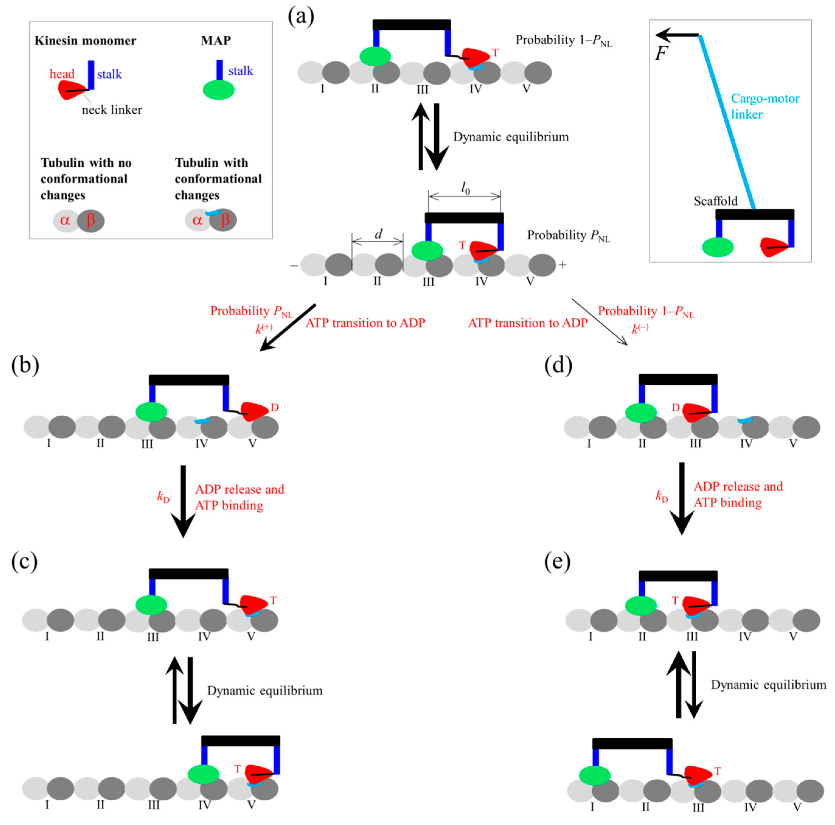
Figure 1. Schematic of the engineered motor composed of a kinesin monomer and a diffusing MAP, where the stalk connecting the monomer with the scaffold and the stalk connecting MAP with the scaffold are short. A backward load F acts on the cargo attached to the engineered motor via a long linker, as in the case of the cargo attached to the WT dimeric kinesin motor via a long stalk. For clarity, the large-sized cargo is not shown, and the load F on the cargo is equivalent to the load F on the cargo-motor linker (right inset). During the stepping of the motor, the two short stalks cannot be stretchable, but the long cargo–motor linker can be stretchable. (ae) Chemomechanical pathway at saturating ATP concentrations (see text for detailed descriptions). Due to the large diffusion constant, MAP can be in either tubulin II or tubulin III in (a), in either tubulin III or tubulin IV in (c), and in either tubulin I or tubulin II in (e). The thickness of an arrow represents the relative magnitude of the probability of the transition between the two states connected by the arrow under no load. At saturating ATP concentrations, the rate of ATP binding is considered to be infinitely large. After ATP transition to ADP, by overcoming the very small affinity Ew1, the monomer can diffuse rapidly (with a timescale of 1 μs) from the local tubulin to the adjacent tubulin, where the monomer has a higher affinity Ew2.
Biophysica 05 00026 g001
Figure 2. Theoretical results for the dynamics of the engineered motor (solid lines). For comparison, the theoretical and prior experimental results for the dynamics of kinesin dimer are also shown (dashed lines for the theoretical results and red dots for the experimental results of Carter and Cross [16]). (a) Velocity versus load. (b) Stepping ratio versus load. (c) Mean number of ATPs consumed per mechanical (either forward or backward) step versus load.
Biophysica 05 00026 g002
Table 1. Parameter values for kinesin.
ParameterDescriptionValue
k(+)Rate of ATP transition to ADP in monomer with forward NL108 s–1
k(–)Rate of ATP transition to ADP in monomer without forward NL3 s–1
E0Energy change of NL docking and large conformational change of ATP monomer3 kBT
Disclaimer/Publisher’s Note: The statements, opinions and data contained in all publications are solely those of the individual author(s) and contributor(s) and not of MDPI and/or the editor(s). MDPI and/or the editor(s) disclaim responsibility for any injury to people or property resulting from any ideas, methods, instructions or products referred to in the content.