Article

Sustained Release of Antibacterial Therapeutic Elements from Functionalized Mesoporous Silica-Coated Silver Nanoparticles for Bone Tissue Engineering

1
School of Queen Mary University of London Engineering, Northwestern Polytechnical University, Xi’an 710072, China
2
State Key Laboratory of Solidification Processing, School of Materials Science and Engineering, Northwestern Polytechnical University, Xi’an 710072, China
3
School of Life Sciences, Northwestern Polytechnical University, Xi’an 710072, China
*
Authors to whom correspondence should be addressed.
Chemistry 2025, 7(5), 146; https://doi.org/10.3390/chemistry7050146
Submission received: 31 July 2025 / Revised: 28 August 2025 / Accepted: 3 September 2025 / Published: 10 September 2025
(This article belongs to the Section Chemistry at the Nanoscale)

Abstract

Applying therapeutic elements to prevent injury from potential infections is a promising avenue in the development of novel bone substitutes; however, achieving controllable delivery of therapeutic ions is crucial to realizing their expected functions. In this study, a Ag nanoparticle core wrapped in an MSN shell was successfully synthesized using a one-pot sol–gel process. Subsequently, the produced Ag@MSN was functionalized with amino and carboxylic groups. The experimental results indicated that these core–shell-structured Ag@MSN spheres had a uniform size of ~60 nm and a specific area of 904.6 m2/g. Their release profiles, influenced by different surface charges, were investigated, with the aim of achieving sustainable release of Ag ions. The concentration-dependent biological effects of Ag@MSNs, including their anti-infection properties and biocompatibility, were comprehensively characterized in vitro, considering their potential for use as bioactive bone substitutes. Functionalized mesoporous silica nanoparticles significantly enhanced the sustained release profile of silver ions, achieving a cumulative release efficiency greater than 50% within 24 h. These nanoparticles also demonstrated exceptional antibacterial efficacy, with an inhibition rate surpassing 98% at a concentration of 30 μg/mL, while concurrently maintaining cell viability above 88%, indicating high biocompatibility. We achieved our goal of effectively decreasing the burst release of Ag to satisfy the intrinsic need for long-term resistance to bacteria in bone substitutes and stimulate osteoblast proliferation.

1. Introduction

The use of artificial bone substitutes for fixing large bone defects or replacing bone is known to be effective for restoring the bones’ mechanical and biological functions. In spite of the notable clinical success of artificial bone substitutes, their long-term availability in the body is restricted, mainly due to weak bone–material contact and risks associated with infection [1,2]. To prevent these negative effects, biomolecules (antibiotics, growth factors, proteins, peptides, and genes) are commonly incorporated into such substitutes and released locally in a controlled manner to regulate cellular behavior and, ultimately, tissue response [3,4]. In addition, different functional biomolecules have been combined in an attempt to realize the multifunctional stimulation of bone regeneration [5,6,7]. However, methods need to be developed to minimize the decomposition of biomolecules during processing, as well as side-effects caused by excessive dosages and leakage.
Recently, researchers used trace elements, such as Ag, Cu and Sr, as therapeutic agents for bone tissue engineering. Growing evidence in the literature indicates that, at the proper dosage, these elements play an important role in antibacterial activity, osteogenesis, and angiogenesis [8,9,10,11,12]. For example, silver has been reported to exhibit broad-spectrum antibacterial activity by increasing membrane permeability and producing reactive oxygen species (ROS) that damage bacterial walls [13,14,15]. Other researchers have found that silicon (Si) participates actively in bone formation and resorption by exerting direct and indirect effects on bone-related cells and bone minerals [16,17]; for example, it has been well established that Si can stimulate osteoblast activity while simultaneously inhibiting osteoclast activity [18,19]. Compared to organic biomolecules used to promote bone regeneration, incorporating trace elements in bone substitute materials possesses many advantages, such as a low cost, high stability during loading and release process and negligible systemic effects. The development of such biologically active materials will pave the way for fabricating a variety of multifunctional biodevices. However, it is still difficult to achieve controllable release of therapeutic ions integrated into transplants to ensure bone regeneration and long-term resistance to bacteria [20,21,22].
The encapsulation of therapeutic agents in nanoparticulate carriers is an effective approach to achieving high drug loading and sustained/controlled drug release [23,24]. Mesoporous silica nanoparticles (MSNs) are a type of inorganic nanocarrier that have been used widely to deliver various bone-forming agents due to their large surface and pore volume, excellent biocompatibility, highly reactive surface, and adjustable pore size [25,26,27,28]. In addition, silicon ions released during MSN degradation have been reported to promote the angiogenesis of endothelial cells [29], which could be beneficial for the further ingrowth of osteoblasts and new bone formation. Therefore, MSNs are considered an ideal platform for delivering therapeutic elements for bone regeneration and other types of medical engineering. Silver nanoparticles encapsulated successfully in MSNs in the form of a core–shell structure, namely Ag@MSNs, were fabricated for antibacterial purposes [30]. An MSN shell will not only prevent the undesirable aggregation of silver nanoparticles but also enable control of the silver ion release rate.
Achieving the controlled release of therapeutic agents from mesoporous silica nanoparticles continues to pose significant challenges, despite receiving considerable research attention. While many existing strategies rely on physically modulating the pore size to tune the release kinetics, this study introduces a novel approach centered on chemical functionalization with tailored organic groups. This method enables the precise regulation of release profiles and markedly enhances both mesoporous silica nanoparticles’ controllability and their applicability as versatile drug delivery platforms. Herein, to address the needs for anti-infection properties in medical engineering, we describe how we grafted a Ag-containing MSN with amidogen and a carboxyl functional group (Ag@MSN-NH2 and Ag@MSN-COOH) to realize the gradual and controllable release of therapeutic ions, as shown schematically in Figure 1. The selection of amino and carboxyl groups was based on their capacity to modulate the surface potential for controlled ion release, as well as their natural abundance in biological systems, which minimizes the risk of immune rejection. Core/shell-structured Ag@MSNs were firstly synthesized using a one-pot sol–gel process. The obtained Ag@MSNs were subjected to functional group modification. By studying the ion release, antibacterial and cell compatibility-related properties of the modified materials, the influence of the functional groups on the inner and outer surfaces of the mesoporous silica nanoparticles (considered the pathways for ion release) on the kinetics of ion release was determined. Meanwhile, we found that Ag@MSNs-NH2 and Ag@MSNs-COOH have excellent antibacterial performance and cell compatibility compared with Ag@MSNs. The findings of this study on structural design and functional group modification provide a theoretical foundation for the design and preparation of a functionalized ion/drug delivery platform.

2. Experimental Section

2.1. Materials

Cetyltrimethylammonium bromide (CTAB, AR), sodium hydroxide (NaOH, AR), a formaldehyde solution (37%, AR), tetraethyl orthosilicate (TEOS, AR), maleic anhydride (99.5%, AR), ammonium nitrate (NH4NO3, AR), N, N-Dimethylformamide (DMF, AR), silver nitrate (AgNO3, 99.95%) and ethanol (99.9%, AR) was purchased from Shanghai Chemical Reagent Co., Ltd., China (Shanghai, China). Luria–Bertani (LB) broth was purchased from Thermo Fisher Oxoid Ltd., UK. (3-aminopropyl) triethoxysilane (APTES, 99%) was purchased from Sigma Aldrich (St. Louis, IL, USA).

2.2. Synthesis of Ag@MSNs

Core–shell-structured Ag@MSNs were synthesized using a sol–gel method. Briefly, an NaOH solution (2 M, 3.5 mL) was added into water (480 mL) containing CTAB (1 g) and thoroughly mixed at 80 °C for 30 min to ensure the complete dissolution of the CTAB. Afterwards, a mixture of formaldehyde (1 M, 3 mL) and a AgNO3 solution (0.1 M, 8 mL) was added dropwise with magnetic stirring. After stirring the mixture for 5 min, TEOS (5 mL) was added slowly, and it was stirred for another 2 h. The reaction temperature was kept at 80 °C for all the steps. The resultant products were collected through centrifugation and washed twice with deionized water and ethanol. The nanoparticles were then transferred to an ethanol solution (100 mL) containing NH4NO3 (0.6 g) and kept in a reflux unit at 80 °C for 24 h. Finally, the sample was washed using the same procedure and dried under vacuum to yield as-prepared Ag@MSNs.

2.3. Functionalization of Ag@MSNs

2.3.1. Synthesis of Amino-Functionalized Ag@MSNs (Ag@MSNs-NH2)

Ag@MSNs (100 mg) were dispersed in isopropyl alcohol (100 mL), and 0.2 mL of APTES was added to the solution. The mixture was heated to 85 °C and stirred for 24 h. Then, the precipitates were collected through centrifugation (10,000 rpm, 5 min) and rinsed with deionized water and ethanol. Finally, the sample was dried under vacuum to yield Ag@MSNs-NH2.

2.3.2. Synthesis of Carboxylic Group-Functionalized Ag@MSNs (Ag@MSNs-COOH)

Ag@MSNs-NH2 (100 mg) were dispersed in N, N-Dimethylformamide (100 mL), and excess maleic anhydride was added to the solution. The mixture was then kept in a reflux unit at 80 °C for 24 h. The solids were collected through centrifugation (10,000 rpm, 5 min) and washed with deionized water and ethanol. Finally, the sample was dried under vacuum to yield Ag@MSNs-COOH.

2.4. Material Characterization

The morphology and mesostructure of the MSNs were observed using scanning (Auriga, Zeiss, Jena, Germany) and transmission electron microscopy (Tecnai G2 F20, FEI, Hillsboro, USA). The phase composition of the silver-containing MSNs was measured using X-ray diffraction (XRD), while high-resolution transmission electron microscopy (HR-TEM) was employed to confirm the presence of silver nanoparticles within the Ag@MSN composite. The Ag@MSNs’ pore structure at different functionalization stages was analyzed using small-angle X-ray diffraction (SAXRD, SmartLab, Rigaku, Japan) with CuKa radiation (40 kV and 40 mA) at a scanning rate of 0.4 °/min and a step width of 0.01 ° over a range of 1–8 °. N2 adsorption–desorption experiments were performed to obtain the surface area and mesopore size distribution (ASAP2020 HD88, Micromeritics, Norcross, USA). The functionalized Ag@MSNs were mixed thoroughly with KBr at a concentration of 2 wt% (w/w) and compressed into pellets for Fourier transform infrared (FTIR, Tensor27, Bruker, Karlsruhe, Germany) spectroscopy analysis. The FTIR spectra were measured in the wavenumber range of 400–4000 cm−1. Meanwhile, the surface charge of the functionalized Ag@MSNs in an ethanol solvent was analyzed using Zetasizer nano ZS equipment (Malvern Instruments, Malvern, UK).

2.5. Assessment of Ag Loading and Release Profiles

The samples used in the ion release tests included Ag@MSNs, Ag@MSNs-NH2 and Ag@MSNs-COOH. The Ag loading percentages were measured before the release test. A total of 1.5 mL of pure HF was mixed with 10 mg of the sample in a centrifuge tube and subjected to stirring for 5 min and then sonication for 30 min until the MSN shell was completely dissolved. The suspension was centrifuged for 5 min at 10,000 rpm, and the precipitate was dried under vacuum for 24 h and recorded as the amount of Ag in the core. For the ion release study, 10 mg of ion-loaded nanoparticles was dispersed in 0.5 mL of deionized water and then transferred into a dialysis bag with a molecular weight cutoff of 8000–14,000 Da. The packed powder was then soaked in deionized water (3.5 mL) and kept in an incubator at 37 °C at a shaking speed of 90 rpm. After the predetermined incubation period, 2 mL of the solution was extracted to test the concentration of the Ag released using an atomic absorption spectrometer. To measure the ion release at a constant volume, 2 mL of fresh media was added after each sampling step. Three replicates were performed, and the results were expressed as the cumulative amount of Ag released as a function of the incubation time.

2.6. Assessment of Antibacterial Properties

E. coli (ATCC: 25922) and S. aureus (ATCC: 27923) were chosen as representative Gram-negative and -positive bacteria for this study. The bacteria were cultured in an LB medium for 18 h at 37 °C, with a rotation speed of 90 rpm and humidity of 80%. Once the OD600 of the culture medium reached 0.6, indicating that the bacteria were in the exponential growth phase, the bacterial suspension was serially diluted to achieve a final concentration of 5 × 10⁵ colony-forming units per ml (CFU/mL) using LB medium. Subsequently, 1 mL of this diluted solution was transferred to a 24-well plate containing varying concentrations of sterilized Ag@MSNs, Ag@MSNs-NH2 and Ag@MSNs-COOH. After 24 h of incubation, the lowest Ag@MSN concentration with no visible growth of the tested organisms was recorded as the minimum inhibitory concentration (MIC). To ensure that the results were reproducible, the MIC test was performed in triplicate. After 8 h of co-culture, 100 μL of the bacterial suspension was collected for colony counting. Specifically, serial dilutions of the bacterial suspension were spread evenly onto solid agar plates. Following incubation, the number of colonies on each plate was enumerated. The original bacterial concentration was then calculated using the dilution factor, and the antibacterial rate was subsequently determined by counting the CFUs on the LB agar plates and using the following equation:
Antibacterial   Rate ( % ) = C F U C o n t r o l C F U M a t e r i a l C F U M a t e r i a l

2.7. In Vitro Study

Cytotoxicity

To assess the cytocompatibility of the material at its maximum tolerable concentration, concentration gradients of 0, 10, 30, 60, 100 and 200 μg/mL were established. For the in vitro biological assessments, following routine protocols, MC3T3-E1 cells were incubated at 37 °C in a humidified 5% CO2 atmosphere using α-MEM supplemented with 10% fetal bovine serum and 0.1% penicillin–streptomycin. The MC3T3-E1 cells were seeded into 96-well plates at a density of 5 × 10³ cells per well. The control group comprised cell-seeded plates not subjected to the drug treatment. After culture for 24 h, sterilized Ag@MSNs, Ag@MSNs-NH2 and Ag@MSNs-COOH with concentrations ranging from 10 to 200 μg/mL were added to the plates, and they were cultured for another 5 d. At the end of the incubation period, CCK testing was conducted. A total of 100 μL of the final mixture was centrifuged (10,000 rpm for 5 min), and the supernatant was subjected to optical density measurement (λ = 450 nm) using a multifunction microplate reader (Synergy HT, BioTek, Winooski, VT, USA).

2.8. Statistical Analysis

Each experiment was at least carried out in triplicate, and the data were presented as the mean ± standard deviation (SD). Statistical analyses between the analyzed groups were carried out using SPSS 16.0 software via one-way ANOVA. Statistical significance was defined as a p < 0.05, 0.01 or 0.001.

3. Results and Discussion

3.1. Characterization of MSNs and Functionalized Ag@MSNs

Synthesis of MSNs has been widely investigated and involves the dynamic electrolysis–polymerization of TEOS around the template, followed by nucleation and growth of spherical nanoparticles [31]. By employing AgNO3 as the silver precursor and HCHO as the reducing agent, the nucleation of the pre-formed silver nanoparticles allowed for further growth of an MSN shell, leading to the formation of core–shell-structured Ag@MSNs [32]. The morphology and mesoporous structure of the MSNs and Ag@MSNs were observed using SEM and TEM, respectively. Figure 2a shows that the as-synthesized MSNs were well-dispersed, with a spherical morphology and uniform diameter of approximately 100 nm. The corresponding high-magnification image in Figure 2a indicates that the MSNs were monodispersed and contained ordered mesopore channels and holes. After incorporating Ag nanoparticles, the obtained nanoparticles exhibited a uniform but substantially reduced particle diameter (Figure 2b). From the high-magnification TEM image in Figure 2b, we can see that monodispersed and core–shell-structured nanoparticles were obtained, showing a core diameter of 13 ± 3 nm. In addition, wormhole-like mesopores arranged radially to the surface were clearly visible. The interplanar spacing of 2.33 Å, measured in the HRTEM analysis, corresponded closely to the (111) lattice planes of metallic silver (d = 2.36 Å), indicating that the nanoparticles consisted predominantly of Ag. According to the further EDX and XRD analyses of the core–shell nanoparticles, as shown in Figure 3e and Figure S1a, the elements Si (45.6 wt.%), O (45.6 wt.%) and Ag (8.8 wt.%) were detected and, more importantly, appearance of peaks corresponding to silver crystals was also observed. Therefore, it can be concluded that Ag-containing MSNs consisting of Ag nanoparticles wrapped in an MSN shell, namely Ag@MSNs, were successfully produced using a modified ‘one-pot’ process. Such a core–shell structure will not only improve the Ag nanoparticles’ colloidal stability but also effectively minimize the toxic effects caused by their aggregation. Moreover, the irregularly arranged mesopore channels in the MSN shell should help to control the release of Ag ions. According to a statistical calculation for 100 different particles shown in Figure S2, the size distributions of the Ag@MSNs, Ag@MSNs-NH2 and Ag@MSNs-COOH were 67 ± 12, 50 ± 9 and 50 ± 10 nm, respectively (Table S1), indicating that the surface functionalization did not significantly influence the particle size and morphology.
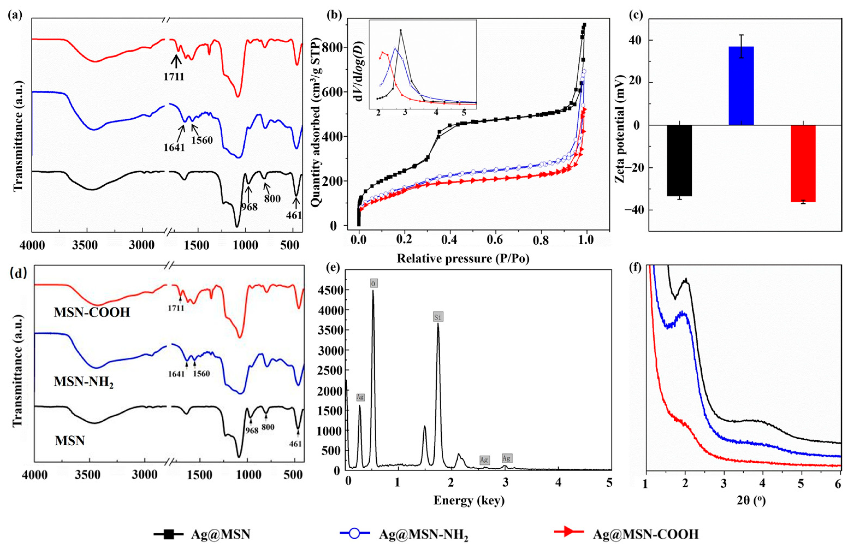
After each functionalization stage, the Ag@MSN powders were further characterized through FTIR analysis, as shown in Figure 3a. The FTIR spectrum of the Ag@MSNs presented absorption peaks typical of the Si−OH bond at 968 cm−1, the Si−O bond at 800 cm−1 and the Si−O−Si bond at 461 cm−1 [32]. The Si–OH band at 960 cm−1 became significantly weaker after functionalization using APTES to graft NH2 groups onto the Ag@MSNs. In addition, vibration of amino groups at 1641 cm−1 and 1560 cm−1 and the C=O bonds in the COOH groups at 1711 cm−1 appeared sequentially after each functionalization step, indicating the successful grafting of the NH2 and COOH groups onto the Ag@MSNs in this study. The presence of the NH2 and COOH groups was also proved using zeta potential measurements taken after each functionalization step. The FTIR spectra of the Ag@MSNs displayed in Figure 3d show no new characteristic peaks nor significant shifts compared to those in the spectra of pure MSNs, indicating the absence of chemical bonds or notable interactions between the silver and the silica host. As shown in Figure 3b, the zeta potential of the as-synthesized Ag@MSNs was −33.4 ± 1.6 mV, which shifted to + 37.0 ± 5.4 mV and then to −36.2 ± 0.8 mV after the amination and carboxylation treatments, respectively.
The mesoporous structure of the Ag@MSNs after each functionalization step was evaluated using small-angle XRD, as shown in Figure 3c. The relative intensity of the diffraction peaks at 1.9° and 3.8° gradually decreased, indicating that the ordered mesoporous structure of the Ag@MSNs was disrupted after the functionalization treatments. In other words, it was confirmed that both the NH2 and COOH groups were successfully grafted onto not only the outer surface of the Ag@MSNs but also the inner surface of the mesopores. The N2 adsorption–desorption isotherms in Figure 3f exhibit significant adsorption behavior at a P/P0 of around 0.35 without a hysteresis loop and are known as type IV isotherms according to the IUPAC classification [28]. Based on these results, the pore size distribution, as well as the specific surface and pore volumes, of the Ag@MSNs after each functionalization step was calculated using the Brunauer–Emmett–Teller (BET) and Barrett–Joyner–Halenda (BJH) models, as shown in Figure 3d and Table 1, respectively. The average pore sizes of the Ag@MSNs, Ag@MSNs-NH2 and Ag@MSNs-COOH were 2.65, 2.44 and 1.98 nm, respectively. In addition, their specific surface areas and pore volumes also decreased significantly after surface functionalization, although all the samples still possessed a high total specific area ranging from 904.6 m2/g to 604.4 m2/g. The change in the mesoporous structure following surface functionalization indicated that a substantial number of functional groups were grafted onto the inner surface of the MSNs, resulting in a narrowed pore diameter and reduced pore volume and surface area. The carboxylation treatment did not change the specific surface area, but the pore volume was reduced by around 22%, indicating that the grafted COOH groups blocked part of the mesopore channel.

3.2. Ag Ion Release Study

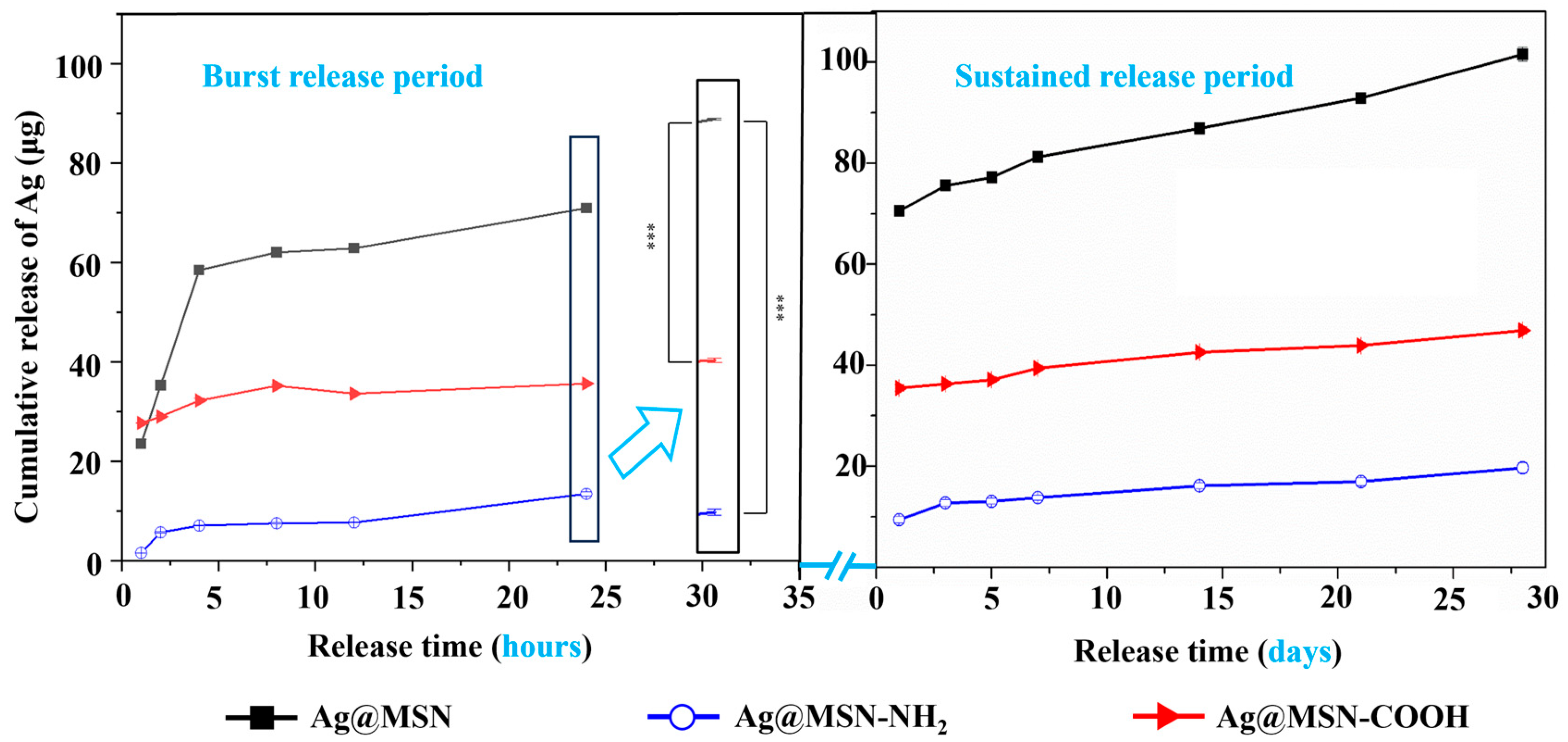
Release of therapeutic ions in a controlled and sustained manner is a prerequisite for them to realize their biological functions. Table S2 lists the loading percentages of Ag in MSNs functionalized with NH2 and COOH groups. The highest loading percentage of Ag was found in the bare Ag@MSNs. The loading percentage of Ag decreased from 6.3 wt% in the bare Ag@MSNs to 5.0 wt% after amination and then slightly decreased to 4.7 wt% after carboxylation, which could be attributed to the leakage of Ag ions during the functionalization treatments. The release profiles of Ag ions from the Ag@MSNs, Ag@MSNs-NH2 and Ag@MSNs-COOH were compared and are plotted in Figure 4. During the burst release period, the functionalized Ag@MSN samples exhibited a significantly minimized silver release rate compared to the bare Ag@MSNs, and no obvious burst release of Ag ions was observed in the Ag@MSN-NH2 sample. During the sustained release period, all the samples exhibited sustained release profiles of up to 672 h (28 days). In addition, the amount released was also controlled through grafting of -NH2/-COOH groups, reducing it by 81% and 50% in the Ag@MSN-NH2 and Ag@MSN-COOH samples, respectively, at 24 h compared to that in the bare Ag@MSNs. Based on the experimental evidence and supporting literature, the observed reduction in both the release rate and total number of Ag⁺ ions released from the functionalized Ag@MSNs can be attributed to three main factors. First, the functionalization process disrupted the ordered mesoporous structure and decreased the pore size, thereby partially obstructing the diffusion of Ag⁺ ions through the channels. Second, chelation and electrostatic interactions involving the introduced -NH2 and -COOH functional groups further retarded the release of ions from the silica carrier. Finally, the formation of coordination bonds between the silver ions and these functional groups may also have contributed to the sustained release behavior [33]. More interestingly, the Ag@MSN-NH2 sample, which possessed a similar specific area and mesoporous structure to the Ag@MSN-COOH sample, exhibited a much lower Ag release rate and cumulative amount released. Therefore, we can conclude that the grafting of NH2 groups is more effective than that of COOH groups in retarding the Ag ion release, which is probably due to the greater strength of the chelation bond between the -NH2 groups and Ag ions than the electrostatic attraction between the -COOH groups and Ag ions.
Based on the Ag release study discussed above, it can be concluded that both the mesoporous structure and surface charge of MSNs play an important role in controlling the release behavior of therapeutic elements. Silver is a well-known antibacterial agent with effective and broad-spectrum antibacterial capability. Compared to other antibacterial agents such as antibiotics, antimicrobial peptides or antibacterial polymers, silver is less expensive and more stable during processing and application. Although it is more advantageous to use therapeutic ions than biological molecules, their biological functions are critically concentration-sensitive, with their concentration needing to be high enough to trigger stimulation but not too high to avoid undesirable side-effects. In this case, controlling the delivery of therapeutic ions from the carrier is of great importance for them to realize their expected functions. Our findings provide simple but effective methods to control the release of silver, which should help in visualizing their potential applications in bone tissue engineering.

3.3. Antibacterial Study

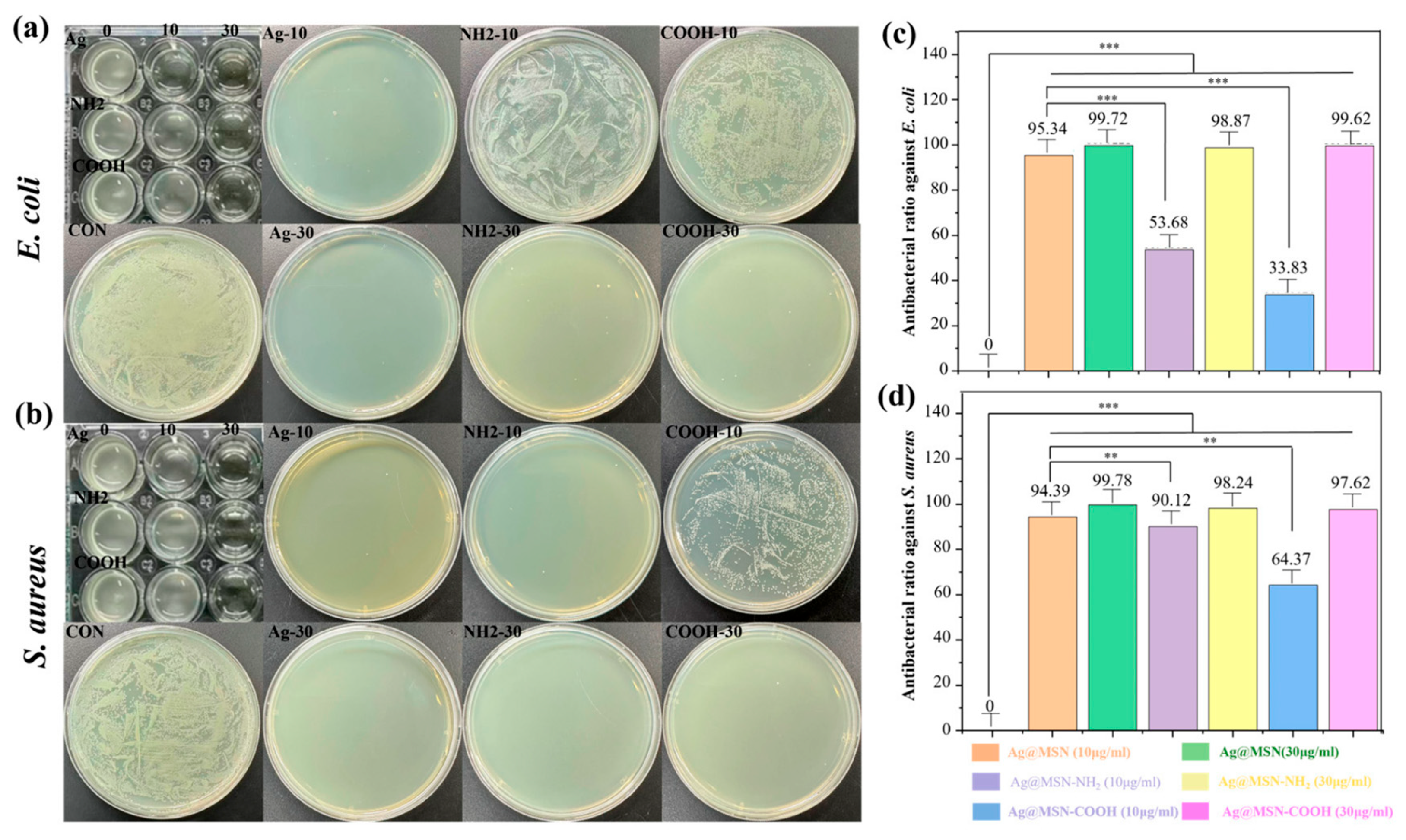
E. coli and S. aureus were selected as model Gram-negative and -positive bacteria to evaluate the in vitro antibacterial activity of the silver-containing samples, including the Ag@MSNs. Moreover, the antibacterial activity of the Ag@MSN-NH2 and Ag@MSN-COOH samples was evaluated to identify the possible effect of functional groups on the Ag@MSNs’ antibacterial properties. The MICs against E. coli and S. aureus that resulted in a transparent bacterial medium were measured as shown in Figure 5a,b. The MIC of the Ag@MSNs against E. coli was 10 μg/mL and increased to 30 μg/mL after amino and carboxylic group functionalization. According to our measurements, the Ag@MSN, Ag@MSN-NH2 and Ag@MSN-COOH samples possessed an MIC of 30 μg/mL against S. aureus. At 30 μg/mL, the Ag@MSN-NH2 and Ag@MSN-COOH samples showed outstanding antibacterial activity. The antibacterial rates were 98.87% and 99.62% against E. coli and 98.24% and 97.62% against S. aureus, respectively (Figure 5c,d). The associated liquid cultures remained clear, and colony counting assays showed almost no bacterial growth on the plates treated with either sample, further demonstrating their exceptional antibacterial activity.
Although the antibacterial mechanism of silver ions is not fully understood due to the complex physicochemical factors involved, it is widely accepted that silver ions can upregulate the local ROS level to kill bacteria indirectly [13,14,15] or damage their cell wall and disrupt the functions of enzymes and DNA through direct contact [34]. As evidenced by the ion release kinetics (Figure 4), surface functionalization effectively attenuated the silver ion release rate (compare the release rate at 24 h for the Ag@MSNs with that for the Ag@MSNs-NH2 and Ag@MSNs-COOH; p < 0.001), leading to a reduced concentration of Ag⁺ in the bacterial culture medium. This result is consistent with the trend observed at a concentration of 10 μg/mL, where the antibacterial rate of the functional group-modified samples (53.68% and 33.83%) remained substantially lower than that of the unmodified group (95.34%). Therefore, we can conclude, based on this study, that the antibacterial effect of the synthesized Ag@MSNs on E. coli is mainly dependent on the concentration of silver ions released. In addition, surface functionalization treatments enhanced the polarity and surface hydration of the sample via electrostatic interactions and hydrogen bonding [35], thus creating a barrier layer to inhibit direct contact between the Ag@MSNs and the cell walls of E. coli and S. aureus. According to the DLS measurements shown in Figure S3, the particle sizes of the Ag@MSNs, Ag@MSNs-NH2 and Ag@MSNs-COOH in water were 151.3, 770.7 and 531.2 nm, respectively, indicating the presence of a thick hydration layer after amino and carboxylic group functionalization. The decreased bactericidal efficacy of the functionalized Ag@MSNs is partly explained by the obstruction of silver ion release caused by this hydration layer.

3.4. Cell Compatibility Study

MC3T3-E1 cells were used as a model to evaluate the cytotoxicity of the Ag@MSNs, Ag@MSNs-NH2 and Ag@MSNs-COOH. Numerous studies have shown that oxidative stress and ROS production are the main processes causing the cytotoxic effects of silver ions and that the level of cytotoxicity is directly correlated with the dose of silver nanoparticles [14]. This study employed materials with nanoparticle concentrations of 10 ug/mL, 30 ug/mL, 60 ug/mL, 100 ug/mL and 200 ug/mL. The viability of cells cultured with different concentrations of the samples for 1, 3 or 5 days are shown in Figure 6. After culture for 1 day, increased particle concentrations in all the samples led to decreased cell viability. The Ag@MSNs-COOH possessed a higher overall cell viability than the Ag@MSNs and Ag@MSNs-NH2, which was probably due to the cells’ negative surface charge [36]. This may have facilitated the uptake of positively charged nanoparticles while minimizing the uptake of negatively charged ones. After culture for 3 days, the viability of the cells further decreased, probably due to the cytotoxic effects of continuous silver ion release. However, the Ag@MSN-COOH samples, which possessed slower silver ion release behaviors, exhibited comparable or even higher cell viability at concentrations of less than 30 μg/mL. After 5 days of co-culture, the cell viability of the Ag@MSNs further decreased to 76.28%, while that of the Ag@MSNs-NH2 and Ag@MSNs-COOH remained as high as 87.11% and 90.76%, respectively. This was attributed to the effective control of silver ion release achieved through functional group modification. Combined with the antibacterial results (Figure 5), we can conclude that 30 μg/mL of functional group-modified Ag@MSNs (-NH2 or -COOH) achieves a balance between excellent antibacterial performance and good biocompatibility.
Previous research has recognized mesoporous silica to be a comparatively effective platform for controlled drug release. However, uncontrolled ion leakage frequently leads to excessive release rates, resulting in substantial cytotoxicity. In this work, we regulated silver ion release through surface functionalization with amino and carboxyl groups, which established an optimal balance between the antibacterial performance and cytocompatibility [23].

4. Conclusions

In this study, core–shell-structured Ag@MSNs with a uniform size (60 nm) and adjustable surface charge (-NH2 or -COOH) were successfully prepared for sustained release of silver ions without disrupting biological functions. Nano-silver significantly inhibited E. coli, and both Ag@MSNs-NH2 and Ag@MSNs-COOH had good biocompatibility. Surface functionalization of the Ag@MSNs with -NH2 and -COOH significantly decreased Ag ion release during both the burst and sustained release periods by reducing the surface area and pore size and forming chelation bonds and electrostatic attraction between the silver ions and functionalization groups. In vitro antibacterial assays demonstrated that the antimicrobial efficacy of the Ag@MSN nanoparticles was concentration-dependent, and surface functionalization was found to attenuate their antibacterial potency. The cell viability decreased in a concentration-dependent manner, according to cytocompatibility experiments. Notably, both amino group-functionalized Ag@MSNs-NH2 and carboxyl group-functionalized Ag@MSNs-COOH attained an ideal balance between their antibacterial activity and biocompatibility at a concentration of 30 μg/mL. This study systematically elucidated the regulatory effects of surface functionalization on both the antibacterial performance and biocompatibility of Ag@MSN materials. More importantly, we developed a tunable nanoplatform that allows functional cargoes to be selectively loaded into either negatively charged, carboxyl group-modified or positively charged, amino group-modified mesoporous channels in Ag@MSNs, depending on the electrostatic properties of the drug molecules. This approach gives these materials additional functionalities and provides new opportunities to create multipurpose antibacterial materials for bone repair.

Supplementary Materials

The following supporting information can be downloaded at: https://www.mdpi.com/article/10.3390/chemistry7050146/s1, Figure S1: Characterization of Ag@MSN. (a) Wide-angle XRD patterns of MSN and Ag@MSN; Figure S2: SEM image of (a) Ag@MSN (b) Ag@MSN-NH2 (c) Ag@MSN-COOH; Figure S3: The DLS measurements of Ag@MSN, Ag@MSN-NH2; Table S1: Particle size distribution; Table S2: The loading percentages of Ag and Sr in bare and functionalized Ag@MSN carriers indicated a slightly decreased loading percentage after functionalization; Table S3: Average particle size.

Author Contributions

Conceptualization, Q.C. and Y.Z. (Yuhan Zhang); methodology, L.H.; software, N.L.; validation, Q.C., Y.Z. (Yuhan Zhang), L.H. and Y.Z. (Yanni Zhang); formal analysis, L.H.; investigation, L.H. and Y.Z. (Yuhan Zhang); resources, Q.C. and Y.Z. (Yanni Zhang); data curation, L.H. and N.L.; writing—original draft preparation, L.H.; writing—review and editing, Y.Z. (Yuhan Zhang) and L.H.; visualization, J.J.; supervision, Q.C.; project administration, Q.C. and Y.Z. (Yuhan Zhang); funding acquisition, Q.C. All authors have read and agreed to the published version of the manuscript.

Funding

The authors received support from the National Natural Science Foundation of China (31800802), Science Foundation for Distinguished Young Scholars of Shannxi Province (2023-JC-JQ-35), and Northwestern Polytechnical University College Student Innovation and Entrepreneurship Training Program (202310699008).

Data Availability Statement

The original contributions presented in this study are included in the article/Supplementary Materials. Further inquiries can be directed to the corresponding authors.

Conflicts of Interest

The authors declare no conflict of interest.

References

  1. Feng, P.; Zhao, R.; Tang, W.; Yang, F.; Tian, H.; Peng, S.; Pan, H.; Shuai, C. Structural and Functional Adaptive Artificial Bone: Materials, Fabrications, and Properties. Adv. Funct. Mater. 2023, 33, 202214726. [Google Scholar] [CrossRef]
  2. Zhao, D.; Zhu, T.; Li, J.; Cui, L.; Zhang, Z.; Zhuang, X.; Ding, J. Poly(lactic-co-glycolic acid)-based composite bone-substitute materials. Bioact. Mater. 2021, 6, 346–360. [Google Scholar] [CrossRef] [PubMed]
  3. Zhang, Y.; Wu, C.; Zhao, Y.; Yu, J.; Han, L.; Jiang, X.; Yang, J.; Zhang, S.; Han, J.; Chen, Q. NIR responsive electrospun composite membranes for on-demand delivery of Ag ions to enhance treatment of bacteria-infected wounds. Chem. Eng. J. 2022, 514, 163428. [Google Scholar] [CrossRef]
  4. Wang, Z.; Liu, X.; Duan, Y.; Huang, Y. Infection microenvironment-related antibacterial nanotherapeutic strategies. Biomaterials 2022, 280, 121249. [Google Scholar] [CrossRef]
  5. Chatterjee, S.; Lou, X.Y.; Liang, F.; Yang, Y.-W. Surface-functionalized gold and silver nanoparticles for colorimetric and fluorescent sensing of metal ions and biomolecules. Coord. Chem. Rev. 2022, 459, 214461. [Google Scholar] [CrossRef]
  6. Liu, J.T.; Huang, J.; Zhang, L.; Lei, J. Multifunctional metal-organic framework heterostructures for enhanced cancer therapy. Chem. Soc. Rev. 2021, 50, 1188–1218. [Google Scholar] [CrossRef]
  7. Xie, Z.; Dai, C.; Gao, X.; Su, X.; Luo, Y.; Guo, X.; Li, Y.; Lin, Z.; Huang, S.; Jiang, W. Bioinspired multifunctional peptides integrated with anti-infective, immunomodulatory and osteogenic activities for infectious bone regeneration. Chem. Eng. J. 2025, 503, 158256. [Google Scholar] [CrossRef]
  8. Yuan, Z.; Wan, Z.; Gao, C.; Wang, Y.; Huang, J.; Cai, Q. Controlled magnesium ion delivery system for in situ bone tissue engineering. J. Control. Release 2022, 350, 360–376. [Google Scholar] [CrossRef]
  9. Liao, J.; Wang, L.; Ding, S.; Tian, G.; Hu, H.; Wang, Q.; Yin, W. Molybdenum-based antimicrobial nanomaterials: A comprehensive review. Nano Today 2023, 50, 101875. [Google Scholar] [CrossRef]
  10. Rajendran, A.K.; Anthraper, M.S.J.; Hwang, N.S.; Rangasamy, J. Osteogenesis and angiogenesis promoting bioactive ceramics. Mater. Sci. Eng. R Rep. 2024, 159, 100801. [Google Scholar] [CrossRef]
  11. Mu, Y.; Du, Z.; Gao, W.; Xiao, L.; Crawford, R.; Xiao, Y. The effect of a bionic bone ionic environment on osteogenesis, osteoimmunology, and in situ bone tissue engineering. Biomaterials 2024, 304, 122410. [Google Scholar] [CrossRef] [PubMed]
  12. Gai, K.; Zhang, T.; Xu, Z.; Li, G.; He, Z.; Meng, S.; Shi, Y.; Zhang, Y.; Zhu, Z.; Pei, X.; et al. Biomimetic management of bone healing stages: MOFs induce tunable degradability and enhanced angiogenesis-osteogenesis coupling. Chem. Eng. J. 2024, 493, 152296. [Google Scholar] [CrossRef]
  13. Jian, Y.; Chen, X.; Ahmed, T.; Shang, Q.; Zhang, S.; Ma, Z.; Yin, Y. Toxicity and action mechanisms of silver nanoparticles against the mycotoxin-producing fungus Fusarium graminearum. J. Adv. Res. 2022, 38, 1–12. [Google Scholar] [CrossRef]
  14. Rengasamy, G.; Mahalingam, S. Multifunctional composite film of chitosan/reduced graphene oxide infused silver nanoparticle for antibacterial and cytotoxic effects on HeLa cells. Int. J. Biol. Macromol. 2025, 321, 146356. [Google Scholar] [CrossRef]
  15. Wang, W.; Yu, Z.L.; Lin, M.S.; Mustapha, A. Toxicity of silver nanoparticle-incorporated bacterial nanocellulose to human cells and intestinal bacteria. Int. J. Biol. Macromol. 2023, 241, 124705. [Google Scholar] [CrossRef]
  16. Dong, Y.S.; Xiao, H.; Wang, J.; Yang, T.; Kang, N.; Huang, J.; Cui, W.; Liu, Y.; Yang, Q.; Wang, S. Sustained release silicon from 3D bioprinting scaffold using silk/gelatin inks to promote osteogenesis. Int. J. Biol. Macromol. 2023, 234, 123659. [Google Scholar] [CrossRef]
  17. Liu, L.; Feng, J.; Lu, Q.; Diao, J.; Kuang, Y.; Zhao, N. 3D printed multi-elemental bioceramics induces favorable osteoimmunomodulation for osteogenesis. Ceram. Int. 2025, 51, 23925–23936. [Google Scholar] [CrossRef]
  18. Ullah, I.; Zhang, W.; Yang, L.; Ullah, M.W.; Atta, O.M.; Khan, S.; Wu, B.; Wu, T.; Zhang, X. Impact of structural features of Sr/Fe co-doped HAp on the osteoblast proliferation and osteogenic differentiation for its application as a bone substitute. Mater. Sci. Eng. C 2020, 110, 110633. [Google Scholar] [CrossRef] [PubMed]
  19. Wirsig, K.; Kilian, D.; von Witzleben, M.; Gelinsky, M.; Bernhardt, A. Impact of Sr2+and hypoxia on 3D triple cultures of primary human osteoblasts, osteocytes and osteoclasts. Eur. J. Cell Biol. 2022, 101, 151256. [Google Scholar] [CrossRef]
  20. Cheng, X.; Tan, B.; Wang, S.; Tang, R.; Bao, Z.; Chen, G.; Chen, S.; Tang, W.; Wang, Z.; Long, C.; et al. Rationally designed protein cross-linked hydrogel for bone regeneration via synergistic release of magnesium and zinc ions. Biomaterials 2021, 274, 120895. [Google Scholar] [CrossRef] [PubMed]
  21. Dai, Q.; Li, M.; Han, X.; Wang, Z.; Ding, Y.; Feng, Q.; Wang, X.; Li, Q.; Cao, X. Co-delivery of Zn ions and resveratrol via bioactive glass-integrated injectable microspheres for postoperative regeneration of bone tumor defects. Compos. Part B Eng. 2024, 272, 111220. [Google Scholar] [CrossRef]
  22. Yu, J.; Zhang, Y.; Guo, J.; Shu, X.; Lu, Q.; Chen, Q. Sand casting-inspired surface modification of 3D-printed porous polyetheretherketone scaffolds for enhancing osteogenesis. Compos. Part A 2024, 179, 108033. [Google Scholar] [CrossRef]
  23. Zhang, Y.; Yu, J.; Wu, C.; Han, L.; Tai, Y.; Wang, B.; Yan, Y.; Liu, Y.; Sun, Y.; Lu, Q.; et al. Electrophoretic deposition of curcumin-loaded mesoporous bioactive glass nanoparticle-chitosan composite coatings on titanium for treating tumor-induced bone defect. Compos. Part B 2025, 289, 111950. [Google Scholar] [CrossRef]
  24. Fernandes, H.R.; Kannan, S.; Alam, M.; Stan, G.E.; Popa, A.C.; Buczyński, R.; Gołębiewski, P.; Ferreira, J.M.F. Two decades of continuous progresses and breakthroughs in the field of bioactive ceramics and glasses driven by CICECO-hub scientists. Bioact. Mater. 2024, 40, 104–147. [Google Scholar] [CrossRef]
  25. Ye, M.; Zhang, W.; Xu, H.; Xie, P.; Song, L.; Sun, X.; Li, Y.; Wang, S.; Zhao, Q. Fe-doped biodegradable dendritic mesoporous silica nanoparticles for starvation therapy and photothermal-enhanced cascade catalysis in tumor therapy. J. Colloid Interface Sci. 2025, 678, 378–392. [Google Scholar] [CrossRef]
  26. Schmid, R.; Neffgen, N.; Linden, M. Straightforward adsorption-based formulation of mesoporous silica nanoparticles for drug delivery applications. J. Colloid Interface Sci. 2023, 640, 961–974. [Google Scholar] [CrossRef] [PubMed]
  27. Dong, J.; Chen, F.; Yao, Y.; Wu, C.; Ye, S.; Ma, Z.; Yuan, H.; Shao, D.; Wang, L.; Wang, Y. Bioactive mesoporous silica nanoparticle-functionalized titanium implants with controllable antimicrobial peptide release potentiate the regulation of inflammation and osseointegration. Biomaterials 2024, 305, 122465. [Google Scholar] [CrossRef] [PubMed]
  28. Jiang, X.; Guo, J.; Zhang, Y.; Zuo, H.; Bao, Y.; Liu, N.; Guo, M.; Wu, R.; Chen, Q. Electrophoretic deposition of Ag-Cu-CTS coatings on porous titanium with photothermal-responsive antibacterial effect. J. Colloid Interface Sci. 2025, 682, 1116–1126. [Google Scholar] [CrossRef]
  29. Lu, T.; Li, G.; Zhang, L.; Yuan, X.; Wu, T.; Ye, J. Optimizing silicon doping levels for enhanced osteogenic and angiogenic properties of 3D-printed biphasic calcium phosphate scaffolds: An in vitro screening and in vivo validation study. Mater. Today Bio 2024, 28, 101203. [Google Scholar] [CrossRef] [PubMed]
  30. Sánchez-Salcedo, S.; García, A.; González-Jiménez, A.; Vallet-Regí, M. Antibacterial effect of 3D printed mesoporous bioactive glass scaffolds doped with metallic silver nanoparticles. Acta Biomater. 2023, 155, 654–666. [Google Scholar] [CrossRef]
  31. Hammond-Pereira, E.; Zhang, X.; Wu, D.; Saunders, S.R. Precise tuning of silica pore length and pore diameter on silica-encapsulated gold core–shell nanoparticles and catalytic impact. Chem. Eng. J. 2023, 475, 146043. [Google Scholar] [CrossRef]
  32. Zhang, Y.; Yu, J.; Guo, J.; He, G.; Zhao, Y.; Lu, Q.; Wu, J.; Shu, X.; Lin, X.; Chen, Q. Improving osteogenic and antibacterial properties of porous titanium scaffolds by facile flow-casting of bioactive glass-Ag@MSN coatings. Surf. Coat. Technol. 2023, 459, 129400. [Google Scholar] [CrossRef]
  33. Rendošová, M.; Gyepes, R.; Kello, M.; Vilkova, M.; Mudroňová, D.; Olejnikova, P.; Cardiano, P.; Gama, S.; Milea, D.; Vargova, Z. Silver(I) pyrrole- and furan-2-carboxylate complexes—From their design and characterization to antimicrobial, anticancer activity, lipophilicity and SAR. J. Inorg. Biochem. 2023, 246, 112266. [Google Scholar] [CrossRef]
  34. Gao, F.; Shao, T.; Yu, Y.; Xiong, Y.; Yang, L. Surface-bound reactive oxygen species generating nanozymes for selective antibacterial action. Nat. Commun. 2021, 12, 745. [Google Scholar] [CrossRef] [PubMed]
  35. Moon, H.; Collanton, R.P.; Monroe, J.I.; Casey, T.M.; Shell, M.S.; Han, S.; Scott, S.L. Evidence for Entropically Controlled Interfacial Hydration in Mesoporous Organosilicas. J. Am. Chem. Soc. 2022, 144, 1766–1777. [Google Scholar] [CrossRef] [PubMed]
  36. Hu, S.; Chen, J.; Hu, Y.; Le, W.; Zhang, P.; Jia, W.; Sun, Q.; Chen, T.; Bao, W.; Cui, Z.; et al. The intensity of cell surface charge defines the malignancy of cancer cells. Chem. Eng. J. 2025, 519, 164948. [Google Scholar] [CrossRef]
Figure 1. Schematic diagram of Ag-containing MSN delivery system and functional group modification.
Chemistry 07 00146 g001
Figure 2. (a) SEM and (b) TEM images showing different morphologies of MSNs, Ag@MSNs, Ag@MSNs-NH2 and Ag@MSNs-COOH. (c) High-resolution TEM characterization and lattice fringes of Ag@MSNs (The distance between the red lines is the crystal plane spacing).
Chemistry 07 00146 g002
Figure 3. (a) FTIR spectra and (b) small-angle XRD patterns. (c) Zeta potential measurements indicate successful grafting of NH2 and COOH groups onto Ag@MSNs. (d) FTIR spectra of MSNs, MSNs-NH2 and MSNs-COOH (e) and EDX spectrum of Ag@MSN nanospheres obtained after removal of CTAB templates. (f) Isotherms based on N2 adsorption–desorption analysis and calculated mesopore size distribution, indicating disordered mesostructure and decreased specific area and pore volume of Ag@MSNs following surface functionalization treatments.
Chemistry 07 00146 g003
Figure 4. Cumulative release of the Ag ions from the Ag@MSNs, Ag@MSNs-NH2 and Ag@MSNs-COOH. Data indicates a sustained release profile of up to 28 days (n = 3). (*** p < 0.001).
Chemistry 07 00146 g004
Figure 5. Evaluation of Ag-containing MSNs’ MICs against (a) E. coli and (b) S. aureus (in units of μg/mL). Antibacterial ratio of Ag-containing MSNs against (c) E. coli and (d) S. aureus. (Group with zero antibacterial efficacy is control group.) (** p < 0.01, *** p < 0.001).
Chemistry 07 00146 g005
Figure 6. Viability of cells cultured with (a) Ag@MSN, (b) Ag@MSN-NH2 and (c) Ag@MSN-COOH for 1, 3 and 5 days, respectively. Error bars show ± SD for n = 3 (0 μg/mL used for control group).
Chemistry 07 00146 g006
Table 1. Specific surface area and pore volume of Ag@MSNs, Ag@MSNs-NH2 and Ag@MSNs-COOH, showing gradual decrease in both after grafting of NH2 and COOH functional groups.
Types of MSNsSBET/m2 g−1DP/nmVP/cm3 g−1
Ag@MSNs904.62.651.15
Ag@MSNs-NH2604.42.440.9
Ag@MSNs-COOH610.81.980.7
Disclaimer/Publisher’s Note: The statements, opinions and data contained in all publications are solely those of the individual author(s) and contributor(s) and not of MDPI and/or the editor(s). MDPI and/or the editor(s) disclaim responsibility for any injury to people or property resulting from any ideas, methods, instructions or products referred to in the content.