

Reports, Volume 8, Issue 1 (March 2025) – 35 articles
Cover Story (view full-size image):
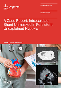
We present the case of a 69-year-old with severe pulmonary hypertension and persistent hypoxia of undetermined etiology with an intracardiac right-to-left shunt from an atrial septal defect. Pulmonary vasodilator therapy initially proved ineffective for the treatment of this patient's hypoxia until a bubble study confirmed the shunt. We describe our diagnostic approach, hemodynamic findings, and clinical intervention, with the patient ultimately stabilizing after the continuation of prostacyclin therapy and adjunctive vasopressor support. The need to consider intracardiac shunting in the evaluation of persistent hypoxia is emphasized, as its management has implications in pulmonary hypertension. View this paper
- Issues are regarded as officially published after their release is announced to the table of contents alert mailing list.
- You may sign up for e-mail alerts to receive table of contents of newly released issues.
- PDF is the official format for papers published in both, html and pdf forms. To view the papers in pdf format, click on the "PDF Full-text" link, and use the free Adobe Reader to open them.
Previous Issue
Next Issue