
Locally Advanced Cervical Cancer in a Patient with Epidermolysis Bullosa Treated with Concurrent Chemoradiotherapy and Electronic Brachytherapy

 , ,
, ,  , and
, and
Abstract
1. Introduction
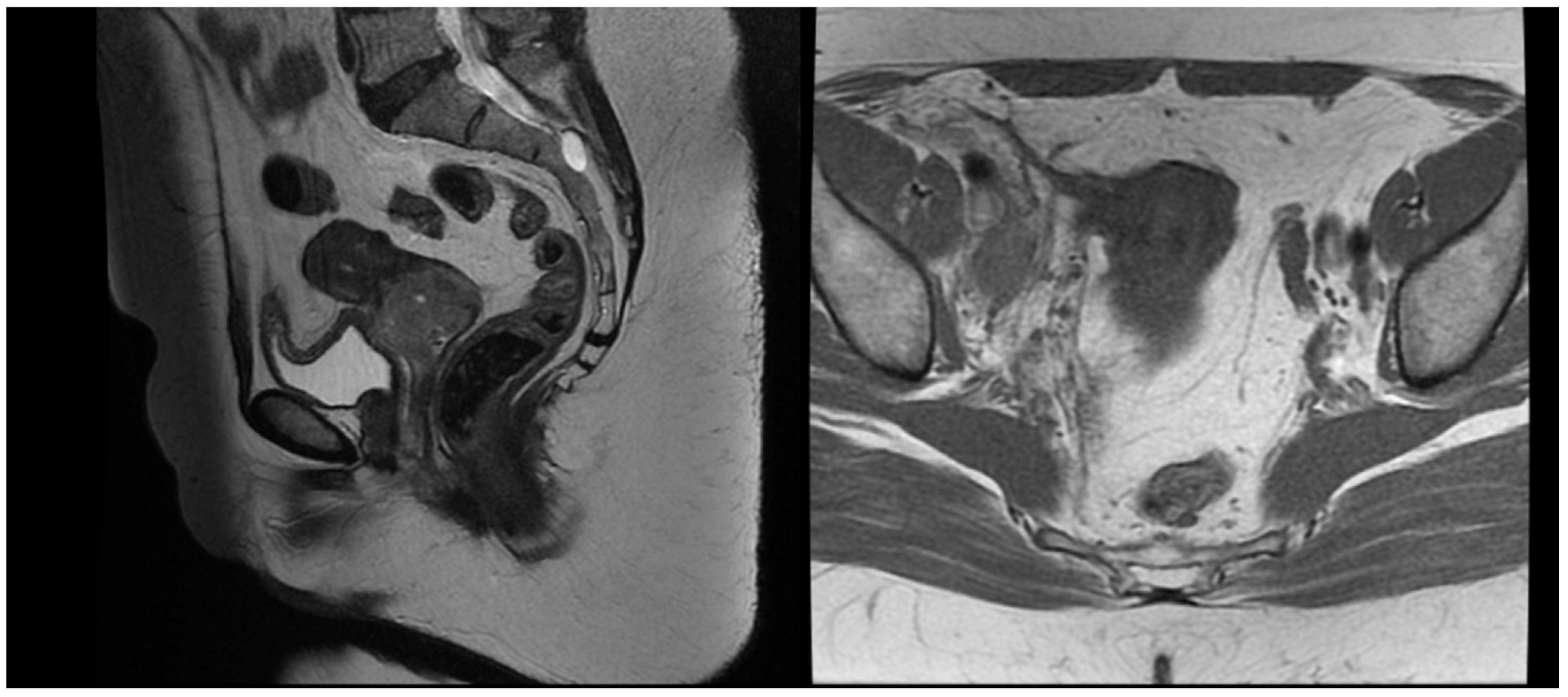
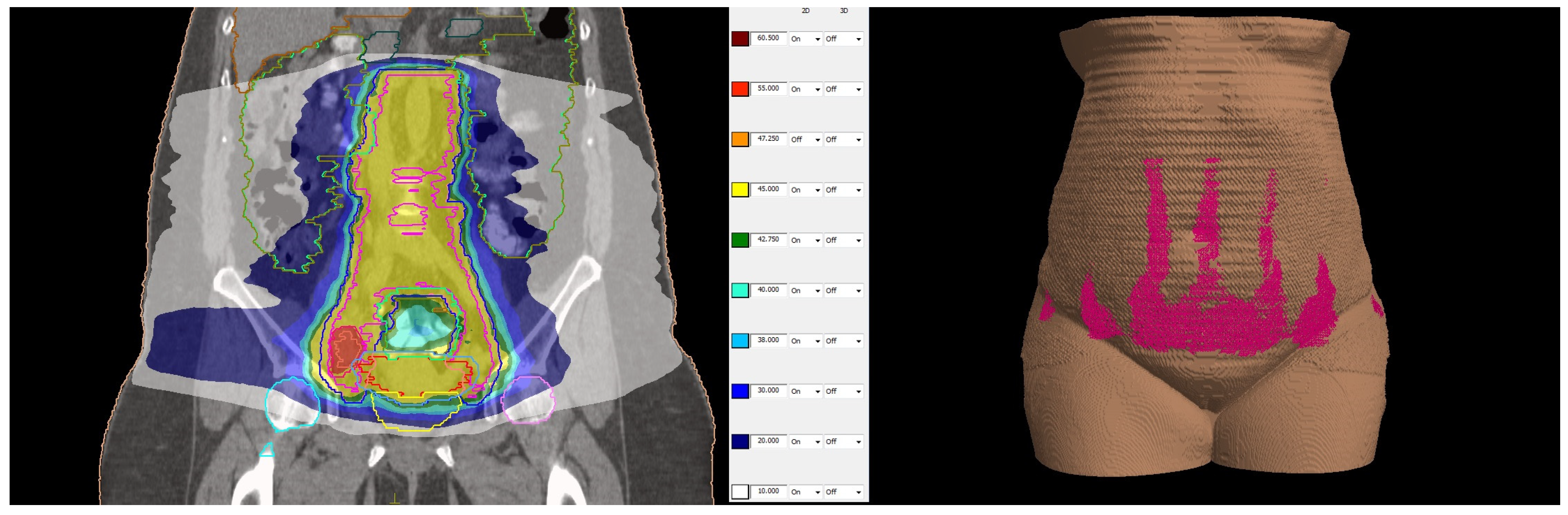
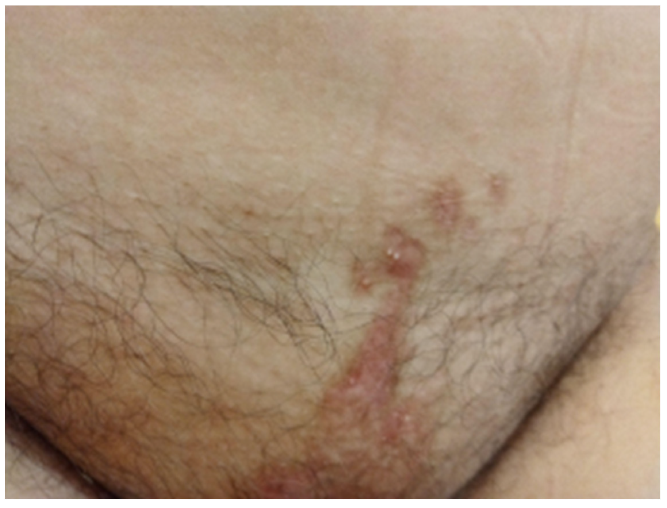
2. Case Description
3. Discussion
4. Conclusions
Author Contributions
Funding
Institutional Review Board Statement
Informed Consent Statement
Data Availability Statement
Conflicts of Interest
References
- Mariath, L.M.; Santin, J.T.; Schuler-Faccini, L.; Kiszewski, A.E. Inherited epidermolysis bullosa: Update on the clinical and genetic aspects. An. Bras. Dermatol. 2020, 95, 551–569. [Google Scholar] [CrossRef] [PubMed]
- Angelis, A.; Kanavos, P.; López-Bastida, J.; Linertová, R.; Oliva-Moreno, J.; Serrano-Aguilar, P.; Posada-De-La-Paz, M.; Taruscio, D.; Schieppati, A.; Iskrov, G.; et al. Social/economic costs and health-related quality of life in patients with epidermolysis bullosa in Europe. Eur. J. Health Econ. 2016, 17, 31–42. [Google Scholar] [CrossRef] [PubMed]
- Fine, J. Epidemiology of Inherited Epidermolysis Bullosa Based on Incidence and Prevalence Estimates From the National Epidermolysis Bullosa Registry. JAMA Dermatol. 2016, 152, 1231–1238. [Google Scholar] [CrossRef] [PubMed]
- Has, C.; Hess, M.; Anemüller, W.; Blume-Peytavi, U.; Emmert, S.; Fölster-Holst, R.; Frank, J.; Giehl, K.; Günther, C.; Hammersen, J.; et al. Epidemiology of inherited epidermolysis bullosa in Germany. J. Eur. Acad. Dermatol. Venereol. 2023, 37, 402–410. [Google Scholar] [CrossRef]
- Yordanova, I. Epidemiology of hereditary bullous epidermolysis in Bulgaria. Social Med. 2001, 3, 20–23. [Google Scholar]
- Warnakulasuriya, S.; Kujan, O.; Aguirre-Urizar, J.M.; Bagan, J.V.; González-Moles, M.; Kerr, A.R.; Lodi, G.; Mello, F.W.; Monteiro, L.; Ogden, G.R.; et al. Oral potentially malignant disorders: A consensus report from an international seminar on nomenclature and classification, convened by the WHO Collaborating Centre for Oral Cancer. Oral. Dis. 2021, 27, 1862–1880. [Google Scholar] [CrossRef]
- Akasaka, E.; Nakano, H.; Sawamura, D. Kindler epidermolysis bullosa associated with oral cancer in the buccal mucosa. JAAD Case Rep. 2022, 26, 13–16. [Google Scholar] [CrossRef]
- Condorelli, A.G.; Dellambra, E.; Logli, E.; Zambruno, G.; Castiglia, D. Epidermolysis bullosa-associated squamous cell carcinoma: From pathogenesis to therapeutic perspectives. Int. J. Mol. Sci. 2019, 20, 5707. [Google Scholar] [CrossRef]
- Tartaglia, G.; Cao, Q.; Padron, Z.M.; South, A.P. Impaired Wound Healing, Fibrosis, and Cancer: The Paradigm of Recessive Dystrophic Epidermolysis Bullosa. Int. J. Mol. Sci. 2021, 22, 5104. [Google Scholar] [CrossRef]
- Bonamonte, D.; Filoni, A.; De Marco, A.; Lospalluti, L.; Nacchiero, E.; Ronghi, V.; Colagrande, A.; Giudice, G.; Cazzato, G. Squamous Cell Carcinoma in Patients with Inherited Epidermolysis Bullosa: Review of Current Literature. Cells 2022, 11, 1365. [Google Scholar] [CrossRef]
- South, A.P.; Laimer, M.; Gueye, M.; Sui, J.Y.; Eichenfield, L.F.; Mellerio, J.E.; Nyström, A. Type VII collagen deficiency in the oncogenesis of cutaneous squamous cell carcinoma in dystrophic epidermolysis bullosa. J. Investig. Dermatol. 2023, 143, 2108–2119. [Google Scholar] [CrossRef] [PubMed]
- Parkin, D.M.; Bray, F.; Ferlay, J.; Pisani, P. Global cancer statistics, 2002. CA Cancer J. Clin. 2005, 55, 74–108. [Google Scholar] [CrossRef] [PubMed]
- Abu-Rustum, N.R.; Yashar, C.M.; Arend, R.; Barber, E.; Bradley, K.; Brooks, R.; Campos, S.M.; Chino, J.; Chon, H.S.; Crispens, M.A.; et al. NCCN Guidelines® Insights: Cervical Cancer, Version 1.2024: Featured Updates to the NCCN Guidelines. J. Natl. Compr. Cancer Netw. 2023, 21, 1224–1233. [Google Scholar] [CrossRef] [PubMed]
- Cibula, D.; Raspollini, M.R.; Planchamp, F.; Centeno, C.; Chargari, C.; Felix, A.; Fischerová, D.; Jahnn-Kuch, D.; Joly, F.; Kohler, C.; et al. ESGO/ESTRO/ESP Guidelines for the management of patients with cervical cancer—Update 2023. Virchows Arch. 2023, 482, 935–966. [Google Scholar] [CrossRef]
- Pötter, R.; Tanderup, K.; Kirisits, C.; De Leeuw, A.; Kirchheiner, K.; Nout, R.; Tan, L.T.; Haie-Meder, C.; Mahantshetty, U.; Segedin, B.; et al. The EMBRACE II study: The outcome and prospect of two decades of evolution within the GEC-ESTRO GYN working group and the EMBRACE studies. Clin. Transl. Radiat. Oncol. 2018, 9, 48–60. [Google Scholar] [CrossRef]
- Mellerio, J.E.; Robertson, S.J.; Bernardis, C.; Diem, A.; Fine, J.; George, R.; Goldberg, D.; Halmos, G.; Harries, M.; Jonkman, M.; et al. Management of cutaneous squamous cell carcinoma in patients with epidermolysis bullosa: Best clinical practice guidelines. Br. J. Dermatol. 2016, 174, 56–67. [Google Scholar] [CrossRef]
- Montaudié, H.; Chiaverini, C.; Sbidian, E.; Charlesworth, A.; Lacour, J.P. Inherited epidermolysis bullosa and squamous cell carcinoma: A systematic review of 117 cases. Orphanet J. Rare Dis. 2016, 11, 1–2. [Google Scholar] [CrossRef]
- Bastin, K.T.; Steeves, R.A.; Richards, M.J. Radiation therapy for squamous cell carcinoma in dystrophic epidermolysis bullosa: Case reports and literature review. Am. J. Clin. Oncol. 1997, 20, 55–58. [Google Scholar] [CrossRef]
- Koulis, T.A.; Herring, C.; Smith, W.; Jon-Paul, V. Adjuvant Radiation Therapy is Feasible in Epidermolysis bullosa: A Case Report. J. Dermatol. Res. Ther. 2015, 1, 1. [Google Scholar]
- Al Shareef, W.; Sayed, S.; Kamel, S.; Alkaf, H.; Bahaj, A.; Amin, R.; Al Herabi, A. Locally advanced tongue squamous cell carcinoma in epidermolysis simplex bullosa patient: A therapeutic conundrum. Ann. R. Coll. Surg. Engl. 2021, 103, e85–e87. [Google Scholar] [CrossRef]
- Ong, W.L.; Bailey, E.; McCormack, C.; Weston, L.; Morgan, V.; McDowell, L. Definitive radiotherapy for Merkel cell carcinoma in the setting of epidermolysis bullosa simplex. Australas. J. Dermatol. 2019, 60, 153–154. [Google Scholar] [CrossRef] [PubMed]
- Bavishi, S.; Wong, K.; Delgardo, T.; Marachelian, A.; Khatua, S. Successful radiation therapy for supratentorial primitive neuroectodermal tumor and epidermolysis bullosa simplex. Pediatr. Blood Cancer 2010, 54, 170–172. [Google Scholar] [CrossRef] [PubMed]
- Mizutani, H.; Masuda, K.; Nakamura, N.; Takenaka, H.; Tsuruta, D.; Katoh, N. Cutaneous and laryngeal squamous cell carcinoma in mixed epidermolysis bullosa, kindler syndrome. Case Rep. Dermatol. 2012, 4, 133–138. [Google Scholar] [CrossRef] [PubMed]
- AlKhawajah, N.M.; AlKhawajah, M.M.; Aljomah, N.A.; Alabduljabbar, A.A.; Alkeraye, S. Epidermolysis bullosa simplex clearance after nasopharyngeal carcinoma treatment. JAAD Case Rep. 2021, 12, 60–63. [Google Scholar] [CrossRef] [PubMed]
- Ray, A.; Bhattacharya, S.; Kumar, A.; Bhattacharya, K. Rare occurrence of carcinoma esophagus in a case of epidermolysis bullosa. Indian J. Cancer 2009, 46, 72–73. [Google Scholar] [CrossRef]
- Lataifeh, I.; Barahmeh, S.; Amarin, Z.; Jaradat, I. Stage III squamous cell carcinoma of the vulva with groin nodes metastasis in a patient with epidermolysis bullosa. J. Obstet. Gynaecol. 2010, 30, 750–752. [Google Scholar] [CrossRef]
- Etienne, A.; Ruffieux, P.; Didierjean, L.; Saurat, J.H. Epidermolyse bulleuse acquise et cancer du col utérin métastatique. Ann. Dermatol. Venereol. 1998, 125, 321–323. (In French) [Google Scholar] [PubMed]
- Lozares-Cordero, S.; Font-Gómez, J.A.; Gandía-Martínez, A.; Miranda-Burgos, A.; Méndez-Villamón, A.; Villa-Gazulla, D.; Alba-Escorihuela, V.; Jiménez-Puertas, S.; González-Pérez, V. Treatment of cervical cancer with electronic brachytherapy. J. Appl. Clin. Med. Phys. 2019, 20, 78–86. [Google Scholar] [CrossRef]


| OARs. | |||||
|---|---|---|---|---|---|
| V50% (%) | V35% (%) | D2cc (Gy) | D1cc (Gy) | D0.1cc (Gy) | |
| Bladder | 0.9 | 2.8 | 3.71 | 4.34 | 5.96 |
| Rectum | 0 | 0 | 0.58 | 0.88 | 0.99 |
| Sigmoid | 0.9 | 4 | 2.32 | 3.66 | 4.08 |
| Reference | Radiotherapy Target | Prescribed Dose | Reported Skin Dose | Outcome |
|---|---|---|---|---|
| Bastin [18] | skin | >45 Gy | moist desquamation, delayed healing | |
| Koulis [19] | pelvic lymph nodes | 48 Gy | <40 Gy | grade 1 erythema |
| Al Shareef [20] | tongue | 30 Gy | ulceration, hemorrhage and necrosis) | |
| Ong [21] | parotid gland, ipsilateral lymph nodes levels IB- V | 60 Gy, 52 Gy | moist desquamation with complete healing | |
| Bavishi [22] | brain temporal lobe, craniospinal irradiation | 32.4 Gy, 23.4 Gy | moist desquamation G2, bullae, hyperpigmentation | |
| Mizutani [23] | larynx | unknown | none reported | |
| AlKhawajah [24] | nasopharynx | 70 Gy with concurrent Cisplatin | clearance of blisters | |
| Ray [25] | esophagus | 61.2 Gy | Grade 2 erythema and desquamation |
Disclaimer/Publisher’s Note: The statements, opinions and data contained in all publications are solely those of the individual author(s) and contributor(s) and not of MDPI and/or the editor(s). MDPI and/or the editor(s) disclaim responsibility for any injury to people or property resulting from any ideas, methods, instructions or products referred to in the content. |
© 2025 by the authors. Licensee MDPI, Basel, Switzerland. This article is an open access article distributed under the terms and conditions of the Creative Commons Attribution (CC BY) license (https://creativecommons.org/licenses/by/4.0/).
Share and Cite
Hitova-Topkarova, D.; Payakova, V.; Yordanov, A.; Kostova-Lefterova, D.; Encheva, E. Locally Advanced Cervical Cancer in a Patient with Epidermolysis Bullosa Treated with Concurrent Chemoradiotherapy and Electronic Brachytherapy. Reports 2025, 8, 12. https://doi.org/10.3390/reports8010012
Hitova-Topkarova D, Payakova V, Yordanov A, Kostova-Lefterova D, Encheva E. Locally Advanced Cervical Cancer in a Patient with Epidermolysis Bullosa Treated with Concurrent Chemoradiotherapy and Electronic Brachytherapy. Reports. 2025; 8(1):12. https://doi.org/10.3390/reports8010012
Chicago/Turabian StyleHitova-Topkarova, Desislava, Virginia Payakova, Angel Yordanov, Desislava Kostova-Lefterova, and Elitsa Encheva. 2025. "Locally Advanced Cervical Cancer in a Patient with Epidermolysis Bullosa Treated with Concurrent Chemoradiotherapy and Electronic Brachytherapy" Reports 8, no. 1: 12. https://doi.org/10.3390/reports8010012
APA StyleHitova-Topkarova, D., Payakova, V., Yordanov, A., Kostova-Lefterova, D., & Encheva, E. (2025). Locally Advanced Cervical Cancer in a Patient with Epidermolysis Bullosa Treated with Concurrent Chemoradiotherapy and Electronic Brachytherapy. Reports, 8(1), 12. https://doi.org/10.3390/reports8010012