Abstract
Diffracted X-ray tracking (DXT) method can trace conformational changes of KcsA potassium ion channel during gating by recording position of diffraction spot from a gold nanocrystal attached to the channel as a movie. For high-resolution imaging under controlled microenvironments for KcsA channels, we report a microfluidic device consisting of two SiN membrane windows bonded with a photo patternable adhesive material. The reduced signal-to-background ratio as well as suitable adhesive material thickness for the microchannel are discussed in the experiment at the synchrotron radiation facility.
1. Introduction
Activity of ion channel is modulated by relevant signals (e.g., chemical or optical) that tightly control the opening and closing of the channel. Crystal structures of several kinds of potassium ion channels have revealed open and closed conformations [1]; however, a mechanism underlying the gating has not been well studied. For further understandings, capturing motion of potassium ion channels at single-molecule level is necessary. Among several imaging techniques (e.g., high-speed atomic force microscopy or nuclear magnetic resonance imaging) [2], diffracted X-ray tracking (DXT) method [3] enabled to record conformational changes of KcsA potassium channel with high spatial and temporal resolutions. In this paper, we propose a new experimental setup for the DXT method that control microenvironments for KcsA channels fixed in an observation chamber, leading to capture conformational change occurring in response to chemicals (e.g., acidic pH or ligands). This observation setup present here differs from the previous work [4] in the specific fabrication for improving signal-to-background (S/B) ratio by assembling SiN membrane windows.
2. Materials and Methods
2.1. DXT Method
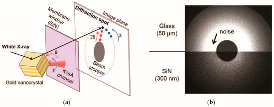
In the DXT method, KcsA potassium channels are fixed on a chemically modified substrate surface inside the observation chamber (Figure 1a). A gold nanocrystal is then attached to a KcsA channel and irradiated with high-flux white X-rays. The position of the diffraction spot on the image plane is recorded through an X-ray camera. The motion of the diffraction spot on the polar co-ordinate represents conformational changes in KcsA channel. In Figure 1a, the position and motion of the diffraction spot on the image plane (the circular polar co-ordinate (angles 2θ and χ) centered on the point of X-ray irradiation) represents posing orientation of the nanocrystal in the three-dimensional space (the spherical polar co-ordinate).

Figure 1.
(a) Principle of the DXT method. White X-rays are diffracted by the gold nanocrystal; (b) Background noise recorded under the same X-rays irradiation condition.
2.2. Device Fabrication
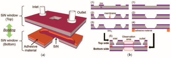
KcsA channels were conventionally fixed on a glass substrate having a thickness of 50 μm, and they were simply covered by thin polyimide film for constructing the observation chamber. Although this observation setup successfully captured the conformational changes of single KcsA channels, high background noise due to X-ray scattering of the observation chamber has limited the record of the diffraction spots, and spatial resolution was not good enough (Figure 1b). In addition, this setup prevents experiments to capture conformational change occurring in response to chemical stimuli. Thus, we have considered a potential of microfabricated SiN membrane window [4] and propose a microfluidic device assembled by a photo patternable adhesive material (Figure 2a).

Figure 2.
(a) Schematic illustration of the microfluidic device consisting of microchannel patterned with a photo patternable adhesive material and SiN membrane windows; (b) Fabrication procedure.
A fabrication of new microfluidic structure in Figure 2b stars with, (1) SiN was patterned by UV lithography for subsequent KOH etching (2), which releases a SiN membrane having a thickness of 300 nm. (3) SiO2 was deposited on the front side of SiN by plasma-enhanced chemical vapor deposition (PECVD) to fix KcsA channels. (4) A photo patternable adhesive material was spin-coated and patterned for a microchannel structure by UV lithography. In order to compare S/B ratio dependence on microchannel height, different thickness of adhesive material was prepared in this study. (5) Finally, two SiN membrane windows were aligned and bonded with applying pressure and thermal heating. An assembling the microfluidic system was simply achieved by pressing the whole microfluidic device in between a 3D-printed polymer holder.
2.3. Evaluation of S/B Ratio
To evaluate S/B ratio of the observation chamber, the fabricated chambers were irradiated with high flux white X-rays from the synchrotron radiation facility (the beamline BL28B2, SPring-8, Kyoto, Japan), and image was recorded with an X-ray camera for 0.02 s at a video rate 5000 frames s−1. The recorded images were further analyzed statistically by fitting multivariate normal distribution. The noise level a can be expressed as:
where xc and yc are the measured position on the image plane (the co-ordinate centered on the point of X-ray irradiation), and σ is the standard deviation.
3. Results and Discussion
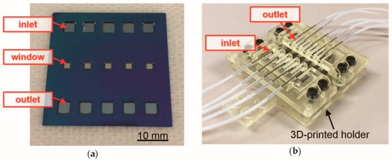
Figure 3 shows an optical image of fabricated microfluidic device. The bonding process using the patterned adhesive material was conducted at the temperature of 160 °C and applied pressure of 126 kPa. Under this optimized bonding condition, the microchannels were bonded enough strong that they could be conducted a KcsA sample preparation without leakage, such as KcsA channels introduction and gold nanocrystal attachment to the KcsA channels [3]. This experimental result indicates that the present fabrication approach is of practical use for the assembling of microfluidic devices.

Figure 3.
Optical image of the fabricated microfluidic device (a) and the assembled system using a 3D-printed polymer holder (b).
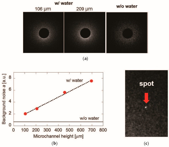
Next, in order to determine a suitable microchannel height (i.e., thickness of adhesive material), background noise generated by observation chamber was evaluated. Figure 4a shows the recorded images of background noise by comparing the different height of microchannel. The background noise of chamber filled with water was evaluated quantitatively by Equation (1). As shown in Figure 4b, the background noise decreases with thinning channel thickness. Furthermore, the microchannel 50 μm in height enabled to reach the background noise level for detectable diffraction spot from a gold nanocrystal attached to KcsA channel (Figure 4c).

Figure 4.
(a) Example images of the recorded background noise; (b) Noise level dependence on thickness for different height of the microchannel evaluated by the average of 100 images captured; (c) Close-up view of the captured the diffraction spot from a gold nanocrystal with the 50 μm-height microchannel.
4. Conclusions
In this paper, we addressed the fabrication of microfluidic device for new experimental setup of the DXT method. To reduce background noise from the observation area, SiN membrane windows having a thickness of 300 nm were used and bonded with the patterned adhesive material for forming the microchannel. The suitable microchannel height to observe diffraction spots as well as the improved S/B ratio were discussed through the experiments. Therefore, we hope to continue our investigation of recording conformational changes of the KcsA channels, particularly with respect to influence of chemical stimuli.
Author Contributions
Conception and Design of Study, Y.H. and H.S.; Acquisition of Experimental Data, Y.H., Y.M., T.T. (Tomoki Tabuchi) and H.S.; Analysis of Data, T.T. (Tomoki Tabuchi) and H.S.; All of the authors discussed the results; Drafting the Manuscript, Y.H.; Review and Editing, H.S. and O.T.; Funding Acquisition, Y.H. and H.S.
Acknowledgments
The present study was supported in part by the JSPS KAKENHI Grant Number 15H04675, 15H01633, 17H05876, and 18H02596, and the Japan Association for Chemical Innovation. Part of the present research was conducted at the Kyoto University Nano Technology Hub as part of the “Nanotechnology Platform Project” sponsored by MEXT, Japan.
Conflicts of Interest
The authors declare no conflict of interest. The founding sponsors had no role in the design of the study; in the collection, analyses, or interpretation of data; in the writing of the manuscript, and in the decision to publish the results.
References
- Hille, B. Ion Channels of Excitable Membranes, 3rd ed.; Sinauer Associates: Sunderland, MA, USA, 2001; ISBN 0878933212. [Google Scholar]
- Oiki, S. Channel function reconstitution and re-animation: a single-channel strategy in the postcrystal age. J. Physiol. 2015, 593, 2553–2573. [Google Scholar] [CrossRef] [PubMed]
- Shimizu, H.; Iwamoto, M.; Konno, T.; Nihei, A.; Sasaki, Y.; Oiki, S. Global twisting motion of single molecular KcsA potassium channel upon gating. Cell 2010, 132, 67–78. [Google Scholar] [CrossRef] [PubMed]
- Tahara, K.; Hirai, Y.; Shimizu, H.; Tsuchiya, T.; Tabata, O. Photoresist micro-chamber for the diffracted X-ray tracking method recording single-molecule conformational changes. Procedia Eng. 2015, 168, 1394–1397. [Google Scholar] [CrossRef]
Publisher’s Note: MDPI stays neutral with regard to jurisdictional claims in published maps and institutional affiliations. |
© 2018 by the authors. Licensee MDPI, Basel, Switzerland. This article is an open access article distributed under the terms and conditions of the Creative Commons Attribution (CC BY) license (https://creativecommons.org/licenses/by/4.0/).