Abstract
Cortisol is the predominant corticosteroid in ray-finned fish since it does not possess the aldosterone synthase necessary to produce specific mineralocorticoids. Cortisol is traditionally believed to function as a fish mineralocorticoid. However, the effects of cortisol are mediated through corticosteroid receptors in other vertebrates, and there is an ongoing debate about whether cortisol acts through the glucocorticoid receptor (GR) or the mineralocorticoid receptor (MR) in teleosts. To investigate this issue, we conducted a study using euryhaline Mozambique tilapia (Oreochromis mossambicus) as the experimental species. The experiment was designed to investigate the effect of cortisol on ionocyte development at both the cellular and gene expression levels in tilapia. We administered exogenous cortisol and receptor antagonists, used immunohistochemistry to quantify ionocyte numbers, and performed real-time PCR to assess the expression of the differentiation factor tumor protein 63 (P63) mRNA, an epidermal stem cell marker. We observed that cortisol increased the number of Na+-K+-ATPase (NKA)-immunoactive ionocytes (increased by 1.6-fold) and promoted the gene expression of P63 mRNA (increased by 1.4-fold). Furthermore, we found that the addition of the mineralocorticoid receptor antagonist Spironolactone inhibited the increase in the number of ionocytes (decreased to the level of the control group) and suppressed the gene expression of P63 (similarly decreased to the level of the control group). We also provided evidence for gr, mr, and p63 localization in epidermal cells. At the transcript level, mr mRNA is ubiquitously expressed in gill sections and present in epidermal stem cells (cells labeled with p63), supporting the antagonism and functional assay results in larvae. Our results confirmed that cortisol stimulates ionocyte differentiation in tilapia through the MR, rather than the GR. Therefore, we provide a new direction for investigating the dual action of osmotic regulation and skin/gill epithelial development in tilapia, which could help resolve previously inconsistent and conflicting findings.
Key Contribution:
Cortisol increases the expression of the differentiation factor P63 gene and promotes the increase of ionocyte number in tilapia via the mineralocorticoid receptor.
1. Introduction
Corticosteroids (CS) are vital hormones for mammals, which are involved in many physiological functions such as ion regulation, fluid constants, energy metabolism, respiration, and immune responses [1,2]. However, ray-finned fish lack the aldosterone synthase to produce a specific mineralocorticoid [3,4]. Therefore, cortisol is currently considered to be the major corticosteroid in fish. Cortisol action is mediated by two corticosteroid receptors (CRs) of the glucocorticoid receptor (GR) and the mineralocorticoid receptor (MR) in mammals [5,6]. The functional regulation of corticosteroids is determined by the complex (CS-GR or CS-MR) that binds the ligand (corticosteroids) to the receptor to initiate the transcription of target cells to regulate diverse physiological phenomenon; that is, what kind of regulation occurs depends on which receptor cortisol binds to [7].
The gills are the major organ for ionoregulation in fish. Before the gills are fully developed, the skin serves as the main organ for ionoregulation at early developmental stages of fish [8,9]. Several studies have investigated the role of cortisol with GR and/or MR in fish osmoregulation; it is an essential hormone for euryhaline fish to adapt to seawater. It also participates in the ion regulation of freshwater acclimation. For instance, ionocytes secrete ions using the Na+-K+-ATPase (NKA) and the Na+-K+-2Cl− cotransporter (NKCC); both the activity of these ion transporters and expression of the encoding genes were found to be regulated by exogenous cortisol [10,11,12]. It was also found that in the acclimation to the two different water environments of seawater and freshwater, the regulation of cortisol should be achieved by regulating the development and cell differentiation of fish gill ionocytes [10,11,12]. Previous studies have shown that the plasma cortisol levels of tilapia increased significantly by 1.8-fold on day 1 of salinity stress [13]. The results of this study demonstrate that changes in salinity and environmental conditions can directly affect the cortisol levels in fish. In recent years, new hematological parameters have been used as important tools to study the salinity adaptation and nutritional status of fish. Changes in environmental salinity can cause significant changes in hematological parameters [14,15]. An increase in water salinity may lead to erythropoiesis as an adaptive process in seawater fish.
The embryonic epidermis in most bony fish comprises three layers: a basal layer, an intermediate stratum, and a microridge-rich superficial layer [16]. When surface epidermal cells in fish are injured or die, they are replaced by cells from the intermediate layer. To ensure the normal functioning of skin tissue, skin cells need to maintain a stable mechanism of proliferation and differentiation [16]. The differentiation mechanisms of ionocytes in zebrafish gills and epidermis have been established by numerous studies in recent years [17,18,19,20]. The tumor protein 63 delta (ΔNp63) is expressed in the monolayer of zebrafish epidermis and plays an important role in maintaining the proliferation state of the epidermis [21]. However, there have been no studies on epidermal cell proliferation and ionocyte differentiation in the euryhaline Mozambique tilapia (Oreochromis mossambicus). Compared to freshwater zebrafish, tilapia is a euryhaline teleost possessing strong salinity acclimation ability. It can not only adapt to various salinities between freshwater and seawater but also tolerate extremely low-ion water and survive in environments with salinity twice that of seawater [22,23,24]. The different types of ionocytes in freshwater and seawater have been well defined [25], making it a suitable euryhaline fish for studying ionocytes regulation. Tilapia is also a maternal mouthbrooder, in which the female carries the fertilized eggs in her mouth until hatching, making it possible to obtain embryos for research purposes throughout the year.
Several studies have investigated the proliferation and differentiation mechanism of ionocytes in fish. In zebrafish, researchers have defined some important transcription factors like tumor protein 63 (P63), Forkhead Box transcription factors I 3a/b (Foxi3a/b), and Glial Cells Missing Transcription Factor 2 (Gcm2) functions mainly controlling the development of epidermal ionocytes. They affect the differentiation and specialization of different types of ionocytes, respectively [17,19,20,26]. Meanwhile, the scientific literature on zebrafish demonstrates that cortisol promotes the development and cell differentiation of embryo ionocytes by modulating differentiation factors via GR. Similarly, studies on medaka have reported analogous findings [27,28,29]. Based on the above studies, cortisol participates in the differentiation of zebrafish and medaka epidermal ionocytes. However, as a primary freshwater fish, it is still being determined whether zebrafish exhibit the same regulatory pathway as euryhaline fish. In addition, recent related studies have found that cortisol may act through the MR in teleosts [30]. The study suggests that cortisol also has a high affinity for MR and affects the expression of ionocytes through MR, leading some researchers to believe that cortisol may act on ionocytes in the gills of teleosts through MR during salinity acclimation [13,31]. In addition, exogenous cortisol treatment can promote calcium absorption and epidermal calcium channel (ECaC) gene expression in tilapia. However, no clear evidence indicates whether this occurs through GR or MR [32]. Therefore, the interaction between cortisol and MR is still controversial.
In recent years, studies on MR have mainly focused on the nervous system and muscles. Some studies have indicated that 11-deoxycorticosterone (DOC), a mineralocorticoid receptor agonist, has a relevant role in the stress response of skeletal muscles in rainbow trout [33]. In addition, cortisol regulates muscle contraction and cell cycle regulation via MR in rainbow trout [34]. Cortisol also regulates neural plasticity and stress responses in Atlantic salmon under low temperature via MR [35]. Other studies suggest that mineralocorticoid signaling may be associated with brain behavior in mudskipper fish [36] and medaka fish [37]. Recent studies have rarely addressed the impact of MR on salinity acclimation and ionocyte development.
Previous studies have generally suggested that cortisol is involved in salinity acclimation of teleosts via GR, rather than MR. However, there is no direct molecular evidence to support the non-involvement of MR in the salinity acclimation of euryhaline fish. This study is the first to investigate the involvement of corticosteroid receptors in ionocyte differentiation at multiple levels, including cell number, gene expression, and tissue staining. In this study, we chose the euryhaline tilapia as the animal model to investigate whether cortisol regulates the gene expression of epidermal stem cells or the number of ionocytes. We further designed experiments to determine whether cortisol affects ionocyte development and function through GR/MR by using the GR antagonist of RU-486 and the MR antagonist of spironolactone. Therefore, we treated tilapia larvae with exogenous cortisol and attempted to understand how cortisol acts on ionocytes (via which corticosteroid receptor) by inhibiting the function of GR/MR with antagonists.
2. Materials and Methods
2.1. Experimental Animals
Mozambique tilapia (Oreochromis mossambicus), 1–60 g in body weight, were obtained from stocks at National University of Tainan, Taiwan. Fish were kept in a freshwater (local tap water) circulating system at 28 ± 0.5 °C under a 14-h:10-h light:dark photoperiod. Tilapia embryos were acquired as follows: fertilized eggs were taken from the mouth of female tilapia when mouthbrooding behavior was observed. Fertilized eggs were incubated in aerated freshwater and were used in the experiments when they hatched immediately. All sampled fish were clinically healthy. All incubation experiments were conducted on larvae, and no feeding occurred. Adult fish and larvae were anesthetized with buffered 0.03% MS-222 (Tricaine, Sigma-Aldrich, Burlington, MA, USA) and then dissected for experiments. The animal-use protocol listed below has been reviewed and approved by the Institutional Animal Care and Use Group (Approval No.: IACUG1050005) at National University of Tainan. All the experimental procedures and the collection of samples were performed in compliance with the ethical considerations of the Taiwan Council of Agriculture Executive Yuan Guideline for the Care and Use of Laboratory Animals (Decree by the Council of Agriculture, Executive Yuan, Taiwan, 2018/06).
2.2. Cortisol and Receptor Antagonist Treatment of Tilapia Larvae
Cortisol dosages were determined according to previous studies [28,32,38,39,40,41]. Cortisol (hydrocortisone, Sigma-Aldrich, Burlington, MA, USA) stock solution was prepared in dimethyl sulfoxide (DMSO) and then diluted to the final working solution (20 mg/L) in aerated tap water. Tilapia larvae were treated with cortisol media immediately after hatching. The incubation media were refreshed daily to ensure consistent cortisol levels were maintained. GR and MR antagonist dosages were determined according to previous studies [32,42,43]. Tilapia embryos were grown in 10 µg/mL RU486 (GR antagonist, Sigma-Aldrich, Burlington, MA, USA) or 10 µg/mL Spironolactone (MR antagonist, Sigma-Aldrich, Burlington, MA, USA) with 20 mg/L cortisol, and the medium was changed every day. In this study, higher dosages of cortisol and antagonists were employed as compared to some previous studies. Nonetheless, no significant mortality or aberrant behavior was noted. The dosages of cortisol and antagonists that we used were proven to work in cultured gills and fish larvae in previous studies [28,32,39,40,41,42,43]. After the experiment, tilapia embryos were anesthetized with buffered 0.03% MS-222 (Tricaine, Sigma-Aldrich, Burlington, MA, USA) for further analysis.
2.3. Total RNA Extraction
After conducting the treatment experiments, three tilapia larvae were collected as a sample, and five replicates (n = 5) were performed. Total RNA was extracted from collected samples using Trizol reagent (Thermo Fisher Scientific, Waltham, MA, USA) as per the manufacturer’s instructions. Briefly, the samples were homogenized in 1 mL Trizol reagent, and 0.2 mL chloroform was added and thoroughly mixed by shaking. The mixture was then centrifuged at 12,000× g at 4 °C for 45 min. An equal volume of isopropanol was added to the sample, and the mixture was centrifuged at 12,000× g at 4 °C for 30 min to precipitate total RNA pellets. The pellets were washed twice with 70% alcohol for 30 min at 12,000× g at 4 °C. The quantity and quality of total RNA were assessed using absorbance at 260 and 280 nm, as well as gel electrophoresis, and then stored at −20 °C until further use.
2.4. Real-Time PCR
The cDNA of tilapia samples was synthesized using the GoScript™ Reverse Transcription System (Promega, Madison, WI, USA) and poly(dT) primers. The mRNA used for cDNA synthesis was treated with DNase I (Promega, Madison, WI, USA) to eliminate genomic DNA contamination. Real-time PCR was carried out using a StepOne Plus Real-Time PCR System (Applied Biosystems, Thermo Fisher Scientific, Waltham, MA, USA) in a final reaction volume of 20 μL, consisting of 10 μL Fast SYBR™ Green Master Mix (Applied Biosystems, Thermo Fisher Scientific, Waltham, MA, USA), 300 nM forward and reverse primers, 20–30 ng cDNA, and nuclease-free water. The primer set for P63 (for detailed sequences of P63, please refer to the Supplementary Materials) was designed to amplify a 175-bp fragment (Accession no. OQ626354, Supplementary Materials) and comprised of the forward (5′- ACAGGCTATGGATTTTTCCC-3′) and reverse (5′- GAAGGAGAAGTCGGACCA-3′) sequences, whereas that for the internal control β-actin, with a 135-bp fragment, consisted of the forward (5′-CGGAATCCACGAAACCACCTA-3′) and reverse (5′-ATCTCCTGCATCCTGTCA-3′) sequences. The β-actin was used as an internal control to normalize mRNA expression and was taken from previous studies [44].
2.5. Whole-Mount Immunohistochemistry (IHC)
After conducting the treatment experiments, one tilapia larvae was collected as a sample, and five replicates (n = 5) were performed. Drug-treated tilapia larvae were collected and fixed with 4% paraformaldehyde in phosphate buffer saline (PBS) solution overnight. After fixation, samples were transferred to 100% methanol and stored at −20 °C until use. The samples were washed several times with PBST (0.1% Tween) and then incubated with 3% bovine serum albumin (BSA) in PBST for 2 h at room temperature to block non-specific binding. Samples were transferred into a Na+-K+-ATPase α5 monoclonal antibody (1:400 dilution; Developmental Studies Hybridoma Bank, University of Iowa, Ames, IA, USA) and incubated overnight at 4 °C. All antibodies were diluted in a blocking PBST solution. The samples were washed with PBST six times for 10 min each and then incubated in Alexa Fluor 488 goat anti-mouse IgG antibodies (Thermo Fisher Scientific, diluted 1:400 with PBS) for 2 h at room temperature. The stained samples were mounted with low-melting agarose (Agarose, II™, VWR Life Science AMRESCO, Radnor, PA, USA) and then examined with a Zeiss LSM 780 confocal microscope.
2.6. RNA Probe Synthesis
RNA probe synthesis for double in situ hybridization was performed following a previous study [19,32,44]. The fragments of tilapia p63, gr, and mr obtained by PCR were inserted into a pGEM-T easy vector (Promega, Madison, WI, USA). The primer set for p63 (for detailed sequences of P63, please refer to the Supplementary Materials) consisted of the forward (5′- CGCCGGCTGCTTGGACTACTTCA -3′) and reverse sequences (5′- ACGCGGTTCCTTTCCATTCACG -3′) (a 693-bp fragment, Accession no. OQ626354, Supplementary Materials), that for gr consisted of the forward (5′- TGATGGCAGGCATGAATCT-3′) and reverse sequences (5′- GACAAACTCGCTGCAAATC -3′) (a 518-bp fragment, Accession no. AB771724.1), and that for mr consisted of the forward (5′- CGAGCAGCACGCCCTTTATCACA -3′) and reverse sequences (5′- TACACGACACGCCGGACAGTTTTT -3′) (a 748-bp fragment, Accession no. AB771726). For double in situ hybridization, two types of RNA probes were synthesized with DIG RNA Labeling Mix or Fluorescein RNA Labeling Mix (Roche, Basel, Switzerland) by in vitro transcription with T7 and M13 RNA polymerase (Promega, Madison, WI, USA). Two types of labeled RNA probes were examined with RNA gels and dot-blot assay to confirm the quality and concentration.
2.7. Double In Situ Hybridization
In situ hybridization was performed following a previous study [19,45]. Briefly, excised gills were fixed overnight with 4% paraformaldehyde in PBS. The fixed gills were washed with PBS and cryoprotected in 30% sucrose prior to embedding in OCT compound embedding medium (Sakura, Tokyo, Japan) at −20 °C. Frozen cross-sections of 10 μm were cut with a CM 3050S rapid sectioning cryostat (Leica, Heidelberg, Germany) and attached to hydrophilic adhesion slides (PLATINUM PRO, MATSUNAMI, Osaka, Japan). Before in situ hybridization, the slide-mounted gill sections were air-dried and rehydrated by a series of methanol and PBST (PBS with 0.1% Tween-20) mixtures. The hybridization mix (HM) contained 50% formamide, 5× saline sodium sitrate (SSC), 9.2 mM citric acid, and 0.1% Tween-20. The slides were washed with PBST several times and then incubated in the pre-hybridization mix (HM+) (HM with additional 500 ng/mL and yeast tRNA and 50 μg/mL heparin) for 2 h and hybridized with RNA probes (200μL HM+ contained 30~50 ng DIG-labeled/Fluorescein-labeled RNA probe) at 70 °C overnight. Next, the hybridized slides were washed at 70 °C for 10 min in 100% HM (without tRNA and heparin), 10 min in 75% HM and 25% 2× saline sodium citrate (SSC), 10 min in 50% HM and 50% 2× SSC, 10 min in 25% HM and 75% 2× SSC, 10 min in 2× SSC, and finally 30 min in 0.2× SSC (this final step was repeated twice). Next, the hybridized slides were washed at room temperature for 10 min in 75% 0.2× SSC and 25% PBST, 10 min in 50% 0.2× SSC and 50% PBST, 10 min in 25% 0.2× SSC and 75% PBST, and 10 min in PBST. The washed slides were incubated for 2 h in blocking buffer containing 5% sheep serum and 2 mg/mL BSA in PBST and then transferred into alkaline phosphatase-conjugated anti-DIG antibody (1:5000 dilution; Roche, Basel, Switzerland) and incubated overnight at 4 °C. Finally, sections were washed with PBST six times for 15 min each and then transferred to alkaline Tris buffer, containing 0.1 M Tris HCl, pH 9.5, 0.05 M MgCl2, 0.1 M NaCl, and 0.1% Tween 20. Tissues were stained with a mixture of 225 μg/mL NBT (Nitro Blue Tetrazolium) and 175 μg/mL BCIP (5-Bromo 4-Chloro 3-indolyl Phosphate) in alkaline Tris buffer. The first labeling reaction would be stopped by a stop solution (containing 1 mM EDTA, 0.1% Tween 20 in 1X PBS, pH 5.5) until the signal was strong enough for analysis. After the first staining step of in situ hybridization, the gill slides were incubated in a stop solution for 1 h to stop the reaction, and then the gill slides were washed with PBST three times for 5 min each. Next, slides were transferred into PBST with 3% H2O2 for 30 min and then washed with PBST four times for 5 min each. Next, slides were transferred into a blocking reagent, containing maleic acid buffer (MABT) (100 mM maleic acid, 150 mM NaCl pH 7.5 and 0.1% Tween 20) with 2% blocking reagent (Roche, Basel, Switzerland) for 1 h at room temperature. Then, slides were transferred into horseradish peroxidase (POD)-conjugated anti-Fluorescein antibody (1:500 dilution; Roche, Basel, Switzerland) and incubated overnight at 4 °C. Finally, sections were washed with MABT four times for 10 min each and then washed with PBST two times for 10 min each. A Tyramide Signal Amplification Plus System (TSA Plus Systems; PerkinElmer, Waltham, MA, USA) was used for the second staining step of in situ hybridization, referring to manufacturer’s protocol. After incubation in TSA-Cyanine 3 reagent for 1 h at 28 °C, sections were washed at room temperature for 5 min in PBST twice, 10 min in 75% PBST and 25% methanol, 10 min in 50% PBST and 50% methanol, 10 min in 25% PBST and 75% methanol, and 10 min in 100% methanol. Then, slides were transferred to methanol with 1% H2O2 for 30 min to inactivate the POD.
2.8. Immunohistochemistry (IHC) of Gill Sections
After double in situ hybridization, the sections were washed at room temperature for 10 min in 25% PBST and 75% methanol, 10 min in 50% PBST and 50% methanol, 10 min in 75% PBST and 25% methanol, and 10 min in PBST; then, they were incubated with 3% bovine serum albumin (BSA) in PBST for 2 h at room temperature. Next, samples were transferred to a Na+-K+-ATPase α5 monoclonal antibody (1:400 dilution; Developmental Studies Hybridoma Bank, University of Iowa, Ames, IA, USA) and incubated overnight at 4 °C. The samples were washed with PBST six times for 10 min each and then incubated in Alexa Fluor 488 goat anti-mouse IgG antibodies (Thermo Fisher Scientific, diluted 1:400 with PBS) for 2 h at room temperature. The stained sections were mounted with Fluoromount-G mounting medium (SouthernBiotech, Birmingham, AL, USA) and then examined with a Zeiss LSM 780 confocal microscope.
2.9. Statistical Analysis
Statistical analysis was done using GraphPad Prism (GraphPad Inc., La Jolla, CA, USA). The normality of the data was tested with the Shapiro-Wilk normality test. The quantification of NKA-immunoreactive ionocyte cell numbers and P63 mRNA expression levels was subjected to statistical analysis using a multi-step ANOVA. This analysis evaluated the significance between the entire treatment group and the control group, as well as between the cortisol group and the combination group. Significance was determined using Dunnett’s multiple comparison test following a one-way ANOVA (p < 0.05).
3. Results
3.1. Effects of GR or MR Antagonist on Ionocyte Differentiation in Tilapia Larvae
To identify the specific pathway through which cortisol controls ionocyte development in tilapia larvae, we exposed 20 mg/L cortisol-treated tilapia larvae with either 10 µg/mL RU486 or spironolactone as GR or MR antagonists, respectively [32]. We used basolateral NKA immunofluorescence staining to locate all four types of ionocytes, which has been reported by Inokuchi and colleagues [46]. Red color staining for NKA-immunoreactive ionocytes allowed us to measure the overall ionocyte number. For facilitating cell quantification more precisely, only those cells located in the caudal fin area were measured and compared (Figure 1A,B). We discovered that the number of NKA-immunoreactive ionocytes was significantly increased after exogenous cortisol treatment (Figure 1D,G) as compared to the control (p < 0.05) (Figure 1C). This result clearly demonstrated that cortisol can promote ionocyte differentiation in tilapia larvae. Later, we treated cortisol-exposed tilapia larvae with the MR antagonist of spironolactone to see whether spironolactone can compete with cortisol for MR. Results show the NKA-immunoreactive ionocytes dramatically decrease after spironolactone treatment (p < 0.05) (Figure 1E,G), supporting the idea that cortisol might activate ionocyte differentiation via MR signaling. On the contrary, treatment with the GR antagonist of RU-486 did not significantly affect NKA-immunoreactive ionocytes (Figure 1F,G). This result suggests that cortisol might not be involved in ionocyte differentiation via GR signaling. The quantification of NKA-immunoreactive ionocyte cell numbers was subjected to statistical analysis using a multi-step ANOVA. This analysis assessed the significance between the entire treatment group and the control group, as well as between the cortisol group and the combination group. Significance was determined using Dunnett’s multiple comparison test following a one-way ANOVA (p < 0.05). The summarized results of this analysis can be found in Figure 1G.
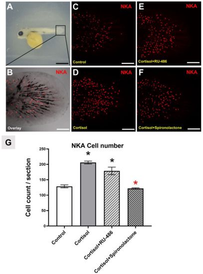
Figure 1.
Effects of exogenous cortisol, glucocorticoid receptor (GR), and mineralocorticoid receptor (MR) antagonists on Na+-K+-ATPase (NKA)-immunoreactive ionocyte number of tilapia. Tilapia (Oreochromis mossambicus) larvae were immersed in exogenous cortisol (20 mg/L), GR antagonist (RU-486) (10 µg/mL), and MR antagonist (spironolactone) (10 µg/mL) immediately after hatching for 3 days. One tilapia larvae were collected as a sample, and five replicates (n = 5) were performed. Whole tilapia larvae were studied 3 days post hatching (dph) (A), and representative images are shown for 3 dph larvae caudal fin NKA ionocytes labeled with anti-NKA α5 antibody (B), control group (C), cortisol treatment (D), cortisol treatment with GR antagonist (E), and cortisol treatment with MR antagonist (F). (G) Quantification of ionocytes in 3 dph tilapia larvae. A multi-step ANOVA was performed to assess the significance between the entire treatment group and the control group, as well as between the cortisol group and the combination group. * Indicates a significant difference (p < 0.05) using Dunnett’s multiple comparison test following a one-way ANOVA. The significance between the whole group and the control group is symbolized by a black star while the combination group with cortisol is symbolized by a red star. Values indicate the mean ± SD (n = 5). Scale bar: 1 mm(A); 500 μm (B–F).
3.2. Localization of GR and MR Expressing Cells in Adult Tilapia Gill
The previous antagonist experiment suggests cortisol affects epidermal ionocyte differentiation through the MR but not the GR in tilapia larvae. To provide more solid data to support this hypothesis, we conducted gr and mr in situ hybridization on adult gill cryosections together with NKA immunostaining to evaluate whether there is any mr or gr mRNA expression in the same NKA-immunoactive ionocytes (Figure 2). Gill tissues were dissected from adult tilapia and subjected to perform gr and mr in situ hybridization (black) and NKA immunofluorescence staining (green). Results demonstrate that gr mRNA is indeed expressed in gill tissue in a salt and pepper pattern (Figure 2B, indicated by black triangles). After comparing with NKA-immunoactive ionocytes (Figure 2A, indicated by red triangles), we found that gr mRNA (Figure 2C, indicated by black triangles) is not colocalized with NKA-immunoactive ionocytes (Figure 2C, indicated by red triangles). We found that mr mRNA is indeed expressed in gill tissue in a salt and pepper pattern (Figure 2E, indicated by black triangles). After comparing with NKA-immunoactive ionocytes in the same tissue section (Figure 2D, indicated by red triangles), we found that mr mRNA (Figure 2F, indicated by black triangles) is not colocalized with NKA-immunoactive ionocytes (Figure 2F, indicated by red triangles). Based on colocalization experiments, we conclude that both mr and gr mRNA are indeed expressed in tilapia gill tissues, but they are not co-localized with NKA-immunoactive ionocytes.
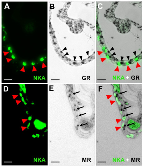
Figure 2.
In situ hybridization and immunohistochemistry of adult tilapia (Oreochromis mossambicus) gill epithelial cryosections. Double labeling with anti-NKA α5 antibody (a ionocyte marker) (A) and gill gr mRNA (B) in the same section. (C) Merged image indicating that the gr (black arrowheads) was not colocalized with NKA (red arrowheads). Double labeling with anti-NKA α5 antibody (D) and gill mr mRNA (E) in the same section. (F) Merged image indicating that the mr (black arrows) was not colocalized with NKA (red arrowheads). Scale bar: 20 μm.
3.3. Localization of gr and mr mRNAs in the Same Tilapia Adult Gill Sections
The non-colocalization nature of gr/mr-expressing cells with NKA-immunoactive ionocytes leads us to design more detailed experiments to determine whether gr and mr mRNAs are co-expressed in the same cells. To reach this goal, we double the in situ hybridization by using different color labeling for mr (black) and gr (red). Three gill filaments in the same cryosection were stained, and results demonstrated that gr-expressing cells did not exhibit mr-expressing signals (Figure 3A–C). Therefore, we conclude that mr and gr mRNA are not colocalized in the same cell.
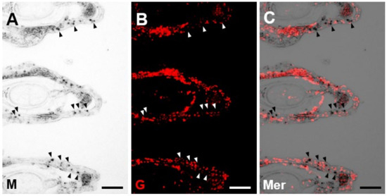
Figure 3.
Double in situ hybridization against mr mRNA and gr mRNA of adult tilapia (Oreochromis mossambicus) gill epithelial cryosections. Double labeling for tilapia mr mRNA (A) and gr mRNA (B) in the same section. (C) Merged image indicating that the mr was not colocalized with gr. Arrowheads indicate the locations of mr signals. Scale bar: 20 μm.
3.4. Localization of mr mRNA, p63 mRNA, and NKA Protein in the Tilapia Adult Gill Sections
The results above indicate that cortisol affects epidermal cell development through the MR but not the GR signaling. However, in gill tissue sections, we failed to find any colocalization pattern between mr/gr-expressing signals with NKA-immunoactive ionocytes. This finding leads us to speculate whether mr/gr-expressing cells belong to other cell categories. To validate this hypothesis, we examined the expression of mr, p63, and NKA protein in the same cryosection of adult tilapia gills. Triple staining was conducted to label mr mRNA (black) and p63 mRNA (red) by in situ hybridization and NKA (green color) proteins by immunostaining. We found mr mRNA was nicely colocalized with some p63 mRNA cells (Figure 4A,B,D), which is supported by evidence from the high magnification zoomed picture displayed in D. In conclusion, we found mr-expressing cells were positive for p63 but negative for NKA staining (Figure 4B,C,E) in triple staining experiments. This result suggests mr/gr mRNA-expressing cells might be derived from p63-expressed stem cells but do not overlap with NKA-immunoreactive ionocytes in tilapia gills. In addition, P63-labeled cells were also observed to co-express NKA at a low level (Figure 4F), which is supported by evidence from the high magnification zoomed picture displayed in Figure 4F. It could be that epidermal stem cells have started to differentiate into ionocytes.
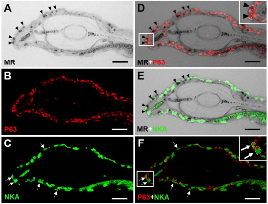
Figure 4.
Double in situ hybridization and immunohistochemistry of adult tilapia (Oreochromis mossambicus) gill epithelial cryosections. Triple labeling for tilapia gill mr mRNA (A), tumor protein 63 (p63) mRNA (epidermal stem cell marker) (B), and anti-NKA α5 antibody (ionocyte marker) (C) in the same section. (D) Merged image indicating that the mr (black arrowheads) was colocalized with p63 mRNA. The inset shows an enlarged picture of two co-stained cells (black arrowheads). (E) Merged image indicating that the mr (black arrowheads) was not colocalized with NKA. (F) Merged image indicating that the p63 (white arrows) was colocalized with NKA. The inset shows an enlarged picture of two co-stained cells (white arrows). Scale bar: 20 μm.
3.5. P63 mRNA Expression after MR and GR Antagonist Exposure
After collecting all the information found in the previous experiment, we hypothesized that cortisol might activate ionocyte differentiation via mr signaling by downregulating the fate of epidermal skin cells. To support this hypothesis, we conducted real-time PCR for measuring the P63 mRNA level after cortisol and MR/GR antagonist treatment. We initially exposed 20 mg/L cortisol-treated tilapia larvae to either RU486 or spironolactone at 10 ug/mL. Later, after either 1- or 3-day exposure, tilapia larvae were subjected to real-time PCR for the mRNA expression measurement (Figure 5). The P63 mRNA expression levels were subjected to statistical analysis using a multi-step ANOVA. This analysis assessed the significance between the entire treatment group and the control group, as well as between the cortisol group and the combination group. Significance was determined using Dunnett’s multiple comparison test following a one-way ANOVA, with a significance threshold set at p < 0.05. Results demonstrate that the P63 mRNA expression level was promoted in tilapia larvae after cortisol exposure (Figure 5B) (p < 0.05), and this upregulation effect can be abolished after being co-treated with MR antagonists after 3-day exposure (Figure 5B) (p < 0.05).
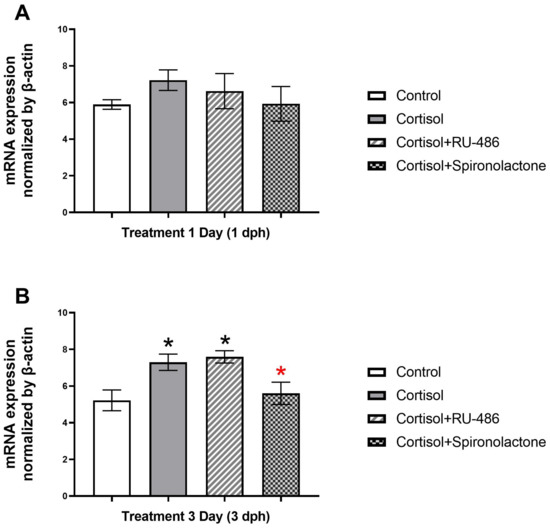
Figure 5.
Effects of exogenous cortisol, and GR and MR antagonists on P63 mRNA expression in tilapia (Oreochromis mossambicus) larvae. Tilapia larvae were immersed in exogenous cortisol (20 mg/L), GR antagonist (RU-486) (10 µg/mL), and MR antagonist (spironolactone) (10 µg/mL) immediately after hatching. Three tilapia larvae were collected as a sample, and five replicates (n = 5) were performed. The experimental samples were treated with the drug for 1 day (1 dph) (A) and 3 days (3 dph) (B). Expression of mRNA was analyzed by real-time PCR, and values were normalized to β-actin. A multi-step ANOVA was performed to assess the significance between the entire treatment group and the control group, as well as between the cortisol group and the combination group. * Indicates a significant difference (p < 0.05) using Dunnett’s multiple comparison test following a one-way ANOVA. The significance between the whole group and the control is symbolized with a black star while the combination group with cortisol is symbolized with a red star. Values indicate the mean ± SD (n = 5).
4. Discussion
In the present study, we found that treatment of tilapia larvae with exogenous cortisol significantly increased the density of ionocytes and P63 expression (p < 0.05). We also provided pharmacological evidence to show that MR, but not GR, might control ionocyte differentiation. We used RNA probes specific to tilapia mr and gr genes to show that gr and mr are not present in NKA-immunoactive ionocytes of gills, and this is inconsistent with the data reported for zebrafish [27,28]. Nonetheless, our antagonist treatment of tilapia larvae validated the major involvement of MR in the NKA-immunoactive ionocytes in tilapia and in affecting their respective roles in P63 mRNA expression. Currently, four types of ionocytes are identified in tilapia, all of which can be labeled with anti-NKA α5 antibody [25,46,47]. Further research is needed to determine which type of ionocyte is primarily increased by exogenous cortisol treatment.
Cortisol is the main corticosteroid hormone, and it may exert its actions through GR and/or MR in fish. Previous studies have shown that cortisol is involved in the adaptive responses of seawater and freshwater fish, while ionocytes in the skin and gills play important roles in osmoregulation and ion regulation [11,12,48,49,50,51]. A series of studies have indicated that changes in the proportion and morphology of ionocytes during the environmental acclimation process of teleost fish are associated with cortisol-induced cell differentiation and proliferation [27,28,29,52,53,54,55,56,57,58,59,60,61,62]. In fact, the administration of exogenous cortisol can increase the number of ionocytes in teleost fish [54,61,62], which is consistent with our experimental findings showing that exogenous cortisol increases the number of NKA-immunoactive ionocytes in tilapia larvae. Prior to this study, it was unclear how cortisol through GR/MR is involved in the differentiation and proliferation of ionocytes in teleost fish other than zebrafish and medaka. In previous studies by Lin and colleagues, tilapia larvae were exposed to cortisol to assess Ca2+ influx and the expression of related ion transporters like ECaC during embryonic development. It has been demonstrated that cortisol can increase the Ca2+ content in tilapia larvae by upregulating the expression of epidermal calcium ion channel proteins, and high calcium environments can enhance the expression of MR [32]. Here, we exposed cortisol-treated tilapia larvae with either GR (RU-486) or MR (spironolactone) antagonists and discovered that spironolactone significantly blocked the effect of exogenous cortisol in increasing the number of epidermal ionocytes, while RU-486 also decreased the number of ionocytes but to a lesser extent than spironolactone. These results suggest that cortisol may primarily regulate the differentiation and proliferation of epidermal ionocytes in tilapia through the MR signaling.
In this study, spironolactone was used as an MR antagonist, but its antagonistic effects have been controversial in different fish models or experimental designs. Spironolactone revealed antagonist properties in the gills of killifish with freshwater acclimation and cultured gills of salmon [42,59]. In previous studies, spironolactone, as an MR antagonist, attenuated the stimulatory effects of exogenous cortisol on ECaC expression [32], and it also acted as an antagonist in the present study. However, although RU-486 influenced the number of ionocytes, it was not as significant as spironolactone, possibly because the high dose of RU-486 with low affinity for MR may also have antagonistic effects on the receptor. Further studies are needed to determine the optimal dosage of antagonists and ligand-receptor affinity by the molecular docking approach in future research. In addition, recent studies have utilized eplerenone as an MR antagonist in fish experiments and have reported improved receptor specificity of this drug compared to spironolactone in these experiments [34,63,64,65]. To the best of our knowledge, there are no other studies that have used eplerenone as an MR antagonist in tilapia. There are reports indicating that eplerenone did not have any effect on cultured trout gill epithelium [66]. This suggests potential species differences in the response of fish to CR antagonists. In future studies, eplerenone could be considered as an antagonist of tilapia MR and compared to spironolactone to determine the optimal choice for this species.
Ionocytes enveloping the embryonic skin are responsible for iono-/osmoregulation of internal fluid homeostasis [67]. During development, ionocytes also appear in functional organs such as the gills of adult fish and eventually become one of the primary regulators of osmotic pressure and ion regulation [8,26,50,51,67,68]. Therefore, it can be expected that the developmental mechanism of ionocytes in tilapia larvae is similar to that of gill in adult tilapia. In this study, at least 3 days of cortisol treatment was required to significantly increase the number of ionocytes in tilapia larvae. This finding reflects the fact that it takes about 3 days of cortisol treatment to induce ionocyte differentiation. This is consistent with our experimental results of gene expression, where P63 expression significantly increased in the 3-day cortisol treatment group. However, as a model organism, tilapia is limited by its biological characteristics and cannot accurately determine the developmental stage of individuals after fertilization. Therefore, experiments can only be conducted from hatching, and there are not as many accumulated research results as for zebrafish. According to Hsiao et al.’s 2007 study, ionocyte development is generally divided into three major events: specification (from 90% epiboly to the 14-somite stage), differentiation (14 s to 36 hpf), and maturation (36 hpf onwards) [19]. Based on a series of previous studies [19,27,28,29], we followed the research results of zebrafish and attempted to understand whether the broad-spectrum Mozambique tilapia has similarities in ionocyte proliferation and differentiation with zebrafish. However, unexpectedly, we found that exogenous cortisol treatment promoted the expression of P63, which differs from the research results of zebrafish [69]. Cortisol controls the epidermal ionocyte differentiation process by regulating ionocyte master regulator Foxi3a and Foxi3b expression [28]. Therefore, we provided new molecular evidence suggesting that cortisol may primarily promote ionocyte differentiation and proliferation through MR activation in p63-expressing stem cells.
To further confirm the experimental results mentioned above, we also conducted in situ hybridization to show that gr expression in the gill tissue of tilapia was different from that of zebrafish. The location of gr in zebrafish was co-localized with NKA-immunoactive ionocytes [27,28]. However, in this study, we found that gr mRNA in tilapia gills was co-localized neither with NKA-immunoactive ionocytes nor with mr mRNA. Furthermore, through triple staining in cryosections of adult tilapia gills, we found that some p63-expressing cells were co-localized with mr-expressing cells, and some p63-expressing cells were co-localized with NKA-immunoactive ionocytes. We suggest that MR is initially co-expressed on p63-positive epidermal stem cells (Figure 4D and Figure 6A). When cortisol binds to MR, it will activate the gene expression of P63, and promote the proliferation and differentiation of epidermal stem cells (Figure 6B). When epidermal stem cells begin to differentiate into ionocytes, MR expression is no longer needed, and only p63 and NKA are co-localized (Figure 4F and Figure 6C). Finally, when ionocytes complete their differentiation and development, p63 expression gradually downregulates and finally disappears (Figure 6D). These results support our previous hypothesis that cortisol may regulate P63 through MR to promote the differentiation and proliferation of ionocytes.
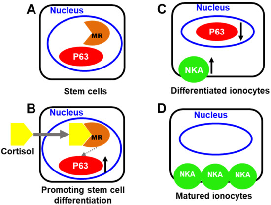
Figure 6.
Model of cortisol regulating the differentiation of epidermal stem cells into ionocytes through the mineralocorticoid receptor in tilapia. MR is initially co-expressed on p63-positive epidermal stem cells (A). Cortisol binds to MR; it will activate the gene expression of p63 and promote the proliferation and differentiation of epidermal stem cells (B). Epidermal stem cells begin to differentiate into ionocytes, mr expression is no longer needed, and only p63 and NKA are co-localized (C). Ionocytes complete their differentiation and development; p63 expression gradually downregulates and finally disappears (D).
In the past, it was commonly believed that ray-finned fish lack aldosterone synthase, so MR may not be a physiologically critical corticosteroid receptor in ray-finned fish. Recent studies have indicated that corticosteroids are involved in various physiological responses through the action of the MR. It has been suggested that MR participates in the stress response of skeletal muscles, muscle contraction, and cell cycle regulation in rainbow trout [33,34]. The study also found that MR plays a crucial role in glucose regulation in zebrafish skeletal muscle [70]. Moreover, MR has been implicated in neural plasticity and stress responses in Atlantic salmon at low temperatures [35], and mineralocorticoid signaling is also thought to be associated with brain behavior of mudskipper fish and medaka fish [36,37]. These studies suggest that MR plays an important role in the physiological actions of teleost fish, but its relevance to salinity acclimation remains unclear.
Cortisol plays a significant role in the osmoregulation and ion regulation necessary for acclimation and survival in fluctuating environments for euryhaline teleost fish [10,11,71,72]. However, the exact pathways by which cortisol mediates these controls, via the GR, MR, or both, have been controversial. A series of experimental results have mainly studied the effects of cortisol on experimental species by detecting mRNA expression levels [42,43,73], and the inconsistent results may be due to different experimental species or experimental designs. Detecting mRNA expression levels can be affected by many factors–––such as experimental methods, individual differences in experimental species, and sample collection time–––and more molecular evidence is needed to support experimental hypotheses and explain results. Currently, only studies on zebrafish as experimental species provide complete data, indicating that cortisol controls osmoregulation and ion regulation mechanisms by binding with GR [27,28]. However, zebrafish are a primary freshwater fish that cannot survive in high-salinity environments or environments with high-salinity fluctuations, and we cannot explain the regulatory mechanisms of euryhaline fish. In addition to demonstrating the effect of cortisol on the mRNA expression levels of the differentiation factor P63, the experimental results of this study provide more complete molecular evidence by counting the number of ionocytes through immunohistochemical staining and molecular labeling on the epidermal of tilapia larvae, which may provide new ideas and research directions for the regulation of salinity acclimation in euryhaline fish in the future. Furthermore, recent research has shown that cortisol regulates acute responses in energy metabolism on the fish gills via GR [74]. Therefore, we propose a hypothesis that cortisol regulates energy metabolism in euryhaline tilapia for salinity acclimation in the short term, to respond to acute environmental changes, and initiate cell differentiation to adapt to long-term survival environments. Further research is needed to explore in detail the differentiation effects of cortisol on the four types of ionocytes and the regulation of downstream differentiation factors.
In summary, our findings indicate that cortisol stimulates ionocyte development in tilapia through MR but not GR. As a result, we propose a new direction for investigating the dual roles of tilapia in osmoregulation and epithelial development of skin/gills.
5. Conclusions
Extensive research has shown that the regulatory mechanism controlling osmotic pressure and ion balance in mammals can be achieved through the action of cortisol in fish. Here, we provide compelling molecular evidence that shows that cortisol influences the differentiation of ionocytes, as treatment with exogenous cortisol promotes gene expression of differentiation factor P63 and increases epidermal ionocytes of tilapia larvae. Co-treatment experiments with an exogenous corticosteroid receptor antagonist also indicate that cortisol regulates epidermal stem cell differentiation and development primarily through the action of MR rather than GR. Moreover, from slice experiments, we found that mr was co-localized in the same cell as p63, i.e., epidermal stem cells, which supports the results of our previous experiments indicating that cortisol regulates ionocyte development via MR. Therefore, we provide a new direction for investigating the dual action of osmotic regulation and skin/gill epithelial development in tilapia, which could help resolve previously inconsistent and conflicting findings. Our findings provide a novel concept that cortisol may regulate various physiological phenomena in fish adaptation to changing environments through both the glucocorticoid receptor (GR) and mineralocorticoid receptor (MR), including energy metabolism and cell differentiation.
Supplementary Materials
The following supporting information can be downloaded at: https://www.mdpi.com/article/10.3390/fishes8060283/s1. Molecular cloning and sequencing of tilapia tumor protein 63 (P63) gene. We used RT-PCR and amplification from the cDNA tail to clone and successfully sequence a partial cDNA of P63 from the gill tissue of tilapia. This sequence consists of 1163 bp with an open reading frame encoding a 344-amino acid protein (Figure S1). It includes the protein’s C-terminal position and a partial 3′-UTR fragment, and the entire sequence contains highly conserved regions (Figure S2). The Genbank accession number for this sequence is OQ626354. Figure S1. Nucleotide and deduced amino acid sequences of tumor protein 63 (P63) cDNA from tilapia gill epithelial cells. # Translational stop codon. Accession no. OQ626354. Figure S2. The nucleotide sequence alignment of zebrafish ΔNp63 (Accession no. AAM48108.1) and tilapia P63. The identical amino acids are indicated by red uplines, and the non-identical amino acids were indicated by blue uplines.
Author Contributions
Conceptualization, C.-Y.W. and D.-Y.T.; methodology, C.-Y.W., D.-Y.T., and T.-H.L.; software, C.-Y.W.; validation, C.-Y.W., D.-Y.T., and T.-H.L.; formal analysis, C.-Y.W.; resources, D.-Y.T. and T.-H.L.; writing—original draft preparation, C.-Y.W.; writing—review and editing, D.-Y.T. and T.-H.L.; supervision and funding acquisition, D.-Y.T. All authors have read and agreed to the published version of the manuscript.
Funding
This study was funded by the grants sponsored by the Ministry of Science and Technology MOST 107-2313-B-024-001 to D.-Y.T.
Institutional Review Board Statement
All protocols and procedures involving tilapia were approved by the Institutional Animal Care and Use Group (Approval No.: IACUG1050005, issue date 12 December 2016) at National University of Tainan. All the experimental procedures and the collection of samples were performed in compliance with the ethical considerations of the Taiwan Council of Agriculture Executive Yuan Guideline for the Care and Use of Laboratory Animals (Decree by the Council of Agriculture, Executive Yuan, Taiwan, 2018/06).
Data Availability Statement
The data presented in this study are available on request from the corresponding author.
Acknowledgments
We thank Y.-H. Huang and T.-F. Kuo for sample collection, and W.-F. Chen, H.-H. Huang, and F.-I. Lu for technical assistance. We would like to express our special thanks to C.-D. Hsiao for providing statistical analysis assistance and valuable suggestions in writing. We gratefully acknowledge the Instrument Development Center at National Cheng Kung University for allowing us to use the Zeiss LSM 780 Confocal Microscope.
Conflicts of Interest
The authors declare no conflict of interest. The funders had no role in the design of the study; in the collection, analyses, or interpretation of data; in the writing of the manuscript; or in the decision to publish the results.
References
- Charmandari, E.; Tsigos, C.; Chrousos, G. Endocrinology of the stress response. Annu. Rev. Physiol. 2005, 67, 259–284. [Google Scholar] [CrossRef]
- McLaughlin, F.; Mackintosh, J.; Hayes, B.P.; McLaren, A.; Uings, I.J.; Salmon, P.; Humphreys, J.; Meldrum, E.; Farrow, S.N. Glucocorticoid-induced osteopenia in the mouse as assessed by histomorphometry, microcomputed tomography, and biochemical markers. Bone 2002, 30, 924–930. [Google Scholar] [CrossRef]
- Baker, M.E. Evolution of Glucocorticoid and Mineralocorticoid Responses: Go Fish. Endocrinology 2003, 144, 4223–4225. [Google Scholar] [CrossRef]
- Takahashi, H.; Sakamoto, T. The role of ‘mineralocorticoids’ in teleost fish: Relative importance of glucocorticoid signaling in the osmoregulation and ‘central’ actions of mineralocorticoid receptor. Gen. Comp. Endocrinol. 2013, 181, 223–228. [Google Scholar] [CrossRef]
- Carroll, S.M.; Ortlund, E.A.; Thornton, J.W. Mechanisms for the Evolution of a Derived Function in the Ancestral Glucocorticoid Receptor. PLoS Genet. 2011, 7, e1002117. [Google Scholar] [CrossRef]
- Bridgham, J.T.; Carroll, S.M.; Thornton, J.W. Evolution of Hormone-Receptor Complexity by Molecular Exploitation. Science 2006, 312, 97–101. [Google Scholar] [CrossRef]
- Gorissen, M.; Flik, G. 3-The Endocrinology of the Stress Response in Fish: An Adaptation-Physiological View. In Fish Physiology; Schreck, C.B., Tort, L., Farrell, A.P., Brauner, C.J., Eds.; Elsevier: Amsterdam, The Netherlands, 2016; Volume 35, pp. 75–111. [Google Scholar]
- Hwang, P.-P.; Lee, T.-H.; Lin, L.-Y. Ion regulation in fish gills: Recent progress in the cellular and molecular mechanisms. Am. J. Physiol. Regul. Integr. Comp. Physiol. 2011, 301, R28–R47. [Google Scholar] [CrossRef]
- Hwang, P.-P.; Tung, Y.-C.; Chang, M.-H. Effect of environmental calcium levels on calcium uptake in tilapia larvae Oreochromis mossambicus. Fish Physiol. Biochem. 1996, 15, 363–370. [Google Scholar] [CrossRef]
- McCormick, S.D.; Bradshaw, D. Hormonal control of salt and water balance in vertebrates. Gen. Comp. Endocrinol. 2006, 147, 3–8. [Google Scholar] [CrossRef]
- McCormick, S.D. Endocrine Control of Osmoregulation in Teleost Fish1. Am. Zool. 2001, 41, 781–794. [Google Scholar] [CrossRef]
- Evans, D.H.; Piermarini, P.M.; Choe, K.P. The Multifunctional Fish Gill: Dominant Site of Gas Exchange, Osmoregulation, Acid-Base Regulation, and Excretion of Nitrogenous Waste. Physiol. Rev. 2005, 85, 97–177. [Google Scholar] [CrossRef] [PubMed]
- Aruna, A.; Nagarajan, G.; Chang, C.-F. Differential expression patterns and localization of glucocorticoid and mineralocorticoid receptor transcripts in the osmoregulatory organs of tilapia during salinity stress. Gen. Comp. Endocrinol. 2012, 179, 465–476. [Google Scholar] [CrossRef]
- Acar, Ü.; Parrino, V.; Kesbiç, O.S.; Lo Paro, G.; Saoca, C.; Abbate, F.; Yılmaz, S.; Fazio, F. Effects of Different Levels of Pomegranate Seed Oil on Some Blood Parameters and Disease Resistance Against Yersinia ruckeri in Rainbow Trout. Front. Physiol. 2018, 9, 596. [Google Scholar] [CrossRef] [PubMed]
- Parrino, V.; Cappello, T.; Costa, G.; Cannavà, C.; Sanfilippo, M.; Fazio, F.; Fasulo, S. Comparative study of haematology of two teleost fish (Mugil cephalus and Carassius auratus) from different environments and feeding habits. Eur. Zool. J. 2018, 85, 193–199. [Google Scholar] [CrossRef]
- Le Guellec, D.; Morvan-Dubois, G.; Sire, J.Y. Skin development in bony fish with particular emphasis on collagen deposition in the dermis of the zebrafish (Danio rerio). Int. J. Dev. Biol. 2004, 48, 217–231. [Google Scholar] [CrossRef] [PubMed]
- Chang, W.-J.; Horng, J.-L.; Yan, J.-J.; Hsiao, C.-D.; Hwang, P.-P. The transcription factor, glial cell missing 2, is involved in differentiation and functional regulation of H+-ATPase-rich cells in zebrafish (Danio rerio). Am. J. Physiol. -Regul. Integr. Comp. Physiol. 2009, 296, R1192–R1201. [Google Scholar] [CrossRef] [PubMed]
- Horng, J.-L.; Lin, L.-Y.; Hwang, P.-P. Functional regulation of H+-ATPase-rich cells in zebrafish embryos acclimated to an acidic environment. Am. J. Physiol. -Cell Physiol. 2009, 296, C682–C692. [Google Scholar] [CrossRef]
- Hsiao, C.-D.; You, M.-S.; Guh, Y.-J.; Ma, M.; Jiang, Y.-J.; Hwang, P.-P. A Positive Regulatory Loop between foxi3a and foxi3b Is Essential for Specification and Differentiation of Zebrafish Epidermal Ionocytes. PLoS ONE 2007, 2, e302. [Google Scholar] [CrossRef]
- Jänicke, M.; Carney, T.J.; Hammerschmidt, M. Foxi3 transcription factors and Notch signaling control the formation of skin ionocytes from epidermal precursors of the zebrafish embryo. Dev. Biol. 2007, 307, 258–271. [Google Scholar] [CrossRef]
- Lee, H.; Kimelman, D. A Dominant-Negative Form of p63 Is Required for Epidermal Proliferation in Zebrafish. Dev. Cell 2002, 2, 607–616. [Google Scholar] [CrossRef]
- Katsuhisa, U.; Toyoji, K.; Hiroaki, M.; Sanae, H.; Tetsuya, H. Excellent Salinity Tolerance of Mozambique Tilapia (Oreochromis mossambicus): Elevated Chloride Cell Activity in the Branchial and Opercular Epithelia of the Fish Adapted to Concentrated Seawater. Zool. Sci. 2000, 17, 149–160. [Google Scholar]
- Stickney, R.R. Tilapia Tolerance of Saline Waters: A Review. Progress. Fish Cult. 1986, 48, 161–167. [Google Scholar] [CrossRef]
- Suresh, A.V.; Lin, C.K. Tilapia culture in saline waters: A review. Aquaculture 1992, 106, 201–226. [Google Scholar] [CrossRef]
- Hiroi, J.; Yasumasu, S.; McCormick, S.D.; Hwang, P.-P.; Kaneko, T. Evidence for an apical Na–Cl cotransporter involved in ion uptake in a teleost fish. J. Exp. Biol. 2008, 211, 2584–2599. [Google Scholar] [CrossRef]
- Hwang, P.-P.; Perry, S.F. Ionic and acid–base regulation. In Fish physiology; Elsevier: Amsterdam, The Netherlands, 2010; Volume 29, pp. 311–344. [Google Scholar]
- Cruz, S.A.; Lin, C.-H.; Chao, P.-L.; Hwang, P.-P. Glucocorticoid Receptor, but Not Mineralocorticoid Receptor, Mediates Cortisol Regulation of Epidermal Ionocyte Development and Ion Transport in Zebrafish (Danio rerio). PLoS ONE 2013, 8, e77997. [Google Scholar] [CrossRef]
- Cruz, S.A.; Chao, P.-L.; Hwang, P.-P. Cortisol promotes differentiation of epidermal ionocytes through Foxi3 transcription factors in zebrafish (Danio rerio). Comp. Biochem. Physiol. Part A Mol. Integr. Physiol. 2013, 164, 249–257. [Google Scholar] [CrossRef]
- Trayer, V.; Hwang, P.-P.; Prunet, P.; Thermes, V. Assessment of the role of cortisol and corticosteroid receptors in epidermal ionocyte development in the medaka (Oryzias latipes) embryos. Gen. Comp. Endocrinol. 2013, 194, 152–161. [Google Scholar] [CrossRef]
- Colombe, L.; Fostier, A.; Bury, N.; Pakdel, F.; Guiguen, Y. A mineralocorticoid-like receptor in the rainbow trout, Oncorhynchus mykiss: Cloning and characterization of its steroid binding domain. Steroids 2000, 65, 319–328. [Google Scholar] [CrossRef]
- Kiilerich, P.; Milla, S.; Sturm, A.; Valotaire, C.; Chevolleau, S.; Giton, F.; Terrien, X.; Fiet, J.; Fostier, A.; Debrauwer, L.; et al. Implication of the mineralocorticoid axis in rainbow trout osmoregulation during salinity acclimation. J. Endocrinol. 2011, 209, 221–235. [Google Scholar] [CrossRef]
- Lin, C.-H.; Kuan, W.-C.; Liao, B.-K.; Deng, A.-N.; Tseng, D.-Y.; Hwang, P.-P. Environmental and cortisol-mediated control of Ca2+ uptake in tilapia (Oreochromis mossambicus). J. Comp. Physiol. B 2016, 186, 323–332. [Google Scholar] [CrossRef]
- Zuloaga, R.; Aravena-Canales, D.; Aedo, J.E.; Osorio-Fuentealba, C.; Molina, A.; Valdés, J.A. Effect of 11-Deoxycorticosterone in the Transcriptomic Response to Stress in Rainbow Trout Skeletal Muscle. Genes 2023, 14, 512. [Google Scholar] [CrossRef] [PubMed]
- Aedo, J.E.; Zuloaga, R.; Aravena-Canales, D.; Molina, A.; Valdés, J.A. Role of glucocorticoid and mineralocorticoid receptors in rainbow trout (Oncorhynchus mykiss) skeletal muscle: A transcriptomic perspective of cortisol action. Front. Physiol. 2023, 13, 2755. [Google Scholar] [CrossRef]
- Tang, P.A.; Stefansson, S.O.; Nilsen, T.O.; Gharbi, N.; Lai, F.; Tronci, V.; Balseiro, P.; Gorissen, M.; Ebbesson, L.O.E. Exposure to cold temperatures differentially modulates neural plasticity and stress responses in post-smolt Atlantic salmon (Salmo salar). Aquaculture 2022, 560, 738458. [Google Scholar] [CrossRef]
- Sakamoto, T.; Mori, C.; Minami, S.; Takahashi, H.; Abe, T.; Ojima, D.; Ogoshi, M.; Sakamoto, H. Corticosteroids stimulate the amphibious behavior in mudskipper: Potential role of mineralocorticoid receptors in teleost fish. Physiol. Behav. 2011, 104, 923–928. [Google Scholar] [CrossRef] [PubMed]
- Sakamoto, T.; Yoshiki, M.; Takahashi, H.; Yoshida, M.; Ogino, Y.; Ikeuchi, T.; Nakamachi, T.; Konno, N.; Matsuda, K.; Sakamoto, H. Principal function of mineralocorticoid signaling suggested by constitutive knockout of the mineralocorticoid receptor in medaka fish. Sci. Rep. 2016, 6, 37991. [Google Scholar] [CrossRef] [PubMed]
- Lin, C.-H.; Hu, H.-J.; Hwang, P.-P. Cortisol regulates sodium homeostasis by stimulating the transcription of sodium-chloride transporter (NCC) in zebrafish (Danio rerio). Mol. Cell. Endocrinol. 2016, 422, 93–102. [Google Scholar] [CrossRef]
- Lin, C.-H.; Shih, T.-H.; Liu, S.-T.; Hsu, H.-H.; Hwang, P.-P. Cortisol Regulates Acid Secretion of H+-ATPase-rich Ionocytes in Zebrafish (Danio rerio) Embryos. Front. Physiol. 2015, 6, 328. [Google Scholar] [CrossRef]
- Lin, C.-H.; Tsai, I.L.; Su, C.-H.; Tseng, D.-Y.; Hwang, P.-P. Reverse Effect of Mammalian Hypocalcemic Cortisol in Fish: Cortisol Stimulates Ca2+ Uptake via Glucocorticoid Receptor-Mediated Vitamin D3 Metabolism. PLoS ONE 2011, 6, e23689. [Google Scholar] [CrossRef]
- Lin, G.R.; Weng, C.F.; Wang, J.I.; Hwang, P.P. Effects of Cortisol on Ion Regulation in Developing Tilapia (Oreochromis mossambicus) Larvae on Seawater Adaptation. Physiol. Biochem. Zool. 1999, 72, 397–404. [Google Scholar] [CrossRef]
- Kiilerich, P.; Kristiansen, K.; Madsen, S.S. Cortisol regulation of ion transporter mRNA in Atlantic salmon gill and the effect of salinity on the signaling pathway. J. Endocrinol. 2007, 194, 417–427. [Google Scholar] [CrossRef]
- Kiilerich, P.; Tipsmark, C.K.; Borski, R.J.; Madsen, S.S. Differential effects of cortisol and 11-deoxycorticosterone on ion transport protein mRNA levels in gills of two euryhaline teleosts, Mozambique tilapia (Oreochromis mossambicus) and striped bass (Morone saxatilis). J. Endocrinol. 2011, 209, 115–126. [Google Scholar] [CrossRef] [PubMed]
- Tseng, Y.-C.; Huang, C.-J.; Chang, J.C.-H.; Teng, W.-Y.; Baba, O.; Fann, M.-J.; Hwang, P.-P. Glycogen phosphorylase in glycogen-rich cells is involved in the energy supply for ion regulation in fish gill epithelia. Am. J. Physiol. Regul. Integr. Comp. Physiol. 2007, 293, R482–R491. [Google Scholar] [CrossRef] [PubMed]
- Han, T.-Y.; Wu, C.-Y.; Tsai, H.-C.; Cheng, Y.-P.; Chen, W.-F.; Lin, T.-C.; Wang, C.-Y.; Lee, J.-R.; Hwang, P.-P.; Lu, F.-I. Comparison of Calcium Balancing Strategies During Hypothermic Acclimation of Tilapia (Oreochromis mossambicus) and Goldfish (Carassius auratus). Front. Physiol. 2018, 9, 1224. [Google Scholar] [CrossRef] [PubMed]
- Inokuchi, M.; Hiroi, J.; Kaneko, T. Why can Mozambique Tilapia Acclimate to Both Freshwater and Seawater? Insights From the Plasticity of Ionocyte Functions in the Euryhaline Teleost. Front. Physiol. 2022, 13, 914277. [Google Scholar] [CrossRef]
- Hiroi, J.; McCormick, S.D.; Ohtani-Kaneko, R.; Kaneko, T. Functional classification of mitochondrion-rich cells in euryhaline Mozambique tilapia (Oreochromis mossambicus) embryos, by means of triple immunofluorescence staining for Na+/K+-ATPase,Na+/K+/2Cl− cotransporter and CFTR anion channel. J. Exp. Biol. 2005, 208, 2023–2036. [Google Scholar] [CrossRef]
- Hwang, P.-P.; Chou, M.-Y. Zebrafish as an animal model to study ion homeostasis. Pflügers Arch. Eur. J. Physiol. 2013, 465, 1233–1247. [Google Scholar] [CrossRef]
- Takei, Y.; Hwang, P.-P. Homeostatic Responses to Osmotic Stress. In Fish Physiology; Elsevier: Amsterdam, The Netherlands, 2016; Volume 35, pp. 207–249. [Google Scholar]
- Guh, Y.-J.; Hwang, P.-P. Insights into molecular and cellular mechanisms of hormonal actions on fish ion regulation derived from the zebrafish model. Gen. Comp. Endocrinol. 2017, 251, 12–20. [Google Scholar] [CrossRef]
- Yan, J.-J.; Hwang, P.-P. Novel discoveries in acid-base regulation and osmoregulation: A review of selected hormonal actions in zebrafish and medaka. Gen. Comp. Endocrinol. 2019, 277, 20–29. [Google Scholar] [CrossRef]
- Perry, S.F.; Laurent, P. Adaptational Responses of Rainbow Trout to Lowered External Nacl Concentration: Contribution of the Branchial Chloride Cell. J. Exp. Biol. 1989, 147, 147–168. [Google Scholar] [CrossRef]
- McCormick, S.D. Cortisol directly stimulates differentiation of chloride cells in tilapia opercular membrane. Am. J. Physiol. -Regul. Integr. Comp. Physiol. 1990, 259, R857–R863. [Google Scholar] [CrossRef]
- Uchida, K.; Kaneko, T.; Tagawa, M.; Hirano, T. Localization of Cortisol Receptor in Branchial Chloride Cells in Chum Salmon Fry. Gen. Comp. Endocrinol. 1998, 109, 175–185. [Google Scholar] [CrossRef] [PubMed]
- Hiroi, J.; Kaneko, T.; Tanaka, M. In vivo sequential changes in chloride cell morphology in the yolk-sac membrane of mozambique tilapia (Oreochromis mossambicus) embryos and larvae during seawater adaptation. J. Exp. Biol. 1999, 202, 3485–3495. [Google Scholar] [CrossRef] [PubMed]
- Wong, C.K.C.; Chan, D.K.O. Chloride cell subtypes in the gill epithelium of Japanese eel Anguilla japonica. Am. J. Physiol. Regul. Integr. Comp. Physiol. 1999, 277, R517–R522. [Google Scholar] [CrossRef] [PubMed]
- Sloman, K.A.; Desforges, P.R.; Gilmour, K.M. Evidence for a mineralocorticoid-like receptor linked to branchial chloride cell proliferation in freshwater rainbow trout. J. Exp. Biol. 2001, 204, 3953–3961. [Google Scholar] [CrossRef] [PubMed]
- Wong, C.K.; Chan, D.K. Effects of cortisol on chloride cells in the gill epithelium of Japanese eel, Anguilla japonica. J. Endocrinol. 2001, 168, 185–192. [Google Scholar] [CrossRef] [PubMed]
- Scott, G.R.; Keir, K.R.; Schulte, P.M. Effects of spironolactone and RU486 on gene expression and cell proliferation after freshwater transfer in the euryhaline killifish. J. Comp. Physiol. B 2005, 175, 499–510. [Google Scholar] [CrossRef]
- Shahsavarani, A.; Perry, S.F. Hormonal and environmental regulation of epithelial calcium channel in gill of rainbow trout (Oncorhynchus mykiss). Am. J. Physiol. -Regul. Integr. Comp. Physiol. 2006, 291, R1490–R1498. [Google Scholar] [CrossRef]
- Madsen, S.S. The role of cortisol and growth hormone in seawater adaptation and development of hypoosmoregulatory mechanisms in sea trout parr (Salmo trutta trutta). Gen. Comp. Endocrinol. 1990, 79, 1–11. [Google Scholar] [CrossRef]
- Laurent, P.; Dunel-Erb, S.; Chevalier, C.; Lignon, J. Gill epithelial cells kinetics in a freshwater teleost, Oncorhynchus mykiss during adaptation to ion-poor water and hormonal treatments. Fish Physiol. Biochem. 1994, 13, 353–370. [Google Scholar] [CrossRef]
- Katsu, Y.; Oana, S.; Lin, X.; Hyodo, S.; Baker, M.E. Aldosterone and dexamethasone activate African lungfish mineralocorticoid receptor: Increased activation after removal of the amino-terminal domain. J. Steroid Biochem. Mol. Biol. 2022, 215, 106024. [Google Scholar] [CrossRef]
- Pippal, J.B.; Cheung, C.M.I.; Yao, Y.-Z.; Brennan, F.E.; Fuller, P.J. Characterization of the zebrafish (Danio rerio) mineralocorticoid receptor. Mol. Cell. Endocrinol. 2011, 332, 58–66. [Google Scholar] [CrossRef] [PubMed]
- Kumai, Y.; Nesan, D.; Vijayan, M.M.; Perry, S.F. Cortisol regulates Na+ uptake in zebrafish, Danio rerio, larvae via the glucocorticoid receptor. Mol. Cell. Endocrinol. 2012, 364, 113–125. [Google Scholar] [CrossRef] [PubMed]
- Kelly, S.P.; Chasiotis, H. Glucocorticoid and mineralocorticoid receptors regulate paracellular permeability in a primary cultured gill epithelium. J. Exp. Biol. 2011, 214, 2308–2318. [Google Scholar] [CrossRef] [PubMed]
- Hwang, P.P.; Lee, T.H.; Weng, C.F.; Fang, M.J.; Cho, G.Y. Presence of Na-K-ATPase in Mitochondria-Rich Cells in the Yolk-Sac Epithelium of Larvae of the Teleost Oreochromis mossambicus. Physiol. Biochem. Zool. 1999, 72, 138–144. [Google Scholar] [CrossRef] [PubMed]
- Hwang, P.P. Salinity effects on development of chloride cells in the larvae of ayu (Plecoglossus altivelis). Mar. Biol. 1990, 107, 1–7. [Google Scholar] [CrossRef]
- Chou, M.-Y.; Hung, J.-C.; Wu, L.-C.; Hwang, S.-P.L.; Hwang, P.-P. Isotocin controls ion regulation through regulating ionocyte progenitor differentiation and proliferation. Cell. Mol. Life Sci. 2011, 68, 2797–2809. [Google Scholar] [CrossRef]
- Faught, E.; Vijayan, M.M. The Mineralocorticoid Receptor Functions as a Key Glucose Regulator in the Skeletal Muscle of Zebrafish. Endocrinology 2022, 163, bqac149. [Google Scholar] [CrossRef]
- Mommsen, T.P.; Vijayan, M.M.; Moon, T.W. Cortisol in teleosts: Dynamics, mechanisms of action, and metabolic regulation. Rev. Fish Biol. Fish. 1999, 9, 211–268. [Google Scholar] [CrossRef]
- Vijayan, M.M.; Pereira, C.; Grau, E.G.; Iwama, G.K. Metabolic Responses Associated with Confinement Stress in Tilapia: The Role of Cortisol. Comp. Biochem. Physiol. Part C Pharmacol. Toxicol. Endocrinol. 1997, 116, 89–95. [Google Scholar] [CrossRef]
- McCormick, S.D.; Regish, A.; O’Dea, M.F.; Shrimpton, J.M. Are we missing a mineralocorticoid in teleost fish? Effects of cortisol, deoxycorticosterone and aldosterone on osmoregulation, gill Na+,K+-ATPase activity and isoform mRNA levels in Atlantic salmon. Gen. Comp. Endocrinol. 2008, 157, 35–40. [Google Scholar] [CrossRef]
- Wu, C.-Y.; Lee, T.-H.; Tseng, D.-Y. Glucocorticoid Receptor Mediates Cortisol Regulation of Glycogen Metabolism in Gills of the Euryhaline Tilapia (Oreochromis mossambicus). Fishes 2023, 8, 267. [Google Scholar] [CrossRef]
Disclaimer/Publisher’s Note: The statements, opinions and data contained in all publications are solely those of the individual author(s) and contributor(s) and not of MDPI and/or the editor(s). MDPI and/or the editor(s) disclaim responsibility for any injury to people or property resulting from any ideas, methods, instructions or products referred to in the content. |
© 2023 by the authors. Licensee MDPI, Basel, Switzerland. This article is an open access article distributed under the terms and conditions of the Creative Commons Attribution (CC BY) license (https://creativecommons.org/licenses/by/4.0/).